Inositol pyrophosphate dynamics reveals control of the yeast phosphate starvation program through 1,5-IP8 and the SPX domain of Pho81
- Département d'immunobiologie, Université de Lausanne, Switzerland
- Institute of Organic Chemistry, Centre for Integrative Biological Signalling Studies, University of Freiburg, Germany
Figures
Figure 1
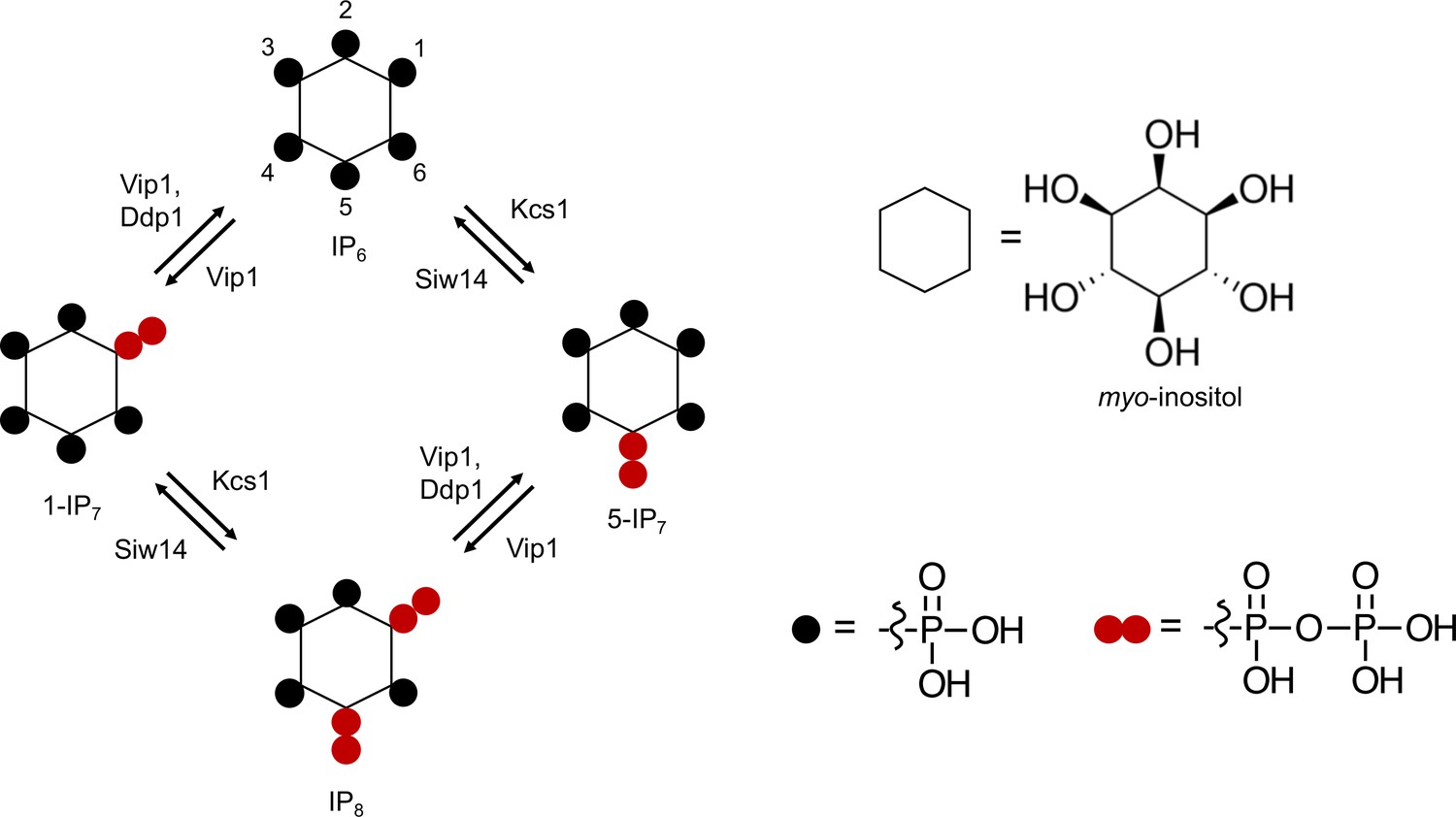
Pathways of inositol pyrophosphate metabolism in S. cerevisiae.
Figure 2 with 3 supplements
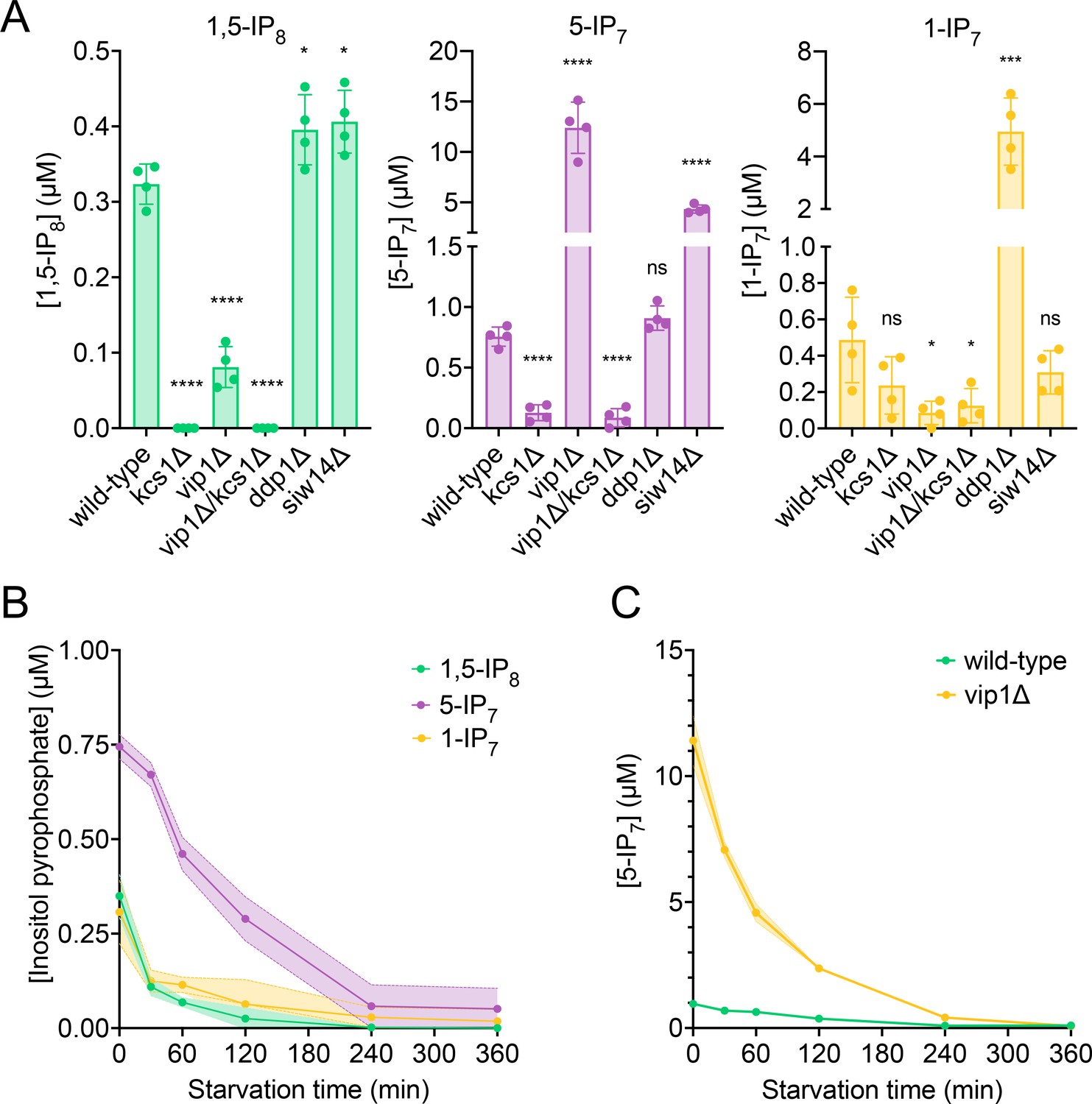
Cytosolic concentrations of 5-IP7, 1-IP7, and 1,5-IP8.
(A) Inositol pyrophosphate concentrations in the cytosol. The indicated strains were grown logarithmically in synthetic complete (SC) medium containing 7.5 mM of inorganic phosphate (Pi) (30°C, 150 rpm, overnight). When OD600nm reached 1 (1 × 10⁷ cells/mL), 1 mL of culture was extracted with perchloric acid and analyzed for inositol pyrophosphates by CE-ESI-MS. The y-axis provides the estimated cytosolic concentrations based on an average cell volume of 42 fL. Means (n=4) and standard deviations are indicated. ****p<0.0001; ***p<0.001; **p<0.01; *p<0.05; n.s. not significant, tested with Student’s t-test. (B) Evolution of inositol pyrophosphate species during Pi starvation. Cells were grown as in A, washed twice with Pi starvation medium, and further incubated in Pi starvation medium. The inoculum for the samples bound to be extracted after different times of further incubation in starvation medium was adjusted such that all samples had similar OD600nm at the time of harvesting (OD600nm=0.5 for 30 min and 60 min samples; OD600nm=0.4 for 120 min and 240 min samples; OD600nm=0.25 for 360 min samples). At the indicated times in starvation media, 1 mL aliquots were extracted and analyzed for inositol pyrophosphates as in A. The data was normalized by the number of cells harvested before calculating cytosolic concentrations. Means and standard deviations are given (n=3). (C) Depletion of 5-IP7 in starving vip1Δ cells. The indicated cells were grown in Pi-replete medium and then transferred to Pi starvation medium as in B. At the indicated times, samples were extracted and analyzed for 5-IP7 as in A. Means and standard deviations (n=4) are shown as solid lines and shaded areas, respectively.
Figure 2—figure supplement 1
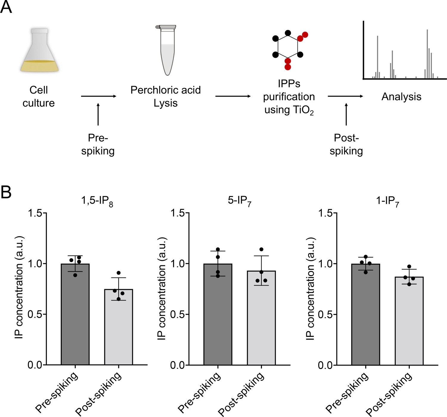
Recovery of inositol pyrophosphates extracted from S. cerevisiae cells.
(A) Illustration of the procedure of inositol pyrophosphate extraction using TiO2 beads. 0.1 µM [13C6]1,5-IP8, 0.5 µM [13C6]5-IP7, and 0.5 µM [13C6]1-IP7 were spiked into the yeast culture immediately before perchloric acid extraction (pre-spiking) or spiked into the inositol pyrophosphate fraction extracted from the cells before the capillary electrophoresis coupled to mass spectrometry (CE-MS) measurement (post-spiking). (B) Recovery. Ratios of the endogenous inositol pyrophosphates over the 13C-labeled standards were determined in the pre- and post-spiked samples. Signals of the pre-spiked samples were set to 1 and served as the reference for the post-spiked samples. Comparison of pre- and post-spiked samples shows 75% of recovery for 1,5-IP8, 93% for 5-IP7, and 87% for 1-IP7. Means (n=4) and standard deviations are indicated.
Figure 2—figure supplement 2

Determination of cell dimensions from wildtype cells stained with trypan blue.
Cells were grown in synthetic complete (SC) medium over night until they reached an OD600nm of 1. 10 µg/mL of trypan blue was added, and the cells were analyzed on a fluorescence microscope. Cells in the acquired images were analyzed by automated image segmentation and their volume was measured using the Nikon NIS Elements General Analysis 3 software package. The script for measuring cell volumes has been deposited at Figshare under the DOI 10.6084 /m9.figshare.c.6700281.
Figure 2—figure supplement 3

Loss of inositol pyrophosphates from siw14Δ cells upon inorganic phosphate (Pi) starvation.
The indicated strains were grown logarithmically in synthetic complete (SC) medium containing 7.5 mM of Pi (30°C, 150 rpm, overnight). The cells were spun down, washed twice with Pi starvation medium, and further incubated in Pi starvation medium or in SC with Pi for 2 hr. The inoculum for the samples for this final 2 hr incubation was adjusted such that all samples had an OD600nm of 1 (107 cells/mL) at the time of harvesting. After the 2 hr incubation, 1 mL of culture was extracted with perchloric acid and analyzed for inositol pyrophosphates by CE-ESI-MS. The data was normalized by the number of cells harvested before calculating cytosolic concentrations. The y-axis provides the estimated cytosolic concentrations based on an average cell volume of 42 fL. Means (n=4) and standard deviations are indicated. Graphs show the content of (A) 1,5-IP8, (B) 5-IP7, and (C) 1-IP7. ****p<0.0001; ***p<0.001; **p<0.01; *p<0.05; n.s. not significant, determined with Student’s t-test.
Figure 3
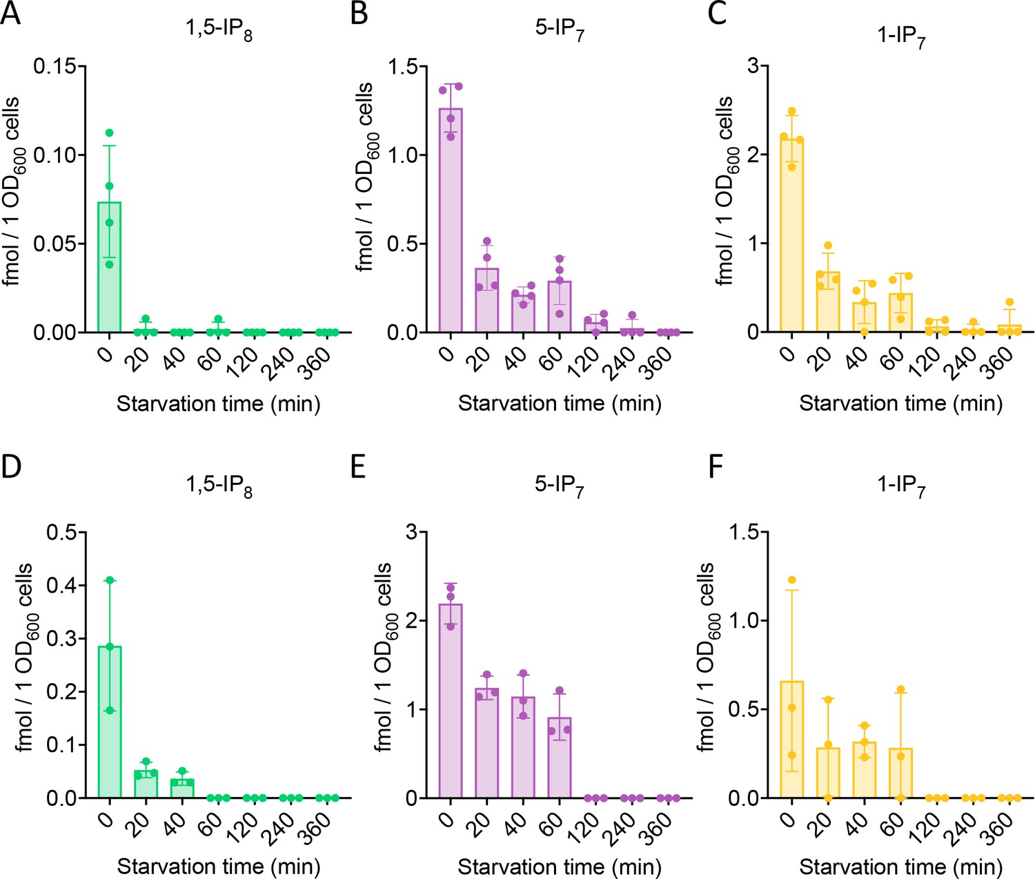
Inositol pyrophosphate analysis in C. neoformans and S. pombe.
Inositol pyrophosphates were measured in C. neoformans (A) and S. pombe (B). Both fungi were logarithmically grown in synthetic complete (SC) medium for 17 h up to an OD600nm of 1. Cells were sedimented by centrifugation, resuspended in SC without Pi, and incubated further. At the indicated times, aliquots were extracted with perchloric acid. Inositol pyrophosphates were enriched on TiO2 beads and analyzed by CE-MS. Concentrations of 1,5-IP8, 5-IP7 and 1-IP7 in the extracts were determined by comparison with added synthetic 13C-labeled inositol pyrophosphate standards. The graphs provide the concentrations in the extracts. n=4 for C. neoformans, and n=3 for S. pombe; means and standard deviations are indicated. The inositol pyrophosphate values were normalized to the OD600 of the culture for every given time point.
Figure 4 with 1 supplement
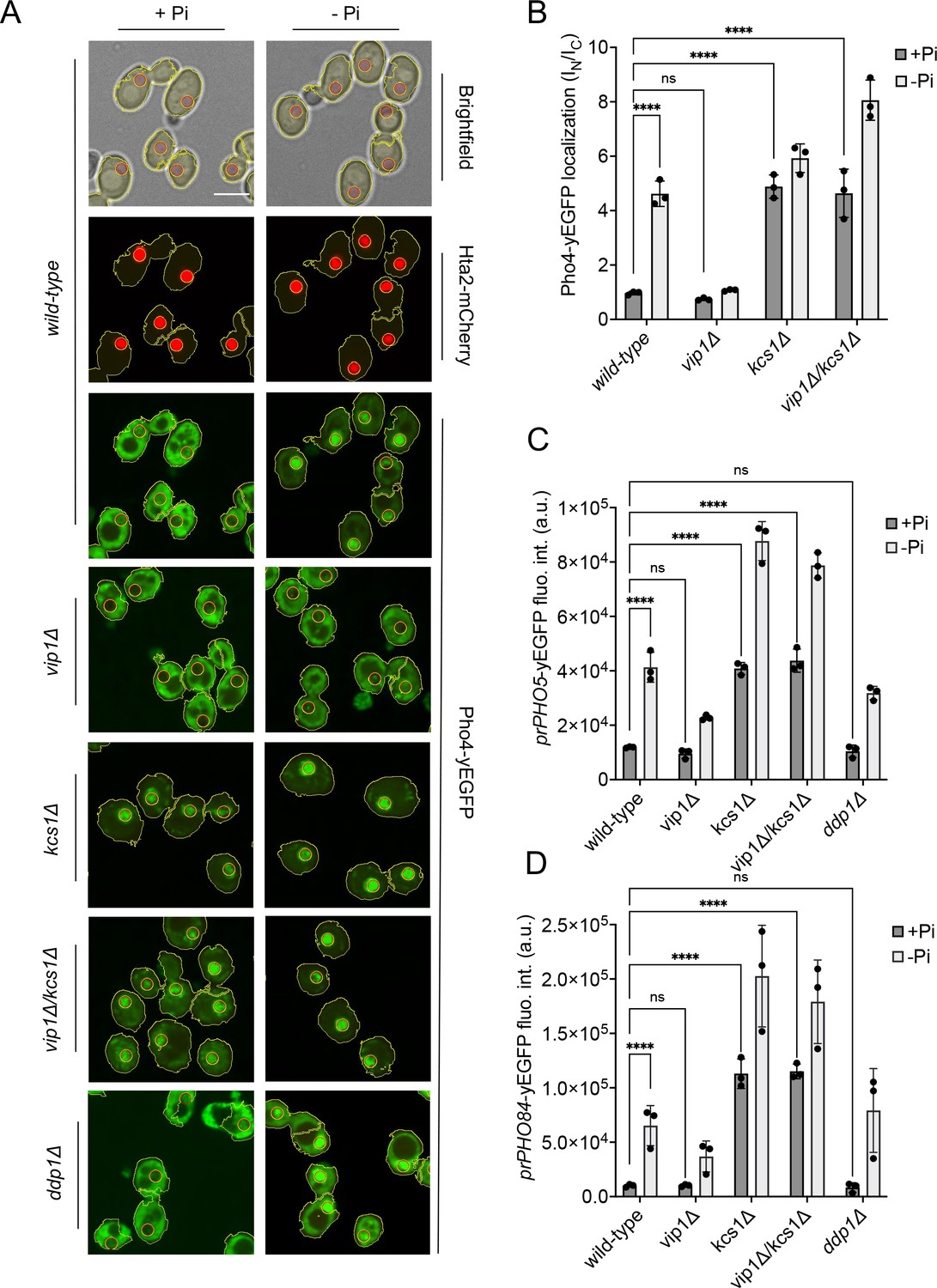
Inhibition of the PHO pathway by excessive 5-IP7.
The indicated cells producing Pho4yEGFP and the histone Hta2mCherry as a nuclear marker were logarithmically grown in inorganic phosphate (Pi)-replete synthetic complete (SC) medium, washed, and transferred to Pi starvation medium as in Figure 2A. (A) Subcellular localization of Pho4yEGFP was analyzed on a spinning disc microscope. Cells are shown in the presence of 7.5 mM of Pi (+Pi) or 30 min after the shift to Pi starvation (- Pi) medium. Yellow lines surrounding the cells illustrate the segmentation performed by the algorithm that was used to quantify Pho4yEGFP distribution in B. Scale bar: 5 μM. λex: 470 nm; λem: 495–560 nm. (B) Average intensity of Pho4yEGFP fluorescence was determined by automated image segmentation and analysis. Pho4yEGFP localization is quantified by the ratio of the average fluorescence intensities in the nucleus over the average fluorescence intensity in the cytosol (IN/IC). 100–200 cells were analyzed per condition and experiment. n=3. Means and standard deviation are indicated. (C) Activation of the PHO5 promotor. Cells expressing the prPHO5-yEGFP reporter construct from a centromeric plasmid were grown in Pi-replete medium (7.5 mM Pi) as in Figure 2A, and then shifted to Pi starvation medium or kept in Pi-replete medium. After 4 hr of further incubation, fluorescence intensity of the same number of cells was measured in a Spectramax EM microplate reader. λex: 480 nm; λem: 510 nm. n=3. Means and standard deviations are indicated. (D) Activation of the PHO84 promotor. Cells expressing the prPho84-yEGFP reporter construct from a centromeric plasmid were treated and analyzed as in C. For B, C, and D: ****p<0.0001; ***p<0.001; **p<0.01; *p<0.05; n.s. not significant, tested with Turkey’s test.
Figure 4—figure supplement 1

Segmentation of fluorescence microscopy time-lapse experiments.
The subcellular localization of Pho4-yEGFP has been quantified from time-lapse fluorescence microscopy experiments. This quantification required the segmentation of microscopy images to discriminate cytosolic and nuclear compartments. To this end, the Hta2 histone of Pho4-yEGFP strains has been tagged with mCherry. Nuclei were segmented using mCherry fluorescence images while the cell contours were segmented using bright-field images. Cell segmentation and the fluorescence quantification were performed using the General Analysis 3 module of the NIS Elements software (Nikon). (A) Bright-field and two fluorescence images of the same field are shown. Yellow lines indicate the boundaries of the cells and nuclei that have been recognized by the algorithm. (B) Flowchart of the commands of the NIS Elements General Analysis 3 suite used for the segmentation. The script has been deposited at Figshare under the doi 10.6084 /m9.figshare.c.6700281.
Figure 5
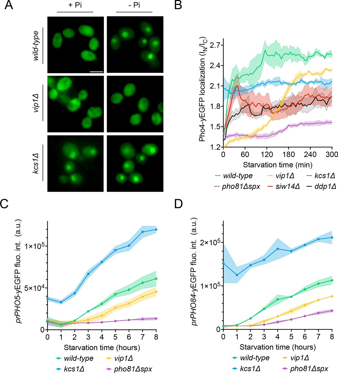
Effect of Vip1, Kcs1, and Pho81 on the time course of Pho4 translocation and PHO-gene activation.
The indicated cells were logarithmically grown in inorganic phosphate (Pi)-replete synthetic complete (SC) medium, washed, and transferred to Pi starvation medium as in Figure 2A. At the indicated times after transfer to Pi-starvation medium, they were analyzed for Pho4 localization, prPHO5-yEGFP expression, or prPHO84-yEGFP expression, using assays as in Figure 4. (A) Cells expressing Pho4yEGFP and the histone Hta2mCherry as a nuclear marker, shown in the presence of 7.5 mM of Pi (+Pi) or after 90 min of Pi starvation (-Pi). Pictures were taken on a widefield fluorescence microscope equipped with a stage-top incubator (kept at 30°C) and an IBIDI flow chamber. Only the GFP channel is shown. Scale bar = 5 μM. λex: 470 nm; λem: 495–560 nm. (B) Distribution of Pho4yEGFP between the nucleus and cytosol was quantified in the cells from A at various timepoints of Pi starvation. 100–200 cells were analyzed per condition and experiment at each timepoint. The solid lines and the shaded areas indicate the means and standard deviation, respectively. (C) Activation of the PHO5 promotor. Cells expressing the prPho5-yEGFP reporter construct from a centromeric plasmid were grown in Pi-replete medium, shifted to Pi starvation medium, and analyzed for GFP fluorescence intensity (as in Figure 4C) at the indicated timepoints of starvation. λex: 480 nm; λem: 510 nm. n=3. The solid lines and the shaded areas indicate the means and standard deviation, respectively. (D) Activation of the PHO84 promotor. As in C, but with cells expressing prPHO84-yEGFP as reporter.
Figure 6 with 1 supplement
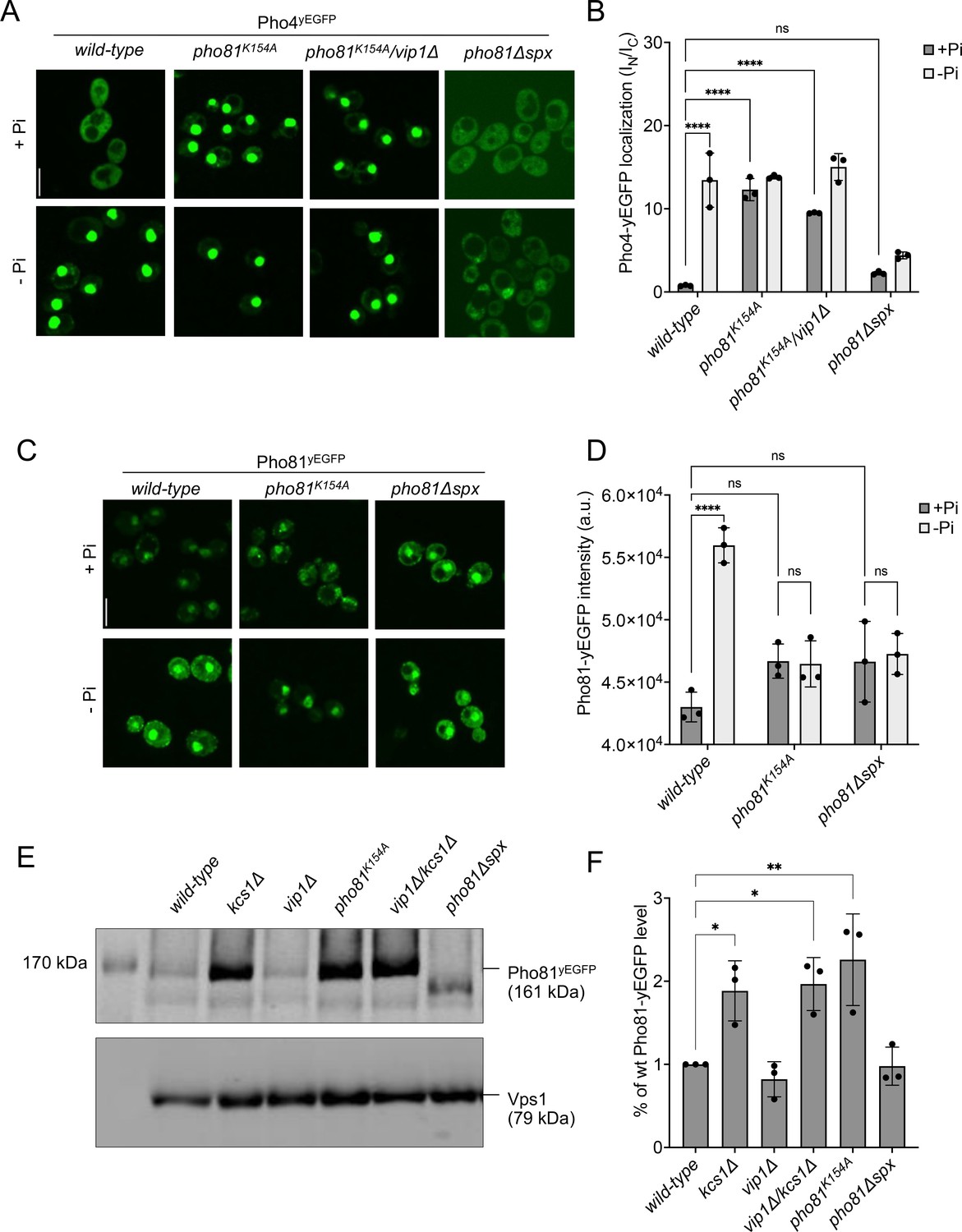
SPX-dependent activation of the PHO pathway.
Cells were logarithmically grown in inorganic phosphate (Pi)-replete synthetic complete (SC) medium as in Figure 2A, washed, and incubated for further 4 hr in medium with 7.5 mM of Pi (+Pi), or in starvation medium (- Pi). (A) Pho4 relocation. The indicated cells expressed Pho4yEGFP from its genomic locus. At the end of the 4 hr starvation period, GFP fluorescence was imaged on a spinning disc microscope. Scale bar: 5 µm. λex: 488 nm; λem: 500–530 nm. (B) Quantification of the nuclear localization of Pho4yEGFP in images from A. Average intensity of Pho4yEGFP fluorescence was determined by automated image segmentation and analysis. Pho4yEGFP localization is quantified by the ratio of the average fluorescence intensities in the nucleus over the average fluorescence intensity in the cytosol (IN/IC). 100–200 cells were analyzed per condition and experiment. n=3. Means and standard deviation are indicated. (C) Pho81yEGFP localization. The cells expressed the indicated variants of Pho81yEGFP from its genomic locus. At the end of the 4 hr growth period, GFP fluorescence was imaged as in A. (D) Quantification of the total cellular fluorescence of Pho81yEGFP. Images from C were subjected to automated segmentation and the average fluorescence intensity of the entire cells was quantified as in Figure 4B. 100–200 cells were quantified per sample. n=3 experiments. Means and standard deviation are indicated. (E) Pho81yEGFP expression assayed by western blotting. Whole-cell protein extracts were prepared from cells expressing the indicated variants of Pho81yEGFP, which had been grown in Pi-replete SC medium as in Figure 2A. Proteins were analyzed by SDS-PAGE and western blotting using antibodies to GFP. Vps1 was decorated as a loading control. (F) Quantification of Pho81yEGFP blotting. Bands from experiments as in E were quantified on a LICOR Odyssey infrared fluorescence imager. The signals from wildtype cells were set to 1. Means and standard deviations are shown. n=3. For B, D, and F: ****p<0.0001; ***p<0.001; **p<0.01; *p<0.05; n.s. not significant, determined with Turkey’s test.
Figure 6—figure supplement 1
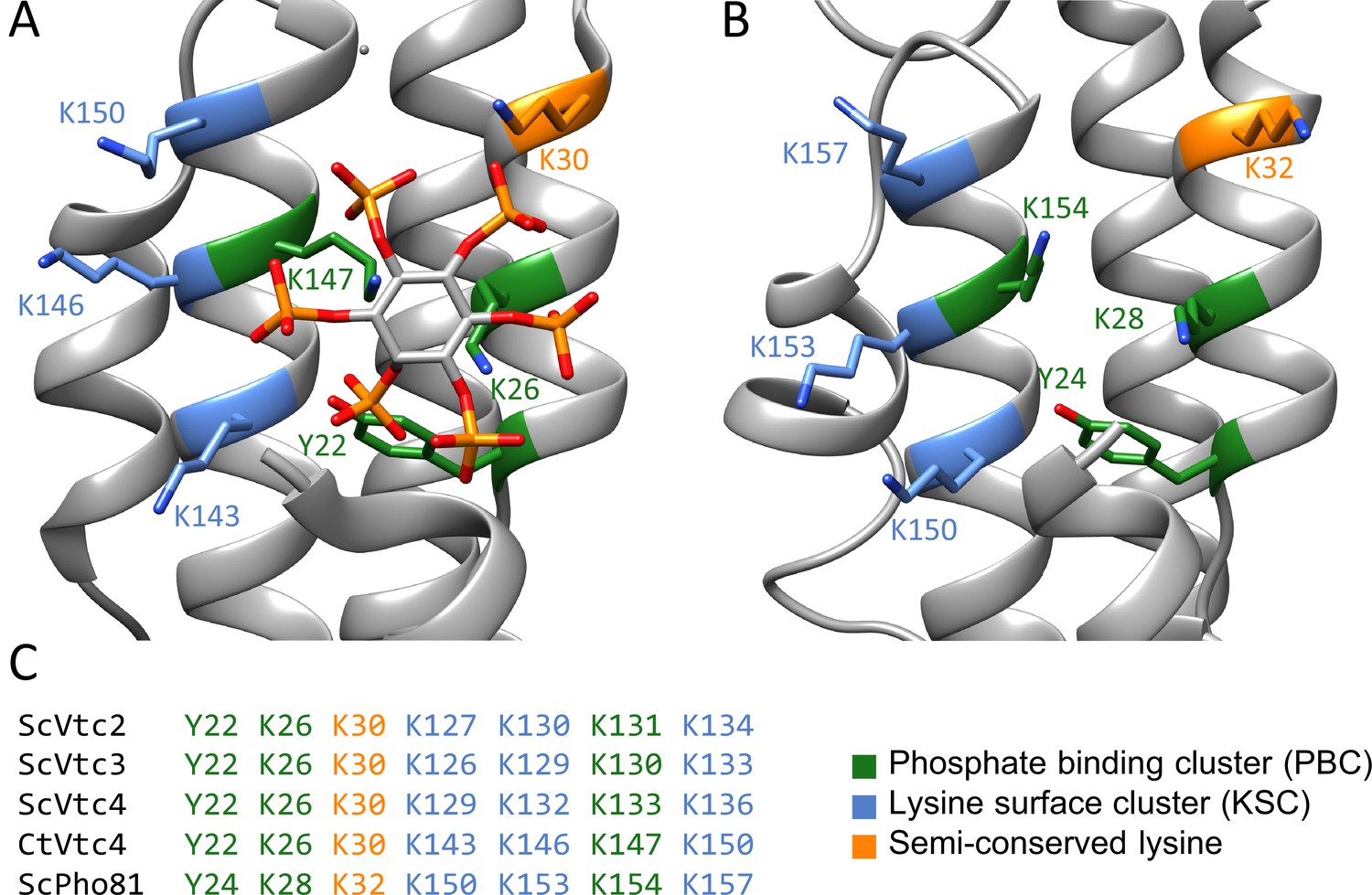
Myo-inositol polyphosphate-binding pocket of yeast SPX domains.
(A) Crystal structure close-up view of Chaetomium thermophilum Vtc4 SPX domain complexing IP6 (5IJP). (B) Structure prediction (generated with SWISS-MODEL) of the S. cerevisiae Pho81 SPX domain revealing the potential conserved PBC and KSC clusters. (C) Alignment of the conserved residues myo-inositol polyphosphate-binding motifs of Pho81 and of the different VTC subunits.
Figure 7
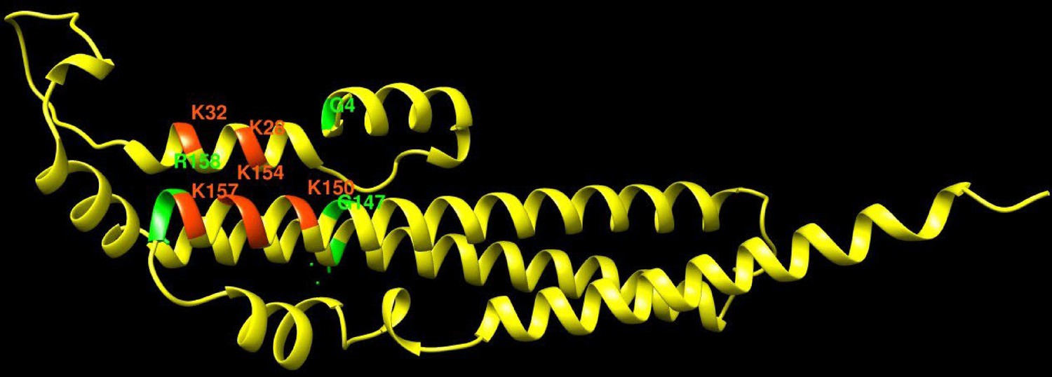
Pho81 residues leading to constitutive activation of the PHO pathway.
The image shows an Alphafold prediction of the Pho81 SPX domain (amino acids 1–215), taken from Alphafold database model AF-P17442-F1 (Varadi et al., 2022), in yellow. Basic residues of the putative inositol pyrophosphate-binding site have been identified by structure matching with the IP6-associated SPX domain of VTC4 from C. thermophilum (PDB 5IJP). They are labeled in red. Residues from random mutagenesis screens, which lead to constitutive activation of the PHO pathway, are labeled in green.
Figure 8
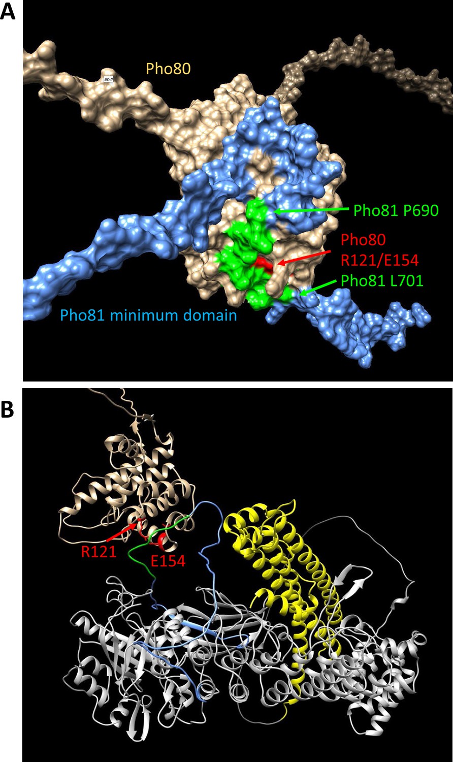
Structure predictions of the minimum domain in complex with Pho80.
Alphafold multimer v3 was used to generate the following structure predictions. (A) Surface representation of Pho80 in complex with a peptide corresponding to the minimum domain of Pho81 (residues 645–724), showing the embedding of critical residues (690–701) of the minimum domain in a groove of Pho80 that contains the salt bridge between R121 and E154. (B) Ribbon representation of Pho80 in complex with full-length Pho81. Coloring: Beige - Pho80; red - E154, R121 of Pho80; blue: Pho81 minimum domain; green - central region of the Pho81 minimum domain that is critical for Pho85-Pho80 inhibition (aa 690–701); yellow: SPX domain of Pho81; gray - rest of Pho81 without assigned function. The complete dataset for both predictions has been deposited at Figshare under the DOI 10.6084 /m9.figshare.c.6700281.
Figure 9
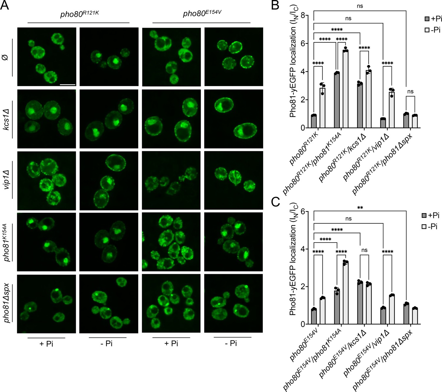
Localization of Pho81yEGFP in pho80R121K and pho80E154V loss-of-affinity mutants.
Cells were logarithmically grown in inorganic phosphate (Pi)-replete synthetic complete (SC) medium as in Figure 2A, washed, and incubated for further 4 hr in medium with 7.5 mM of Pi (+Pi), or in starvation medium (-Pi). (A) Pho81 imaging. Cells expressing the indicated variants of Pho81yEGFP and Pho80 from their genomic loci were imaged on a spinning disc microscope after 4 hr of growth in the presence of 7.5 mM Pi (+Pi), or after 4 hr of Pi starvation (- Pi). Scale bar = 5 μM. λex: 488 nm; λem: 500–530 nm. Note that the fluorescent dots visible in pho81Δspx cells are not nuclei because they are too small and at positions where nuclei are not found. Their location was not further investigated because it is not essential for this study. (B) Quantification of the nuclear localization of Pho81yEGFPin pho80R121K cells. Images from A were subjected to automated segmentation and quantification of the average fluorescence intensity in the nucleus and cytosol as in Figure 4B. 100–200 cells were quantified per sample. n=3 experiments. (C) Quantification of the nuclear localization of Pho81yEGFP in pho80E154V mutants was performed as in B. For B and C: ****p<0.0001; ***p<0.001; **p<0.01; *p<0.05; n.s. not significant, determined with Turkey’s test.
Figure 10
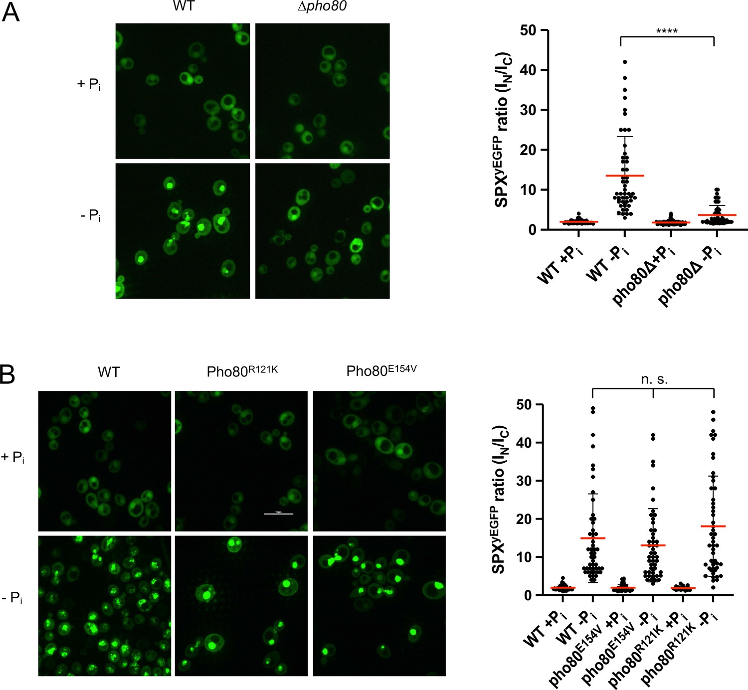
Recruitment of Pho81SPXyEGFP to the nucleus by Pho85-Pho80.
The indicated wildtype or isogenic mutant cells expressing the SPX domain of Pho81 as a yEGFP fusion (Pho81SPXyEGFP) from a centromeric plasmid under the ADH promotor were logarithmically grown in synthetic complete (SC), sedimented in a tabletop centrifuge, and transferred into SC with (+Pi) or without Pi (-Pi). After 3 hr of further cultivation, cells were imaged by spinning disc confocal microscopy. 50 cells per condition were analyzed from two independent experiments. Regions of interest were defined manually, and the fluorescence contained in the nuclei (IN) or the cytosol (IC) was integrated using ImageJ. Scale bar: 5 µm. Indicated pairwise differences were evaluated by a Mann-Whitney test. ****p<0.0001; n.s. not significant. (A) WT (BY4742) and isogenic pho80Δ cells expressing PHO81SPXyEGFP. (B) WT (BY4741) and isogenic cells expressing pho80R121K or pho80E154V from their genomic locus and Pho81SPXyEGFP from the plasmid.
Figure 11
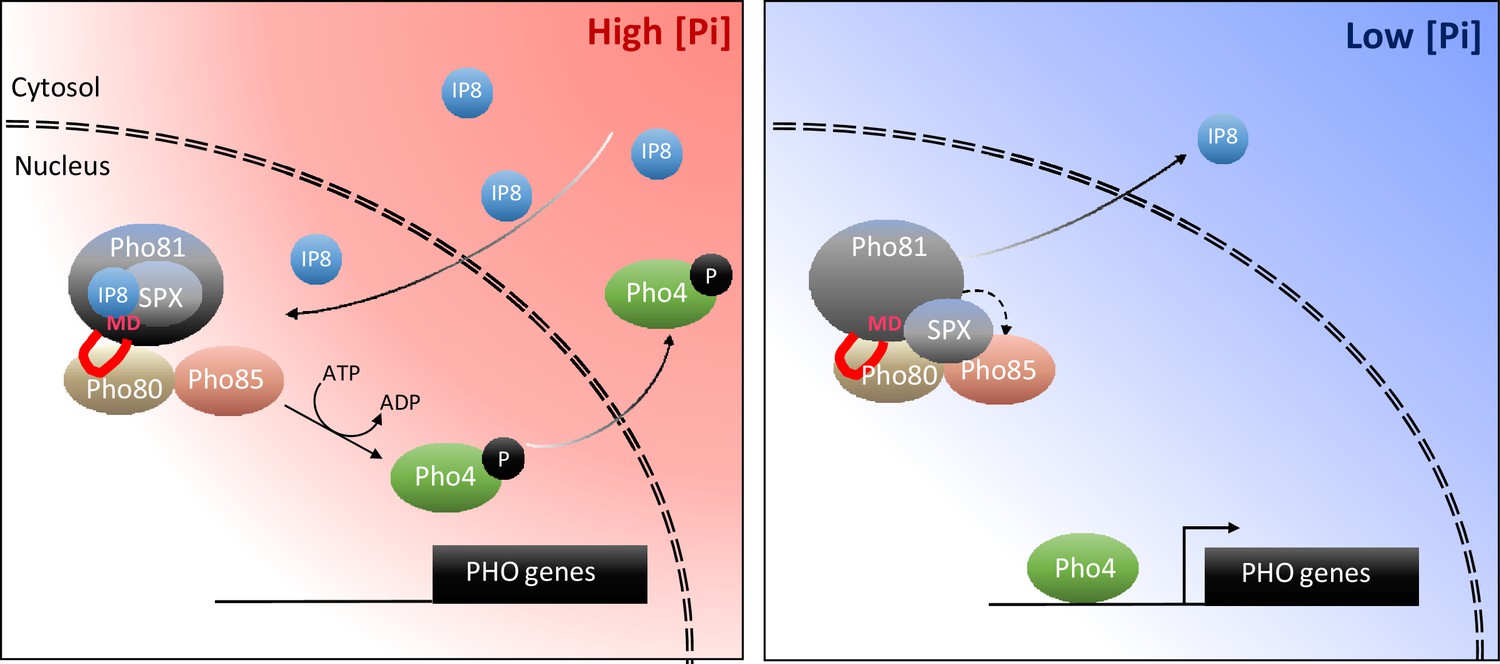
Working model on the control of the PHO pathway through 1,5-IP8 and Pho81.
At high Pi concentrations, inositol pyrophosphates accumulate. 1,5-IP8 binds the SPX domain of Pho81, which labilizes the interaction of Pho81 with Pho80 and prevents Pho81 from inhibiting Pho85-Pho80 kinase. In low Pi, 1,5-IP8 declines. Liberation of the SPX domain from this ligand allows this domain to interact with Pho85-Pho80. This, and the interaction of the minimum domain with a critical groove of Pho80, allow Pho81 to inhibit Pho80-85. The resulting dephosphorylation of Pho4 triggers its concentration in the nucleus and activation of the PHO pathway. This inhibition is reinforced by increased expression of PHO81, which itself is a PHO pathway-controlled gene.
Tables
Table 1
Parameters for MRM transitions.
| Compound | Precursor ion | Product ion | dwell | CE (V) | Cell acc (V) | Polarity |
|---|---|---|---|---|---|---|
| [13C6]IP8 | 411.9 | 362.9 | 80 | 10 | 1 | Negative |
| IP8 | 408.9 | 359.9 | 80 | 10 | 1 | Negative |
| [13C6]IP7 | 371.9 | 322.9 | 80 | 10 | 1 | Negative |
| IP7 | 368.9 | 319.9 | 80 | 10 | 1 | Negative |
| [13C6]IP6 | 331.9 | 486.9 | 80 | 17 | 1 | Negative |
| IP6 | 328.9 | 480.9 | 80 | 17 | 1 | Negative |
Additional files
-
MDAR checklist
- https://cdn.elifesciences.org/articles/87956/elife-87956-mdarchecklist1-v1.docx
-
Supplementary file 1
This supplementary file contains information on the genetic constitution of the strains used in this study, their origin, and on the plasmids and primers used to generate them.
(a) List of strains. (b) List of plasmids. (c) List of primers used for genetic manipulations.
- https://cdn.elifesciences.org/articles/87956/elife-87956-supp1-v1.docx
Download links
A two-part list of links to download the article, or parts of the article, in various formats.
Downloads (link to download the article as PDF)
Open citations (links to open the citations from this article in various online reference manager services)
Cite this article (links to download the citations from this article in formats compatible with various reference manager tools)
Inositol pyrophosphate dynamics reveals control of the yeast phosphate starvation program through 1,5-IP8 and the SPX domain of Pho81
eLife 12:RP87956.
https://doi.org/10.7554/eLife.87956.3


