Ulk4 promotes Shh signaling by regulating Stk36 ciliary localization and Gli2 phosphorylation
- Department of Molecular Biology, University of Texas Southwestern Medical Center, United States
- Department of Pharmacology, University of Texas Southwestern Medical Center, United States
Figures
Figure 1 with 2 supplements

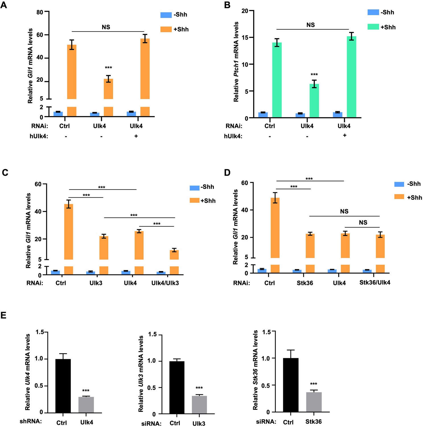
Ulk4 acts in conjunction with Stk36 to promote Shh-mediated Gli activation.
(A, B) Relative Gli1 (A) and Ptch1 (B) mRNA levels measured by RT-qPCR in NIH3T3 cells expressing the indicated shRNA and a human Ulk4 (hUlk4) lentiviral construct and treated with or without Shh-N. Of note, hUlk4 is insensitive to RNAi, which targets mouse Ulk4. (C, D) Relative Ptch1 mRNA levels measured by RT-qPCR in NIH3T3 cells treated with the indicated RNAi and with or without Shh-N. (E, E’) Western blot analysis (E) and quantification (E’) of Myc-Gli2 phosphorylation in NIH3T3 cells expressing the indicated shRNA and hUlk4 lentiviral construct and treated with or without Shh-N. (F–G’) Western blot analysis (F, G) and quantification (F’, G’) of Myc-Gli2 phosphorylation in NIH3T3 cells with the indicated RNAi in the presence or absence of Shh-N treatment. (H) Schematic diagram showing the functional relationship among Ulk4, Stk36, and Ulk3 in Shh-induced Gli activation. The cells (A–G) were first starved in serum-free medium for 12 hr, then cultured in the same medium with or without Shh-N fragment for another 12 hr before they are subjected to RNA preparation (A–D) or western blot analysis (E–G). Data in (A–D) are mean ± SD from three independent experiments. Data in (E’, F’, G’) are mean ± SD from two independent experiments. ***p<0.001 (Student’s t-test). NS, not significant.
-
Figure 1—source data 1
Source data for western blots in Figure 1E, F, and G.
- https://cdn.elifesciences.org/articles/88637/elife-88637-fig1-data1-v1.zip
Figure 1—figure supplement 1
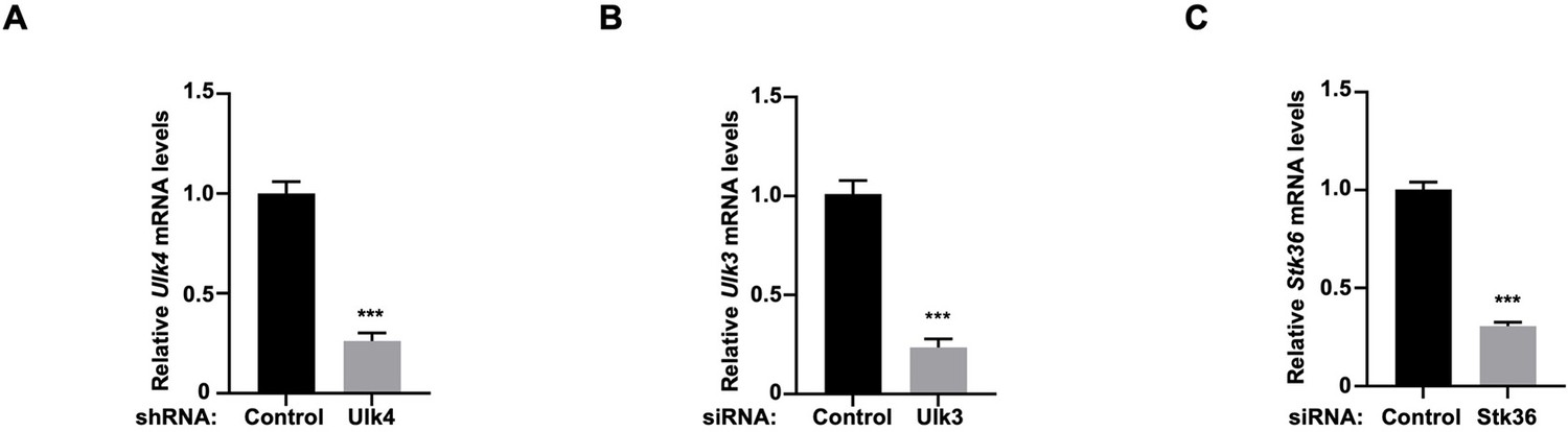
The knockdown efficiency of Ulk4, Ulk3, and Stk36 in NIH3T3 cells.
(A–C) Relative Ulk4 (A), Ulk3 (B), and Stk36 (C) mRNA levels measured by RT-qPCR in NIH3T3 cells treated with the indicated shRNA or siRNA. Data are mean ± SD from three independent experiments. ***p<0.001 (Student’s t-test).
Figure 1—figure supplement 2
Ulk4 acts in conjunction with Stk36 to regulate Shh-induced expression of Gli1 and Ptch1 in mouse embryonic fibroblasts (MEFs).
(A, B) Relative Gli1 (A) and Ptch1 (B) mRNA levels measured by RT-qPCR in MEF cells expressing the indicated shRNA and hUlk4 lentiviral constructs with or without Shh-N treatment. (C, D) Relative Gli1 mRNA levels measured by RT-qPCR in MEF cells with the indicated RNAi knockdown in the presence or absence of Shh-N treatment. (E) Relative Ulk4, Ulk3, and Stk36 mRNA levels measured by RT-qPCR in MEF cells treated with the indicated shRNA or siRNA. The cells were first starved in serum-free medium for 12 hr, then cultured in the same medium with or without Shh-N fragment for another 12 hr before they are subjected to RNA preparation. Data are mean ± SD from three independent experiments. ***p<0.001 (Student’s t-test).
Figure 2
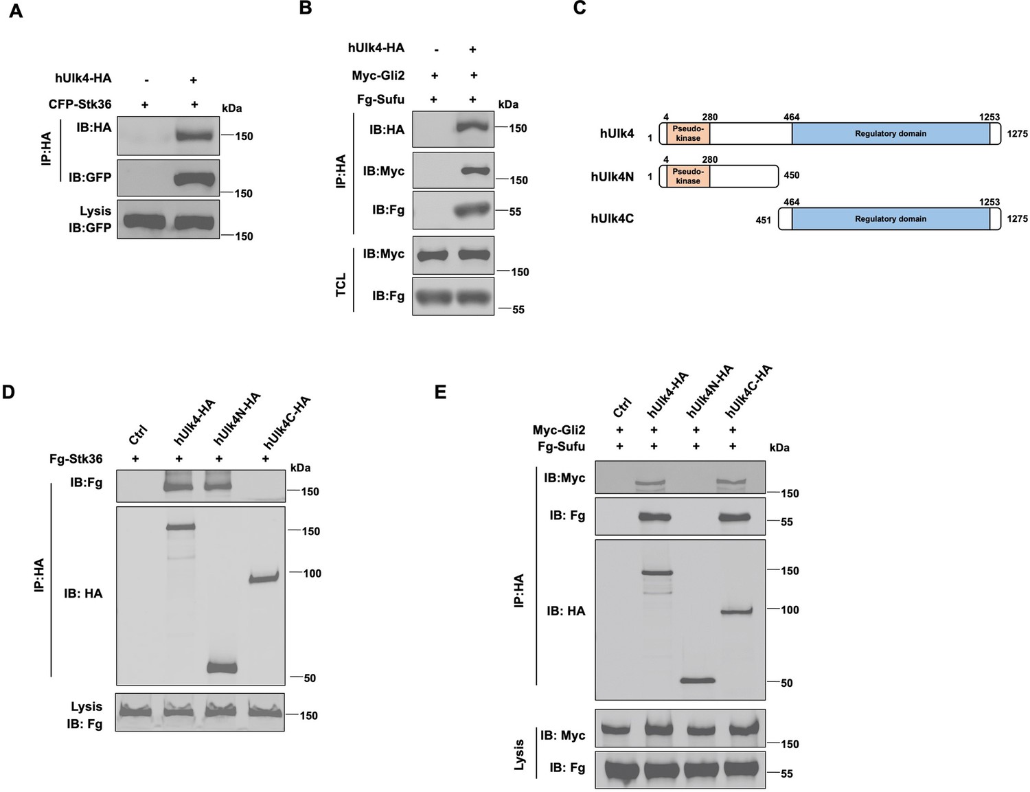
Ulk4 forms a complex with Stk36 and Gli2.
(A) Ulk4 forms a complex with Stk36. HEK293T cells were transfected with hUlk4-HA and CFP-Stk36 constructs, followed by co-immunoprecipitation (Co-IP) and western blot analyses with the indicated antibodies. (B) Ulk4 forms a complex with Gli2 and Sufu. HEK293T cells were transfected with hUlk4-HA, Myc-Gli2, and Fg-Sufu constructs, followed by Co-IP and western blot analyses with the indicated antibodies. (C) Ulk4 domain structure and deletion mutants used for Co-IP experiments. (D, E) Ulk4 interacted with Stk36 through its N-terminal domain (D) and with Myc-Gli2/Fg-Sufu through its C-terminal region (E). HEK293T cells were transfected with the indicated hUlk4, Fg-Stk36, Myc-Gli2, and Fg-Sufu constructs, followed by Co-IP and western blot analyses.
-
Figure 2—source data 1
Source data for western blots in Figure 2A, B, D, and E.
- https://cdn.elifesciences.org/articles/88637/elife-88637-fig2-data1-v1.zip
Figure 3

Ulk4 is dispensable for Stk36 kinase activation.
(A) In vitro kinase assay using the immunopurified Fg-Stk36 or Fg-Stk36/hUlk4-HA as kinase and GST-Gli2NWT or GST-Gli2NS230A as substrate. pIMAGO was used to detect GST-Gli2N phosphorylation. The levels of Fg-Stk36/hUlk4-HA and GST/GST-Gli2N were analyzed by western blot and Coomassie blue (CB) staining, respectively. (B) The schematic diagram showing the sequence alignment of the activation segments of Drosophila Fu, mouse Stk36 (mStk36), and human Stk36 (hStk36). The conserved Ser/Thr residues in the activation loop are color-coded in green. The residues used as antigen for developing pT158/pS159 are colored in yellow. Amino acid substitutions for Stk36 variants are indicated. (C) Shh induced Stk36 phosphorylation on T158/S159. NIH3T3 cells expressing CFP-Stk36 lentiviral construct were treated with or without Shh-N, followed by. IP with a GFP antibody and western blot analysis using an antibody against the phosphorylated T158/S159 (pT158/pS159). (D) Shh induced Stk36 phosphorylation on T158/S159 depending on Stk36 kinase activity. Ulk3 and Stk36 double knockout NIH3T3 cells expressing CFP-Stk36WT or CFP-Stk36KR (kinase dead) lentiviral construct were treated with or without Shh-N, followed by IP with a GFP antibody and western blot analysis with the pT158/pS159 antibody. (E, F) Western blot analysis (E) and quantification (F) of T158/S159 phosphorylation in Fg-Stk36WT, Fg-Stk36EE, or Fg-Stk36AA. HEK293T cells were transfected with the indicated Fg-Stk36 constructs, followed by IP with anti-Flag antibody and western blot analysis with the indicated antibodies. (G, H) Western blot analysis (G) and quantification (H) of Myc-Gli2 phosphorylation on S230/S232 by the wild type (WT) or indicated Stk36 variants in HEK293T cells. (I) Ulk4 is not required for Shh-induced Stk36 kinase activation. NIH3T3 cells expressing the indicated shRNA and CFP-Stk36 lentiviral construct were treated with or without Shh-N, followed by IP with a GFP antibody and western blot analysis with the pT158/pS159 antibody (right). The knockdown efficiency of Ulk4 was determined by RT-qPCR (left). Data are mean ± SD from three independent experiments. **p<0.01, ***p<0.001 (Student’s t-test).
-
Figure 3—source data 1
Source data for western blots in Figure 3A, C–E, G, and I.
- https://cdn.elifesciences.org/articles/88637/elife-88637-fig3-data1-v1.zip
Figure 4
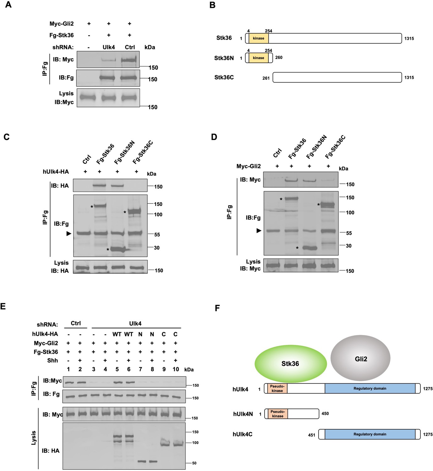
Ulk4 mediates the interaction between Stk36 and Gli2.
(A) Depletion of Ulk4 diminished the association between Stk36 and Gli2. HEK293T cells were transfected with the indicated shRNA and Fg-Stk36 and Myc-Gli2 constructs, followed by co-immunoprecipitation (Co-IP) and western blot analysis with the indicated antibodies. (B) Domain structure and deletion mutants of human Stk36 used for the Co-IP experiments. (C, D) Stk36 binds to Ulk4 and Gli2 through its N-terminal kinase domain. HEK293T cells were transfected with Ulk4-HA (C) or Myc-Gli2 (D) and the indicated Fg-Stk36 constructs, followed by Co-IP and western blot analyses with the indicated antibodies. Black triangles indicate the heavy chain of IgG, and asterisks indicate the protein bands produced by individual Stk36 constructs. (E) Both N- and C-terminal domains of Ulk4 are required for mediating Stk36/Gli2 association. NIH3T3 cell lines stably expressing control (Ctrl) or Ulk4 shRNA were infected with lentiviruses expressing the indicated hUlk4 constructs (diagrams shown on F, right), Fg-Stk36 and Myc-Gli2, and treated with or without the Shh-N, followed by Co-IP and western blot analysis with the indicated antibodies. (F) Schematic diagram showing the interaction relationship between Ulk4 and Stk36, Gli2.
-
Figure 4—source data 1
Source data for western blots in Figure 4A and C–E.
- https://cdn.elifesciences.org/articles/88637/elife-88637-fig4-data1-v1.zip
Figure 5 with 1 supplement

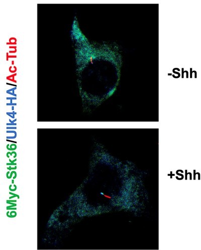
Shh stimulates ciliary tip accumulation of both Ulk4 and Stk36.
(A–D) Representative images of immunostaining (A, C) and quantification (B, D) of ciliary tip localized Myc-Stk36 (green in A) or Ulk4-HA (green in C) and Gli2 (blue in A and C) in NIH3T3 cells infected with the Myc-Stk36 or Ulk4-HA lentivirus and treated with or without Shh-N. Primary cilia are marked by acetylated tubulin (Ac-tub) staining (red in A and C). (E, F) Representative images of immunostaining (E) and quantification (F) of ciliary tip localized Myc-Stk36 (green) and Ulk4-HA (blue) in NIH3T3 cells co-infected with Myc-Stk36 and Ulk4-HA lentiviruses and treated with or without Shh-N. Primary cilia are marked by Ac-Tub staining (red). The cells are starved in serum-free medium for 12 hr to allow ciliation and cultured in the same medium with or without Shh-N fragment for another 12 hr before they are subjected into immunostaining assay. The intensity of ciliary-localized Myc-Stk36, Ulk4-HA, and Gli2 was measured by ImageJ. Twenty cells were randomly selected and counted for each group. Data are mean ± SD from three independent experiments. ***p<0.001 (Student’s t-test). Scale bars are 2 μM.
Figure 5—figure supplement 1
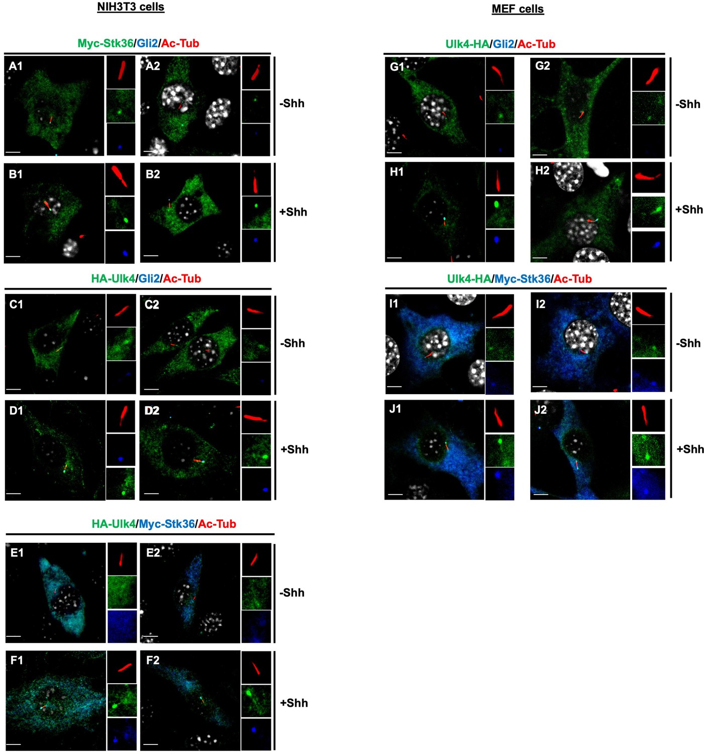
Shh stimulates ciliary tip accumulation of both Ulk4 and Stk36.
(A1–D2) Representative images of immunostaining ciliary tip localized Myc-Stk36 (green in A1–B2) or Ulk4-HA (green in C1–D2) and Gli2 (blue) in NIH3T3 cells infected with the Myc-Stk36 or Ulk4-HA lentivirus and treated with or without Shh-N. (E1–F2) Representative images of immunostaining of ciliary tip localized Myc-Stk36 (blue) and Ulk4-HA (green) in NIH3T3 cells co-infected with Myc-Stk36 and Ulk4-HA lentiviruses and treated with or without Shh-N. (G1–J2) Representative images of immunostaining of ciliary tip localized Myc-Stk36 (green in G1–H2) and Gli2 (blue in G1–H2) or Ulk4-HA (green in I1–J2) and Myc-Stk36 (blue in I1–J2) in mouse embryonic fibroblasts (MEFs) infected with either Myc-Stk36 (G1–H2) or both Myc-Stk36 and Ulk4-HA lentiviruses and treated with or without Shh-N. Primary cilia are marked by acetylated tubulin (Ac-tub) staining (red in A1–J2). Insets show the enlarged images of primary cilia with individual channels shown separately. Scale bars are 2 μM.
Figure 6
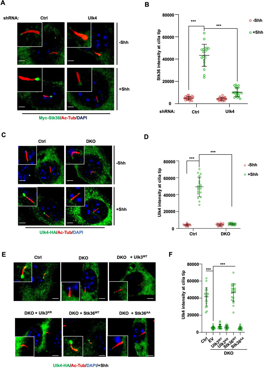
Stk36 and Ulk4 depend on each other for their ciliary tip accumulation.
(A, B) Representative images of immunostaining (A) and quantification (B) of ciliary tip localized Myc-Stk36 in NIH3T3 cells infected with the indicated shRNA and Myc-Stk36 lentivirus in the presence or absence of Shh-N. Primary cilia are marked by Ac-Tub staining (red); Myc-Stk36 is marked by Myc staining (green); nuclei are marked by DAPI (Blue). (C, D) Representative images of immunostaining (C) and quantification (D) of ciliary tip localized Ulk4-HA (green) in wild type (Ctrl) or Ulk3 and Stk36 double knockout (DKO) NIH3T3 cells treated with or without Shh-N. Primary cilia are marked by Ac-Tub staining (red) and nuclei by DAPI (blue). (E, F) Representative images of immunostaining (E) and quantification (F) of ciliary-localized Ulk4-HA (green) in Shh-N-treated control or DKO NIH3T3 cells infected with or without lentiviruses expressing the indicated Ulk3 or Stk36 constructs. The cells are starved in serum-free medium for 12 hr to allow ciliation and cultured in the same medium with or without Shh-N fragment for another 12 hr before they are subjected into immunostaining assay. The intensity of ciliary-localized Myc-Stk36 or Ulk4-HA was measured by ImageJ. Twenty cells were randomly selected and counted for each group. Data are mean ± SD from three independent experiments. ***p<0.001 (Student’s t-test). Scale bars are 2 μM.
Figure 7
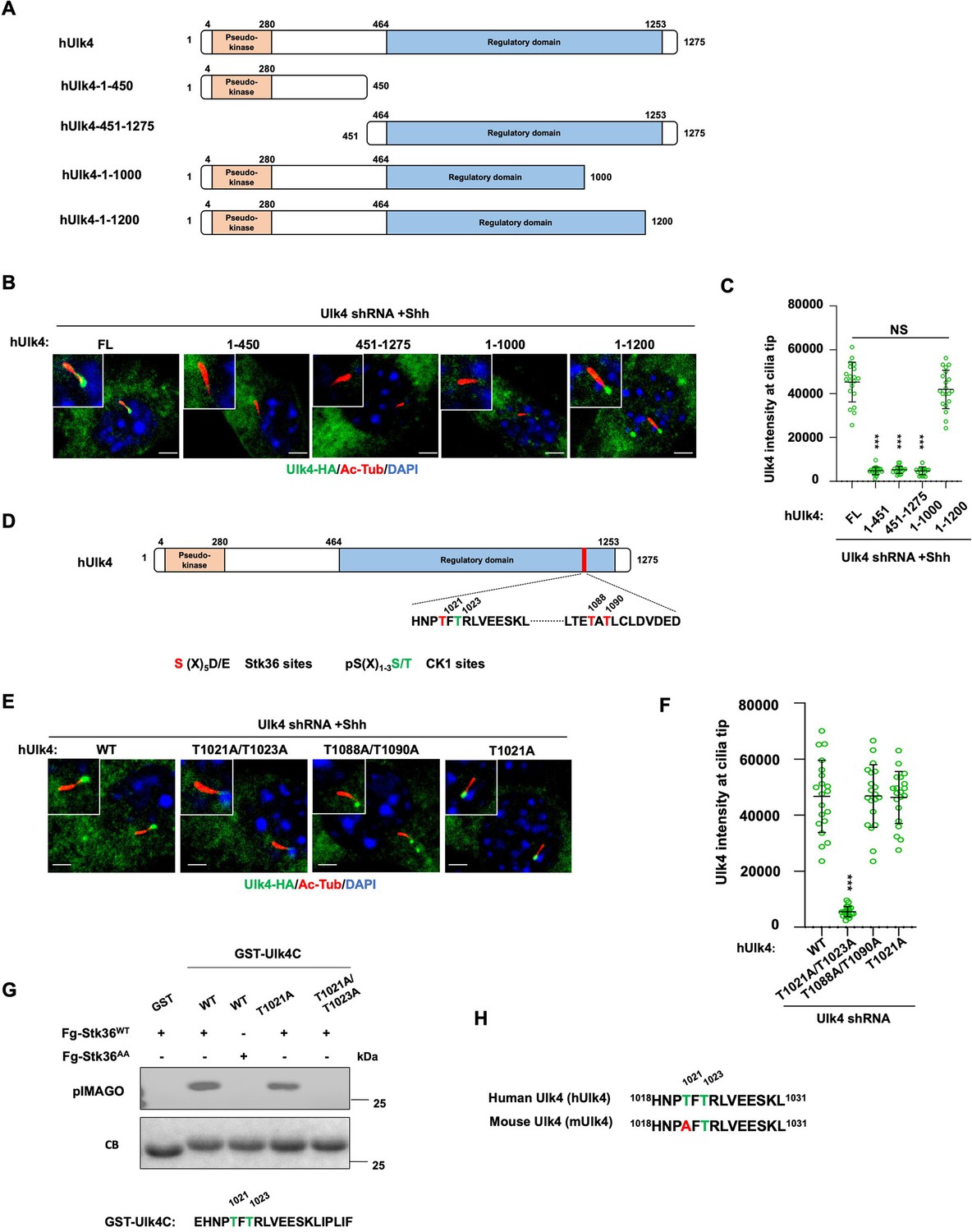
Ulk4 ciliary tip accumulation is promoted by its phosphorylation by Stk36.
(A) Diagrams of hUlk4 deletion constructs. (B, C) Representative images of immunostaining (B) and quantification (C) of ciliary tip localized full-length or truncated hUlk4-HA in Ulk4 knockdown cells treated with Shh-N. (D) Stk36 and CK1 phosphorylation sites in the C-terminal region of hUlk4. (E, F) Representative images of immunostaining (E) and quantification (F) of ciliary tip localized wild type (WT) or mutant hUlk4-HA baring the indicated amino acid substitutions in Ulk4 knockdown NIH3T3 cells treated with Shh-N. (G) In vitro kinase assay using the immunopurified Fg-Stk36WT or Fg-Stk36AA as kinase and the indicated GST-Ulk4C fusion proteins as substrates. Phosphorylation was detected by the pIMAGO system. (H) Schematic diagram showing the sequence alignment of C-terminal phosphorylation sites of mouse Ulk4 (mUlk4) and human Ulk4 (hUlk4). The Thr residues phosphorylated by Stk36 are color-coded in green. The cells are starved in serum-free medium for 12 hr to allow ciliation and cultured in the same medium with or without Shh-N fragment for another 12 hr before they are subjected into immunostaining assay. The intensity of ciliary-localized WT and mutant Ulk4-HA was measured by ImageJ. Twenty cells were randomly selected and counted for each group. Data are mean ± SD from three independent experiments. ***p<0.001 (Student’s t-test). Scale bars are 2 μM.
-
Figure 7—source data 1
Source data for western blots in Figure 7G.
- https://cdn.elifesciences.org/articles/88637/elife-88637-fig7-data1-v1.zip
Figure 8
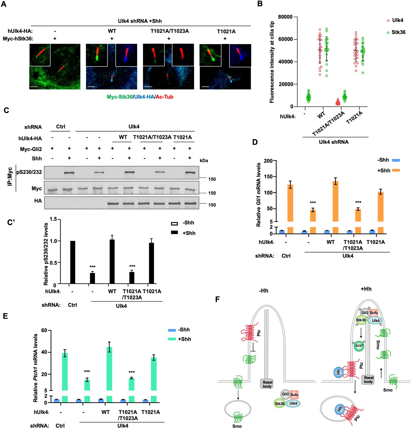
Ciliary tip localization of Ulk4 is required for Shh signal transduction.
(A, B) Representative images of immunostaining (A) and quantification (B) of ciliary tip localized Myc-Stk36 and hUlk4 in Ulk4-depleted NIH3T3 cells expressing the indicated hUlk4 constructs and treated with Shh. (C, C’) Western blot analysis (C) and quantification (C’) of Myc-Gli2 phosphorylation on S230/S232 in NIH3T3 cells expressing the indicated shRNA and hUlk4 lentiviral constructs and treated with or without Shh-N. Data are mean ± SD from two independent experiments. ***p<0.001 (Student’s t-test). (D, E) Relative Gli1 (D) and Ptch1 (E) mRNA levels in NIH3T3 cells expressing the indicated shRNA and hUlk4 lentiviral constructs treated with or without Shh-N. (F) Model for how Ulk4 participates in Shh signaling. See text for details. The cells are starved in serum-free medium for 12 hr to allow ciliation and cultured in the same medium with or without Shh-N fragment for another 12 hr before they are subjected into RNA preparation, western blot, and immunostaining assay. The intensity of ciliary-localized WT and mutant Ulk4-HA was measured by ImageJ. Twenty cells were randomly selected and counted for each group. Data are mean ± SD from three independent experiments. ***p<0.001 (Student’s t-test). Scale bars are 2 μM.
-
Figure 8—source data 1
Source data for western blots in Figure 8C.
- https://cdn.elifesciences.org/articles/88637/elife-88637-fig8-data1-v1.zip
Author response image 1
Additional files
Download links
A two-part list of links to download the article, or parts of the article, in various formats.
Downloads (link to download the article as PDF)
Open citations (links to open the citations from this article in various online reference manager services)
Cite this article (links to download the citations from this article in formats compatible with various reference manager tools)
Ulk4 promotes Shh signaling by regulating Stk36 ciliary localization and Gli2 phosphorylation
eLife 12:RP88637.
https://doi.org/10.7554/eLife.88637.3


