Chromosome-specific maturation of the epigenome in the Drosophila male germline
- Basic Sciences Division, Fred Hutchinson Cancer Center, United States
- Howard Hughes Medical Institute, United States
Figures
Figure 1
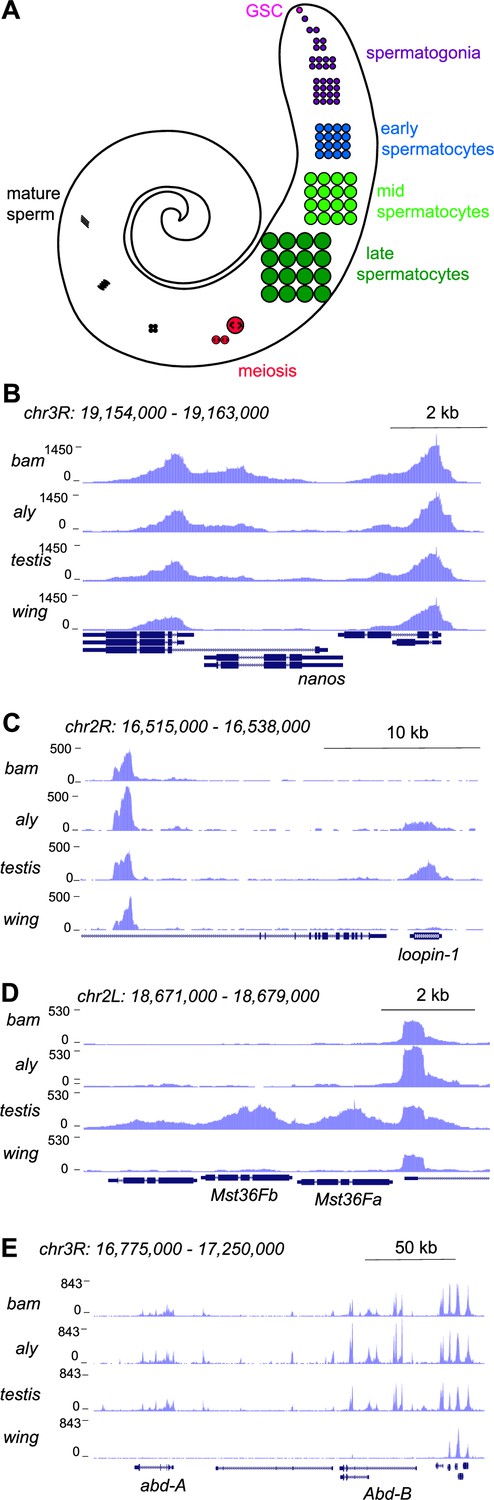
Profiling of the histone H3K4me2 modification in the Drosophila testis.
(A) Schematic of male germline stages in Drosophila. Germline stem cells (GSCs, fuschia) are located in the apical tip of the testis. After an asymmetric division a progeny spermatogonium (purple) undergoes four rounds of mitotic divisions. After one last S phase cells grow over ~3 days as spermatocytes (blue, light green, dark green) before meiosis (red). Post-meiotic differentiation produces mature sperm (black) with elongated nuclei. Somatic cell types of the testis are not shown. (B–E) Distribution of the H3K4me2 modification in testes from bam mutants, from aly mutants, from wildtype animals, and from wing imaginal discs. (B) H3K4me2 around the GSC-expressed nanos gene. Neighboring genes show peaks in all samples, while low signal across nanos is highest in testes from bam mutants, and apparent in all three testes samples. (C) H3K4me2 around the spermatocyte-expressed loopin-1 gene. H3K4me2 signal appears in aly mutant samples (which contain early spermatocytes) and reach high levels in wildtype testes (which include later stages of spermatogenesis). (D) H3K4me2 around the meiotically expressed genes Mst36Fa and Mst36Fb genes. Signal across these genes only appears in wildtype testes. (E) H3K4me2 around the abd-A and Abd-B genes, which are expressed in somatic cells of the testis.
Figure 2 with 1 supplement
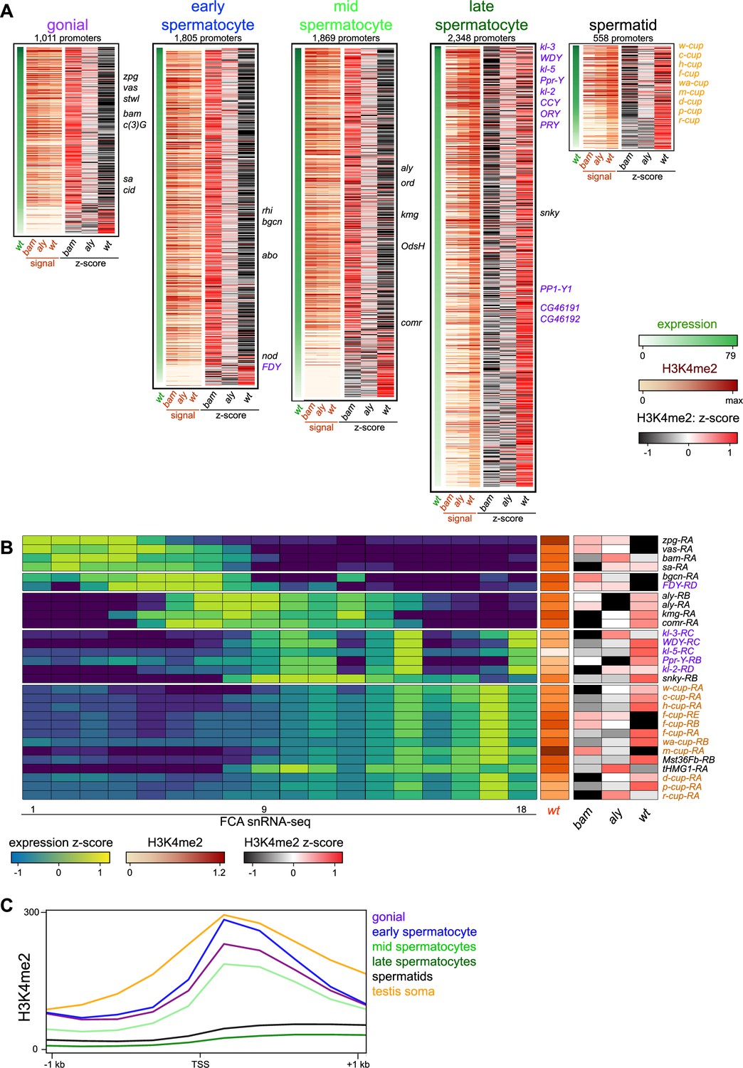
Changes in the histone H3K4me2 modification in germline-expressed genes.
H3K4me2 signal around gene promoters (–200 to +500 bp) with transcripts enriched in specific germline cell types. Transcript expression was derived from FCA snRNA-seq clustering (Raz et al., 2023). (A) Expression (snRNA-seq) in wildtype testes and H3K4me2 enrichment in bam mutant, in aly mutant, and in wildtype testes in germline stages. Z-scores for H3K4me2 signal were calculated between the three genotypes. Notable germline-enriched genes are indicated, including those expressed in GSCs, in meiosis, or for cell division (black), linked to the Y chromosome (purple), or expressed post-meiotically (orange). (B) Selected examples of promoters with germline-enriched expression. Expression z-scores from FCA snRNA-seq across 18 germline clusters (Raz et al., 2023) and H3K4me2 enrichment in bam mutant, in aly mutant, and in wildtype testes. (C) Distribution of H3K4me2 around promoters with germline-enriched expression. The testis somatic category comprises the top tercile of promoters with somatic cell-type expression in snRNA-seq data (Raz et al., 2023). Only promoters with no promoter of a second gene within 1 kb upstream are shown.
Figure 2—figure supplement 1
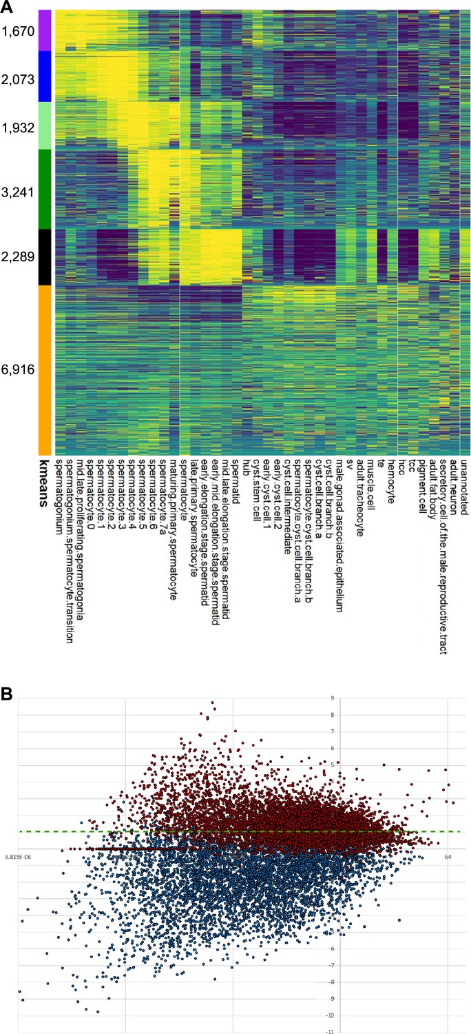
Genes with germline-enriched expression in testes.
(A) FCA clustering. Number of promoters in each cluster is marked on the left. (B) MA plot and threshold for germline-enriched genes (snRNA-seq data showing gexp/sexp with >1 cut-off). Red marks genes in germline-expressed clusters 1–5, and blue marks genes in testes somatic clusters 6–10. The threshold for assigning genes with germline-enriched expression is in green.
Figure 3 with 1 supplement
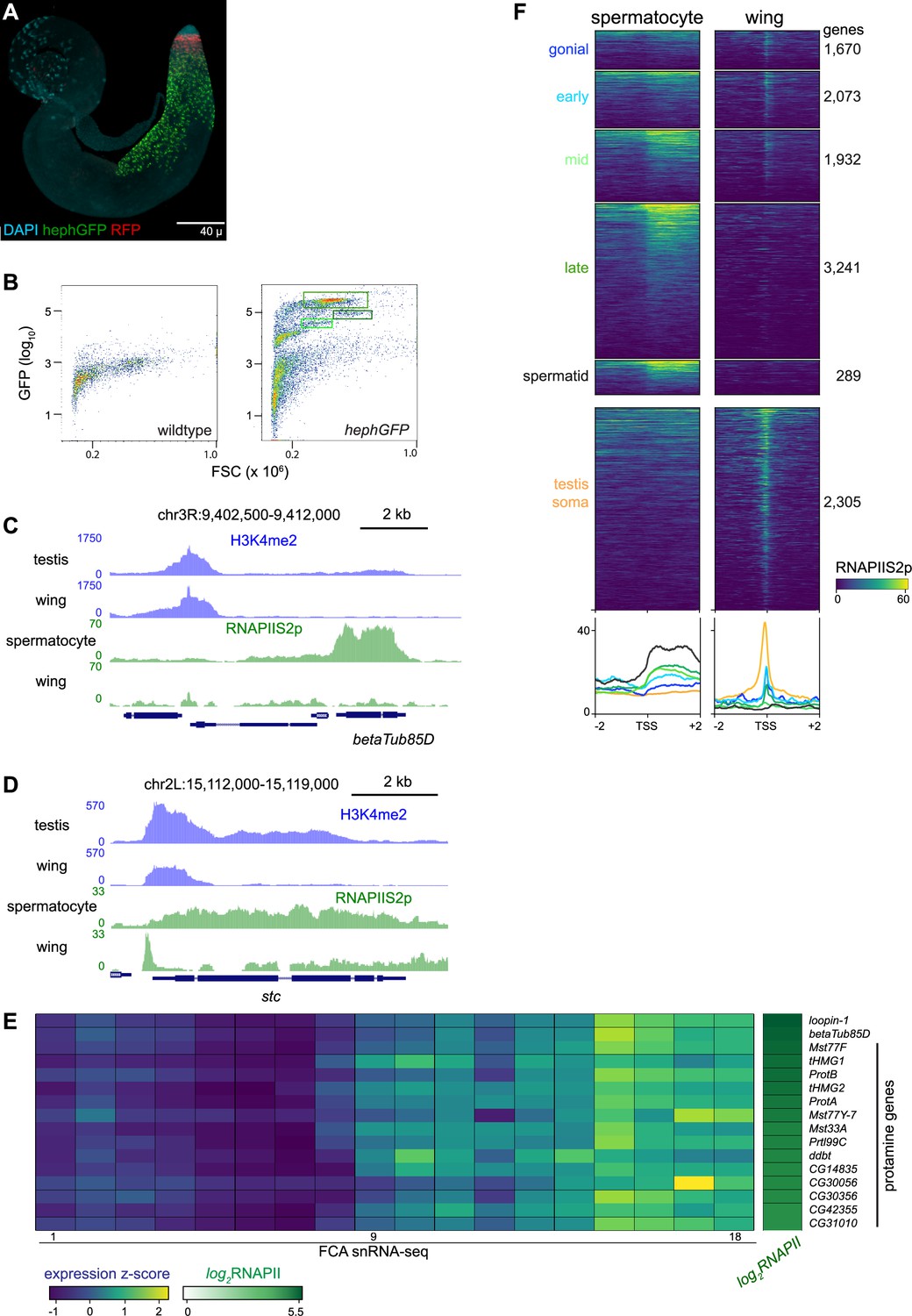
Profiling RNA polymerase II in isolated spermatocytes.
(A) An adult testis carrying a UASRFP construct induced by a bamGAL4 driver and a hephGFP construct. Gonial cells are labeled red while spermatocytes are labeled green with fluorescent proteins. (B) Fluorescence-activated cell sorting (FACS) plots of recorded events from dissociated testes for forward scatter (FSC) and GFP signal. Boxes indicate events collected for chromatin profiling. (C) Distribution of RNAPIIS2p at the spermatocyte-expressed betaTub85D gene in isolated spermatocytes and in wing imaginal discs. (D) Distribution of RNAPIIS2p at the broadly expressed stc gene in isolated spermatocytes and in wing imaginal discs. RNAPIIS2p is strongly localized at the stc promoter in wing imaginal discs, but more evenly distributed in spermatocytes. (E) Selected examples of genes with late germline expression and for protamines. Expression z-scores from FCA snRNA-seq across 18 germline clusters (Raz et al., 2023) and RNAPIIS2p enrichment in isolated spermatocytes. (F) Enrichment of RNAPIIS2p in isolated spermatocytes and in wing imaginal discs across genes with germline-enriched transcripts.
Figure 3—figure supplement 1

Distribution of elongating RNAPII in spermatocytes and in somatic cells.
Heatmap of RNAPIIS2p signal around gene TSSs in spermatocytes, in dissociated testes, and in wing imaginal discs.
Figure 4

Chromosomal distribution of RNA polymerase II and H4K16ac in isolated spermatocytes.
(A) CIRCOS plot of RNAPIIS2p across Drosophila chromosomes. The signal (black) in IgG controls, in wing imaginal discs, and in isolated spermatocytes is shown in internal rings, and the log2fold-change of signals between spermatocytes and wings is shown in the outer ring. (B) Enrichment of RNAPIIS2p across gene bodies in wing imaginal discs, in whole dissociated testes, and in isolated spermatocytes separated by chromosomal location. Scores are scaled to the median score on the second and third chromosomes. (C) CIRCOS plot of the dosage-compensation marker histone H4K16ac across Drosophila chromosomes. Signal (black) in IgG controls, in wing imaginal discs, and in isolated spermatocytes is shown in internal rings, and the log2fold-change of signals between spermatocytes and wings is shown in the outer ring.
Figure 5
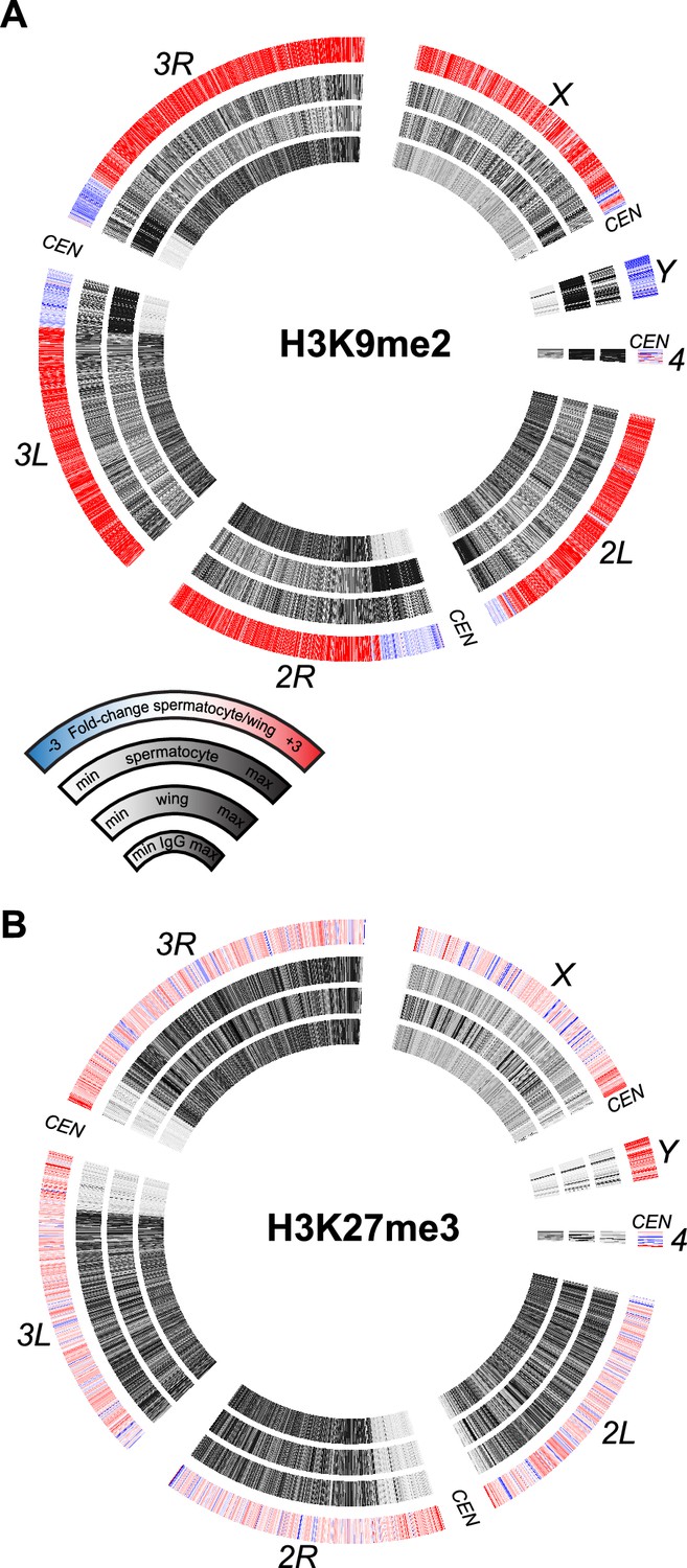
Chromosomal distribution of repressive histone modifications in isolated spermatocytes.
CIRCOS plots across Drosophila chromosomes show signal (black) in IgG controls, in wing imaginal discs, and in isolated spermatocytes in internal rings, and the log2fold-change of signals between spermatocytes and wings in the outer ring. (A) Distribution of the heterochromatin-silencing marker H3K9me2. (B) Distribution of the Polycomb-silencing marker H3K27me3.
Figure 6 with 1 supplement
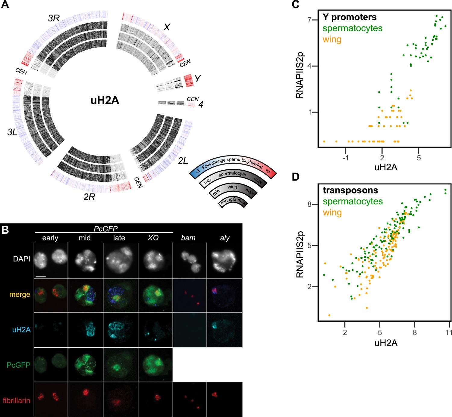
Chromosomal distribution of ubiquitinylated histone H2A in isolated spermatocytes.
(A) CIRCOS plots of uH2A across Drosophila chromosomes in IgG controls, in wing imaginal discs, and in isolated spermatocytes. (B) Immunostaining of uHA (blue) and the nucleolar marker fibrillarin (red) on germline nuclei. Early-, mid-, and late-spermatocyte stages were identified by Polycomb-GFP (PcGFP) (green) localization pattern in wildtype spermatocytes and in X/O spermatocytes. Testes from bam mutants contain gonial cells, while testes from aly mutants contain mostly early spermatocytes. All images are displayed at the same magnification, and a 100 µm scale bar is shown in the top left section. (C) Correspondence of uH2A and RNAPIIS2p signals around the promoters of genes on the Y chromosome in wing imaginal discs and in isolated spermatocytes. (D) Correspondence of uH2A and RNAPIIS2p signals across transposon consensus sequences in wing imaginal discs and in isolated spermatocytes.
Figure 6—figure supplement 1
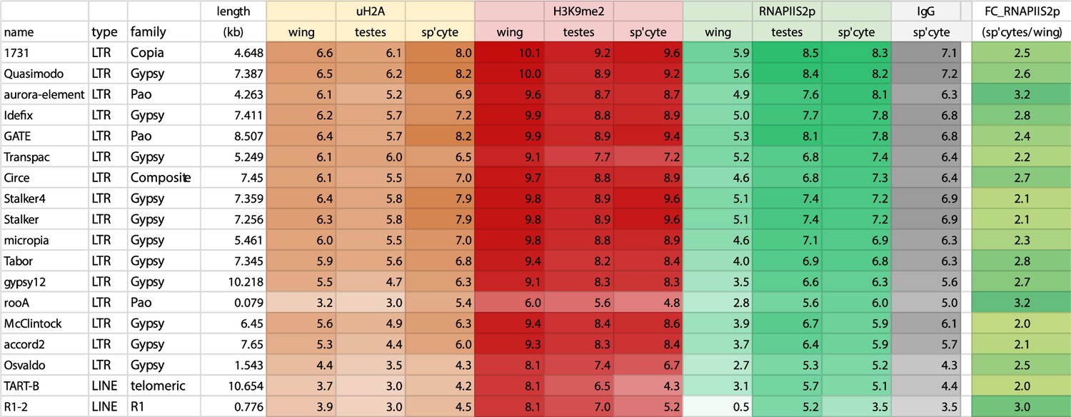
Chromatin features across transposon consensus sequences in spermatocytes and in somatic cells.
Selected transposons with increased RNAPIIS2p signal in spermatocytes versus wing imaginal discs (log2FC >2).
Tables
Key resources table
| Reagent type (species) or resource | Designation | Source or reference | Identifiers | Additional information |
|---|---|---|---|---|
| Antibody | Anti-H3-K4-dimethyl (rabbit monoclonal) | Epicypher | Cat. No. 13–0027 RRID: AB_3068541 | CUT&Tag (1:100) |
| Antibody | Anti-RNAPII-Serine-2-phosphorylation (rabbit monoclonal) | Cell Signalling Technology | Cat. No. 13499 RRID: AB_2798238 | CUT&Tag (1:100) |
| Antibody | Anti- H2A-K119-ubiquitinylation (rabbit monoclonal) | Cell Signalling Technology | Cat. No. 8240 RRID: AB_10891618 | CUT&Tag (1:100) IFF (1:100) |
| Antibody | Anti-H3-K9-dimethyl (mouse monoclonal) | EMD Millipore | Cat. No. 05–1249 RRID: AB_11210998 | CUT&Tag (1:100) |
| Antibody | Anti-H3-K27-trimethyl (rabbit monoclonal) | Cell Signalling Technology | Cat. No. 9733 RRID: AB_2616029 | CUT&Tag (1:100) |
| Antibody | Anti-H4-K16-acetyl (rabbit monoclonal) | Abcam | Cat. No. ab109463 RRID: AB_10858987 | CUT&Tag (1:100) |
| Antibody | ab5821 anti-fibrillarin (rabbit polyclonal) | Abcam | Cat. No. ab5821 RRID: AB_2105785 | IFF (1:100) |
| Genetic reagent (Drosophila melanogaster) | w1118 | Bloomington Drosophila Stock Center | BDSC:3605; RRID:BDSC_3605 | |
| Genetic reagent (D. melanogaster) | bamD86 | Bloomington Drosophila Stock Center | BDSC:5427; RRID:BDSC_5427 | |
| Genetic reagent (D. melanogaster) | Df(3R)FDD-0089346 | Bloomington Drosophila Stock Center | BDSC:27402; RRID:BDSC_27402 | Deficiency including bam |
| Genetic reagent (D. melanogaster) | aly1 | Bloomington Drosophila Stock Center | BDSC:1148; RRID:BDSC_1148 | |
| Genetic reagent (D. melanogaster) | Df(3L)BSC428 | Bloomington Drosophila Stock Center | BDSC:24932; RRID:BDSC_24932 | Deficiency including aly |
| Genetic reagent (D. melanogaster) | Heph-GFP | Bloomington Drosophila Stock Center | BDSC:51540; RRID:BDSC_51540 | P[PTT-GC]hephCC00664 |
| Genetic reagent (D. melanogaster) | bam-GAL4 | Bloomington Drosophila Stock Center | BDSC:80579; RRID:BDSC_80579 | P[bam-GAL4:VP16,w+]1 |
| Genetic reagent (D. melanogaster) | UAS-RFP | Bloomington Drosophila Stock Center | BDSC:30556; RRID:BDSC_30556 | P[UAS-RFP,w+]2 |
| Genetic reagent (D. melanogaster) | Pc-GFP | Bloomington Drosophila Stock Center | BDSC:9593; RRID:BDSC_9593 | P[Pc-eGFP,w+]3 |
| Biological sample (D. melanogaster) | Wing imaginal discs | This paper | Dissected tissue | |
| Biological sample (D. melanogaster) | Adult testes | This paper | Dissected tissue | |
| Biological sample (D. melanogaster) | Spermatocytes | This paper | Dissected tissue |
Additional files
-
Supplementary file 1
Samples and sequencing results.
(a) Sample IDs and sequencing results. (b) snRNA-seq gene expression scores and H3K4me2 enrichment at Drosophila promoters. This lists unique TSSs in the Flybase dm6 r6.31 assembly release and associated gene expression scores (derived from Raz et al., 2023) and H3K4me2 signal (in counts per million [CPM]) in a –200 to +500 bp window around each TSS. (c) Enrichment of RNAPIIS2p, H3K9me2, H3K27me3, and uH2A across Drosophila genes in dissociated testes, in isolated spermatocytes, and in wing imaginal discs. This lists unique genes in the Flybase dm6 r6.31 assembly release and associated gene expression scores (derived from Raz et al., 2023) and chromatin profiling signals (in counts per kilobase per million [CPKM]) across each gene length. (d) Enrichment of RNAPIIS2p, H3K9me2, H3K27me3, and uH2A across consensus transposon sequences in dissociated testes, in isolated spermatocytes, and in wing imaginal discs. This lists a subset of transposon consensus sequences (https://github.com/bergmanlab/drosophila-transposons/blob/master/misc/D_mel_transposon_sequence_set.fa) and associated chromatin profiling signals (in CPKM) across each consensus length.
- https://cdn.elifesciences.org/articles/89373/elife-89373-supp1-v1.xlsx
-
MDAR checklist
- https://cdn.elifesciences.org/articles/89373/elife-89373-mdarchecklist1-v1.docx
Download links
A two-part list of links to download the article, or parts of the article, in various formats.
Downloads (link to download the article as PDF)
Open citations (links to open the citations from this article in various online reference manager services)
Cite this article (links to download the citations from this article in formats compatible with various reference manager tools)
Chromosome-specific maturation of the epigenome in the Drosophila male germline
eLife 12:RP89373.
https://doi.org/10.7554/eLife.89373.3


