Hundreds of myosin 10s are pushed to the tips of filopodia and could cause traffic jams on actin
- Department of Biochemistry and Molecular Biology, University of Chicago, United States
- Department of Biochemistry and Molecular Biology, The Institute for Biophysical Dynamics, University of Chicago, United States
Figures
Figure 1 with 3 supplements
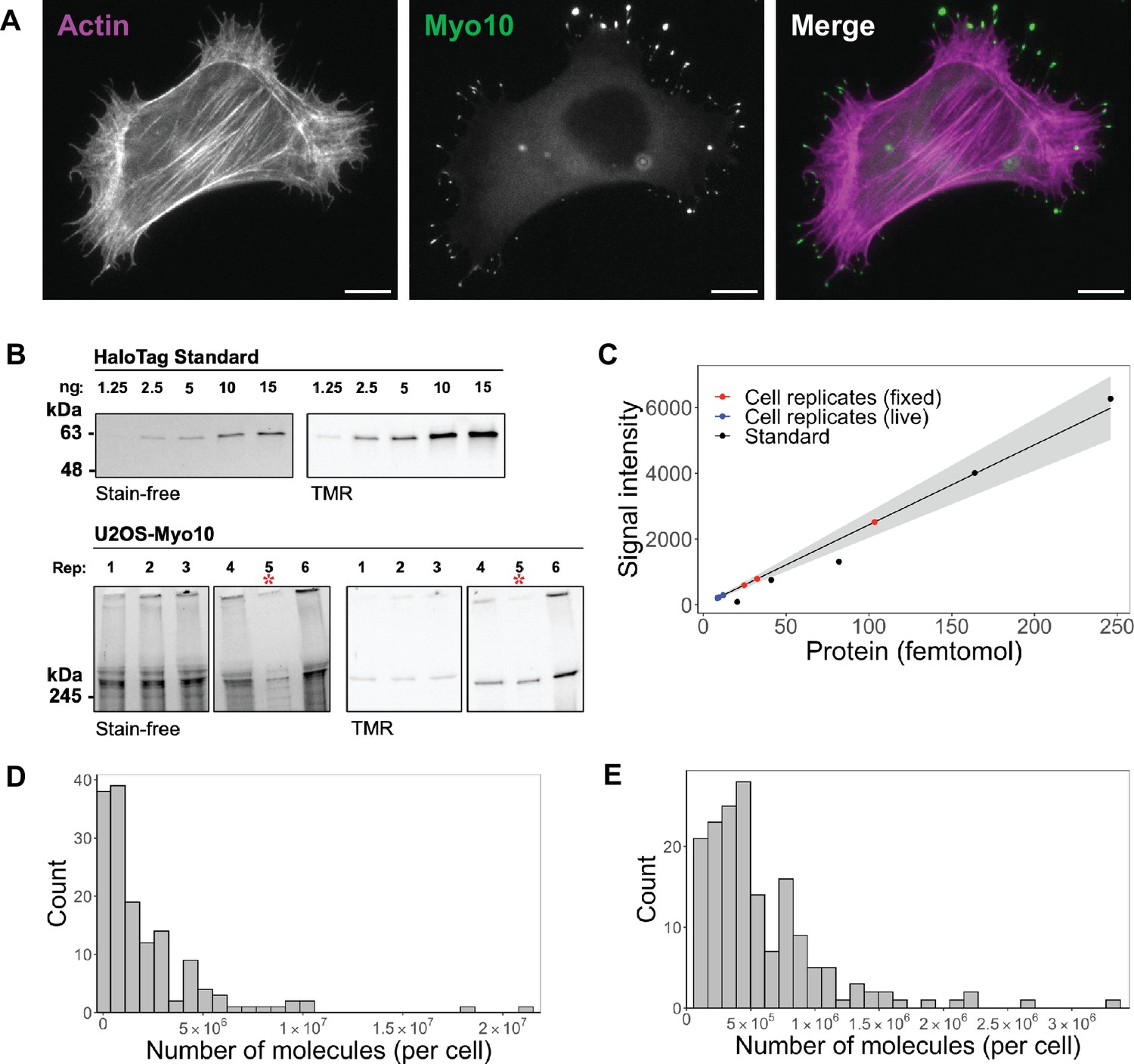
Hundreds of thousands of Myo10 monomer molecules found in Myo10-transfected U2OS cells.
(A) Epifluorescence image of exogenously expressed HaloTag-Myo10 in U2OS cells. Actin is labeled with phalloidin-AF633 (magenta). Myo10 is labeled with HaloTag ligand-TMR (green). Scale bar = 10 µm. (B) The top SDS-PAGE lanes show the indicated quantity (in ng) of HaloTag standard protein. The bottom SDS-PAGE lanes from the same gel show 50,000 cells from six separate U2OS transient transfections (except bioreplicate 5, indicated by a red asterisk (*), has 10,000 cells). Bioreplicates 1, 2, and 3 are from live-cell analysis, while bioreplicates 4, 5, and 6 are from fixed-cell analysis. Stain-free shows total protein signal, while TMR shows only TMR-HaloTag-Myo10 signal. Signal was integrated for full-length Myo10 (at ~250 kDa) and any Myo10 aggregated in the wells at the top of the panels. (C) Standard curve for TMR fluorescence signal of HaloTag standard protein (black dots) compared to signal from HaloTag-Myo10 U2OS cells (red dots = fixed-cell experiments, blue dots = live-cell experiments). The linear fit is y=24.32x, where the y-intercept is set to 0. R2=0.98. Standard error of slope = 1.42. Gray shading indicates the 95% confidence interval. (D) Distribution of the number of Myo10 molecules per fixed cell (N=150 cell images; min = 39,000, 95% CI: 33,000–46,000; median = 1,000,000, 95% CI: 870,000–1,200,000; max = 21,000,000, 95% CI: 18,000,000–25,000,000; bins = 30). (E) Distribution of the number of Myo10 molecules per live cell as determined by quantification of the first frame of N=168 cell movies (min = 79,000, 95% CI: 67,000–92,000; median = 450,000, 95% CI: 370,000–520,000; max = 3,300,000, 95% CI: 2,800,000–3,800,000; bins = 30).
-
Figure 1—source data 1
Total Myo10 cell signal from all three live-cell bioreplicates.
- https://cdn.elifesciences.org/articles/90603/elife-90603-fig1-data1-v1.zip
-
Figure 1—source data 2
Uncropped and labeled gels for Figure 1.
- https://cdn.elifesciences.org/articles/90603/elife-90603-fig1-data2-v1.zip
-
Figure 1—source data 3
Raw unedited gels for Figure 1.
- https://cdn.elifesciences.org/articles/90603/elife-90603-fig1-data3-v1.zip
Figure 1—figure supplement 1
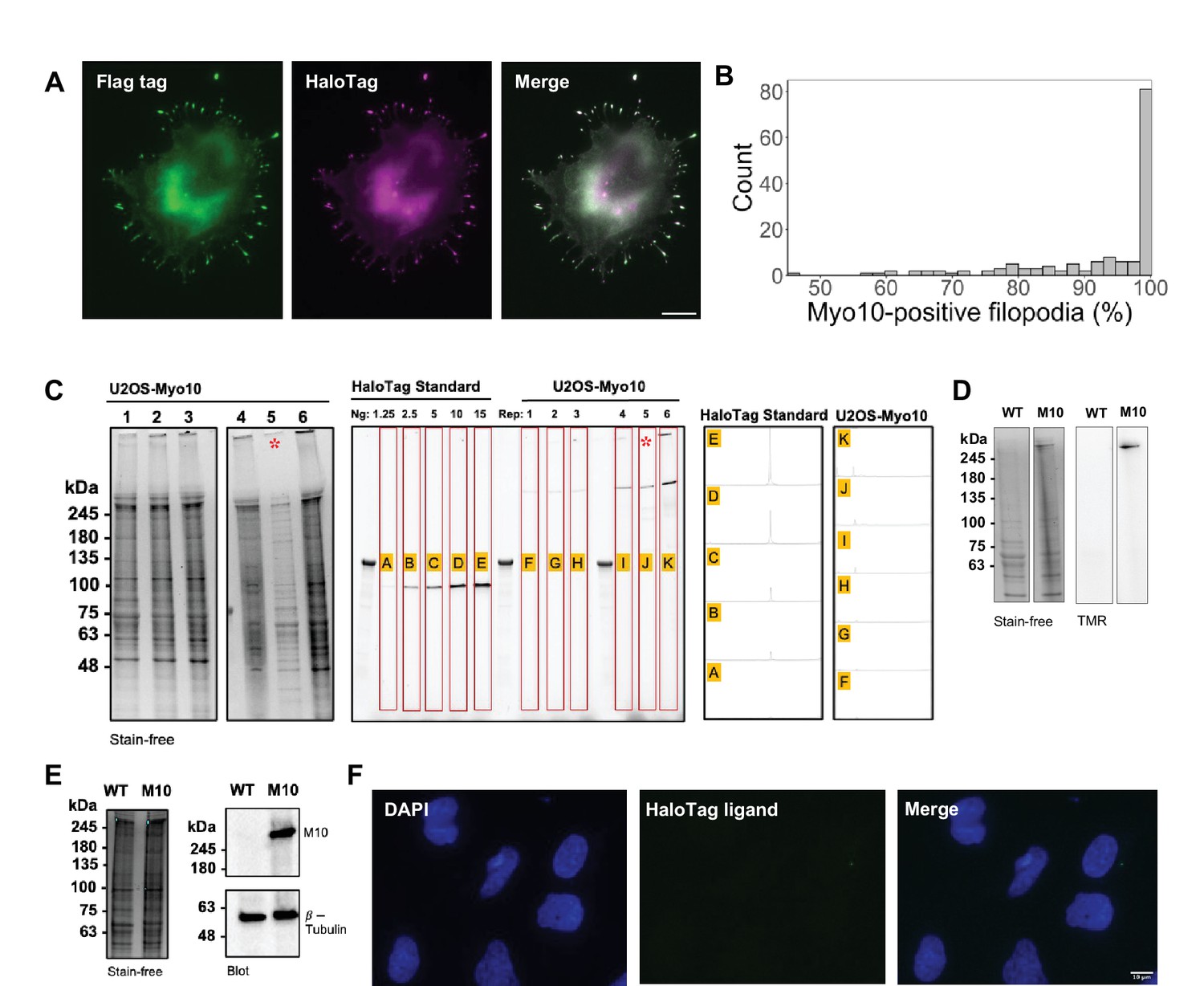
HaloTag ligand-TMR specifically labels HaloTag-Myo10 and reliably reflects the total Myo10 present in U2OS cells.
(A) Epifluorescence image of exogenously expressed HaloTag-Myo10 in U2OS cells. Flag-tag was labeled with monoclonal anti-Flag M2 antibody (Sigma, F1804) and goat anti-mouse AF647 (green). HaloTag was labeled with HaloTag ligand-TMR (magenta). Merged image shows high colocalization between labeling of both tags. Scale bar = 10 µm. (B) In fixed cells: distribution of the percent of total filopodia (manually counted) per cell that were Myo10-positive as determined by quantification of 150 cell images. Min = 46.67%, median = 92.35%, max = 100%. (C) Left SDS-PAGE lanes: full-length image of Figure 1B. 50,000 cells from six separate U2OS transient transfections. Bioreplicate 5 has 10,000 cells loaded, indicated by a red asterisk (*). Bioreplicates 1, 2, and 3: for live-cell analysis. Bioreplicates 4, 5, and 6: for fixed-cell analysis. Stain-free shows total protein signal, and TMR illumination shows only HaloTag-Myo10 signal. Right SDS-PAGE lanes: HaloTag standard and U2OS lysates were imaged on the same gel. Signal was integrated for full-length Myo10 (at ~250 kDa) and Myo10 aggregated in the wells. Integrations of band intensity was done using ImageJ’s Gel Analysis plug-in. (D) SDS-PAGE gel of wildtype U2OS (WT) labeled with HaloTag ligand-TMR vs. HaloTag-Myo10 overexpressed in U2OS cells (M10). 50,000 cells of each sample were loaded onto the same gel. Stain-free shows total protein signal, and TMR illumination shows only HaloTag-Myo10 signal. Negative control WT indicates no nonspecific HaloTag ligand-TMR labeling. (E) Left: SDS-PAGE gel of wildtype U2OS (WT) labeled with HaloTag ligand-TMR vs. HaloTag-Myo10 overexpressed in U2OS cells (M10). 50,000 cells of each sample loaded. Right: Immunoblot for Myo10 (NBP1-87748) and β-tubulin (Invitrogen 22833). (F) Epifluorescence image of wildtype U2OS cells labeled with HaloTag ligand-TMR. Negative control microscopy sample indicates no nonspecific HaloTag ligand-TMR labeling.
-
Figure 1—figure supplement 1—source data 1
Uncropped and labeled gels for Figure 1—figure supplement 1.
- https://cdn.elifesciences.org/articles/90603/elife-90603-fig1-figsupp1-data1-v1.zip
-
Figure 1—figure supplement 1—source data 2
Raw unedited gels for Figure 1—figure supplement 1.
- https://cdn.elifesciences.org/articles/90603/elife-90603-fig1-figsupp1-data2-v1.zip
Figure 1—figure supplement 2
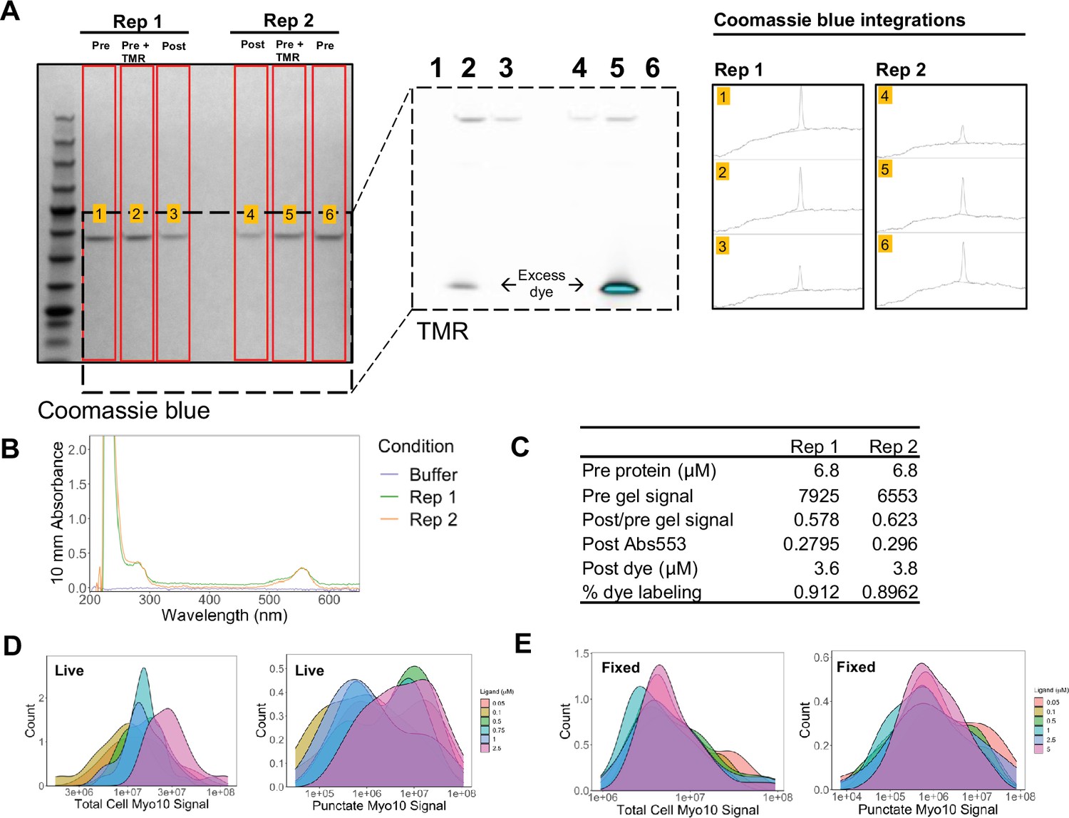
HaloTag ligand-TMR robustly labels HaloTag-Myo10.
(A) Left SDS-PAGE lanes: Coomassie blue shows HaloTag standard protein samples prepared to test HaloTag ligand-TMR labeling efficiency. ‘Pre’ refers to protein prior to exposure with TMR ligand and bio-beads. ‘Pre+TMR’ refers to the protein after TMR ligand labeling prior to bio-beads. ‘Post’ refers to protein after exposure with TMR ligand and bio-beads. The zoomed-in inset (middle) shows TMR illumination. Dye at the bottom of the gel indicates TMR ligand was added in excess (cyan indicates detector saturation). Rightmost: Integrations of Coomassie blue band intensities were done using ImageJ’s Gel Analysis plug-in. To account for protein that may have nonspecifically bound to the beads, the signal intensities of pre- and post-bead gel bands were compared. (B) The absorbance curve of the post-bead HaloTag standard protein samples. (C) Values used to determine HaloTag ligand-TMR labeling efficiency of the HaloTag standard protein. All Myo10 molecule measurements were adjusted assuming 90% HaloTag ligand-TMR labeling efficiency. (D) In live cells: effect of HaloTag ligand-TMR concentrations on the total intracellular Myo10 signal (left) and total punctate Myo10 signal (right). There was higher, uneven background in the higher HaloTag ligand-TMR live samples that falsely inflated final Myo10 intracellular signal. Plotting only punctate Myo10 signal highlights that signal saturation occurs at 0.5 µM in living cells. (E) In fixed cells: effect of TMR-HaloTag ligand concentrations on the total intracellular Myo10 signal (left) and total punctate Myo10 signal (right). Signal saturation occurs at 0.5 µM in fixed cells.
-
Figure 1—figure supplement 2—source data 1
Uncropped and labeled gels for Figure 1—figure supplement 2.
- https://cdn.elifesciences.org/articles/90603/elife-90603-fig1-figsupp2-data1-v1.zip
-
Figure 1—figure supplement 2—source data 2
Raw unedited gels for Figure 1—figure supplement 2.
- https://cdn.elifesciences.org/articles/90603/elife-90603-fig1-figsupp2-data2-v1.zip
Figure 1—figure supplement 3
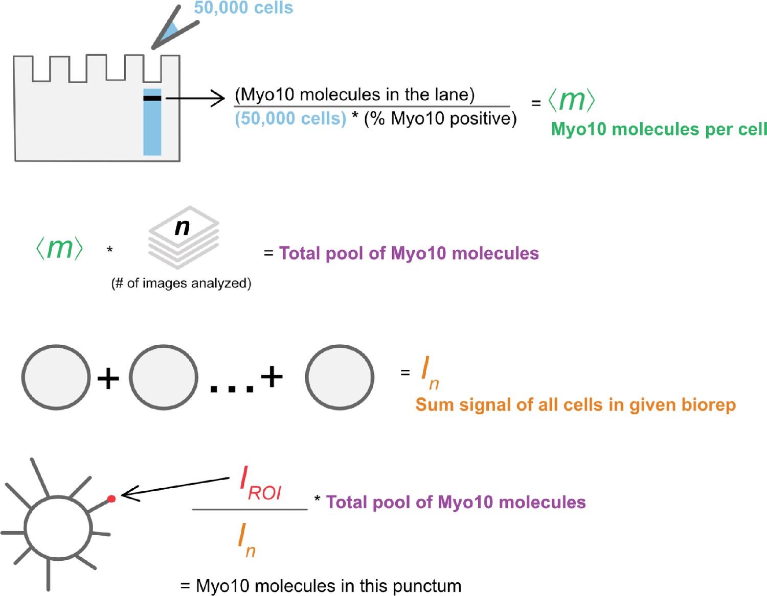
Calculations to convert signal from SDS-PAGE and epifluorescence microscopy to Myo10 molecule counts.
Figure 2 with 2 supplements
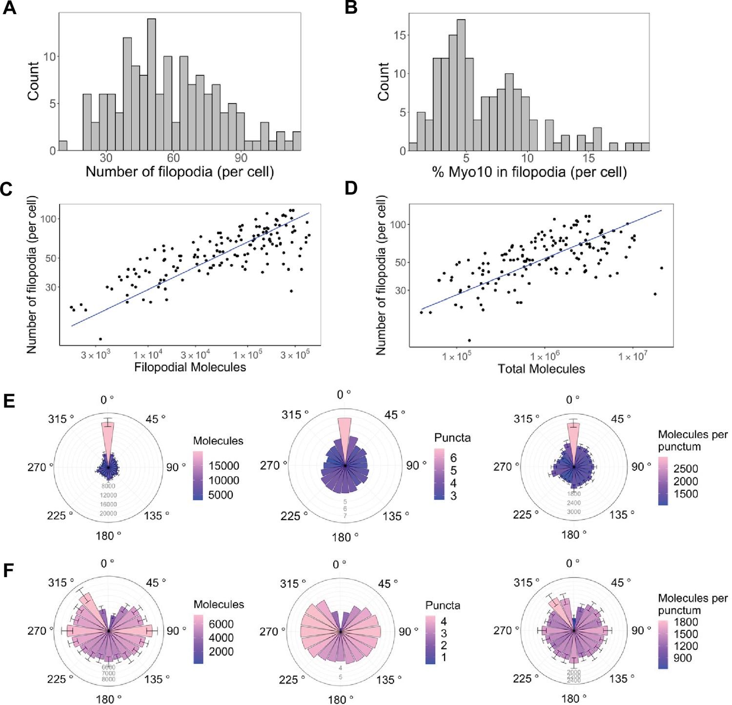
Only a small portion of intracellular Myo10 is activated and enters filopodia, and Myo10 is unevenly distributed around the cell.
The following values are from fixed-cell images. (A) Distribution of the number of Myo10-positive filopodia per cell (N=8733 Myo10-positive filopodia, 150 cells, min = 12, median = 55, max = 116). (B) Distribution of the percent of Myo10 localized in the filopodia per cell (N=150 cells, min = 0.86%, median = 5.35%, max = 19.87%). (C) Correlation between number of Myo10-positive filopodia in a cell and the filopodial number of Myo10 molecules in the cell. The slope of power law function is 0.36. (D) Correlation between number of Myo10-positive filopodia in a cell and the total number of Myo10 molecules in the cell. The slope of power law function is 0.29. (E) Spatial correlation of Myo10-rich regions of the cell edge. Each cell was divided into 20 angular sections, and the section with the most molecules was aligned to 0°. Section quantities were then averaged across cells. Molecules, puncta, and molecules per punctum are shown. (F) Spatial correlation of Myo10-poor regions of the cell edge. As in E, but the section with the fewest molecules was aligned to 0° for each cell’s rose plot. If >1 section contained no Myo10, a randomly selected empty Myo10 section was aligned to 0°. Error bars in E, F are the standard error of the mean for 500 bootstrapped samples of the 150 cells. Note the correlation of both molecules and puncta at opposite ends of cells (0°, 180°), and the anticorrelation with the two sides (90°, 270°).
-
Figure 2—source data 1
Filopodial Myo10 signal, cell body Myo10 signal, and number of filopodia for all three fixed-cell bioreplicates.
- https://cdn.elifesciences.org/articles/90603/elife-90603-fig2-data1-v1.xlsx
-
Figure 2—source data 2
Data for spatial correlation of Myo10-rich regions of the cell edge for all three fixed-cell bioreplicates.
- https://cdn.elifesciences.org/articles/90603/elife-90603-fig2-data2-v1.zip
-
Figure 2—source data 3
Data for spatial correlation of Myo10-poor regions of the cell edge for all three fixed-cell bioreplicates.
- https://cdn.elifesciences.org/articles/90603/elife-90603-fig2-data3-v1.zip
Figure 2—figure supplement 1
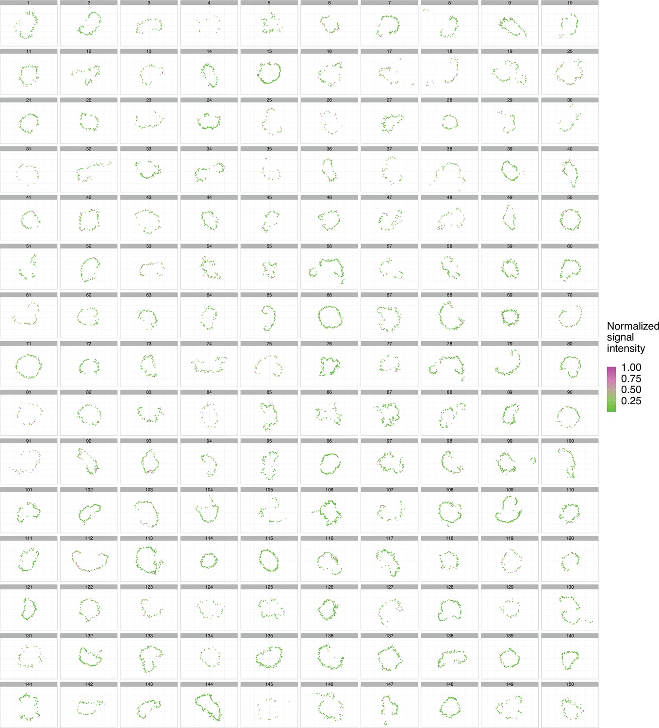
Myo10 is irregularly distributed across the plasma membrane.
In fixed cells: filopodial Myo10 signal distribution in 150 cells. The center of each Myo10 punctum was plotted after two-dimensional PCA. Each Myo10 punctum is colored by signal intensity (log-transformed), where magenta = high signal and green = low signal. Cells display periodic stretches of higher Myo10 signal along the cell membrane. Even cells with sparse Myo10 show uneven Myo10 filopodial distribution. Some cells even have membrane patches of no Myo10 signal.
Figure 2—figure supplement 2
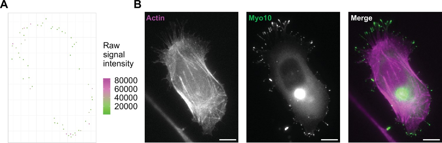
Example cell displaying Myo10 density pattern.
(A) Cell 14 from Figure 2—figure supplement 1, though raw signal is plotted. Eight values >100,000 raw signal counts not shown. (B) The fixed-cell image of (A). Myo10 often concentrates in zones at opposing sides of the cell. Filopodia with high Myo10 signal are next to filopodia also high in Myo10. Actin is labeled with phalloidin-AF633 (magenta). Myo10 is labeled with HaloTag ligand-TMR (green). Scale bar = 10 µm.
Figure 3 with 3 supplements
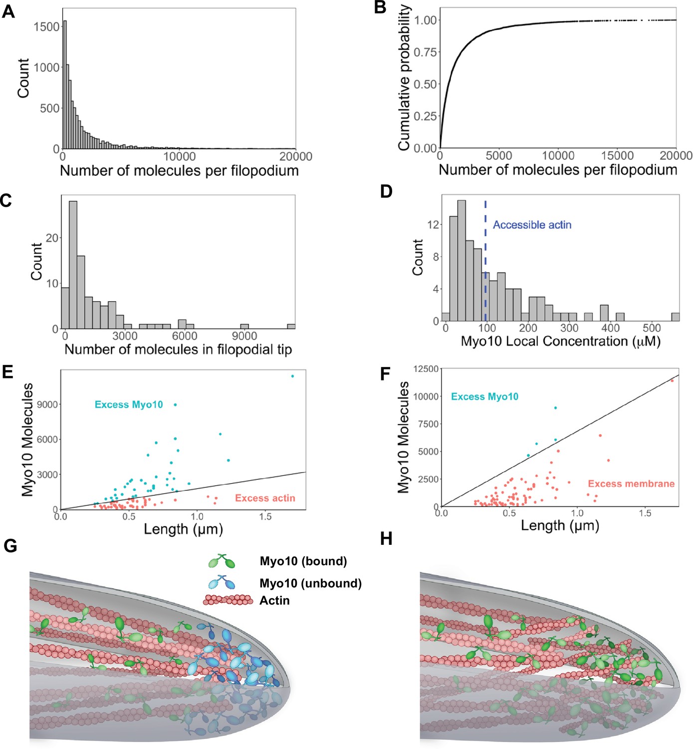
Hundreds of Myo10 molecules are found in a filopodium, potentially in excess over available actin at the filopodial tip.
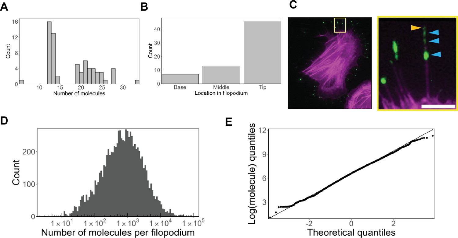
The following values are from fixed-cell images. (A) Distribution of the number of Myo10 molecules per filopodium (N=150 cells, 8733 filopodia; min = 6, 95% CI: 5–7; median = 730, 95% CI: 610–850; max = 80,000, 95% CI: 67,000–93,000; 62 values >20,000 not shown; bins = 100). (B) Cumulative distribution function plot of data in part A. (C) Distribution of the number of Myo10 molecules at the filopodial tip (90 randomly chosen filopodia tip-localized Myo10 puncta from nine different cell images; min = 66, 95% CI: 55–76; median = 788, 95% CI: 660–915; max = 11,000, 95% CI: 9600–13,000; bins = 30). (D) The local concentration of Myo10 at a filopodial tip. To estimate the volume, we measured the length of the filopodia tip-localized Myo10 puncta from part C in ImageJ. We then modeled filopodium as a cylinder of radius = 0.1 µm (published average). Min = 6.2 µM, 95% CI: 5.2–7.2; median = 84 µM, 95% CI: 70–97; max = 560 µM, 95% CI: 470–650, bins = 30. Blue dashed vertical line indicates the concentration of F-actin accessible for Myo10 binding in a filopodium (~96 µM). (E) Scatterplots of molecules vs. length for the puncta from part C. The phase boundary shows the 96 µM threshold from part D. (F) As in part E, but the line represents an estimate of allowable Myo10 on the filopodial tip membrane area. See Figure 3—figure supplement 2 for membrane occupancy estimates. (G) Model of a Myo10 traffic jam at the filopodium tip. Not enough available actin monomers results in a population of free Myo10 (in blue). The free Myo10 is detached from actin but potentially still membrane-associated. (H) Model of frayed actin filaments at the filopodium tip. If actin filaments are not neatly packed into parallel bundles at the filopodium tip, disorganized and frayed actin filaments yield more accessible binding sites to Myo10.
-
Figure 3—source data 1
Signal in segmented filopodial Myo10 puncta for all three fixed-cell bioreplicates.
- https://cdn.elifesciences.org/articles/90603/elife-90603-fig3-data1-v1.xlsx
-
Figure 3—source data 2
Data for local concentration of Myo10 at filopodial tips for all three fixed-cell bioreplicates.
- https://cdn.elifesciences.org/articles/90603/elife-90603-fig3-data2-v1.zip
Figure 3—figure supplement 1
HaloTag ligand-TMR labeling to determine Myo10 distributions along filopodia.
(A) In fixed cells: dim Myo10 puncta in filopodia, identified as those containing <35 Myo10 molecules, were examined. 135 total dim puncta across 26 cells were identified. Of the 135 puncta, 7 puncta were hard to visually interpret, 7 puncta were image noise, and 56 puncta were over-segmented (e.g. not actual distinct puncta but rather part of continuous Myo10 signal in filopodia). The distribution of the remaining 66 true dim puncta is displayed. Min = 6 (95% CI: 5–7), median = 19 (95% CI: 16–22), max = 34 (95% CI: 28–39). Bins = 30. (B) Filopodial localization of dim Myo10 puncta containing <35 molecules. The 66 true dim puncta from (A) were analyzed. (C) Example image demonstrating how Myo10 punctum position was determined in filopodia analyzed in (B). Actin was labeled with phalloidin-AF633 (magenta). HaloTag was labeled with HaloTag ligand-TMR (green). Zoomed-in inset features a filopodium displaying segmented Myo10 puncta localized in the middle (blue arrows) vs. at the tip (orange arrow); zoomed-in inset was contrast-adjusted to maximally highlight dim puncta. Scale bar = 5 µm. (D) Log-transformed distribution of the number of Myo10 molecules per filopodium. 8733 total filopodia across 150 cell images analyzed. Bins = 100. (E) Quantile-quantile (QQ) plot for the log-transformed distribution of Myo10 molecules in filopodia.
Figure 3—figure supplement 2
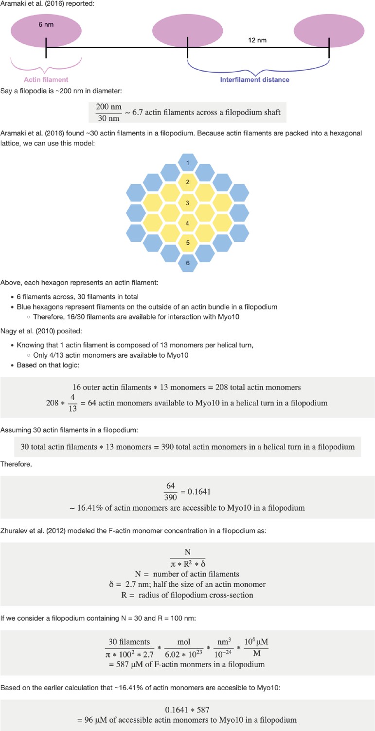
96 µM of actin monomers are accessible to Myo10 in a filopodium.
Figure 3—figure supplement 3

Plasma membrane at the filopodial tip can accommodate a portion of Myo10 unbound to actin.
Figure 4 with 5 supplements
Myo10 dynamics from live-cell movies.
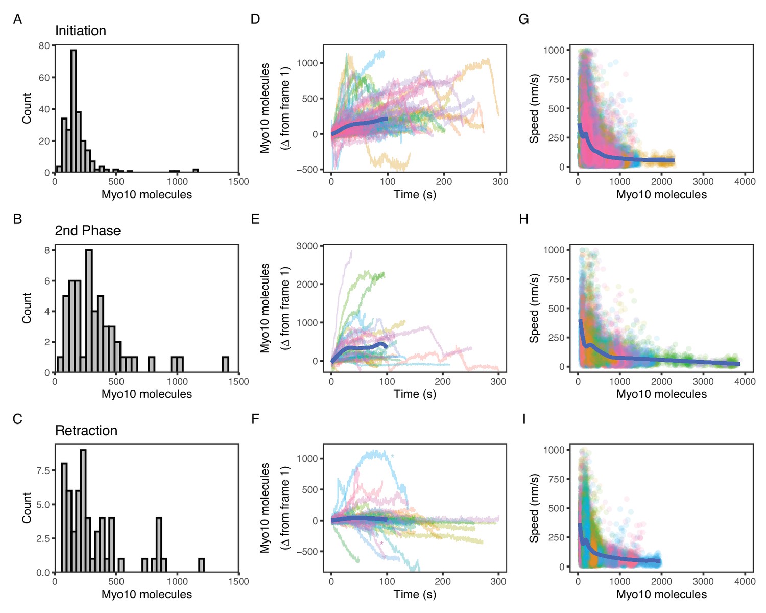
(A–C) Dense Myo10 puncta appear at the start of each phase of the filopodial lifecycle. Distributions of the number of Myo10 molecules in puncta upon: (A) Filopodial initiation (min = 52, 95% CI: 44–61; median = 160, 95% CI: 140–190; max = 1200, 95% CI: 970–1300). (B) Second-phase elongation (min = 65, 95% CI: 54–75; median = 290, 95% CI: 240–340; max = 1400, 95% CI: 1200–1600). (C) Filopodial retraction (min = 71, 95% CI: 60–82; median = 240, 95% CI: 200–280; max = 1200, 95% CI: 1000–1400). Values are the means of the first two frames after spot detection and identification of filopodial lifecycle stage. Histograms A–C have 30 bins each. (D–F) Accumulation of Myo10 in puncta after filopodial initiation or second-phase elongation, but not after retraction. Evolution of number of molecules for each filopodial phase over time for: (D) filopodial initiation. (E) Second-phase elongation. (F) Filopodial retraction. Starting values from (A–C) were subtracted from all traces to obtain delta over time. The generalized additive model (GAM) trend lines (blue) exclude long times (>100 s) with few surviving trajectories. The outlier trajectory indicated by the magenta asterisk is from Figure 4—video 3, and the cyan asterisk is from Figure 4—video 4. (G–I) Myo10 punctum speeds are inversely correlated with the number of Myo10 molecules. Plots of instantaneous speeds vs. molecules for: (G) filopodial initiation (min = 0.7, median = 160, max = 2,200 nm/s, Spearman’s ρ = –0.51, p<2.2*10–16). (H) Second-phase elongation (min speed = 0.4, median = 110, max = 3000 nm/s, Spearman’s ρ = –0.55, p<2.2*10–16). (I) Filopodial retraction (min speed = 1.2, median = 140, max = 1900 nm/s, Spearman’s ρ = –0.45, p<2.2*10–16). Color signifies Myo10 puncta belonging to the same trajectory within each event type. Colors are independent in each panel. Blue lines are GAM trend lines. Panels A, D, G: 237 trajectories from 31 cells; B, E, H: 51 trajectories from 19 cells; C, F, I: 58 trajectories from 26 cells.
-
Figure 4—source data 1
Trajectories of Myo10 puncta upon retraction from live-cell bioreplicate 1.
- https://cdn.elifesciences.org/articles/90603/elife-90603-fig4-data1-v1.zip
-
Figure 4—source data 2
Trajectories of Myo10 puncta upon second-phase elongation from live-cell bioreplicate 1.
- https://cdn.elifesciences.org/articles/90603/elife-90603-fig4-data2-v1.zip
-
Figure 4—source data 3
Trajectories of Myo10 puncta upon filopodial initiation from live-cell bioreplicate 1.
- https://cdn.elifesciences.org/articles/90603/elife-90603-fig4-data3-v1.zip
-
Figure 4—source data 4
Trajectories of Myo10 puncta upon retraction from live-cell bioreplicate 2.
- https://cdn.elifesciences.org/articles/90603/elife-90603-fig4-data4-v1.zip
-
Figure 4—source data 5
Trajectories of Myo10 puncta upon second-phase elongation from live-cell bioreplicate 2.
- https://cdn.elifesciences.org/articles/90603/elife-90603-fig4-data5-v1.zip
-
Figure 4—source data 6
Trajectories of Myo10 puncta upon filopodial initiation from live-cell bioreplicate 2.
- https://cdn.elifesciences.org/articles/90603/elife-90603-fig4-data6-v1.zip
-
Figure 4—source data 7
Trajectories of Myo10 puncta upon retraction from live-cell bioreplicate 3.
- https://cdn.elifesciences.org/articles/90603/elife-90603-fig4-data7-v1.zip
-
Figure 4—source data 8
Trajectories of Myo10 puncta upon second-phase elongation from live-cell bioreplicate 3.
- https://cdn.elifesciences.org/articles/90603/elife-90603-fig4-data8-v1.zip
-
Figure 4—source data 9
Trajectories of Myo10 puncta upon filopodial initiation from live-cell bioreplicate 3.
- https://cdn.elifesciences.org/articles/90603/elife-90603-fig4-data9-v1.zip
Figure 4—figure supplement 1
Myo10 is found along the filopodium shaft and can undergo a second elongation.
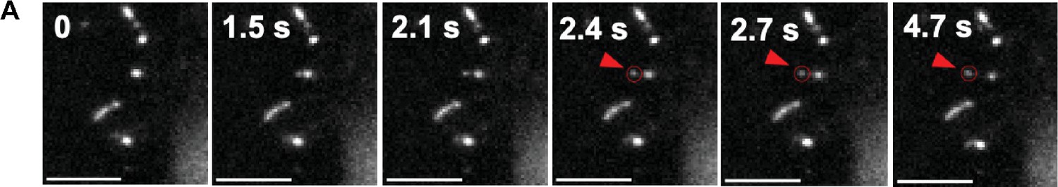
(A) Frame stills from Figure 4—video 2. Red arrow points to a Myo10 punctum (red circle) right after emergence from an existing filopodium. Elapsed seconds from the first frame is depicted. Scale bar = 5 µm.
Figure 4—video 1
Myo10 punctum initiates a nascent filopodium.
Example of a Myo10 punctum (red circle) right as the filopodium shoots from the cell body. 300 ms exposure, 10 fps.
Figure 4—video 2
Myo10 punctum can undergo a second elongation.
Example of a Myo10 punctum (red circle) right after emergence from an existing filopodium. 300 ms exposure, 10 fps.
Figure 4—video 3
Example of Myo10 punctum decreasing in intensity as it returns to cell body.
The trajectory indicated by the blue asterisk in Figure 3E. Example of a Myo10 punctum (purple circle) that decreases in intensity before becoming the site of new filopodium growth. 300 ms exposure, 30 fps.
Figure 4—video 4
Example of Myo10 punctum in a ‘snapping’ filopodium as it returns to cell body.
The trajectory indicated by the magenta asterisk in Figure 3E. Example of a Myo10 punctum (purple circle) that ‘snaps’ toward the cell body and then diffuses into the cytoplasm. 300 ms exposure, 30 fps.
Tables
Key resources table
| Reagent type (species) or resource | Designation | Source or reference | Identifiers | Additional information |
|---|---|---|---|---|
| Cell line (human) | U2OS cells | ATCC | HTB-96 | |
| Transfected construct (human) | HaloTag-Myo10-Flag (plasmid) | This paper | See supplemental for DNA sequence | |
| Antibody | Anti-Myosin10 antibody (rabbit polyclonal) | Novus | NBP1-87748 (RRID:AB_11035627) | 1:10,000 for western blot |
| Antibody | Anti-beta-tubulin antibody (mouse monoclonal) | Invitrogen | 22833 (RRID:AB_2533072) | 1:10,000 for western blot |
| Antibody | Anti-Rabbit-HRP antibody (goat polyclonal) | Cell Signaling | 7074 (RRID:AB_2099233) | 1:10,000 for western blot |
| Antibody | Anti-Mouse-HRP antibody (horse polyclonal) | Cell Signaling | 7076 (RRID:AB_330924) | 1:10,000 for western blot |
| Peptide, recombinant protein | Laminin | Sigma-Aldrich | CC095-M | |
| Peptide, recombinant protein | HaloTag standard protein | Promega | G4491 | |
| Commercial assay or kit | SuperSignal West Femto chemiluminescent substrate | Thermo Scientific | 34094 | |
| Other | Gibco 1x DMEM | Thermo Fisher | 11995073 | Cell media |
| Other | Accutase | Corning | MT25058CI | Enzyme cell detachment media |
| Other | FuGENE HD Transfection reagent | Promega | E2311 | Non-liposomal transfection reagent |
| Other | Lipofectamine 2000 | Invitrogen | 11668-027 | Cationic-lipid transfection reagent |
| Other | #1.5 coverglass bottom 35 mm Petri dishes | Cellvis, MatTek | D35-10-1.5-N, P35G-1.5-14-C | Coverglass for live-cell imaging |
| Other | Ibidi eight-well chamber slides | Ibidi | 80807 | Coverglass for fixed-cell imaging |
| Other | 4–20% Mini-PROTEAN TGX Stain-Free protein gel | Bio-Rad | 4568095 | Stain-free precast gels for SDS-PAGE |
| Other | TMR-HaloLigand | Promega | G8251 | Fluorophore-labeled HaloLigand used for visualizing Myo10 |
| Other | Alex Fluor 647 Phalloidin | Invitrogen | A22287 | 1 mM working concentration to label actin |
| Other | Bio-Beads SM-2 | Bio-Rad | 152-8920 | Resin to remove excess dye when testing HaloLigand labeling efficiency |
Additional files
-
MDAR checklist
- https://cdn.elifesciences.org/articles/90603/elife-90603-mdarchecklist1-v1.pdf
-
Source code 1
A descriptive text file explaining each subfolder is included within the zip file.
- https://cdn.elifesciences.org/articles/90603/elife-90603-code1-v1.zip
Download links
A two-part list of links to download the article, or parts of the article, in various formats.
Downloads (link to download the article as PDF)
Open citations (links to open the citations from this article in various online reference manager services)
Cite this article (links to download the citations from this article in formats compatible with various reference manager tools)
Hundreds of myosin 10s are pushed to the tips of filopodia and could cause traffic jams on actin
eLife 12:RP90603.
https://doi.org/10.7554/eLife.90603.4


