Lateral/caudal ganglionic eminence makes limited contribution to cortical oligodendrocytes
- State Key Laboratory of Medical Neurobiology and MOE Frontiers Center for Brain Science, Institutes of Brain Science, and Department of Neurology, Zhongshan Hospital, Fudan University, China
- Department of Anesthesiology, Shuguang Hospital Affiliated with Shanghai University of Traditional Chinese Medicine, China
- Center for Clinical Research and Translational Medicine, Yangpu Hospital, School of Medicine, Tongji University, China
Figures
Figure 1 with 1 supplement
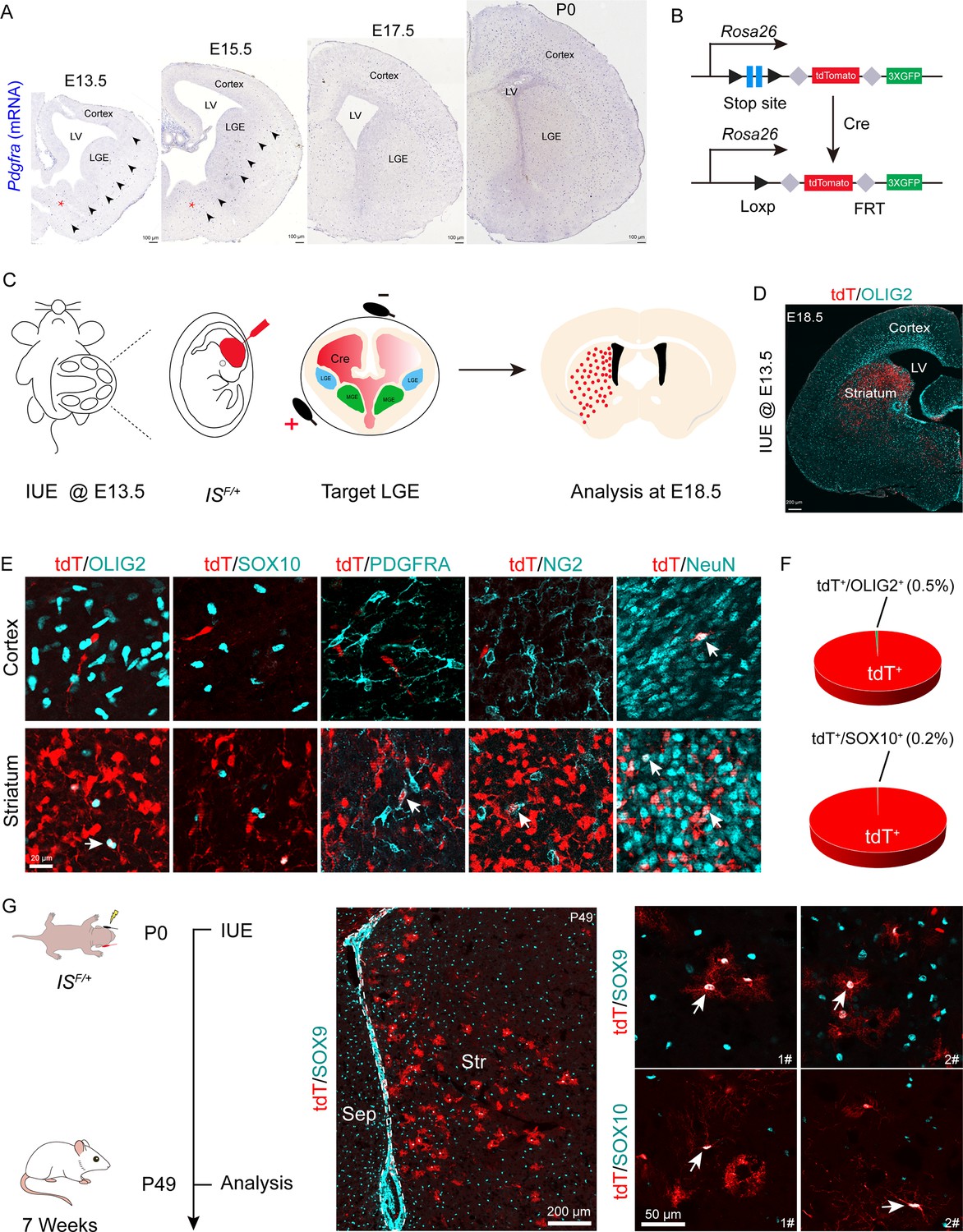
Fate mapping of lateral ganglionic eminence (LGE)-derived oligodendrocyte precursor cells (OPCs) by combining in utero electroporation (IUE) with a Cre recombinase-dependent IS reporter.
(A) In situ hybridization showing Pdgfra expression from embryonic day 13.5 (E13.5) to postnatal day 0 (P0). Arrowheads indicated the Pdgfra+ cell migration stream from the medial ganglionic eminence (MGE)/anterior entopeduncular area (AEP) to the cortex. (B) Scheme of the IS reporter lines. (C) Experimental schedule to trace LGE-derived OPCs. (D) Representative coronal sections showing the distribution of tdT+ cells. (E) tdT+ cells expressed NeuN but not OLIG2, SOX10, PDGFRA, or NG2 in the cortex. In contrast, the tdT+ cells expressed all of these markers (OLIG2, SOX10, PDGFRA, NG2, and NeuN) in the striatum. (F) The ratio of the OLIG2+ and SOX10+ cells of the electroporated cells in the cortex. N=4 mice per group. (G) Fate mapping of LGE-derived cells at P0. tdT+ cells expressed SOX10 and SOX9 in the striatum at P49.
-
Figure 1—source data 1
The raw data for the visualization of data presented in Figure 1F.
- https://cdn.elifesciences.org/articles/94317/elife-94317-fig1-data1-v1.xlsx
Figure 1—figure supplement 1

Lineage tracing of lateral ganglionic eminence (LGE)-derived oligodendrocyte precursor cells (OPCs) by combining in utero electroporation (IUE) with a Cre recombinase-dependent IS reporter.
(A) Experimental schedule for fate mapping of LGE-derived OPCs. (B) Representative coronal sections showing the distribution of the tdT+ cells at P10. (C) Nearly all tdT+ cells expressed NeuN but not OLIG2 in the cortex. tdT+ cells expressed NeuN and OLIG2 in the striatum. (D) tdT+ cells did not express OLIG2, SOX10, PDGFRA, or ALDH1L1. Instead, they expressed NeuN in the cortex. In sharp contrast, tdT+ cells in the striatum expressed OLIG2, SOX10, PDGFRA, ALDH1L1, and NeuN at P10. (E) 3D reconstruction of consecutive brain sections demonstrated that the traced cells were mainly located in the striatum. N=4 mice per group.
Figure 2
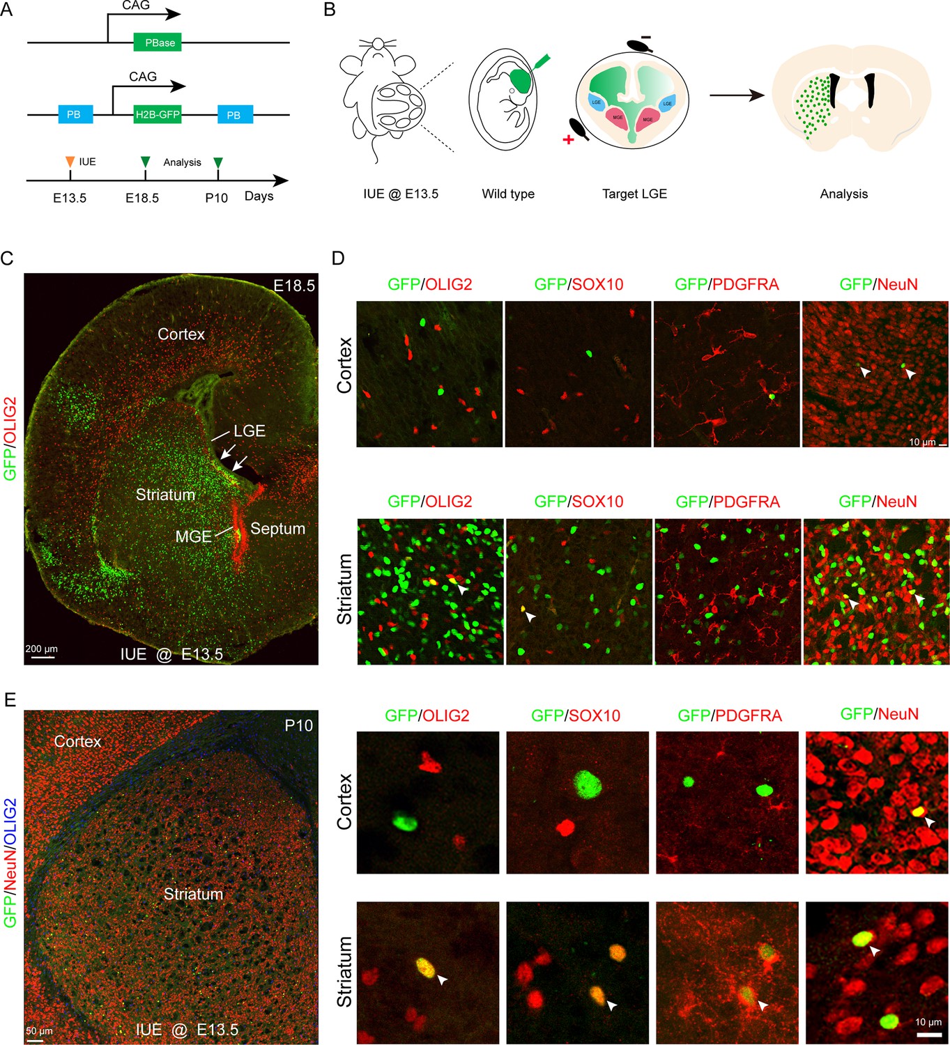
Fate mapping of lateral ganglionic eminence (LGE)-derived oligodendrocyte precursor cells (OPCs) by combining in utero electroporation (IUE) with PiggyBac transposon system.
(A) Schematic of the PiggyBac transposon reporter system used in this study. (B) The experimental workflow. (C) Representative coronal sections showing the distribution of GFP+ cells at embryonic day 18.5 (E18.5). (D) GFP+ cells expressed NeuN but not glial cell markers, such as OLIG2, SOX10, and PDGFRA in the cortex. However, GFP+ cells expressed NeuN, OLIG2, SOX10, and PDGFRA in the striatum. (E) GFP+ cells expressed NeuN but not glial cell markers, such as OLIG2, SOX10, and PDGFRA in the cortex and GFP+ cells expressed NeuN, OLIG2, SOX10, and PDGFRA in the striatum at postnatal day 10 (P10). N=4 per group.
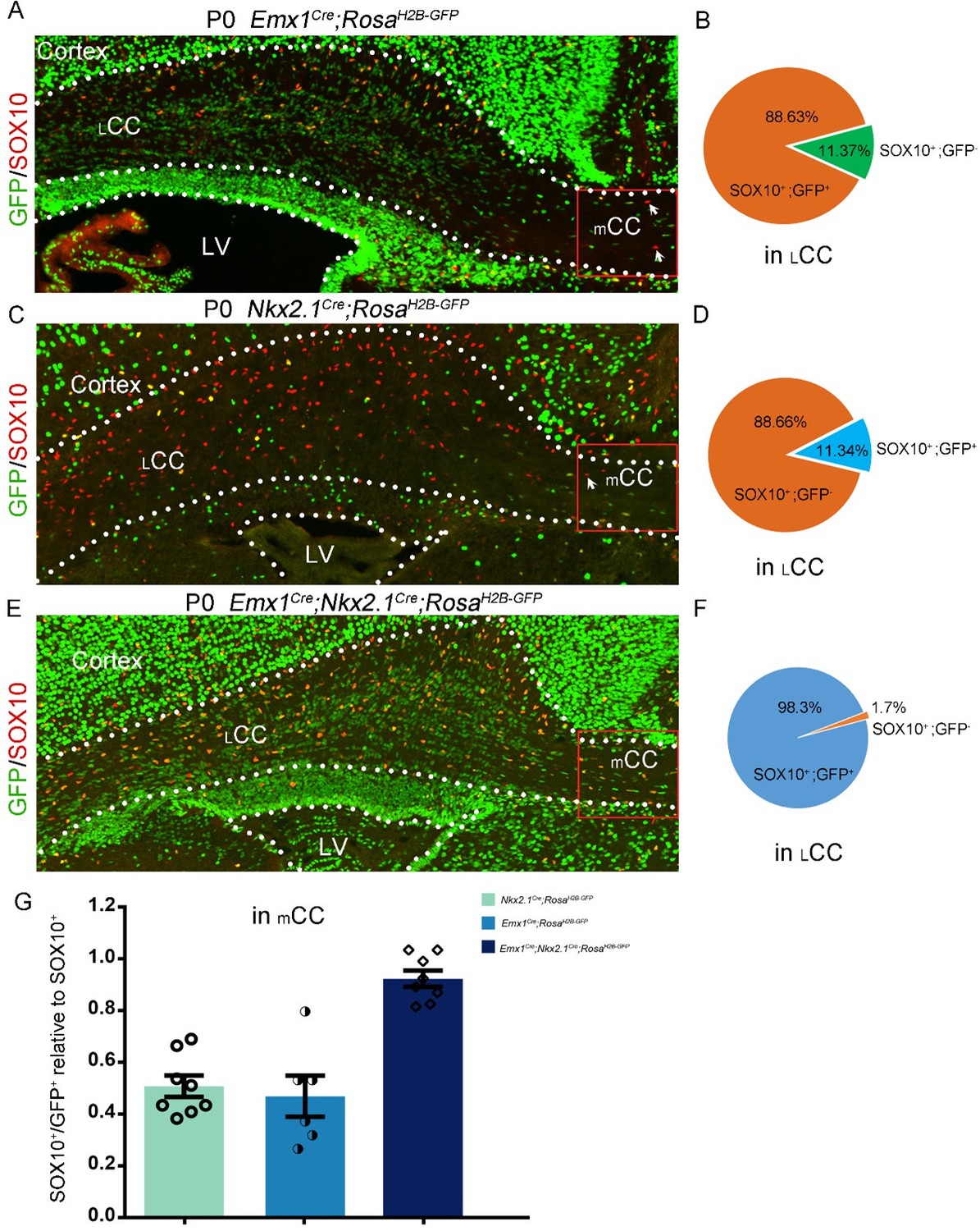
Figure 3 with 1 supplement
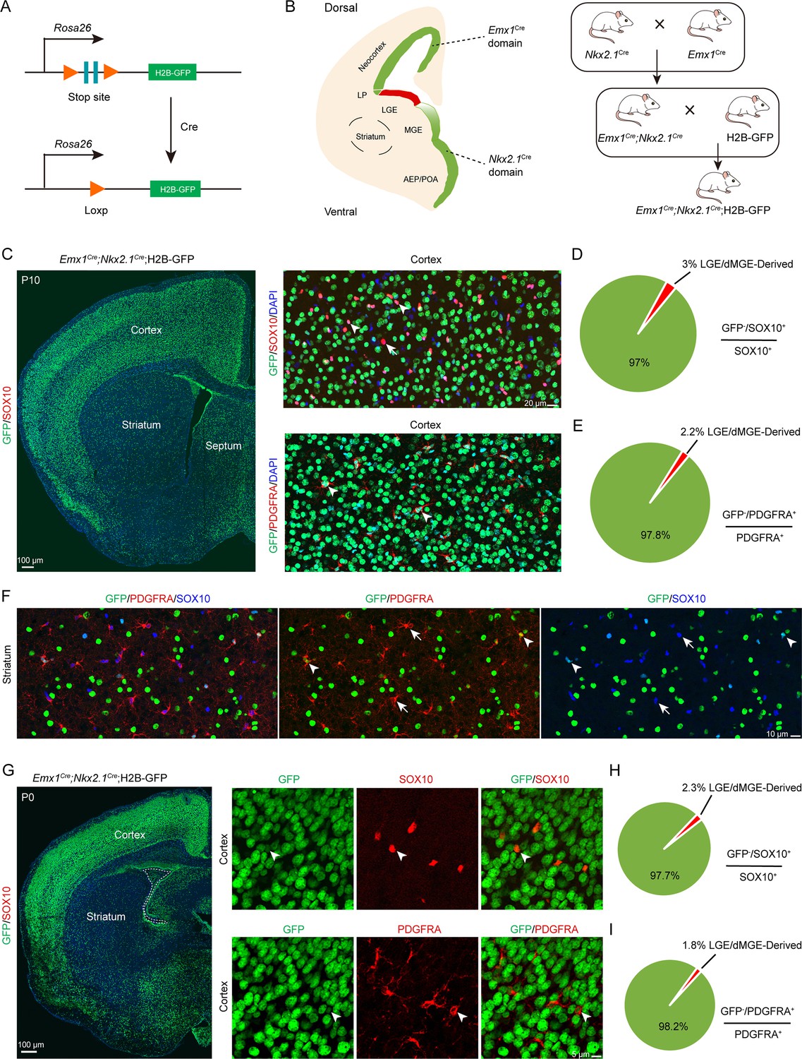
An exclusion strategy showed that lateral/caudal ganglionic eminence (LGE/CGE) contribution to cortical oligodendrocyte precursor cells (OPCs) is minimal.
(A) Scheme of the H2B-GFP reporter lines. (B) Experimental design of the exclusion strategy to trace the lineage of LGE radial glial cells (RGCs). (C) Representative coronal sections showing the traced cells in the forebrain. The majority of SOX10- and PDGFRA-positive cells were GFP+ cells in the cortex at postnatal day 10 (P10). (D–E) The pie chart shows that the percentage of LGE/dMGE-derived cortical OPCs was approximately 3%. N=6 mice per group. (F) Many GFP+ cells expressed SOX10 and PDGFRA in the striatum at P10. (G) Nearly all SOX10 and PDGFRA expressed GFP in the cortex at P0. (H–I) The pie chart shows that the percentage of LGE/dMGE-derived cortical OPCs was less than 3%. N=6 mice per group.
-
Figure 3—source data 1
The raw data for the visualization of data presented in Figure 3D, E, H, I.
- https://cdn.elifesciences.org/articles/94317/elife-94317-fig3-data1-v1.xlsx
Figure 3—figure supplement 1
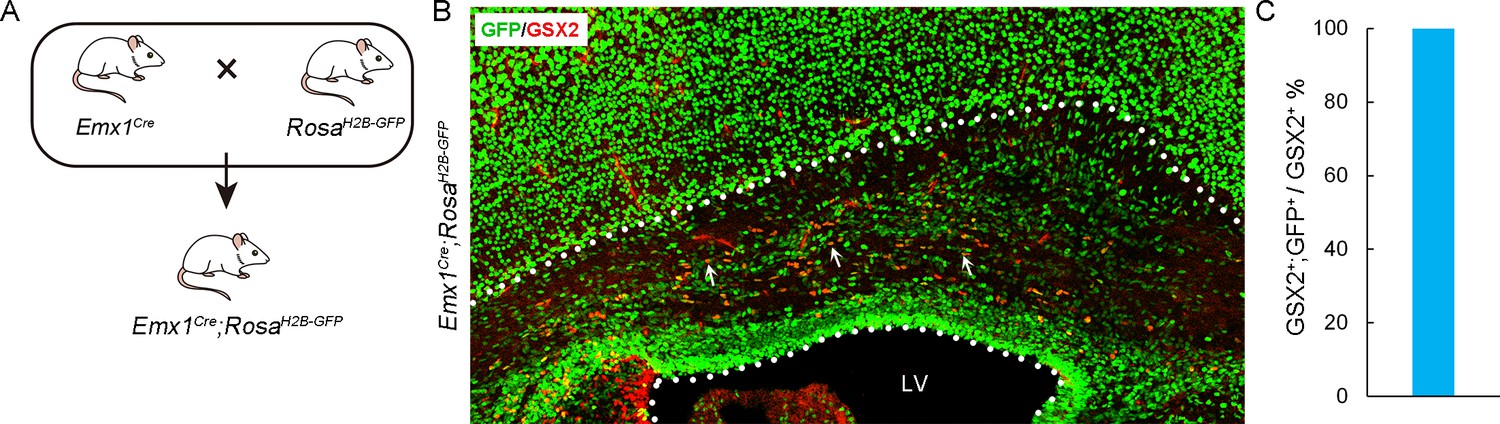
Lineage tracing of Emx1Cre derived cortical cells.
(A) Experimental design for the generation of Emx1Cre; H2B-GFP mice. (B) Double immunostaining of GSX2 with GFP in the Emx1Cre; H2B-GFP cortex at postnatal day 0 (P0). (C) The statistics show that nearly all GSX2+ cells co-labeled with GFP in the cortical SVZ. N=4 per group.
-
Figure 3—figure supplement 1—source data 1
The raw data for the visualization of data presented in Figure 3—figure supplement 1C.
- https://cdn.elifesciences.org/articles/94317/elife-94317-fig3-figsupp1-data1-v1.xlsx
Figure 4
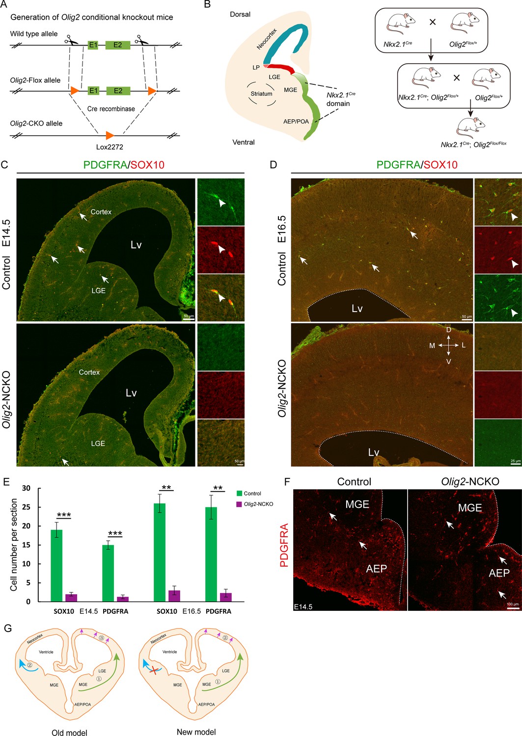
The medial ganglionic eminence (MGE) may be the sole ventral source of cortical oligodendrocyte precursor cells (OPCs).
(A) The CRISPR/Cas9 technique was used to generate an Olig2 conditional knockout allele. (B) Experimental design for the generation of Olig2-NCKO mice. (C–D) The expression of SOX10 and PDGFRA was significantly reduced in the cortex of Olig2-NCKO mice compared with that of control mice. (E) The number of SOX10- and PDGFRA-positive cells was significantly reduced in Olig2-NCKO mice compared with control mice. Student’s t-test, **p<.01, ***p<.001, N≥5 mice per group, mean ± SEM. (F) Few PDGFRA-positive cells were detected in the MGE/anterior entopeduncular area (AEP) of the Olig2-NCKO mice. (G) A new model of the developmental origins of cortical OPCs.
-
Figure 4—source data 1
The raw data for the visualization of data presented in Figure 4E.
- https://cdn.elifesciences.org/articles/94317/elife-94317-fig4-data1-v1.xlsx
Author response image 1
Tables
Key resources table
| Reagent type (species) or resource | Designation | Source or reference | Identifiers | Additional information |
|---|---|---|---|---|
| Genetic reagent (Mus musculus) | Emx1Cre | The Jackson Laboratory | Stock No. 005628 | |
| Genetic reagent (Mus musculus) | Nkx2.1Cre | The Jackson Laboratory | Stock No. 008661 | |
| Genetic reagent (Mus musculus) | IS reporter | The Jackson Laboratory | Stock No. 028582 | |
| Genetic reagent (Mus musculus) | Olig2F/+ | Our lab | N/A; available from the authors | |
| Genetic reagent (Mus musculus) | C57BL/6 | Department of Laboratory Animal Science at Fudan University | http://10.107.12.196/ | |
| Genetic reagent (Mus musculus) | ICR | Department of Laboratory Animal Science at Fudan University | http://10.107.12.196/ | |
| Antibody | Anti-GFP (Chicken polyclonal) | Aves Labs | Cat# GFP-1020, RRID: AB_10000240 | IF (1:3000) |
| Antibody | Anti-ALDH1L1 (Rabbit polyclonal) | Abcam | Cat# ab87117, RRID: AB_10712968 | IF (1:1000) |
| Antibody | Anti-OLIG2 (Rabbit polyclonal) | Oasis Biofarm | Cat# OB-PRB009, RRID: AB_2934240 | IF (1:500) |
| Antibody | Anti-OLIG2 (Rat polyclonal) | Oasis Biofarm | Cat# OB-PRT020, RRID: AB_2934241 | IF (1:500) |
| Antibody | Anti-OLIG2 (Mouse polyclonal) | Millipore | Cat# MABN50, RRID: AB_10807410 | IF (1:1000) |
| Antibody | Anti-OLIG2 (Rabbit polyclonal) | Millipore | Cat# AB9610, RRID: AB_570666 | IF (1:1000) |
| Antibody | Anti-SOX10 (Goat polyclonal) | R&D Systems | Cat# ab442208, RRID: unknown | IF (1:500) |
| Antibody | Anti-SOX10 (Guinea Pig polyclonal) | Oasis Biofarm | Cat# OB-PGP001, RRID: AB_2934230 | IF (1:1000) |
| Antibody | Anti-PDGFRA (Rabbit polyclonal) | Oasis Biofarm | Cat# OB-PRB051, RRID: AB_2938684 | IF (1:800) |
| Antibody | Anti-NeuN (Rabbit unknown) | Bioscience | Cat# R-3770–100, RRID: AB_2493045 | IF (1:1000) |
| Antibody | Anti-NeuN (Rabbit polyclonal) | Oasis Biofarm | Cat# OB-PRB039, RRID: AB_2934232 | IF (1:1000) |
| Antibody | Anti-NG2 (Guinea Pig polyclonal) | Oasis Biofarm | Cat# OB-PGP002, RRID: AB_2938678 | IF (1:1000) |
| Antibody | Alexa488-Conjugated Affinipure Donkey Anti-Chicken IgY++ (IgG) (H+L) | Jackson ImmunoResearch Labs | Cat# 703-545-155, RRID: AB_2340375 | IF (1:500) |
| Antibody | CyTM3-Conjugated Affinipure Donkey Anti-Rabbit IgG (H+L) | Jackson ImmunoResearch Labs | Cat# 711-165-152, RRID: AB_2307443 | IF (1:500) |
| Antibody | Alexa647-Conjugated Affinipure Donkey Anti-Rat IgG (H+L) | Jackson ImmunoResearch Labs | Cat# 712-605-153, RRID: AB_2340694 | IF (1:500) |
| Antibody | Alexa647-Conjugated Affinipure Donkey Anti-Goat IgG (H+L) | Jackson ImmunoResearch Labs | Cat# 705-605-147, RRID: AB_2340437 | IF (1:500) |
| Antibody | Alexa488-Conjugated Affinipure Donkey Anti-Rabbit IgG (H+L) | Jackson ImmunoResearch Labs | Cat# 711-545-152, RRID: AB_2313584 | IF (1:500) |
| Antibody | Cy3-AffiniPure Donkey Anti-Rat IgG (H+L) | Jackson ImmunoResearch Labs | Cat# 712-165-150, RRID: AB_2340666 | IF (1:500) |
| Antibody | Cy3-AffiniPure Donkey Anti-Guinea Pig IgG (H+L) | Jackson ImmunoResearch Labs | Cat# 706-165-148, RRID: AB_2340460 | IF (1:500) |
| Recombinant DNA reagent | pCAGIG (plasmid) | Addgene | Cat# 11159 | |
| Recombinant DNA reagent | pGL4.10 (plasmid) | Promega | Cat# E6651 | |
| Recombinant DNA reagent | pGL4.23 (plasmid) | Promega | Cat# E8411 |
Additional files
Download links
A two-part list of links to download the article, or parts of the article, in various formats.
Downloads (link to download the article as PDF)
Open citations (links to open the citations from this article in various online reference manager services)
Cite this article (links to download the citations from this article in formats compatible with various reference manager tools)
Lateral/caudal ganglionic eminence makes limited contribution to cortical oligodendrocytes
eLife 13:RP94317.
https://doi.org/10.7554/eLife.94317.3


