Oxydifficidin, a potent Neisseria gonorrhoeae antibiotic due to DedA-assisted uptake and ribosomal protein RplL sensitivity
- Laboratory of Genetically Encoded Small Molecules, The Rockefeller University, United States
- Graduate Center, City University of New York, United States
- Brooklyn College, City University of New York, United States
- Laboratoire Jean Perrin, UMR 8237 Sorbonne Université/CNRS, France
Figures
Figure 1

Oxydifficidin isomers inhibit the growth of N. gonorrhoeae.
(a) Discovery of a contaminant (Bacillus amyloliquefaciens BK) that inhibited the growth of N. gonorrhoeae. (b) Example of known oxydifficidin isomers. (c) Minimum inhibitory concentration (MIC) of oxydifficidin against bacteria (n = 2). Genome-based phylogenetic tree was built by Genome Clustering of MicroScope using neighbor-joining method.
Figure 2
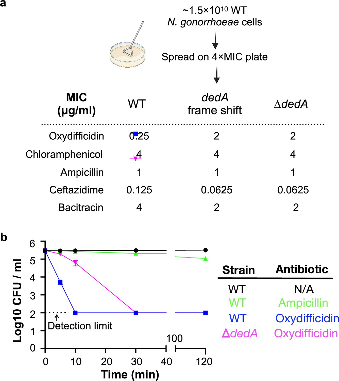
Oxydifficidin-resistant N. gonorrhoeae mutant development and corresponding susceptibilities.
(a) Schematic representation of N. gonorrhoeae mutant development that identified dedA. Activity of different antibiotics against MS11 and dedA gene disrupted N. gonorrhoeae MS11. (b) Time-dependent antibiotic killing assay of N. gonorrhoeae strains. Each antibiotic was tested at 8× its minimum inhibitory concentration (MIC) for the specific strain being examined (MS11 Ampicillin: 8 μg/ml; MS11 Oxydifficidin: 2 μg/ml; MS11 ∆dedA Oxydifficidin: 16 μg/ml). ∆dedA indicates N. gonorrhoeae MS11 dedA deletion mutant (n = 3). Bars represent mean CFU values ± SD.
Figure 3
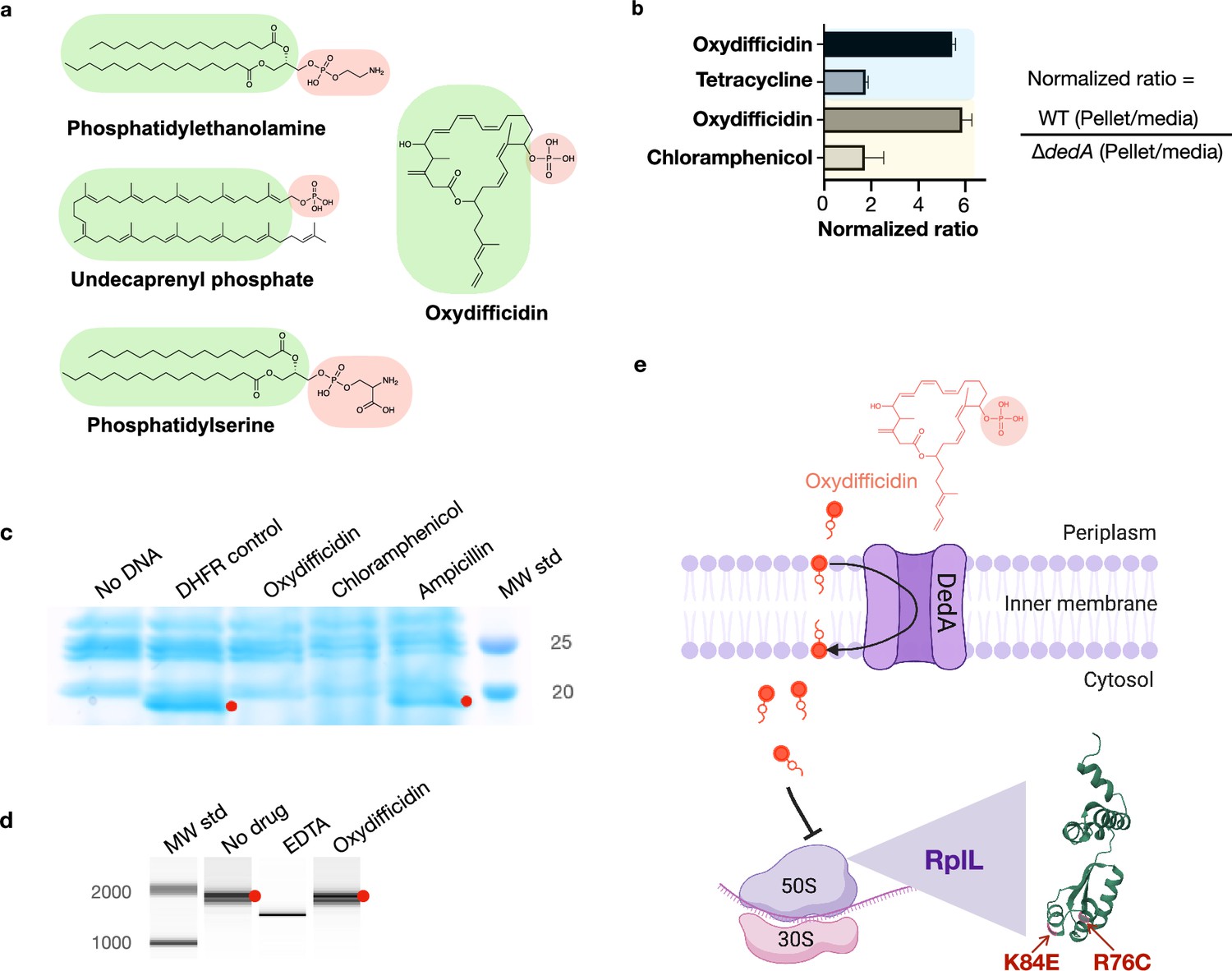
Oxydifficidin’s anti-N. gonorrhoeae activity arises from a combination of DedA flippase-assisted uptake and ribosomal protein L7/L12 (RplL) sensitivity.
(a) Structure of oxydifficidin compared to that of the known substrates for DedA homologs. (b) Comparison of antibiotic accumulation in MS11 and MS11 dedA knockout cells. Blue and yellow highlighted sections represent independent experiments (oxydifficidin and tetracycline: n = 2; oxydifficidin and chloramphenicol: n = 3). (c) In vitro coupled transcription/translation assay. The effect of oxydifficidin and other antibiotics on in vitro protein production using a coupled transcription/translation system was monitored by SDS–PAGE. Red dots indicate in vitro production of dihydrofolate reductase (18 kDa) from the DHFR gene. MW std: kDa molecular weight standard. (d) In vitro transcription assay. Red dots indicate in vitro production of a 1704-bp RNA from the FLuc gene. A reaction containing 20 mM of EDTA was used as an inhibition control. MW std: bp molecular weight standard. (e) Model explaining oxydifficidin’s potent activity in N. gonorrhoeae. In this model DedA flips oxydifficidin across the inner membrane to assist its uptake and oxydifficidin then inhibits protein synthesis through either a direct or indirect interaction with L7/L12 (RplL). Two spontaneous mutations (K84E and R76C) in the RplL (L7/L12) protein were found to confer resistance to oxydifficidin. Created using BioRender.com. Bars represent mean ratio values ± SD.
-
Figure 3—source data 1
Original SDS–PAGE picture for Figure 3c.
- https://cdn.elifesciences.org/articles/99281/elife-99281-fig3-data1-v1.zip
-
Figure 3—source data 2
Original SDS–PAGE picture for Figure 3c, labeled.
- https://cdn.elifesciences.org/articles/99281/elife-99281-fig3-data2-v1.zip
-
Figure 3—source data 3
Original BioAnalyzer report for Figure 3d.
- https://cdn.elifesciences.org/articles/99281/elife-99281-fig3-data3-v1.zip
-
Figure 3—source data 4
Original BioAnalyzer report for Figure 3d, labeled.
- https://cdn.elifesciences.org/articles/99281/elife-99281-fig3-data4-v1.zip
Appendix 1—figure 1
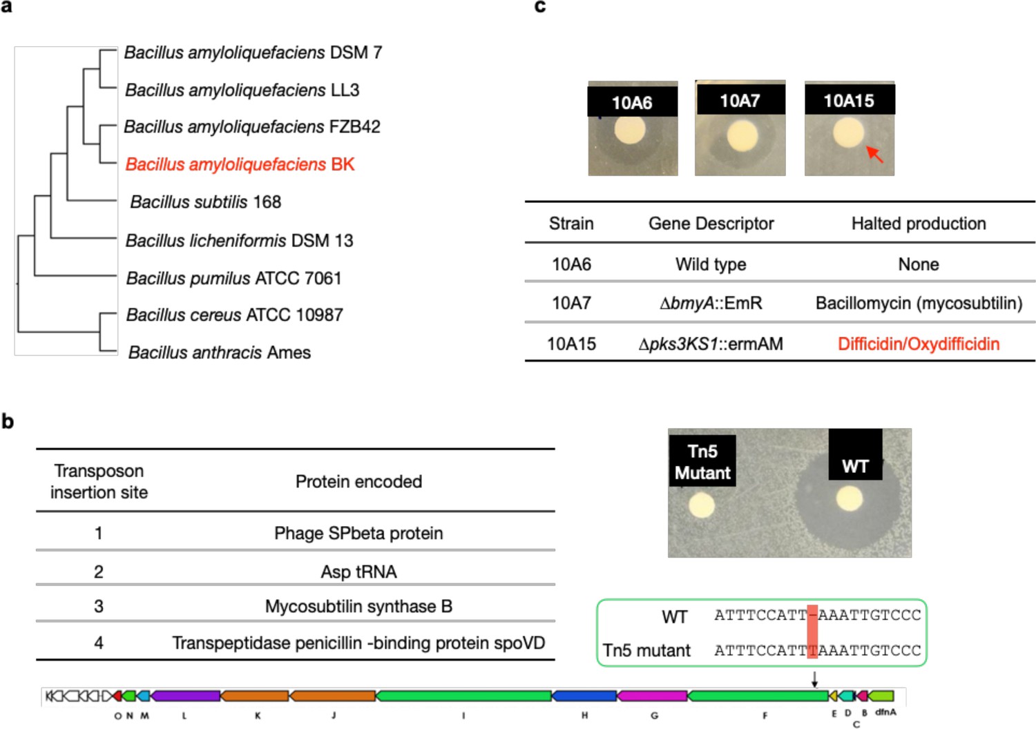
Mutagenesis results of oxydifficidin-producing Bacillus spp.
(a) Genome-based phylogenetic tree containing Bacillus amyloliquefaciens BK and closely related Bacillus spp. The tree was built by Genome Clustering of MicroScope using neighbor-joining method. The NCBI accession numbers of Bacillus strains used in the tree are GCA_000196735.1, GCA_000204275.1, GCA_000015785.2, GCA_019093835.1, GCA_000009045.1, GCA_000011645.1, GCA_000172815.1, GCA_000008005.1, and GCA_000007845.1 (from top to bottom). (b) Disc diffusion assay of a methanol extract from cultures of WT B. amyloliquefaciens BK (WT) and a Tn5 mutant. The test lawn was N. gonorrhoeae. The table shows all transposon insertion sites in the Tn5 mutant strain. The Tn5 strain also contains a frame-shift mutation in the difF gene; red box highlights the location of frame-shift mutation in the oxydifficidin biosynthetic gene cluster (BGC). (c) Disc diffusion assay of a methanol extract from cultures of WT and BGC knockout strains of Bacillus amyloliquefaciens FZB42. The test lawn was N. gonorrhoeae. Strain genotypes are shown in the table. Red arrow indicates that only strain 10A15 no longer produce the anti-N. gonorrhoeae compound.
Appendix 1—figure 2

Phylogenetic tree of 15,825 bacterial DedA family proteins.
The tree was built by MUSCLE v5 and FastTree and visualized using iTOL. N. gonorrhoeae NGFG_RS04905 highlighted in red represents the DedA gene associated with the activity of oxydifficidin. N. gonorrhoeae NGFG_RS07420 and N. gonorrhoeae NGFG_RS00755 represents two other DedA family proteins in N. gonorrhoeae.
Appendix 1—figure 3
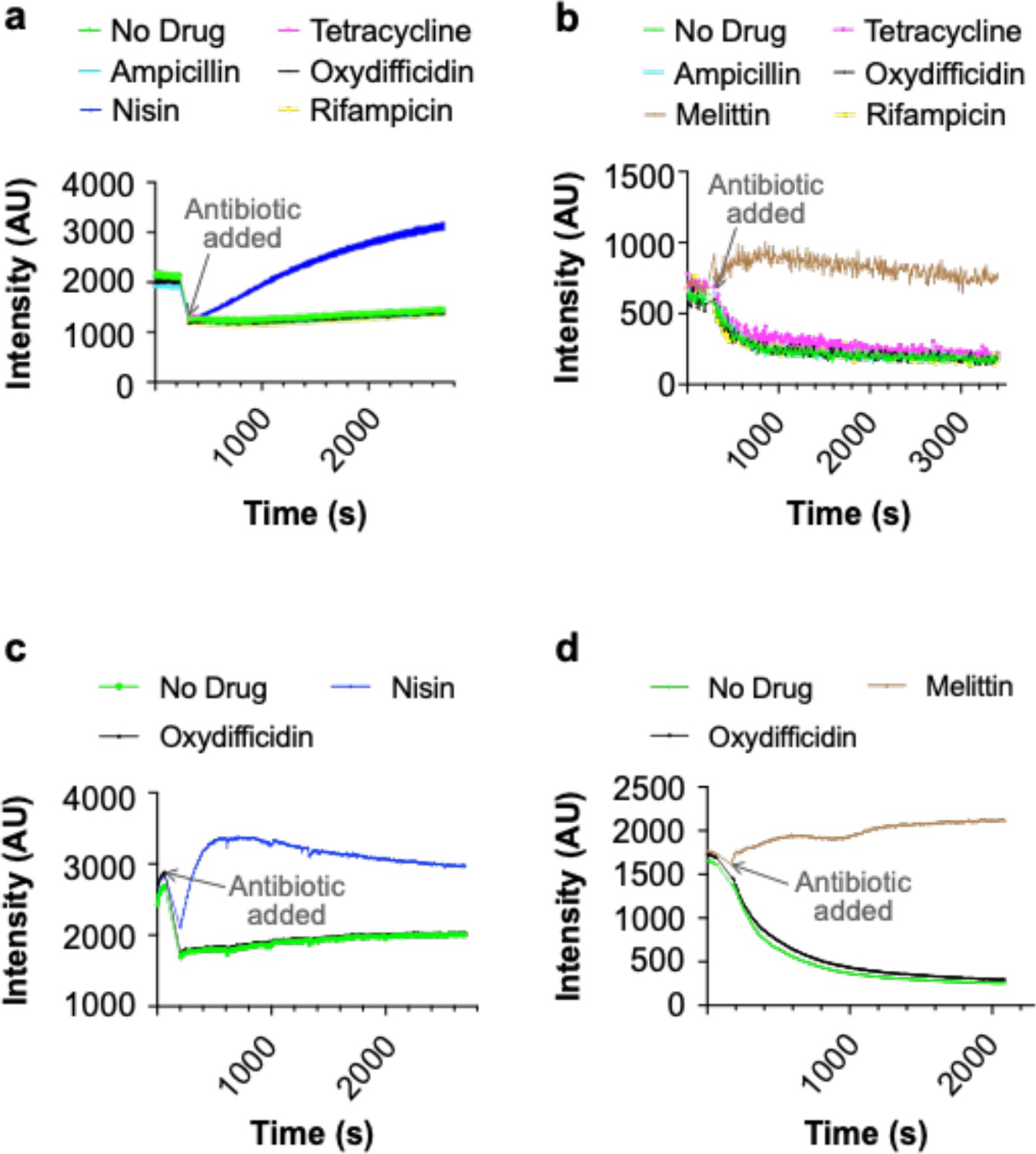
Oxydifficidin does not lyse or depolarize the membrane of N. gonorrhoeae.
(a) Lysis assay using SYTOX green dye and 8× the minimum inhibitory concentration (MIC) of each antibiotic. (b) Depolarization assay using DiSC3(5) dye and 8× the MIC of each antibiotic. (c) Lysis assay using SYTOX green dye with 100× the MIC of oxydifficidin and 32× the MIC of nisin. (d) Depolarization assay using DiSC3(5) dye with 100× the MIC of oxydifficidin and the 32× the MIC of melittin (n = 3 for all assays).
Appendix 1—figure 4
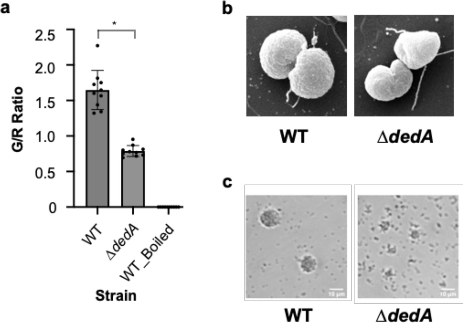
Mutations in dedA affect cell morphology and pili functionality of N. gonorrhoeae.
(a) Membrane integrity assay of N. gonorrhoeae WT and dedA deletion mutant (∆dedA) cells using SYTO 9 and propidium iodide. Cell integrity was assessed using the ratio of green- to red-stained cell count. *p < 0.05. (b) Scanning electron microscope pictures of N. gonorrhoeae WT and ∆dedA cells. (c) Micro-colony formation assay of N. gonorrhoeae WT and ∆dedA cells.
Appendix 1—figure 5
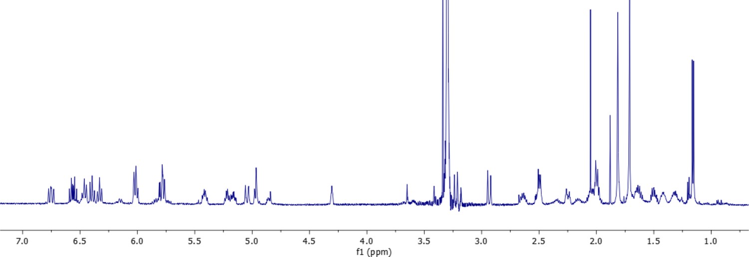
1H-NMR spectrum of oxydifficidin (800 MHz, 298 K, CD3OD–D2O (1:1)).
Appendix 1—figure 6
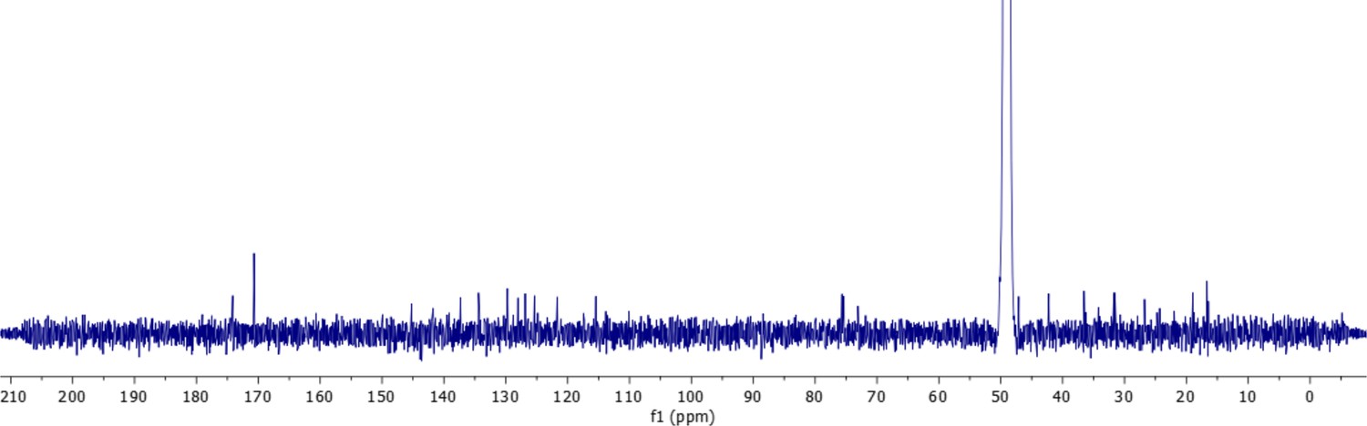
13C-NMR spectrum of oxydifficidin (800 MHz, 298 K, CD3OD–D2O (1:1)).
Appendix 1—figure 7
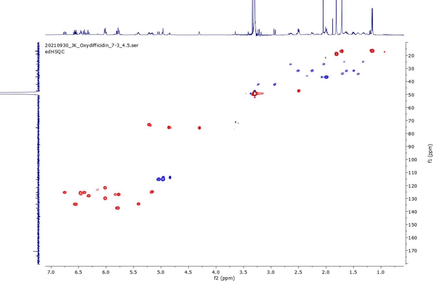
edHSQC spectrum (800 MHz, 298 K, CD3OD–D2O (1:1)) of oxydifficidin.
Appendix 1—figure 8

COSY spectrum (800 MHz, 298 K, CD3OD–D2O (1:1)) of oxydifficidin.
Appendix 1—figure 9

1H–13C HMBC spectrum (800 MHz, 298 K, CD3OD–D2O (1:1)) of oxydifficidin.
Appendix 1—figure 10
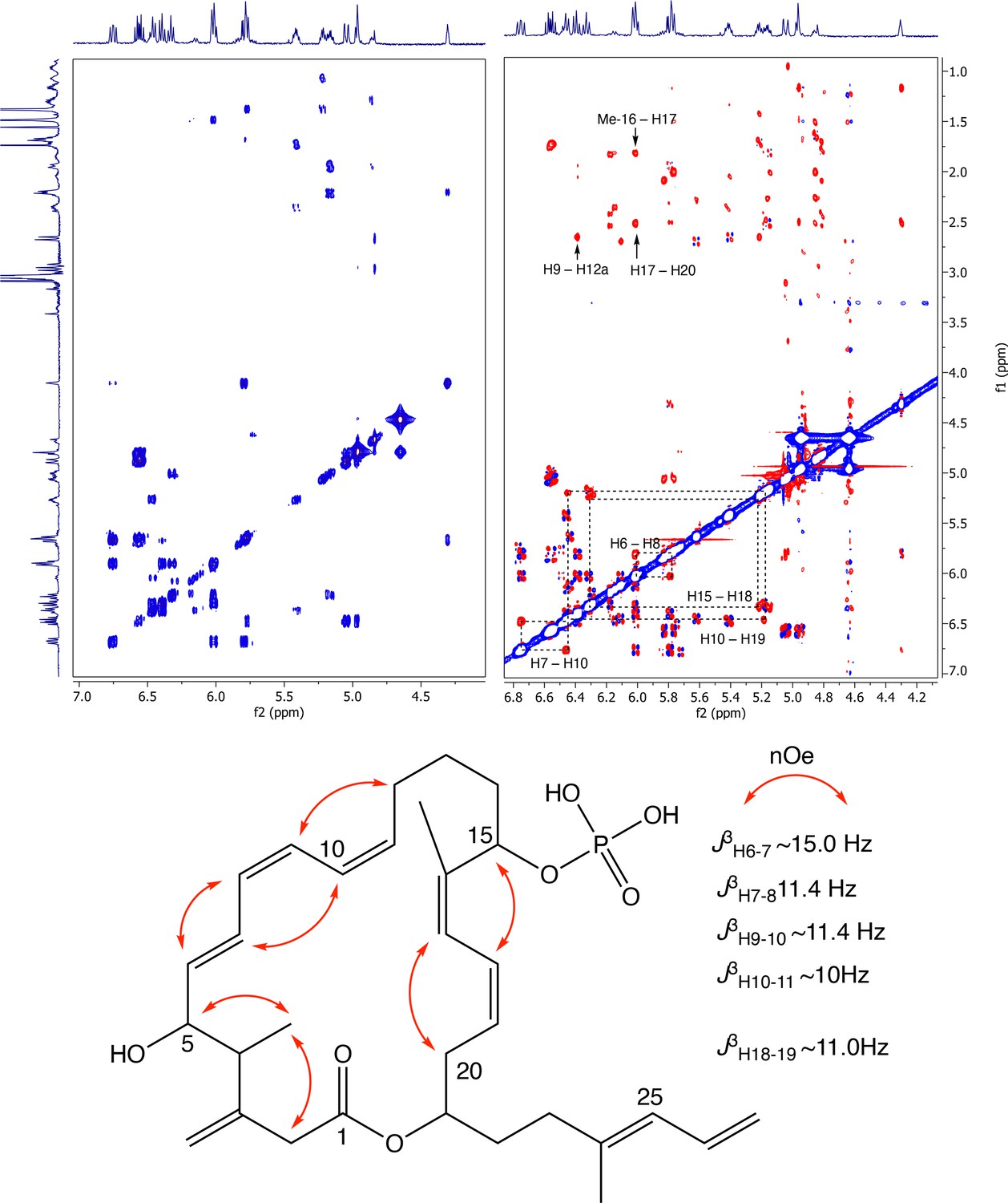
Partial COSY (left) and ROESY (right) comparison and key ROESY correlations of oxydifficidin (800 MHz, 298 K, CD3OD–D2O (1:1)).
Appendix 1—figure 11
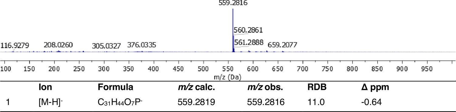
Full HRMS and annotation of oxydifficidin [M–H]– parental ion.
Appendix 1—figure 12

ESI MS/MS spectrum and fragment annotation of oxydifficidin [M–H]– ion.
Tables
Table 1
Susceptibilities of N. gonorrhoeae to antibiotics.
| Clinically relevant antibiotic | MIC (μg/ml) | |||
|---|---|---|---|---|
| MS11 | H041 | AR#1280 | AR#1281 | |
| Ceftriaxone | 0.125 | 1 | 1 | 1 |
| Azithromycin | 0.25 | 0.5 | 0.5 | 1 |
| Ciprofloxacin | 0.031 | 32 | 16 | 16 |
| Gentamicin | 8 | 8 | 8 | 8 |
| Tetracycline | 1 | 1 | 1 | 1 |
| Mode of action relevant antibiotic | ||||
| Oxydifficidin | 0.25 | 0.125 | 0.125 | 0.125 |
| Ceftazidime | 0.125 | 16 | 1 | 8 |
| Ampicillin | 1 | 8 | 2 | >64 |
| Chloramphenicol | 4 | 4 | 4 | 4 |
| Rifampicin | 0.25 | 0.25 | 0.125 | 0.125 |
| Nalidixic acid | 16 | 16 | 16 | 16 |
| Irgasan | 0.5 | 0.5 | 0.5 | 0.5 |
| Vancomycin | >64 | >64 | 64 | >64 |
| Polymyxin B | >64 | >64 | 64 | >64 |
| Melittin | 2 | 2 | 1 | 2 |
| Nisin | 4 | 4 | 4 | 4 |
| Bacitracin | 4 | 4 | 1 | 4 |
| Daptomycin | >64 | >64 | >64 | >64 |
-
n = 2.
Table 2
Activity of antibiotics against N. gonorrhoeae rplL mutant.
| Antibiotic | MIC (μg/ml) | |
|---|---|---|
| MS11 | MS11 RplL_R76C (Engineered) | |
| Oxydifficidin | 0.25 | 2 |
| Ampicillin | 1 | 1 |
| Ceftazidime | 0.125 | 0.125 |
| Bacitracin | 4 | 4 |
| Ribosome-targeting antibiotic | ||
| Chloramphenicol | 4 | 4 |
| Spectinomycin | 16 | 16 |
| Tetracycline | 1 | 1 |
| Erythromycin | 0.5 | 0.5 |
| Gentamicin | 8 | 8 |
| Avilamycin | 4 | 4 |
| Thiostrepton | 0.125 | 0.125 |
-
n = 2.
Additional files
-
Supplementary file 1
Activity of oxydifficidin against of Neisseria mutants.
- https://cdn.elifesciences.org/articles/99281/elife-99281-supp1-v1.xlsx
-
Supplementary file 2
1H- and 13C-NMR data of oxydifficidin (800 MHz, 298 K, CD3OD–D2O (1:1)).
- https://cdn.elifesciences.org/articles/99281/elife-99281-supp2-v1.xlsx
-
Supplementary file 3
Primer sequences used in this study.
- https://cdn.elifesciences.org/articles/99281/elife-99281-supp3-v1.xlsx
-
MDAR checklist
- https://cdn.elifesciences.org/articles/99281/elife-99281-mdarchecklist1-v1.docx
Download links
A two-part list of links to download the article, or parts of the article, in various formats.
Downloads (link to download the article as PDF)
Open citations (links to open the citations from this article in various online reference manager services)
Cite this article (links to download the citations from this article in formats compatible with various reference manager tools)
Oxydifficidin, a potent Neisseria gonorrhoeae antibiotic due to DedA-assisted uptake and ribosomal protein RplL sensitivity
eLife 13:RP99281.
https://doi.org/10.7554/eLife.99281.3


