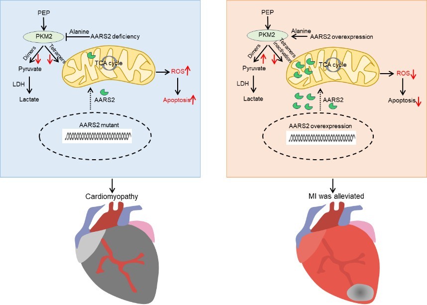
AARS2 ameliorates myocardial ischemia via fine-tuning PKM2-mediated metabolism
- Beijing Key Laboratory of Cardiometabolic Molecular Medicine, Institute of Molecular Medicine, College of Future Technology, Academy for Advanced Interdisciplinary Studies, and State Key Laboratory of Natural and Biomimetic Drugs, Peking University, China
- School of Basic Medical Sciences and The Second Affiliated Hospital, Jiangxi Medical College, Nanchang University, China
- Obstetrics and Gynecology Hospital of Fudan University, State Key Lab of Genetic Engineering, School of Life Sciences and Institutes of Biomedical Sciences, Fudan University, China
Figures
Figure 1 with 1 supplement
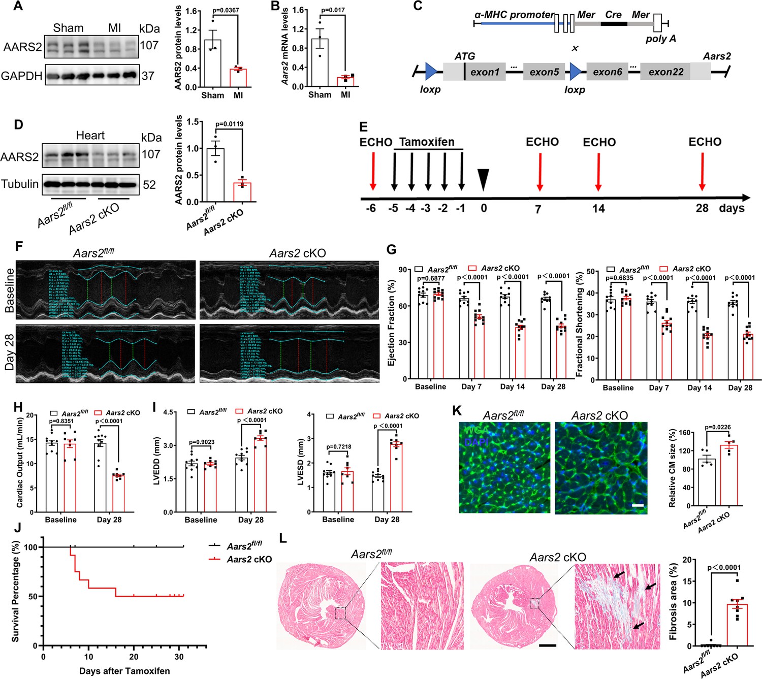
Cardiomyocyte-specific knockout of alanyl-tRNA synthetase (AARS2) leads to cardiac dysfunction and fibrosis in mice.
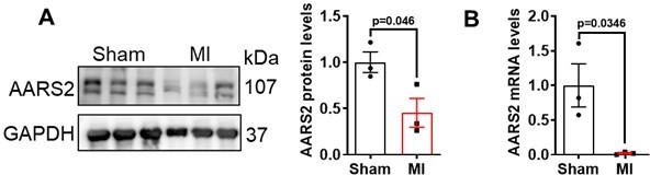
(A, B) Western blot and quantitative real-time PCR (qRT-PCR) analysis showing reduced expression of AARS2 proteins (A) or mRNA (B) of 3 d myocardial infarction (MI) hearts compared with sham hearts (n=3). (C) Construction diagram of α-MHC-MerCreMer (upper) and Aars2fl/fl mice (lower). (D) Western blots showing reduced AARS2 proteins in Aars2 cKO hearts compared with Aars2fl/fl hearts (n=3). (E) Schematic timelines of tamoxifen treatment and echocardiography (ECHO). (F) Representative M-mode tracings of ECHO in control and conditioned knockout (cKO) hearts before and after tamoxifen treatment. (G) Ejection fraction (EF) and fractional shortening (FS) of Aars2fl/fl and Aars2 cKO hearts at different time points after tamoxifen induction (n=10–11). (H) Cardiac output of Aars2fl/fl and Aars2 cKO mice at 28 d after tamoxifen induction (n=8–10). (I) Left ventricular end-diastolic diameter (LVEDD) and left ventricular end-systolic diameter (LVESD) in Aars2fl/fl and Aars2 cKO hearts at 28 d after tamoxifen induction (n=8–10). (J) Survival percentage of Aars2fl/fl and Aars2 cKO mice at 28 d after tamoxifen treatment (n=8–10). (K) WGA immunofluorescence showing cardiomyocyte hypertrophy on heart slices of Aars2 cKO group compared with Aars2fl/fl group at 28 d after tamoxifen induction (scale bar, 100 μm; n=5). (L) Masson staining and quantitative analysis showing increased cardiac fibrosis in Aars2 cKO hearts compared with Aars2fl/fl control hearts at 28 d after tamoxifen induction (scale bar, 1 mm; n=8). Mean ± s.e.m.
-
Figure 1—source data 1
PDF file containing original western blots for Figure 1A and D.
- https://cdn.elifesciences.org/articles/99670/elife-99670-fig1-data1-v1.zip
-
Figure 1—source data 2
Original files for western blot analysis displayed in Figure 1A and D.
- https://cdn.elifesciences.org/articles/99670/elife-99670-fig1-data2-v1.zip
Figure 1—figure supplement 1
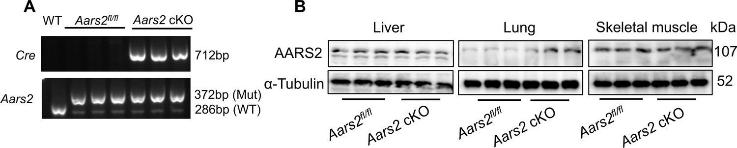
Genotyping of cardiomyocyte-specific Aars2 knockout mice.
(A) Genotyping of Aars2fl/fl and Aars2 cKO mice (n=3; Mut, mutant). (B) Western Blot showing normal expression of AARS2 proteins in the liver, lung, and skeletal muscle of Aars2fl/fl and Aars2 cKO mice (n=3).
-
Figure 1—figure supplement 1—source data 1
PDF file containing original western blots for Figure 1—figure supplement 1A and B.
- https://cdn.elifesciences.org/articles/99670/elife-99670-fig1-figsupp1-data1-v1.zip
-
Figure 1—figure supplement 1—source data 2
Original files for western blot analysis displayed in Figure 1—figure supplement 1A and B.
- https://cdn.elifesciences.org/articles/99670/elife-99670-fig1-figsupp1-data2-v1.zip
Figure 2 with 1 supplement
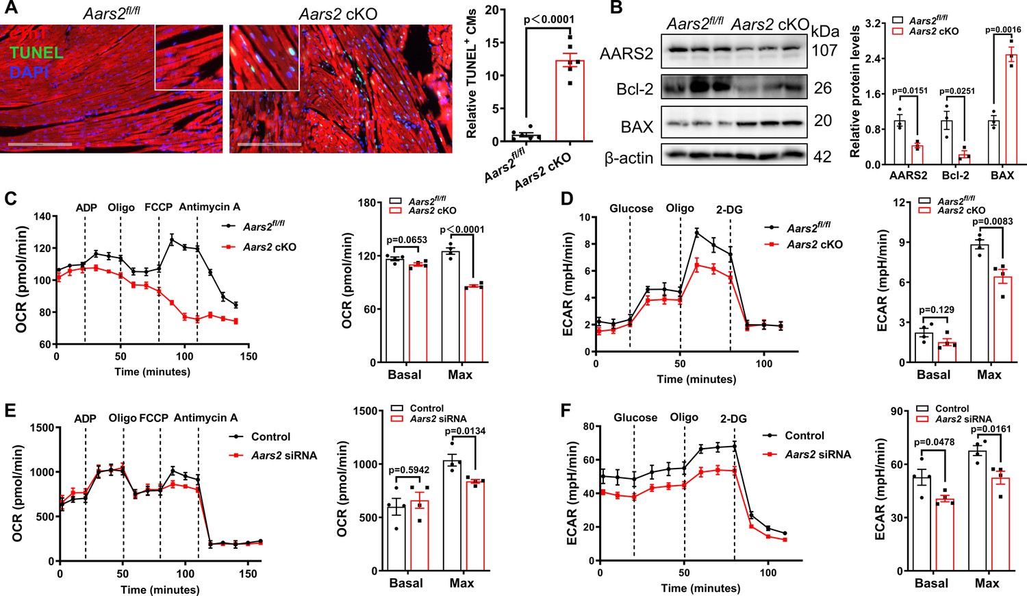
Cardiomyocyte-specific conditioned knockout (cKO) of alanyl-tRNA synthetase (AARS2) results in cardiomyocyte apoptosis and energy metabolism deficiency.
(A, B) Immunofluorescence staining showing increased numbers of cTnT+ TUNEL+ cardiomyocytes (A) and Western blot showing reduced anti-apoptotic protein Bcl-2 and increased pro-apoptotic protein BAX (B) in Aars2 cKO hearts compared with Aars2fl/fl control hearts at 28 d post-tamoxifen treatment (scale, 200 μm; n=6 for panel A; n=3 for panel B). (C) Seahorse analysis showing reduced oxygen consumption rate (OCR) of cardiac mitochondria in Aars2 cKO hearts compared with Aars2fl/fl control hearts at 28 d after tamoxifen induction (n=4). (D) Seahorse analysis showing reduced extracellular acidification rate (ECAR) of adult mouse cardiomyocytes in Aars2 cKO hearts compared with Aars2fl/fl control hearts at 28 d after tamoxifen induction (n=4). (E–F) Seahorse analysis showing reduced OCR (E) and ECAR (F) of NRCMs in AARS2 siRNA group compared with control group at 3 d after transfection (n=4). Mean ± s.e.m.
-
Figure 2—source data 1
PDF file containing original western blots for Figure 2B.
- https://cdn.elifesciences.org/articles/99670/elife-99670-fig2-data1-v1.zip
-
Figure 2—source data 2
Original files for western blot analysis displayed in Figure 2B.
- https://cdn.elifesciences.org/articles/99670/elife-99670-fig2-data2-v1.zip
Figure 2—figure supplement 1
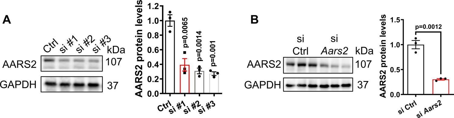
Evaluating Aars2 siRNA for knockout efficiency of alanyl-tRNA synthetase (AARS2) proteins in neonatal rat cardiomyocytes (NRCMs).
(A) Western blots showing knockdown efficiency of three different Aars2 siRNAs in NRCMs, respectively (n=3). (B) Western blots confirming knockdown efficiency of Aars2 siRNA#3 in NRCMs (n=3). Ctrl, control; si, small interfering; mean ± s.e.m.
-
Figure 2—figure supplement 1—source data 1
PDF file containing original western blots for Figure 2—figure supplement 1A and B.
- https://cdn.elifesciences.org/articles/99670/elife-99670-fig2-figsupp1-data1-v1.zip
-
Figure 2—figure supplement 1—source data 2
Original files for western blot analysis displayed in Figure 2—figure supplement 1A and B.
- https://cdn.elifesciences.org/articles/99670/elife-99670-fig2-figsupp1-data2-v1.zip
Figure 3 with 2 supplements
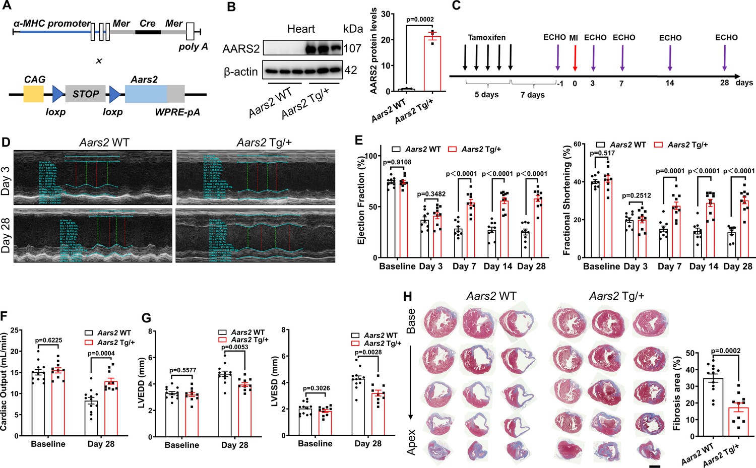
Cardiomyocyte-specific alanyl-tRNA synthetase (AARS2) overexpression improves cardiac function and decreases cardiac fibrosis in mice post-MI.
(A) Schematic diagram of α-MHC-MerCreMer, and CAG-Aars2 mice that is driven by the CAG promoter. (B) Western blots showing transgenic overexpression of AARS2 proteins in the hearts of Aars2 Tg/+compared with Aars2 WT control mice (n=3). (C) Experimental protocols for Tamoxifen induction for 5 d, and then recovery for 7 d before echocardiography (ECHO) and myocardial infarction (MI). (D) Representative M-mode of ECHO in control and Aars2 Tg/+mouse hearts at 3 d or 28 d post-MI. (E) Ejection fraction (EF) and fractional shortening (FS) of the Aars2 WT and Aars2 Tg/+mouse hearts were measured at different time points before and after MI (n=10–11). (F) The cardiac output of Aars2 WT and Aars2 Tg/+mice was measured before MI and 28 d after MI (n=10–11). (G) Left ventricle end-diastolic diameter (LVEDD) and left ventricle end-systolic diameter (LVESD) of Aars2 WT and Aars2 Tg/+ mice before MI and 28 d after MI (n=10–11). (H) Masson’s staining showing decreased fibrotic area in the hearts of Aars2 Tg/+compared with Aars2 WT mice at 28 d after MI (scale bar, 1 mm, n=10–11). Mean ± s.e.m.
-
Figure 3—source data 1
PDF file containing original western blots for Figure 3B.
- https://cdn.elifesciences.org/articles/99670/elife-99670-fig3-data1-v1.zip
-
Figure 3—source data 2
Original files for western blot analysis displayed in Figure 3B.
- https://cdn.elifesciences.org/articles/99670/elife-99670-fig3-data2-v1.zip
Figure 3—figure supplement 1
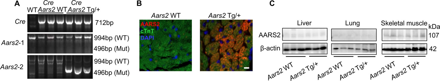
Cardiomyocyte-specific overexpression of alanyl-tRNA synthetase (AARS2) in the heart but not in the liver, lung, and skeletal muscle.
(A) Genotyping of wild-type (WT), α-MHC-MerCreMer; Aars2 WT (Aars2 WT) control, and α-MHC-MerCreMer; Aars2 Tg/+ (Aars2 Tg/+) mice (n=3). (B) Immunofluorescence staining showing ectopic AARS2 proteins in cardiomyocytes of Aars2 Tg/+transgenic mice compared with Aars2 WT control mice (scale bar, 50 μm). (C) Western blots showing comparable expression of AARS2 proteins in the liver, lung, and skeletal muscle of Aars2 WT control and Aars2 Tg/+ transgenic mice (n=3). Mean ± s.e.m.
-
Figure 3—figure supplement 1—source data 1
PDF file containing original western blots for Figure 3—figure supplement 1A and B.
- https://cdn.elifesciences.org/articles/99670/elife-99670-fig3-figsupp1-data1-v1.zip
-
Figure 3—figure supplement 1—source data 2
Original files for western blot analysis displayed in Figure 3—figure supplement 1A and B.
- https://cdn.elifesciences.org/articles/99670/elife-99670-fig3-figsupp1-data2-v1.zip
Figure 3—figure supplement 2
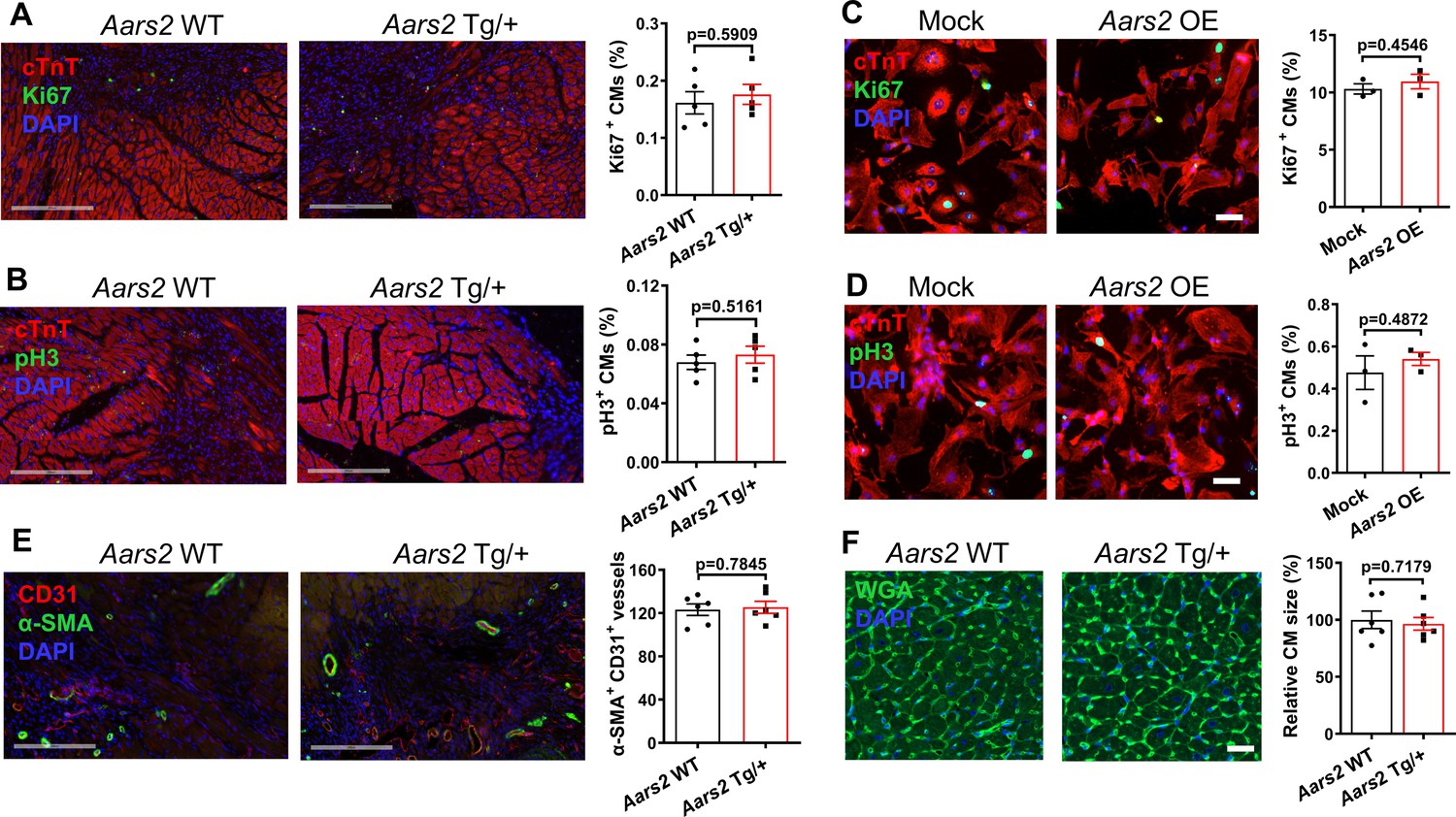
Overexpression of alanyl-tRNA synthetase (AARS2) in cardiomyocytes has no apparent effect on cardiomyocyte proliferation, hypertrophy, and angiogenesis after myocardial infarction (MI).
(A–B) At 7 d after MI, immunofluorescence staining showing comparable cTnT+/Ki67+ (A) and cTnT+/pH3+ (B) cardiomyocytes in the heart sections of Aars2 WT and Aars2 Tg/+groups (scale, 200 μm; n=5). (C–D) Immunofluorescence staining showing comparable numbers of cTnT+/Ki67+ (C) and cTnT+/pH3+ (D) cardiomyocytes at 48 h after infection of neonatal rat cardiomyocytes (NRCMs) with Mock lentivirus or Aars2 lentivirus (scale, 50 μm; n=3). (E) At 7 d after MI, immunofluorescence staining showing comparable CD31+/α-SMA+ coronary vessels in the heart sections of Aars2 WT and Aars2 Tg/+groups (scale, 200 μm; n=5). (F) Wheat germ agglutinin (WGA) staining showing no evident cardiomyocyte hypertrophy in heart slices of Aars2 WT and Aars2 Tg/+groups at 28 d after MI (scale, 100 μm; n=6). Mean ± s.e.m.
Figure 4
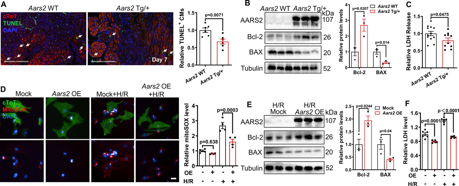
Overexpression of alanyl-tRNA synthetase (AARS2) attenuates cardiomyocyte apoptosis.
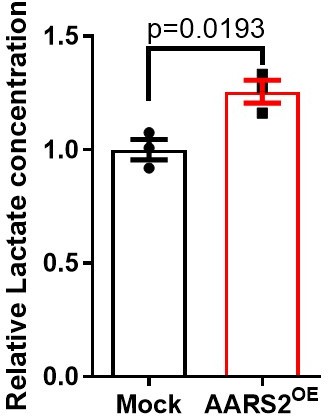
(A) Immunofluorescence staining showing reduced cTnT+/TUNEL+ cardiomyocytes in Aars2 Tg/+hearts compared with Aars2 WT control hearts at 7 d after myocardial infarction (MI) (scale bar, 200 μm, n=6). (B) Western blots showing increased anti-apoptotic protein Bcl-2 and decreased pro-apoptotic protein BAX in Aars2 Tg/+compared with Aars2 WT control hearts at 7 d after MI (n=3). (C) The serum level of lactate dehydrogenase (LDH) decreased in Aars2 Tg/+ hearts compared with Aars2 WT control hearts at 28 d after MI (n=10). (D) Immunofluorescence staining and quantitative analysis showing reduced MitoSOX in Aars2 OE neonatal rat cardiomyocytes (NRCMs) after 12 hr of hypoxia followed by 1 hr of reoxygenation (H/R, scale bar, 20 μm; n=4). (E) Western blots showing increased Bcl-2 and decreased BAX in NRCMs overexpressing Aars2 (Aars2 OE) compared with control NRCMs after 12 hr of hypoxia followed by 1 hr of reoxygenation (n=3). (F) The level of LDH decreased in Aars2 OE NRCMs after 12 hr of hypoxia followed by 1 hr of reoxygenation (n=6). Mean ± s.e.m.
-
Figure 4—source data 1
PDF file containing original western blots for Figure 4B and E.
- https://cdn.elifesciences.org/articles/99670/elife-99670-fig4-data1-v1.zip
-
Figure 4—source data 2
Original files for western blot analysis displayed in Figure 4B and E.
- https://cdn.elifesciences.org/articles/99670/elife-99670-fig4-data2-v1.zip
Figure 5
Cardiomyocyte overexpression of alanyl-tRNA synthetase (AARS2) regulates cardiac metabolism.
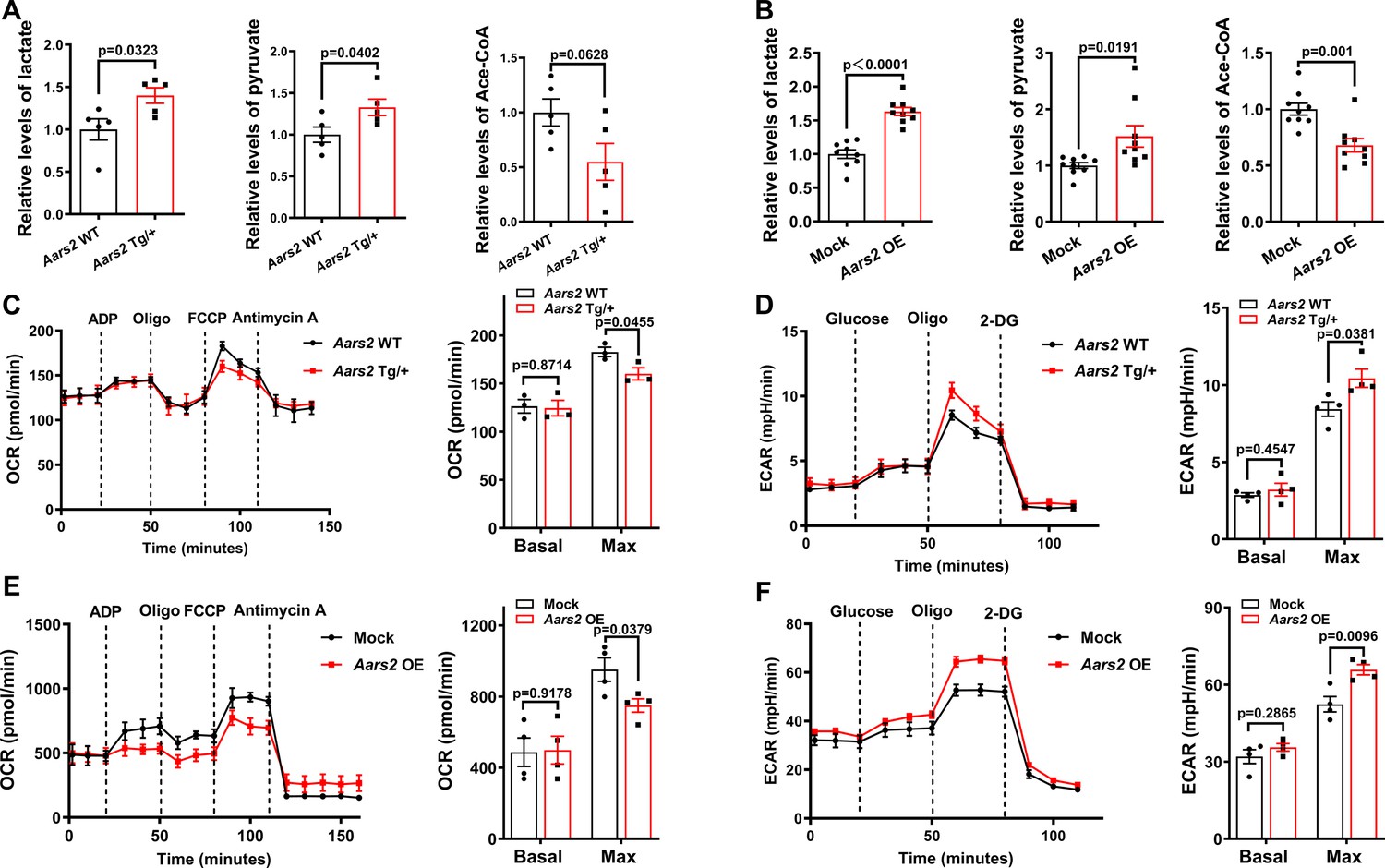
(A) Mass spectrometry showing increased lactate and pyruvate but reduced acetyl-CoA in Aars2 Tg/+ hearts compared with Aars2 WT hearts after 7 d of myocardial infarction (MI) (n=5). (B) Mass spectrometry showing increased lactate and pyruvate but decreased acetyl-CoA in neonatal rat cardiomyocytes (NRCMs) overexpressing Aars2 (Aars2 OE) for 3 d (n=9). (C) Seahorse analysis showing oxygen consumption rate (OCR) of cardiac mitochondria in Aars2 WT and Aars2 Tg/+ mice at 28 ds after tamoxifen induction (n=3). (D) Seahorse analysis showing extracellular acidification rate (ECAR) and quantitative analysis of adult mouse cardiomyocytes in Aars2 WT and Aars2 Tg/+ mice at 28 d after tamoxifen induction (n=4). (E–F) Seahorse analysis showing OCR (E) and ECAR (F) of NRCMs in Mock control and Aars2 OE groups at 3 d after transfection (n=4). Mean ± s.e.m.
Figure 6 with 1 supplement
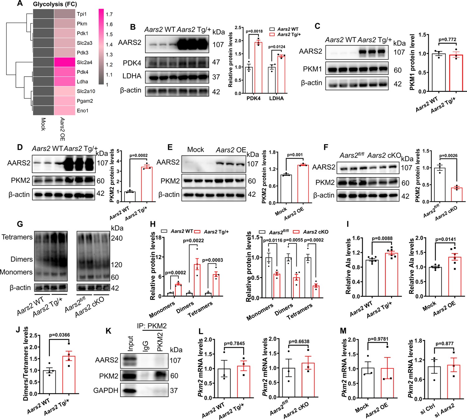
Overexpression of alanyl-tRNA synthetase (AARS2) increases the protein level of glycolytic pyruvate kinase M2 (PKM2) via enhancing PKM2 translation.
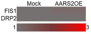
(A) Ribosome RNA-seq showing elevated translation of signaling pathways of glycolysis in the Aars2 OE NRCMs compared to the Mock neonatal rat cardiomyocytes (NRCMs). (B) Western Blots showing the level of AARS2, PDK4, and LDHA proteins in the hearts of Aars2 WT control and Aars2 Tg/+ transgenic mice (n=3). (C) Western Blots showing the level of AARS2 and PKM1 proteins in the hearts of Aars2 WT control and Aars2 Tg/+ transgenic mice (n=3). (D–F) Western Blots showing the level of AARS2 and PKM2 proteins in the hearts of Aars2 WT control and Aars2 Tg/+ transgenic mice (n=3) (D), in Mock control and Aars2 OE NRCMs (E) (n=3), and in the hearts of Aars2fl/fl and Aars2 cKO mice (F) (n=3). (G–H) Western Blots by non-denatured gels (G) and statistics (H) showing the amounts of PKM2 monomers, dimers, and tetramers in the hearts of Aars2 WT control and Aars2 Tg/+transgenic mice, and in the hearts of Aars2fl/fl and Aars2 cKO mice (n=4). (I) Mass spectrometry analysis measuring the amounts of alanine (Ala) from homogenates of heart tissues (n=6) and NRCM lysates (n=6). (J) Ratio of quantitative results of PKM2 dimers and tetramers in the hearts of Aars2 WT control and Aars2 Tg/+ transgenic mice of panel H (n=4). (K) Co-immunoprecipitation reveals no evident interactions between PKM2 and AARS2 in NRCMs. (L–M) qRT-PCR showing the comparative level of Pkm2 mRNA in the hearts of control sibling and Aars2 Tg/+transgenic hearts; control sibling and Aars2 cKO hearts (L); and in control, Aars2 OE, and AARS2siRNA NRCMs (M) (n=3). FC, Fold changes; Mean ± s.e.m.
-
Figure 6—source data 1
PDF file containing original western blots for Figure 6B–G and K.
- https://cdn.elifesciences.org/articles/99670/elife-99670-fig6-data1-v1.zip
-
Figure 6—source data 2
Original files for western blot analysis displayed in Figure 6B–G and K.
- https://cdn.elifesciences.org/articles/99670/elife-99670-fig6-data2-v1.zip
Figure 6—figure supplement 1
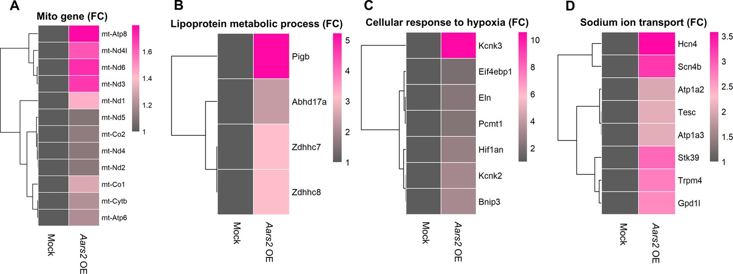
Overexpression of alanyl-tRNA synthetase (AARS2) increases the translation level of some cellular proteins.
(A-D) Ribosome RNA-Seq showing elevated translation of signaling pathways of genes encoded by mitochondria(A), lipoprotein metabolic process (B), cellular response to hypoxia (C), and sodium ion transport (D) in the Aars2 OE neonatal rat cardiomyocytes (NRCMs) compared to the Mock NRCMs. FC, fold changes.
Figure 7
Pyruvate kinase M2 (PKM2) activator TEPP-46 improves cardiomyopathy in Aars2 conditioned knockout (cKO) mice.
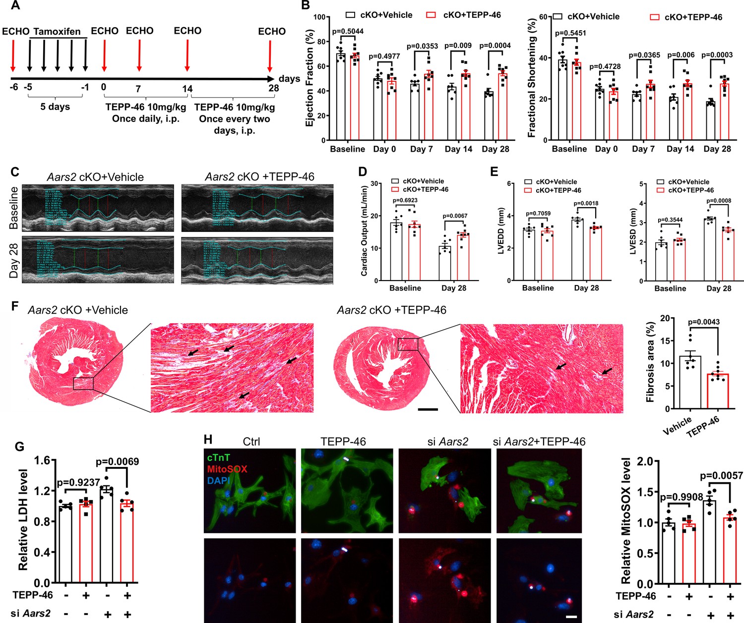
(A) Experimental scheme and time points for tamoxifen induction, echocardiography (ECHO), and TEPP-46 administration. (B) Ejection fraction (EF) and fractional shortening (FS) of Aars2 cKO mouse hearts at different time points after administration of control solvent and TEPP-46 (n=7–8). (C) Representative M-mode of ECHO in different groups of 4 wk mice. (D) Cardiac outputs of Aars2 cKO mouse hearts at different time points after administration of control solvent and TEPP-46 (n=7–8). (E) Left ventricular end-diastolic diameter (LVEDD) and left ventricular end-systolic diameter (LVESD) at 28 d of Aars2 cKO mice after administration of control solvent and TEPP-46 (n=7–8). (F) Masson staining showing cardiac fibrosis of Aars2 cKO mouse hearts at 28 d after administration of either control solvent or TEPP-46 (scale bar, 1 mm; n=7–8). (G) Measurements of lactate dehydrogenase (LDH) release in neonatal rat cardiomyocytes (NRCMs) from the control group and TEPP-46 group (20 μM) after Aars2 siRNA or control siRNA treatment for 72 hr (n=5). (H) Quantitative analysis of MitoSOX immunofluorescence in NRCMs from the control group and TEPP-46 group (20 μM) after Aars2 siRNA or control siRNA treatment for 72 hr (scale bar, 20 μm; n=5). Mean ± s.e.m.
Author response image 1

Author response image 2
Author response image 3
Author response image 4

Author response image 5
Author response image 6

Author response image 7
Tables
Key resources table
| Reagent type (species) or resource | Designation | Source or reference | Identifiers | Additional information |
|---|---|---|---|---|
| Genetic reagent (M. musculus) | Aars2flox/flox | Zhao Lab, Fudan University | N/A | C57BL/6 J |
| Genetic reagent (M. musculus) | Aars2 Transgenic | Zhao Lab, Fudan University | N/A | C57BL/6 J |
| Genetic reagent (M. musculus) | α-MHC-MerCreMer | Xiong Lab, Peking University | RRID:IMSR_GPT:T060079 | C57BL/6 J |
| Biological sample (rat) | Primary neonatal rat cardiomyocytes | Charles River | N/A | CD(SD) IGS Rat, 3 d |
| Antibody | anti-PKM2 (Rabbit polyclonal) | CST | 4053 S RRID:AB_1904096 | WB (1:1000) |
| Antibody | anti-AARS2 (Rabbit polyclonal) | Abcam | ab197367 RRID:AB_2943036 | WB (1:1000) |
| Antibody | anti-Bcl2 (Rabbit polyclonal) | CST | 3498 S RRID:AB_1903907 | WB (1:1000) |
| Antibody | anti-BAX (Rabbit polyclonal) | CST | 14796 S RRID:AB_2716251 | WB (1:1000) |
| Antibody | anti-PKM1 (Rabbit polyclonal) | Proteintech | 15821–1-AP RRID:AB_2163820 | WB (1:1000) |
| Antibody | anti-PDK4 (Rabbit polyclonal) | Bioss | bs-0682R RRID:AB_10856420 | WB (1:1000) |
| Antibody | anti-LDHA (Rabbit polyclonal) | Bioss | bs-34202R This paper | WB (1:1000) |
| Antibody | Goat Anti-Rabbit IgG (H&L)-HRP Conjugated | EASYBIO | BE0101-100 RRID:AB_3083002 | WB (1:10000) |
| Antibody | HRP-conjugated GAPDH | Proteintech | HRP-60004 RRID:AB_2737588 | WB (1:10000) |
| Antibody | HRP-conjugated Beta Actin | Proteintech | HRP-60008 RRID:AB_2819183 | WB (1:10000) |
| Antibody | HRP-conjugated Alpha Tubulin | Proteintech | HRP-66031 RRID:AB_2687491 | WB (1:10000) |
| Antibody | anti-Ki67 (Rabbit polyclonal) | CST | 12075 S RRID:AB_2728830 | IF (1:300) |
| Antibody | anti-pH3 (Rabbit polyclonal) | CST | 53348 S RRID:AB_2799431 | IF (1:300) |
| Antibody | anti-cTnT (Mouse polyclonal) | Abcam | ab8295 RRID:AB_306445 | IF (1:300) |
| Antibody | anti-α-SMA (Mouse polyclonal) | CST | 48938 S RRID:AB_2799347 | IF (1:300) |
| Antibody | anti-CD31 (Rabbit polyclonal) | Abcam | ab182981 RRID:AB_2920881 | IF (1:300) |
| Antibody | Goat Anti-Rabbit IgG H&L (Alexa Fluor 488) | Abcam | ab150077 RRID:AB_2630356 | IF (1:500) |
| Antibody | Goat Anti-Mouse IgG H&L (Alexa Fluor 555) | Abcam | ab150118 RRID:AB_2714033 | IF (1:500) |
| Recombinant DNA reagent | pLenti-CMV-MCS-PGK-Puro-WPRE (plasmid) | This paper | N/A | CMV-MCS-PGK-Puro |
| Software, algorithm | Zen | Zeiss | RRID:SCR_013672 | https://www.zeiss.com/microscopy/en/products/software/zeiss-zen.html |
| Software, algorithm | GraphPad Prism | GraphPad | RRID:SCR_002798 | http://www.graphpad.com/ |
| Software, algorithm | Seahorse Wave | Agilent | RRID:SCR_014526 | http://www.agilent.com/en-us/products/cell-analysis-(seahorse)/software-download-for-wave-desktop |
| Software, algorithm | ImageJ | ImageJ | RRID:SCR_003070 | https://imagej.nih.gov/ij/ |
Additional files
-
Supplementary file 1
Aars2 siRNA sequences, genotyping primers, and real-time PCR primers for mouse and rat models.
- https://cdn.elifesciences.org/articles/99670/elife-99670-supp1-v1.docx
-
MDAR checklist
- https://cdn.elifesciences.org/articles/99670/elife-99670-mdarchecklist1-v1.pdf
Download links
A two-part list of links to download the article, or parts of the article, in various formats.
Downloads (link to download the article as PDF)
Open citations (links to open the citations from this article in various online reference manager services)
Cite this article (links to download the citations from this article in formats compatible with various reference manager tools)
AARS2 ameliorates myocardial ischemia via fine-tuning PKM2-mediated metabolism
eLife 13:RP99670.
https://doi.org/10.7554/eLife.99670.3


