Scientists have developed a way to image sexual reproduction in living flowers, according to a study published today in the open-access journal eLife.
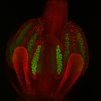
An Arabidopsis floral bud with pollen mother cells highlighted in green. Credit: Valuchova, Mikulkova et al. (CC BY 4.0)
The new technique, originally reported on bioRxiv*, records for the first time movies of fundamental processes in flower development and opens up new avenues for research on plant sexual reproduction.
Plant reproduction occurs in the anthers and ovaries of developing flowers, resulting in the formation of pollen and an embryo sac that holds male and female germ cells. The production of germ cells involves both types of cell division called meiosis and mitosis. Following production, these cells fuse together during fertilisation to produce a cell that develops into a plant.
Much of our understanding of plant reproductive processes has come from studying dissected samples of plants under a microscope and by examining aberrations arising from mutations in plant genes involved in plant reproduction. However, these methods do not provide information about where and when different events happen during reproduction. Live imaging provides a way of capturing this important detail.
“Live imaging has been instrumental in research into root growth and development, but live-cell imaging of cell processes within the flower is technically much more challenging,” explains co-first author Sona Valuchova, a postdoctoral researcher at the Central European Institute of Technology at Masaryk University, Czech Republic. “There is a need to develop imaging methods in the context of whole organs or plants.”
Valuchova and her colleagues used a technique called light sheet fluorescence microscopy (LSFM), where a sample is moved through a thin sheet of laser light, and a detector picks up three-dimensional image data. In this way, an entire flower that has been embedded in agar can be imaged quickly by moving it through the plane of light. The resulting 3D model shows intricate detail of the flower structure and can be used to track the fate of individual germ cells within it.
Having shown that LSFM could provide high-resolution images of flowers, the team’s next goal was to establish that it could detect specific events in reproduction. To achieve this, they used flowers that had been engineered to have fluorescent labels on key molecules involved in meiosis and mitosis. They were able to capture the entire process of meiosis in male germ cells by detecting changes in the amount and location of a molecule called ASY1 every hour for four days.
The team went on to show that live imaging could be successfully used to study plant hormone levels during different stages of flower development and to watch the movement of chromosomes across the cell during cell division.
One of the most neglected areas of plant reproduction research is the production of female germ cells during female meiosis. Most studies on plant meiosis have focused on male germ cells because the female equivalent – called the megaspore mother cell – are incredibly rare and look very similar to other cells, making them hard to study. To overcome this, the team developed a version of live imaging specifically for female meiosis. “This required careful dissection of the flower bud to reveal the ovules, which were then passed through the laser light every 10 minutes over 24 hours to create a 3D film,” says co-first author Pavlina Mikulkova, also a senior scientist at the Central European Institute of Technology at Masaryk University. “Using this technique, we were able to record the two phases of female meiosis and determine how long each one lasted.”
“This work demonstrates the power of LSFM to provide novel information about plant reproduction that could not previously be studied by other types of microscopy,” concludes senior researcher Karel Riha, Deputy Director for Research at the Central European Institute of Technology at Masaryk University. “Our success in developing a live imaging protocol for female meiosis represents a major technical advancement in plant cell biology.”
*This study was originally published on bioRxiv at https://www.biorxiv.org/content/10.1101/774299v2.
Media contacts
Emily Packer
eLife
e.packer@elifesciences.org
+441223855373Ester Jarour
CEITEC MU
ester.jarour@ceitec.muni.cz
+420775351405
About
About eLife
eLife is a non-profit organisation inspired by research funders and led by scientists. Our mission is to help scientists accelerate discovery by operating a platform for research communication that encourages and recognises the most responsible behaviours in science. We publish important research in all areas of the life and biomedical sciences, including Cell Biology and Plant Biology, which is selected and evaluated by working scientists and made freely available online without delay. eLife also invests in innovation through open-source tool development to accelerate research communication and discovery. Our work is guided by the communities we serve. eLife is supported by the Howard Hughes Medical Institute, the Max Planck Society, the Wellcome Trust and the Knut and Alice Wallenberg Foundation. Learn more at https://elifesciences.org.
To read the latest Cell Biology research published in eLife, visit https://elifesciences.org/subjects/cell-biology.
And for the latest in Plant Biology, see https://elifesciences.org/subjects/plant-biology.
About CEITEC Masaryk University
CEITEC MU – Central European Institute of Technology at Masaryk University – a member of the CEITEC consortium, is the leading life science research institute of the Czech Republic. CEITEC is based in Brno, where G.J. Mendel once laid the groundwork for modern genetics. The young and dynamic interdisciplinary centre is focused on plant science, structural biology, molecular medicine, and neuroscience. CEITEC attracts top EU grants and has become an integral part of distinguished European scientific networks. Its success can be attributed not only to its modern, efficiently managed shared laboratories, but also to its people. The centre employs excellent scientists from all over the world, who are committed to fulfil CEITEC’s mission to improve quality of life and human health. CEITEC strives to provide a motivating and innovative scientific environment, state-of-the-art research infrastructure, and a supportive culture of open communication and equal opportunities. Learn more at https://www.ceitec.eu.


