Peer review process
Not revised: This Reviewed Preprint includes the authors’ original preprint (without revision), an eLife assessment, public reviews, and a provisional response from the authors.
Read more about eLife’s peer review process.Editors
- Reviewing EditorDion DickmanUniversity of Southern California, Los Angeles, United States of America
- Senior EditorSonia SenTata Institute for Genetics and Society, Bangalore, India
Reviewer #1 (Public review):
The authors of this study use electron microscopy and 3D reconstruction techniques to study the morphology of distinct classes of Drosophila sensory neurons *across many neurons of the same class.* This is a comprehensive study attempting to look at nearly all the sensory neurons across multiple sensilla to determine a) how much morphological variability exists between and within neurons of different and similar sensory classes, and 2) identify dendritic features that may have evolved to support particular sensory functions. This study builds upon the authors' previous work, which allowed them to identify and distinguish sensory neuron subtypes in the EM volumes without additional staining so that reconstructed neurons could reliably be placed in the appropriate class. This work is unique in looking at a large number of individual neurons of the same class to determine what is consistent and what is variable about their class-specific morphologies.
This means that in addition to providing specific structural information about these particular cells, the authors explore broader questions of how much morphological diversity exists between sensory neurons of the same class and how different dendritic morphologies might affect sensory and physiological properties of neurons.
The authors found that CO2-sensing neurons have an unusual, sheet-like morphology in contrast to the thin branches of odor-sensing neurons. They show that this morphology greatly increases the surface area to volume ratio above what could be achieved by modest branching of thin dendrites, and posit that this might be important for their sensory function, though this was not directly tested in their study. The study is mainly descriptive in nature, but thorough, and provides a nice jumping-off point for future functional studies. One interesting future analysis could be to examine all four cell types within a single sensilla together to see if there are any general correlations that could reveal insights about how morphology is determined and the relative contributions of intrinsic mechanisms vs interactions with neighboring cells. For example, if higher than average branching in one cell type correlated with higher than average branching in another type, if in the same sensilla. This might suggest higher extracellular growth or branching cues within a sensilla. Conversely, if higher branching in one cell type consistently leads to reduced length or branching in another, this might point to dendrite-dendrite interactions between cells undergoing competitive or repulsive interactions to define territories within each sensilla as a major determinant of the variability.
Reviewer #2 (Public review):
Summary:
The manuscript employs serial block‐face electron microscopy (SBEM) and cryofixation to obtain high‐resolution, three‐dimensional reconstructions of Drosophila antennal sensilla containing olfactory receptor neurons (ORNs) that detect CO2. This method has been used previously by the same lab in Gonzales et. al, 2021. (https://elifesciences.org/articles/69896), which had provided an exemplary model by integrating high-resolution EM with electrophysiology and cell-type-specific labeling. The previous study ended up correlating morphology with activity for multiple olfactory sensillar types. Compared to the 2021 study, this current manuscript appears somewhat incomplete and lacks integration with activity.
In fact older studies have also reported two-dimensional TEM images of the putative CO2 neuron in Drosophila (Shanbhag et al., 1999) and in mosquitoes (McIver and Siemicki, 1975; Lu et al, 2007), and in these instances reported that the dendritic architecture of the CO2 neuron was somewhat different (circular and flattened, lamellated) from other olfactory neurons.
The authors claim that this approach offers an artifact‐minimized ultrastructural dataset compared to earlier. In this study, not only do they confirm this different morphology but also classify it into distinct subtypes (loosely curled, fully curled, split, and mixed). This detailed morphological categorization was not provided in prior studies (e.g., Shanbhag et al., 1999 ). The authors would benefit from providing quantitative thresholds or objective metrics to improve reproducibility and to clarify whether these structural distinctions correlate with distinct functional roles.
Strengths:
The study makes a convincing case that ab1C neurons exhibit a unique, flattened dendritic morphology unlike the cylindrical dendrites found in ab1D neurons. This observation extends previous qualitative TEM findings by not only confirming the presence of flattened lamellae in CO₂ neurons but also quantifying key morphometrics such as dendritic length, surface area, and volume, and calculating surface area-to-volume ratios. The enhanced ratios observed in the flattened segments are speculated to be linked to potential advantages in receptor distribution (e.g., Gr21a/Gr63a) and efficient signal propagation.
Weaknesses:
While the manuscript offers valuable ultrastructural insights and reveals previously unappreciated heterogeneity among CO₂-sensing neurons, several issues warrant further investigation in addition to the points made above.
(1) Although this quantitative approach is robust compared to earlier descriptive reports, its impact is somewhat limited by the absence of direct electrophysiological data to confirm that ultrastructural differences translate into altered neuronal function. A direct comparison or discussion of how the present findings align with the functional data obtained from electrophysiology would strengthen the overall argument.
(2) Clarifying the criteria for dendritic subtype classification with quantitative parameters would enhance reproducibility and interpretability. Moreover, incorporating electrophysiological recordings from ab1C neurons would provide compelling evidence linking structure and function, and mapping key receptor proteins through immunolabeling could directly correlate receptor distribution with the observed morphological diversity.
(3) Even though Cryofixation is claimed to be superior to chemical fixation for generating fewer artifacts, authors need to confirm independently the variation observed in the CO2 neuron morphologies across populations. All types of fixation in TEMs cause some artifacts, as does serial sectioning. Without understanding the error rates or without independent validation with another method, it is hard to have confidence in the conclusions drawn by the authors of the paper.
Addressing these concerns and integrating additional experiments would significantly bolster the manuscript's completeness and advancement.
Reviewer #3 (Public review):
Summary:
In the current manuscript entitled "Population-level morphological analysis of paired CO2- and odor-sensing olfactory neurons in D. melanogaster via volume electron microscopy", Choy, Charara et al. use volume electron microscopy and neuron reconstruction to compare the dendritic morphology of ab1C and ab1D neurons of the Drosophila basiconic ab1 sensillum. They aim to investigate the degree of dendritic heterogeneity within a functional class of neurons using ab1C and ab1D, which they can identify due to the unique feature of ab1 sensilla to house four neurons and the stereotypic location on the third antennal segment. This is a great use of volumetric electron imaging and neuron reconstruction to sample a population of neurons of the same type. Their data convincingly shows that there is dendritic heterogeneity in both investigated populations, and their sample size is sufficient to strongly support this observation. This data proposes that the phenomenon of dendritic heterogenity is common in the Drosophila olfactory system and will stimulate future investigations into the developmental origin, functional implications, and potential adaptive advantage of this feature.
Moreover, the authors discovered that there is a difference between CO2- and odour-sensing neurons of which the first show a characteristic flattened and sheet-like structure not observed in other sensory neurons sampled in this and previous studies. They hypothesize that this unique dendritic organization, which increases the surface area to volume ratio, might allow more efficient Co2 sensing by housing higher numbers of Co2 receptors. This is supported by previous attempts to express Co2 sensors in olfactory sensory neurons, which lack this dendritic morphology, resulting in lower Co2 sensitivity compared to endogenous neurons.
Overall, this detailed morphological description of olfactory sensory neurons' dendrites convincingly shows heterogeneity in two neuron classes with potential functional impacts for odour sensing.
Strength:
The volumetric EM imaging and reconstruction approach offers unprecedented details in single cell morphology and compares dendrite heterogeneity across a great fraction of ab1 sensilla.
The authors identify specific shapes for ab1C sensilla potentially linked to their unique function in CO2 sensing.
Weaknesses:
While the morphological description is highly detailed, no attempts are made to link this to odour sensitivity or other properties of the neurons. It would have been exciting to see how altered morphology impacts physiology in these olfactory sensory cells.
Author Response:
Reviewer #1 (Public review):
The authors of this study use electron microscopy and 3D reconstruction techniques to study the morphology of distinct classes of Drosophila sensory neurons *across many neurons of the same class.* This is a comprehensive study attempting to look at nearly all the sensory neurons across multiple sensilla to determine a) how much morphological variability exists between and within neurons of different and similar sensory classes, and 2) identify dendritic features that may have evolved to support particular sensory functions. This study builds upon the authors' previous work, which allowed them to identify and distinguish sensory neuron subtypes in the EM volumes without additional staining so that reconstructed neurons could reliably be placed in the appropriate class. This work is unique in looking at a large number of individual neurons of the same class to determine what is consistent and what is variable about their class-specific morphologies.
This means that in addition to providing specific structural information about these particular cells, the authors explore broader questions of how much morphological diversity exists between sensory neurons of the same class and how different dendritic morphologies might affect sensory and physiological properties of neurons.
The authors found that CO2-sensing neurons have an unusual, sheet-like morphology in contrast to the thin branches of odor-sensing neurons. They show that this morphology greatly increases the surface area to volume ratio above what could be achieved by modest branching of thin dendrites, and posit that this might be important for their sensory function, though this was not directly tested in their study. The study is mainly descriptive in nature, but thorough, and provides a nice jumping-off point for future functional studies. One interesting future analysis could be to examine all four cell types within a single sensilla together to see if there are any general correlations that could reveal insights about how morphology is determined and the relative contributions of intrinsic mechanisms vs interactions with neighboring cells. For example, if higher than average branching in one cell type correlated with higher than average branching in another type, if in the same sensilla. This might suggest higher extracellular growth or branching cues within a sensilla. Conversely, if higher branching in one cell type consistently leads to reduced length or branching in another, this might point to dendrite-dendrite interactions between cells undergoing competitive or repulsive interactions to define territories within each sensilla as a major determinant of the variability.
We thank the reviewer for the insightful comments and appreciation for our study.
Reviewer #2 (Public review):
Summary:
The manuscript employs serial block‐face electron microscopy (SBEM) and cryofixation to obtain high‐resolution, three‐dimensional reconstructions of Drosophila antennal sensilla containing olfactory receptor neurons (ORNs) that detect CO2. This method has been used previously by the same lab in Gonzales et. al, 2021. (https://elifesciences.org/articles/69896), which had provided an exemplary model by integrating high-resolution EM with electrophysiology and cell-type-specific labeling.
We thank the reviewer for expressing appreciation for our published study.
The previous study ended up correlating morphology with activity for multiple olfactory sensillar types. Compared to the 2021 study, this current manuscript appears somewhat incomplete and lacks integration with activity.
We thank the reviewer for their feedback. However, we would like to clarify that our previous study did not correlate morphology with activity to a greater extent than the current study. Both employed the same cryofixation, SBEM-based approach without recording odor-induced activity, but the focus of the current work is fundamentally different. While the previous study examined multiple sensillum types, the current study concentrates on a single sensillum type to address a distinct biological question regarding morphological heterogeneity. We appreciate the opportunity to clarify this distinction, and we hope that the revised manuscript more clearly conveys the unique scope and contributions of this study.
In fact older studies have also reported two-dimensional TEM images of the putative CO2 neuron in Drosophila (Shanbhag et al., 1999) and in mosquitoes (McIver and Siemicki, 1975; Lu et al, 2007), and in these instances reported that the dendritic architecture of the CO2 neuron was somewhat different (circular and flattened, lamellated) from other olfactory neurons.
We thank the reviewer for pointing this out. As noted in both the Introduction and Discussion sections, previous studies—including those cited by the reviewer—suggested that CO2-sensing neurons may have a distinct dendritic morphology. However, those earlier studies lacked the means to definitively link the observed morphology to CO2 neuron identity.
In contrast, our study assigns neuronal identity based on quantitative morphometric measurements, allowing us to confidently associate the unique dendritic architecture with CO2 neurons. Furthermore, we extend previous observations by providing full 3D reconstructions and nanoscale morphometric analyses, offering a much more comprehensive and definitive characterization of these neurons. We believe this represents a significant advancement over earlier work.
The authors claim that this approach offers an artifact‐minimized ultrastructural dataset compared to earlier. In this study, not only do they confirm this different morphology but also classify it into distinct subtypes (loosely curled, fully curled, split, and mixed). This detailed morphological categorization was not provided in prior studies (e.g., Shanbhag et al., 1999 ).
We thank the reviewer for acknowledging the significance of our study.
The authors would benefit from providing quantitative thresholds or objective metrics to improve reproducibility and to clarify whether these structural distinctions correlate with distinct functional roles.
We thank the reviewer for raising this point. However, we would like to clarify that assigning neurons to strict morphological subtypes was not the primary aim of our study. In practice, dendritic architectures can be highly complex, with individual neurons often displaying features characteristic of multiple subtypes. This is precisely why we included a “mixed” subtype category—to acknowledge and capture this morphological heterogeneity rather than impose rigid classification boundaries.
Our intent in defining subtypes was not to imply discrete functional classes, but rather to highlight the range of morphological variation observed across ab1C neurons. While we agree that exploring potential correlations between structure and function is an important future direction, the current study focuses on characterizing this diversity using 3D reconstruction and morphometric analysis. We hope this clarifies the purpose and scope of our morphological categorization.
Strengths:
The study makes a convincing case that ab1C neurons exhibit a unique, flattened dendritic morphology unlike the cylindrical dendrites found in ab1D neurons. This observation extends previous qualitative TEM findings by not only confirming the presence of flattened lamellae in CO₂ neurons but also quantifying key morphometrics such as dendritic length, surface area, and volume, and calculating surface area-to-volume ratios. The enhanced ratios observed in the flattened segments are speculated to be linked to potential advantages in receptor distribution (e.g., Gr21a/Gr63a) and efficient signal propagation.
We thank the reviewer for appreciating the significance our current study.
Weaknesses:
While the manuscript offers valuable ultrastructural insights and reveals previously unappreciated heterogeneity among CO₂-sensing neurons, several issues warrant further investigation in addition to the points made above.
(1) Although this quantitative approach is robust compared to earlier descriptive reports, its impact is somewhat limited by the absence of direct electrophysiological data to confirm that ultrastructural differences translate into altered neuronal function. A direct comparison or discussion of how the present findings align with the functional data obtained from electrophysiology would strengthen the overall argument.
We thank the reviewer for this comment. We would like to clarify, however, that our study does not claim that the observed morphological heterogeneity necessarily leads to functional diversity. Rather, we consider this as a possible implication and discuss it as a potential question for future research. This idea is raised only in the Discussion section, and we are carefully not to present functional diversity as a conclusion of our study. Nonetheless, we have reviewed the relevant paragraph to ensure the language remains cautious and does not overstate our interpretation.
We also acknowledge the significance of directly linking ultrastructural features to neuronal function through electrophysiological recordings. However, at present, it is technically challenging to correlate the nanoscale morphology of individual ORNs with their functional activity, as this would require volume EM imaging of the very same neurons that were recorded via electrophysiology. Currently, there is no dye-labeling method compatible with single-sensillum recording and SBEM sample preparation that allows for unambiguous identification and segmentation of recorded ORNs at the necessary ultrastructural resolution.
To acknowledge this important limitation, we have added a paragraph in the Discussion section, as suggested, to clarify the current technical barriers and to highlight this as a promising direction for future methodological advances.
(2) Clarifying the criteria for dendritic subtype classification with quantitative parameters would enhance reproducibility and interpretability. Moreover, incorporating electrophysiological recordings from ab1C neurons would provide compelling evidence linking structure and function, and mapping key receptor proteins through immunolabeling could directly correlate receptor distribution with the observed morphological diversity.
Please see our response to the comment regarding the technical limitations of directly correlating ultrastructure with electrophysiological data.
In addition, we would like to address the suggestion of using immunolabeling to map receptor distribution in relation to the 3D EM models. Currently, antibodies against Gr21a or Gr63a (the receptors expressed in ab1C neurons) are not available. Even if such antibodies were available, immunogold labeling for electron microscopy requires harsh detergent treatment to increase antibody permeability, damaging morphological integrity. These treatments would compromise the very morphological detail that our study aims to capture and quantify.
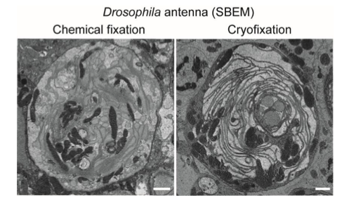
(3) Even though Cryofixation is claimed to be superior to chemical fixation for generating fewer artifacts, authors need to confirm independently the variation observed in the CO2 neuron morphologies across populations. All types of fixation in TEMs cause some artifacts, as does serial sectioning. Without understanding the error rates or without independent validation with another method, it is hard to have confidence in the conclusions drawn by the authors of the paper.
We thank the reviewer for raising concerns regarding potential artifacts in morphological analyses. However, we would like to clarify that cryofixation is widely regarded as a gold standard for ultrastructural preservation and minimizing fixation-induced artifacts, as supported by extensive literature. This is why we adopted high-pressure freezing and freeze substitution in our study.
We have also published a separate methods paper (Tsang et al., eLife, 2018) directly comparing our cryofixation-based protocol with conventional chemical fixation, demonstrating substantial improvements in morphological preservation (see the image below, adapted from Figure 2 of our 2018 eLife paper). This provides strong empirical support for the reliability of our approach.
Author response image 1.
Regarding the suggestion to validate observed morphological variation across populations: we note that determining the presence of artifacts requires a known ground truth, which is inherently unavailable as we could not measure the morphometrics of fly olfactory receptor neurons in their native state. In the absence of such a benchmark, we have instead prioritized using the best-available preparation methods and high-resolution imaging to ensure structural integrity.
Addressing these concerns and integrating additional experiments would significantly bolster the manuscript's completeness and advancement.
We appreciate the reviewer’s feedback. As discussed in our responses to the specific comments above, certain suggested experiments are currently limited by technical constraints, particularly in the context of high-resolution volume EM for insect tissues enclosed in cuticles.
Nevertheless, we have carefully addressed the reviewer’s concerns to the fullest extent possible within the scope of this study. We have revised the manuscript to clarify methodological limitations, added new explanatory content where appropriate, and ensured that our interpretations remain well grounded in the data. We hope these revisions strengthen the clarity and completeness of the manuscript.
Reviewer #3 (Public review):
Summary:
In the current manuscript entitled "Population-level morphological analysis of paired CO2- and odor-sensing olfactory neurons in D. melanogaster via volume electron microscopy", Choy, Charara et al. use volume electron microscopy and neuron reconstruction to compare the dendritic morphology of ab1C and ab1D neurons of the Drosophila basiconic ab1 sensillum. They aim to investigate the degree of dendritic heterogeneity within a functional class of neurons using ab1C and ab1D, which they can identify due to the unique feature of ab1 sensilla to house four neurons and the stereotypic location on the third antennal segment. This is a great use of volumetric electron imaging and neuron reconstruction to sample a population of neurons of the same type. Their data convincingly shows that there is dendritic heterogeneity in both investigated populations, and their sample size is sufficient to strongly support this observation. This data proposes that the phenomenon of dendritic heterogenity is common in the Drosophila olfactory system and will stimulate future investigations into the developmental origin, functional implications, and potential adaptive advantage of this feature.
Moreover, the authors discovered that there is a difference between CO2- and odour-sensing neurons of which the first show a characteristic flattened and sheet-like structure not observed in other sensory neurons sampled in this and previous studies. They hypothesize that this unique dendritic organization, which increases the surface area to volume ratio, might allow more efficient Co2 sensing by housing higher numbers of Co2 receptors. This is supported by previous attempts to express Co2 sensors in olfactory sensory neurons, which lack this dendritic morphology, resulting in lower Co2 sensitivity compared to endogenous neurons.
Overall, this detailed morphological description of olfactory sensory neurons' dendrites convincingly shows heterogeneity in two neuron classes with potential functional impacts for odour sensing.
Strength:
The volumetric EM imaging and reconstruction approach offers unprecedented details in single cell morphology and compares dendrite heterogeneity across a great fraction of ab1 sensilla.
The authors identify specific shapes for ab1C sensilla potentially linked to their unique function in CO2 sensing.
We thank the reviewer for the insightful comments and appreciation for our study.
Weaknesses:
While the morphological description is highly detailed, no attempts are made to link this to odour sensitivity or other properties of the neurons. It would have been exciting to see how altered morphology impacts physiology in these olfactory sensory cells.
We agree that linking morphological variation to physiological properties, such as odor sensitivity, would be a highly valuable direction for future research. However, the aim of the current study is to provide an in-depth nanoscale characterization based on a substantial proportion of ab1 sensilla, highlighting morphological heterogeneity among homotypic ORNs.
At present, it is technically challenging to correlate the nanoscale morphology of individual ORNs with their physiological responses, as this would require volume EM imaging of the exact neurons recorded via single-sensillum electrophysiology. Currently, no dye-labeling method exists that is compatible with both single-sensillum recording and the stringent requirements of SBEM sample preparation to allow for unambiguous identification and segmentation of recorded ORNs.
To acknowledge this important limitation, we have added a paragraph in the Discussion section clarifying the current technical barriers and highlighting this as a promising area for future methodological development. Please also see our responses to the reviewer’s 4th comment below, where we present preliminary experiments examining whether odor sensitivity varies among homotypic ORNs.


