
Peer review process
Revised: This Reviewed Preprint has been revised by the authors in response to the previous round of peer review; the eLife assessment and the public reviews have been updated where necessary by the editors and peer reviewers.
Read more about eLife’s peer review process.Editors
- Reviewing EditorJonathan CooperFred Hutch Cancer Center, Seattle, United States of America
- Senior EditorJonathan CooperFred Hutch Cancer Center, Seattle, United States of America
Reviewer #1 (Public review):
Summary:
This study identifies a mechanism responsible for the accumulation of the MET receptor in invadopodia, following stimulation of Triple-negative breast cancer (TNBC) cells with HGF. HGF-driven accumulation and activation of MET in invadopodia causes the degradation of the extracellular matrix promoting cancer cell invasion, a process here investigated using gelatine-degradation and spheroid invasion assays.
Mechanistically, HGF stimulates the recycling of MET from RAB14-positive endodomes to invadopodia, increasing their formation. At invadopodia, MET induces matrix degradation via direct binding with the metallo protease MT1-MMP.
The delivery of MET from the recycling compartment to invadopodia is mediated by RCP which facilitates the colocalization of MET to RAB14 endosomes. On this compartment, HGF induces the recruitment of the motor protein KIF16B promoting the tubulation of the RAB14-MET recycling endosomes to the cell surface.
This pathway is critical for the HGF-driven invasive properties of TNBC cells as it is impaired upon silencing of RAB14.
Strengths:
The study is well organized and executed using state of the art technology. The effects of MET recycling in the formation of functional invadopodia are carefully studied taking advantage of mutant forms of the receptor that are degradation-resistant or endocytosis-defective.
Data analyses are rigorous and appropriate controls are used in most of the assays to assess the specificity of the scored effects. Overall, the quality of the research is high.
The conclusions are well supported by the results and the data and methodology are of interest for a wide audience of cell biologists.
Previous Weaknesses:
The role of the MET receptor in invadopodia formation and cancer cell dissemination has been intensively studied in many settings including Triple Negative breast cancer cells. The novelty of the present study mostly consists in the detailed molecular description of the underlying mechanism based on HGF-driven MET recycling. The question of whether the identified pathway is specific for TNBC cells or represents a general mechanism of HGF-mediated invasion detectable in other cancer cells is not addressed or at least discussed.
Comments on revised version:
The authors have partially replied to my previous concerns.
Reviewer #2 (Public review):
Summary:
In this manuscript, Khamari and colleagues investigate how HGF-MET signaling and the intracellular trafficking of the MET receptor tyrosine kinase influence invadopodia formation and invasion in triple-negative breast cancer (TNBC) cells. They show that HGF stimulation enhances both the number of invadopodia and their proteolytic activity. Mechanistically, the authors demonstrate that HGF-induced, RAB4- and RCP-RAB14-KIF16B-dependent recycling routes deliver MET to the cell surface specifically at sites where invadopodia form. Moreover, they report that MET physically interacts with MT1-MMP - a key transmembrane metalloproteinase required for invadopodia function- and that these two proteins co-traffic to invadopodia upon HGF stimulation.
Although the HGF-MET axis has previously been implicated in invadopodia regulation (e.g., by Rajadurai et al., Journal of Cell Science 2012), studies directly linking ligand-induced MET trafficking with the spatial regulation of MT1-MMP localization and activity have been lacking.
Overall, the manuscript addresses a relevant and timely topic and provides several novel insights.
Comments on revised version:
I appreciate the authors' efforts to revise the manuscript and address the reviewers' comments. While the revised version includes additional experiments and several improvements in data presentation, the major methodological and conceptual concerns raised in the initial review remain largely unresolved. In my opinion, these issues critically undermine the central mechanistic conclusions of the study.
(1) Inappropriate experimental design for studying MET trafficking
A major concern remains the use of prolonged HGF stimulation times (2-6 hours) to study MET endocytosis and recycling. This is not an appropriate experimental design for investigating receptor tyrosine kinase trafficking dynamics. Ligand-induced internalization of MET occurs within minutes, with maximal endosomal accumulation typically observed within 5-15 minutes, whereas recycling occurs over approximately 15-60 minutes.
Importantly, the authors have not included short stimulation time points or any kinetic analysis that would allow a proper assessment of MET internalization or recycling. The additional surface biotinylation experiment does not address this issue, as it still does not provide temporal information regarding receptor trafficking.
Therefore, the current data do not support the conclusions regarding MET endocytosis or recycling, and this major methodological concern has not been adequately addressed in the revised manuscript.
(2) Insufficient validation of antibody specificity in immunofluorescence
The validation of antibody specificity for MET, phospho-MET, and MT1-MMP in immunofluorescence experiments remains insufficient. While the authors demonstrate knockdown efficiency by immunoblotting and show some reduction in fluorescence signal, they do not provide rigorous evidence that the immunofluorescence signal is specifically abolished upon gene silencing under identical imaging conditions. Such validation is essential, particularly because the manuscript relies heavily on imaging-based localization and colocalization analyses. Without these controls, it cannot be excluded that the observed signal represents non-specific staining.
Importantly, the authors attempt to justify antibody specificity primarily by citing previous publications that used the same antibodies. However, this is not an adequate substitute for experimental validation within the current study. Previous reports do not guarantee specificity under the present experimental conditions, particularly in immunofluorescence, where staining patterns can be strongly influenced by fixation procedures, antibody concentrations, imaging settings, and cell type. Moreover, those studies may themselves lack sufficiently rigorous validation of antibody specificity. Therefore, antibody specificity should be demonstrated directly in the experimental system used in this manuscript, especially given that the principal conclusions rely extensively on the subcellular localization of MET, phospho-MET, and MT1-MMP.
(3) Questionable MET localization in TIRF microscopy
The presence of punctate MET signal in TIRF microscopy under unstimulated conditions raises additional concerns. Under basal conditions, MET is generally expected to exhibit a predominantly diffuse distribution at the plasma membrane, whereas prominent punctate structures are typically associated with ligand-induced clustering, endocytosis, or trafficking events.
The observation of numerous MET-positive puncta in unstimulated cells, together with the insufficient validation of antibody specificity, raises the possibility that at least part of the observed signal represents non-specific staining or imaging artefacts rather than bona fide MET localization. This concern is further compounded by the lack of rigorous immunofluorescence antibody validation discussed above and significantly undermines the interpretation of all TIRF-based trafficking analyses presented in the manuscript.
(4) The evidence supporting a MET-specific role in invadopodia remains unconvincing
The authors argue that the role of MET in invadopodia formation is validated using three independent approaches: shRNA-mediated knockdown, SMARTpool siRNA-mediated knockdown, and pharmacological inhibition with PHA665752. However, I do not agree that these constitute three independent orthogonal validations of MET function.
First, the shRNA-mediated knockdown presented in this study achieves only modest depletion of MET protein. The authors themselves acknowledge this limitation and therefore selected cells with visibly reduced MET staining for imaging. Consequently, the shRNA experiments cannot be considered a robust or independent validation of MET function.
Second, although pooled SMARTpool siRNAs are widely used to improve knockdown efficiency, they cannot exclude off-target effects, as each individual guide RNA contributes its own potential off-target profile. Therefore, pooled siRNAs cannot by themselves establish that an observed phenotype is specifically attributable to depletion of the intended target and do not replace validation using independent individual siRNAs or rescue experiments.
Third, the pharmacological data should also be interpreted with caution. Throughout the manuscript, PHA665752 is presented as a MET inhibitor supporting the specificity of the observed phenotype. However, there is essentially no such thing as a truly selective receptor tyrosine kinase inhibitor. PHA665752 inhibits multiple kinases in addition to MET, particularly at concentrations commonly used in cell-based assays. Consequently, the inhibitor cannot be considered an independent validation of MET-specific function.
Importantly, the newly added siRNA experiments do not resolve my original concern regarding the role of MET in invadopodia formation. Although siRNA-mediated MET depletion is substantially more efficient than the shRNA-mediated knockdown presented in the original manuscript, this marked difference in MET depletion is not accompanied by a correspondingly stronger inhibition of invadopodia formation or ECM degradation. If MET were indeed the principal driver of the observed phenotype, one would expect the magnitude of the biological effect to correlate with the efficiency of MET depletion. This inconsistency raises the possibility that the observed phenotype is not solely attributable to MET depletion and calls into question the specificity of the proposed mechanism.
Taken together, the three perturbation approaches used by the authors cannot be regarded as independent orthogonal validation of MET function. One approach provides only modest target depletion, another relies on pooled RNAi reagents that cannot exclude off-target effects, and the third employs a multi-kinase inhibitor rather than a MET-specific compound. Collectively, these limitations substantially weaken the conclusion that the reduction in invadopodia formation is specifically attributable to loss of MET. A convincing demonstration of MET-specific function would require rescue experiments or another truly orthogonal validation strategy.
(5) Weak evidence for MET-MT1-MMP interaction
The evidence supporting a physical interaction between MET and MT1-MMP remains unconvincing. The newly added co-immunoprecipitation experiment does not reveal a convincing MET-MT1-MMP interaction, and I am unable to appreciate a specific co-immunoprecipitated MT1-MMP signal in the presented blot. As presented, these data do not convincingly demonstrate a specific or functionally relevant interaction. Given that this interaction constitutes a central component of the proposed mechanistic model, this remains a major weakness of the study.
(6) Overinterpretation of the data
Taken together, the study proposes a mechanistic model linking MET trafficking to MT1-MMP localization and invadopodia function. However, the experimental evidence largely supports correlative observations rather than demonstrating a direct mechanistic relationship.
Specifically, MET endocytosis and recycling are not properly demonstrated because of the inappropriate temporal resolution of the trafficking experiments; the localization data remain uncertain owing to insufficient validation of the immunofluorescence reagents; and the proposed interaction between MET and MT1-MMP is not convincingly demonstrated. Consequently, the manuscript establishes correlation rather than causality, and the central mechanistic conclusions appear to be substantially overstated relative to the presented data.
Conclusion:
While the manuscript addresses an interesting and biologically relevant question, the current experimental evidence does not adequately support the proposed mechanistic model. The combination of inappropriate experimental design for trafficking studies, insufficient validation of key imaging reagents, questionable interpretation of the localization data, lack of convincing evidence for the proposed MET-MT1-MMP interaction, and the absence of a clear relationship between the degree of MET depletion and the biological phenotype substantially limits the reliability of the conclusions.
In my opinion, these issues cannot be addressed by further revision of the current manuscript, as they require substantial additional experimentation, including appropriately designed trafficking assays with short kinetic time points, rigorous validation of antibody specificity for immunofluorescence, and stronger mechanistic evidence linking MET trafficking to MT1-MMP-dependent invadopodia function.
Author response:
The following is the authors’ response to the current reviews.
We thank the editor and the reviewers for their comments on the revised manuscript. Based on the comments, we decided to go for a minor revision which will address all the comments of reviewer 1.
Towards the comments of the reviewer 2, we would like to state that we already provided the results from the new experiments and the reasons why we did not perform some of the suggested ones. Interestingly, we have not deviated from standard practices in the field in our approaches. Yet, the reviewer is not convinced and raised concern about the robustness of our observations. We therefore decided to carry out a few more control experiments which in our opinion are redundant as they already were carried out multiple times by us as well as by the other field experts under identical conditions and using the identical cell lines.
Reviewer #1 (Public review):
Summary:
This study identifies a mechanism responsible for the accumulation of the MET receptor in invadopodia, following stimulation of Triple-negative breast cancer (TNBC) cells with HGF. HGF-driven accumulation and activation of MET in invadopodia causes the degradation of the extracellular matrix promoting cancer cell invasion, a process here investigated using gelatine-degradation and spheroid invasion assays.
Mechanistically, HGF stimulates the recycling of MET from RAB14-positive endodomes to invadopodia, increasing their formation. At invadopodia, MET induces matrix degradation via direct binding with the metallo protease MT1-MMP.
The delivery of MET from the recycling compartment to invadopodia is mediated by RCP which facilitates the colocalization of MET to RAB14 endosomes. On this compartment, HGF induces the recruitment of the motor protein KIF16B promoting the tubulation of the RAB14-MET recycling endosomes to the cell surface.
This pathway is critical for the HGF-driven invasive properties of TNBC cells as it is impaired upon silencing of RAB14.
Strengths:
The study is well organized and executed using state of the art technology. The effects of MET recycling in the formation of functional invadopodia are carefully studied taking advantage of mutant forms of the receptor that are degradation-resistant or endocytosisdefective.
Data analyses are rigorous and appropriate controls are used in most of the assays to assess the specificity of the scored effects. Overall, the quality of the research is high.
The conclusions are well supported by the results and the data and methodology are of interest for a wide audience of cell biologists.
Previous Weaknesses:
The role of the MET receptor in invadopodia formation and cancer cell dissemination has been intensively studied in many settings including Triple Negative breast cancer cells. The novelty of the present study mostly consists in the detailed molecular description of the underlying mechanism based on HGF-driven MET recycling. The question of whether the identified pathway is specific for TNBC cells or represents a general mechanism of HGFmediated invasion detectable in other cancer cells is not addressed or at least discussed.
Comments on revised version:
The authors have partially replied to my previous concerns.
We sincerely thank the reviewer for careful evaluation of our manuscript and recognizing the strength of our study. We are grateful for the positive assessment about the well-executed methods, rigorous data analysis, usage of appropriate controls, and for acknowledging that we have addressed, at least in part, the concerns raised in the previous round of review. We appreciate the reviewer’s constructive comments, which have helped us further clarify the scope and significance of our findings.
Reviewer #1 (Recommendations for the authors):
The authors have partially replied to my previous concerns. The following points still have to be addressed.
(1) Despite many TNBC tumours present high expression of the EGFR, trials with EGFR inhibitors have been very disappointing (please read PMID: 41651315). EGFR inhibition has been extensively attempted with negative outcome, and this is well known.
In the clinical practise, Triple Negative breast cancer patients are commonly treated with chemotherapy, not with EGFR or MET inhibitors.
Line 31-38 are misleading at best and should be removed and the incipit of the study modified. The biology of this study is sound there is no need to push the clinical relevance with instances that are notoriously not applicable.
We are thankful to the reviewer for bringing up this point. We will modify the manuscript as per the reviewer’s suggestion.
(2) In reply to point 4, the authors claim that they checked MMP2 and the experiment is shown in FigS5I, which is not......
We sincerely apologise for uploading an incorrect file. The correct file will be uploaded.
(3) I noticed that, in the previous version of the manuscript in Fig. S4A the panel showing the mutation frequency was erroneously indicated in the legend as referring to MET.
The authors replied that they fixed this but actually the legend is still wrong...
We thank the reviewer for bringing this error into our attention. We will attentively correct the legend in the revised version of the manuscript.
Reviewer #2 (Public review):
Summary:
In this manuscript, Khamari and colleagues investigate how HGF-MET signaling and the intracellular trafficking of the MET receptor tyrosine kinase influence invadopodia formation and invasion in triple-negative breast cancer (TNBC) cells. They show that HGF stimulation enhances both the number of invadopodia and their proteolytic activity. Mechanistically, the authors demonstrate that HGF-induced, RAB4- and RCP-RAB14KIF16B-dependent recycling routes deliver MET to the cell surface specifically at sites where invadopodia form. Moreover, they report that MET physically interacts with MT1MMP - a key transmembrane metalloproteinase required for invadopodia function- and that these two proteins co-traffic to invadopodia upon HGF stimulation.
Although the HGF-MET axis has previously been implicated in invadopodia regulation (e.g., by Rajadurai et al., Journal of Cell Science 2012), studies directly linking ligandinduced MET trafficking with the spatial regulation of MT1-MMP localization and activity have been lacking.
Overall, the manuscript addresses a relevant and timely topic and provides several novel insights.
Comments on revised version:
I appreciate the authors' efforts to revise the manuscript and address the reviewers' comments. While the revised version includes additional experiments and several improvements in data presentation, the major methodological and conceptual concerns raised in the initial review remain largely unresolved. In my opinion, these issues critically undermine the central mechanistic conclusions of the study.
We thank the reviewer for critically re-evaluating our manuscript and acknowledging the novel insights and relevance of our study.
We thank the reviewer for pointing out the study by Rajadurai et al., Journal of Cell Science, 2012, which provided crucial evidence about the role of MET signaling in invadopodia formation [1]. However, the experimental system largely used by Rajadurai et al. is fundamentally different from the receptor trafficking mechanism investigated in the present study. The group have mostly used overexpression of Tpr-MET, which is a cytosolic MET mutant, that does not undergo the canonical ligand-induced RTK endocytosis and subsequent degradation or recycling. Our study did not only establish another link between MET signaling and invadopodia formation; rather, we identified a trafficking-dependent mechanism whereby HGF stimulation regulates the spatial redistribution and recycling of full-length MET to invadopodia, thus providing more physiologically relevant insights.
We also respectfully disagree that the methodological concerns raised critically undermine our mechanistic conclusions. Although other approaches as suggested by the reviewer could provide complementary information, we believe that the methods used in our study are appropriate for assessing invasive behaviour of the TNBC cells. These methods have been used by us and other research groups in the filed as reflected from the existing literature [2–6].
In this context, we would also like to add that the study referred by the reviewer above, has used a more off target prone approach (SiGenome Smartpool) compared to ONTARGETplus (chemically modified SiRNA pool for minimizing off target effect) in addition to the same small molecule inhibitor used in our study. Additionally, we also used a shRNA-based silencing to verify the phenotype. Since the silencing was not as pronounced as the siRNA-mediated knockdown, the reviewer has expressed concern.
We would like to point out that the antibody we used in IF for MT1-MMP (MMP14) have been published in multiple peer-reviewed journals by various research groups using the identical cell lines (MDA-MB-231, ATCC- HTB-26) [6–8]. MET antibodies used in the study (CST, D1C2 XP & L6E7) has been validated in MDA-MB-231 and MET-depleted cells [9-12]. Since these antibodies have been utilized for IF since a long time across various research groups under identical laboratory/experimental conditions, the exercise of validation suggested by the reviewer is surprising.
(1) Inappropriate experimental design for studying MET trafficking
A major concern remains the use of prolonged HGF stimulation times (2-6 hours) to study MET endocytosis and recycling. This is not an appropriate experimental design for investigating receptor tyrosine kinase trafficking dynamics. Ligand-induced internalization of MET occurs within minutes, with maximal endosomal accumulation typically observed within 5-15 minutes, whereas recycling occurs over approximately 15-60 minutes.
Importantly, the authors have not included short stimulation time points or any kinetic analysis that would allow a proper assessment of MET internalization or recycling. The additional surface biotinylation experiment does not address this issue, as it still does not provide temporal information regarding receptor trafficking.
Therefore, the current data do not support the conclusions regarding MET endocytosis or recycling, and this major methodological concern has not been adequately addressed in the revised manuscript.
We want to clarify that, our prime objective is to determine how MET trafficking is regulated at the time points at which we observe the functional effects of HGF on invadopodia formation and matrix degradation. Since our functional assays were performed following 2-3 h of HGF stimulation, we specifically examined MET localization and trafficking at these same time points.
Though shorter time points could provide information on the kinetics of MET trafficking, but their absence does not invalidate our conclusions regarding the role of MET trafficking in HGF-induced invasive function. In other words, our conclusions are made for the time points for which we have conducted the experiments. It is needless to add that different cargo molecule will show different kinetics.
In summary, we wanted to study the MET trafficking at the late hours in accordance with our functional assays and accordingly designed our experiments. Also, we have supported our results through biochemical methods which is considered to be one of the gold standards in the field.
(2) Insufficient validation of antibody specificity in immunofluorescence
The validation of antibody specificity for MET, phospho-MET, and MT1-MMP in immunofluorescence experiments remains insufficient. While the authors demonstrate knockdown efficiency by immunoblotting and show some reduction in fluorescence signal, they do not provide rigorous evidence that the immunofluorescence signal is specifically abolished upon gene silencing under identical imaging conditions. Such validation is essential, particularly because the manuscript relies heavily on imaging-based localization and colocalization analyses. Without these controls, it cannot be excluded that the observed signal represents non-specific staining.
Importantly, the authors attempt to justify antibody specificity primarily by citing previous publications that used the same antibodies. However, this is not an adequate substitute for experimental validation within the current study. Previous reports do not guarantee specificity under the present experimental conditions, particularly in immunofluorescence, where staining patterns can be strongly influenced by fixation procedures, antibody concentrations, imaging settings, and cell type. Moreover, those studies may themselves lack sufficiently rigorous validation of antibody specificity. Therefore, antibody specificity should be demonstrated directly in the experimental system used in this manuscript, especially given that the principal conclusions rely extensively on the subcellular localization of MET, phospho-MET, and MT1-MMP.
We understand the reviewer’s concern regarding antibody specificity and agree that appropriate validation is important for imaging-based analyses. However, we strongly disagree with the statement that the MT1-MMP, MET antibody were not adequately validated. The specificity of the MT1-MMP antibody was independently validated by both siRNA- and sgRNA-mediated gene silencing, where we observed a substantially diminished MT1-MMP signal by immunoblotting (Fig S5I, M’). Moreover, the antibody has been used for IF in the same cell line by multiple research groups [6–8]. So, in our opinion, this validation is completely redundant.
We have validated the MET staining/ signal using the antibody in gene-silenced cells by immunoblotting and immunofluorescence (Fig S1I, K, L), as also acknowledged by the reviewer in comment-4. In addition, we would also like to clarify that the references cited in support of antibody specificity were not selected simply because they used the same antibodies. They include studies that provide experimental validation of the antibodies by gene silencing.
To further confirm the antibody specificity, we will add immunofluorescence images of MET or MT1-MMP silenced cells stained with respective antibodies. However, we may not want to add these data to the manuscript as they do not carry any additional values to the manuscript.
(3) Questionable MET localization in TIRF microscopy
The presence of punctate MET signal in TIRF microscopy under unstimulated conditions raises additional concerns. Under basal conditions, MET is generally expected to exhibit a predominantly diffuse distribution at the plasma membrane, whereas prominent punctate structures are typically associated with ligand-induced clustering, endocytosis, or trafficking events.
The observation of numerous MET-positive puncta in unstimulated cells, together with the insufficient validation of antibody specificity, raises the possibility that at least part of the observed signal represents non-specific staining or imaging artefacts rather than bona fide MET localization. This concern is further compounded by the lack of rigorous immunofluorescence antibody validation discussed above and significantly undermines the interpretation of all TIRF-based trafficking analyses presented in the manuscript.
We would like to highlight the apparent similarities between Fig 2A, B and the unstimulated condition in Fig. 2H. In figure 2A, B, MET is detected using an anti-MET antibody, whereas in Figure 2H, GFP-MET is imaged under live cell condition. We believe the reviewer would agree that imaging GFP-MET in live cells avoids fixation- and antibody-related artifacts. The comparable localization observed using these two independent approaches therefore provides additional support that the MET distribution shown in Fig. 2A, B reflects genuine receptor localization rather than an imaging or staining artefact.
(4) The evidence supporting a MET-specific role in invadopodia remains unconvincing
The authors argue that the role of MET in invadopodia formation is validated using three independent approaches: shRNA-mediated knockdown, SMARTpool siRNA-mediated knockdown, and pharmacological inhibition with PHA665752. However, I do not agree that these constitute three independent orthogonal validations of MET function.
First, the shRNA-mediated knockdown presented in this study achieves only modest depletion of MET protein. The authors themselves acknowledge this limitation and therefore selected cells with visibly reduced MET staining for imaging. Consequently, the shRNA experiments cannot be considered a robust or independent validation of MET function.
Second, although pooled SMARTpool siRNAs are widely used to improve knockdown efficiency, they cannot exclude off-target effects, as each individual guide RNA contributes its own potential off-target profile. Therefore, pooled siRNAs cannot by themselves establish that an observed phenotype is specifically attributable to depletion of the intended target and do not replace validation using independent individual siRNAs or rescue experiments.
Third, the pharmacological data should also be interpreted with caution. Throughout the manuscript, PHA665752 is presented as a MET inhibitor supporting the specificity of the observed phenotype. However, there is essentially no such thing as a truly selective receptor tyrosine kinase inhibitor. PHA665752 inhibits multiple kinases in addition to MET, particularly at concentrations commonly used in cell-based assays. Consequently, the inhibitor cannot be considered an independent validation of MET-specific function.
Importantly, the newly added siRNA experiments do not resolve my original concern regarding the role of MET in invadopodia formation. Although siRNA-mediated MET depletion is substantially more efficient than the shRNA-mediated knockdown presented in the original manuscript, this marked difference in MET depletion is not accompanied by a correspondingly stronger inhibition of invadopodia formation or ECM degradation. If MET were indeed the principal driver of the observed phenotype, one would expect the magnitude of the biological effect to correlate with the efficiency of MET depletion. This inconsistency raises the possibility that the observed phenotype is not solely attributable to MET depletion and calls into question the specificity of the proposed mechanism.
Taken together, the three perturbation approaches used by the authors cannot be regarded as independent orthogonal validation of MET function. One approach provides only modest target depletion, another relies on pooled RNAi reagents that cannot exclude off-target effects, and the third employs a multi-kinase inhibitor rather than a MET-specific compound. Collectively, these limitations substantially weaken the conclusion that the reduction in invadopodia formation is specifically attributable to loss of MET. A convincing demonstration of MET-specific function would require rescue experiments or another truly orthogonal validation strategy.
We had adopted three independent approaches to validate the phenotype. All three approaches are well practiced in the field. The small molecule inhibitor has been used in the study by Rajadurai et al, J Cell Science, 2012 and it is very much accepted in studying cellular kinases [1].
We agree that even though the smart pool has always chance of off-target effects, the OnTargetPlus Smart pool has the minimum chance of off-target effects because of the patented chemical modifications, compared to the individual oligos and SiGenome SMARTpool, which was used by Rajadurai, et. al. in their study [1].
The shRNA mediated silencing resulted in less reduction in the MET level (~50-60%) but is it scientifically not acceptable, particularly when it showed similar phenotype over n=3 sets of experiments?
The arguments made by the reviewer in this context seems to be harsh. However, we decided to carry out MET silencing using two independent oligos from the SMART pool.
(5) Weak evidence for MET-MT1-MMP interaction
The evidence supporting a physical interaction between MET and MT1-MMP remains unconvincing. The newly added co-immunoprecipitation experiment does not reveal a convincing MET-MT1-MMP interaction, and I am unable to appreciate a specific coimmunoprecipitated MT1-MMP signal in the presented blot. As presented, these data do not convincingly demonstrate a specific or functionally relevant interaction. Given that this interaction constitutes a central component of the proposed mechanistic model, this remains a major weakness of the study.
We have detected the interaction in both GFP pulldown assay in 4 different cell lines and the corresponding reverse His-Ni-NTA pulldown assay, providing complementary evidence for their physical association (Fig 6E, S5F, G). We have also clearly stated in the manuscript that this interaction is weak in nature and have not claimed it to be a strong interaction. While we acknowledge that the signal is modest, disregarding reproducible positive results would not be an appropriate interpretation of the pulldown assays. We believe the reproducibility of these findings supports a genuine MET and MT1-MMP association, and we have reported it accordingly.
(6) Overinterpretation of the data
Taken together, the study proposes a mechanistic model linking MET trafficking to MT1MMP localization and invadopodia function. However, the experimental evidence largely supports correlative observations rather than demonstrating a direct mechanistic relationship.
Specifically, MET endocytosis and recycling are not properly demonstrated because of the inappropriate temporal resolution of the trafficking experiments; the localization data remain uncertain owing to insufficient validation of the immunofluorescence reagents; and the proposed interaction between MET and MT1-MMP is not convincingly demonstrated. Consequently, the manuscript establishes correlation rather than causality, and the central mechanistic conclusions appear to be substantially overstated relative to the presented data.
We agree that there is scope for further investigation of MET and MT1-MMP cotrafficking, however, we have provided preliminary evidence demonstrating the cotrafficking of MET and MT1-MMP at the cell surface (Fig 6F). Importantly, MET and MT1-MMP co-trafficking represents only one component of the manuscript and not a central mechanistic conclusion. As the title of the study indicates, the major component of the study is focused on MET trafficking and its implication in invadopodia-associated TNBC invasion, for which we have provided direct experimental evidence. Therefore, we believe that describing the overall study as primarily overstated and correlative underestimates the extent of the experimental evidence supporting our mechanistic conclusions.
Conclusion:
While the manuscript addresses an interesting and biologically relevant question, the current experimental evidence does not adequately support the proposed mechanistic model. The combination of inappropriate experimental design for trafficking studies, insufficient validation of key imaging reagents, questionable interpretation of the localization data, lack of convincing evidence for the proposed MET-MT1-MMP interaction, and the absence of a clear relationship between the degree of MET depletion and the biological phenotype substantially limits the reliability of the conclusions.
In my opinion, these issues cannot be addressed by further revision of the current manuscript, as they require substantial additional experimentation, including appropriately designed trafficking assays with short kinetic time points, rigorous validation of antibody specificity for immunofluorescence, and stronger mechanistic evidence linking MET trafficking to MT1-MMP-dependent invadopodia function.
We thank the reviewer for outlining the remaining concerns. We will address the points raised by performing additional antibody validation and independent oligo-mediated MET silencing experiment, providing further support for the specificity and robustness of our findings.
However, we respectfully disagree, that the conclusions require the extensive additional experimentation suggested by the reviewer. As clarified above, our trafficking experiments were designed around the time points at which the functional invasive phenotype is observed, rather than to define the kinetics of MET internalization.
In conclusion, we would expect that the views of the reviewer 2 towards the manuscript should change and the reliability of our manuscript to the public should improve.
References:
(1) Rajadurai CV, Havrylov S, Zaoui K, Vaillancourt R, Stuible M, Naujokas M, Zuo D, Tremblay ML, Park M. Met receptor tyrosine kinase signals through a cortactinGab1 scaffold complex, to mediate invadopodia. J Cell Sci. 2012 Jun 15;125(Pt 12):2940-53. doi: 10.1242/jcs.100834. Epub 2012 Feb 24. PMID: 22366451; PMCID: PMC3434810.
(2) Sharma P, Parveen S, Vinod Shah L, Mukherjee M, Kalaidzidis Y, Joseph Kozielski A, et al. Title: SNX27-retromer assembly directs MT1-MMP trafficking to invadopodia and promotes breast cancer metastasis.
(3) Mader CC, Oser M, Magalhaes MAO, Bravo-Cordero JJ, Condeelis J, Koleske AJ, et al. Molecular and Cellular Pathobiology An EGFR-Src-Arg-Cortactin Pathway Mediates Functional Maturation of Invadopodia and Breast Cancer Cell Invasion [Internet]. doi:10.1158/0008-5472.CAN-10-1432
(4) Parveen S, Khamari A, Raju J, Coppolino MG, Datta S. Syntaxin 7 contributes to breast cancer cell invasion by promoting invadopodia formation. J Cell Sci. 2022 Jun 15;135(12). doi:10.1242/jcs.259576 PubMed PMID: 35762511.
(5) Joffre C, Barrow R, Ménard L, Calleja V, Hart IR, Kermorgant S. A direct role for Met endocytosis in tumorigenesis. Nat Cell Biol. 2011 Jun 5;13(7):827–37. doi:10.1038/ncb2257 PubMed PMID: 21642981.
(6) Monteiro P, Rossé C, Castro-Castro A, Irondelle M, Lagoutte E, Paul-Gilloteaux P, et al. Endosomal WASH and exocyst complexes control exocytosis of MT1-MMP at invadopodia. J Cell Biol. 2013 Dec 23;203(6):1063–79. doi:10.1083/jcb.201306162 PubMed PMID: 24344185.
(7) Wenzel EM, Pedersen NM, Elfmark LA, Wang L, Kjos I, Stang E, et al. Intercellular transfer of cancer cell invasiveness via endosome-mediated protease shedding. Nat Commun. 2024 Feb 10;15(1):1277. doi:10.1038/s41467-024-45558-8
(8) Pedersen NM, Wenzel EM, Wang L, Antoine S, Chavrier P, Stenmark H, Raiborg C. Protrudin-mediated ER-endosome contact sites promote MT1-MMP exocytosis and cell invasion. J Cell Biol. 2020 Aug 3;219(8):e202003063. doi: 10.1083/jcb.202003063. PMID: 32479595; PMCID: PMC7401796.
(9) Duan Q, Jia HR, Chen W, Qin C, Zhang K, Jia F, Fu T, Wei Y, Fan M, Wu Q, Tan W. Multivalent Aptamer-Based Lysosome-Targeting Chimeras (LYTACs) Platform for Mono- or Dual-Targeted Proteins Degradation on Cell Surface. Adv Sci (Weinh). 2024 May;11(17):e2308924. doi: 10.1002/advs.202308924. Epub 2024 Feb 29. PMID: 38425146; PMCID: PMC11077639.
(10) Wei J, Wang J, Guan W, Li J, Pu T, Corey E, Lin TP, Gao AC, Wu BJ. PlexinD1 is a driver and a therapeutic target in advanced prostate cancer. EMBO Mol Med. 2025 Feb;17(2):336-364. doi: 10.1038/s44321-024-00186-z. Epub 2025 Jan 2. PMID: 39748059; PMCID: PMC11822115.
(11) Yamasaki A, Miyake R, Hara Y, Okuno H, Imaida T, Okita K, Okazaki S, Akiyama Y, Hirotani K, Endo Y, Masuko K, Masuko T, Tomioka Y. Dual-targeting therapy against HER3/MET in human colorectal cancers. Cancer Med. 2023 Apr;12(8):9684-9696. doi: 10.1002/cam4.5673. Epub 2023 Feb 7. PMID: 36751113; PMCID: PMC10166911.
(12) Soonnarong R, Putra ID, Sriratanasak N, Sritularak B, Chanvorachote P. Artonin F Induces the Ubiquitin-Proteasomal Degradation of c-Met and Decreases AktmTOR Signaling. Pharmaceuticals (Basel). 2022 May 21;15(5):633. doi: 10.3390/ph15050633. PMID: 35631459; PMCID: PMC9145792.
The following is the authors’ response to the original reviews.
We sincerely thank the editor and reviewers for thoroughly evaluating the manuscript. Following the comments from the reviewers we caried out four major sets of experiments and added the results and the conclusions derived from them in the revised manuscript. We also modified the abstract and the introduction. As suggested by the reviewers, we have rewritten the discussion. All the mislabelling and typing errors have been corrected, and representative graphs has been replaced as suggested. The list of the newly carried out experiments are -
(i) In the original submission, we carried out a microscopy-based study to investigate the recycling of MET. We now added surface biotinylation approach to show the RCP or KIF16B-mediated surface delivery of MET (Fig. 5J).
(ii) To demonstrate the functional effect of KIF16B silencing on TNBC invasion we have performed ECM degradation assay with depleted KIF16B cells (Fig. S4K-L). Further, the MET degradation in KIF16B-silenced cells has also been investigated using immunoblotting (Fig. S4H).
(iii) To rule out the off-target effect of the siRNA used in this study, we have validated the invadopodia-associated function using 2 individual siRNAs for RAB14 and RCP (Fig. S4K-L).
(iv) We have now introduced MMP2 as a positive control as a substrate of MT1-MMP to show the effect of silencing of the protease on its cleavage (Fig. S5I).
We also incorporated following changes, largely additions of new plots, data in the revised manuscript.
(i) RAB4 and RAB14 colocalization with MET in BT-549 cell line has also been added in Fig. 4 (A, B, C).
(ii) Data showing MET silencing using siRNA and its effect on invadopodia has been added to Fig 1D.
(iii) Graph showing percentage of cells forming invadopodia has added to Fig. S1G.
(iv) Line intensity plots of MET-containing invadopodia has been added in Fig. 2G’.
(v) We have added quantification of all the blots to the figures.
(vi) A graph representing MET degradation kinetics with HGF over 3 experiments has been added to Fig S2F.
(vii) The full field of view of Fig 1F has been added in the Fig S1L. A quantification of the gelatin degradation has been added to Fig 1F.
(viii) The blot for loading control of Fig S1K has been changed from Actin to Vinculin.
(ix) The blot showing expression of MT1-MMP in the SCR and knockout cells has been added to Fig S5M’.
(x) The survival plot for patients with altered or unaltered MET and RCP has been removed. The graph showing frequency alteration of MET has also been removed.
(xi) Additional immunoblots associated with all the figures are now provided in a newly added supplementary figure (Fig S6).
Reviewer #1 (Public review):
Summary:
This study identifies a mechanism responsible for the accumulation of the MET receptor in invadopodia, following stimulation of Triple-negative breast cancer (TNBC) cells with HGF. HGF-driven accumulation and activation of MET in invadopodia causes the degradation of the extracellular matrix, promoting cancer cell invasion, a process here investigated using gelatin-degradation and spheroid invasion assays.
Mechanistically, HGF stimulates the recycling of MET from RAB14-positive endosomes to invadopodia, increasing their formation. At invadopodia, MET induces matrix degradation via direct binding with the metalloprotease MT1-MMP. The delivery of MET from the recycling compartment to invadopodia is mediated by RCP, which facilitates the colocalization of MET to RAB14 endosomes. In this compartment, HGF induces the recruitment of the motor protein KIF16B, promoting the tubulation of the RAB14-MET recycling endosomes to the cell surface. This pathway is critical for the HGF-driven invasive properties of TNBC cells, as it is impaired upon silencing of RAB14.
Strengths:
The study is well-organized and executed using state-of-the-art technology. The effects of MET recycling in the formation of functional invadopodia are carefully studied, taking advantage of mutant forms of the receptor that are degradation-resistant or endocytosis-defective.
Data analyses are rigorous, and appropriate controls are used in most of the assays to assess the specificity of the scored effects. Overall, the quality of the research is high.
The conclusions are well-supported by the results, and the data and methodology are of interest for a wide audience of cell biologists.
We sincerely thank the reviewer for the positive feedback and for considering our study to be well executed and rigorous. The valuable suggestions and comments certainly improved the understanding of the role of the RAB14-RCP-KIF16B axis in MET trafficking and breast cancer invasion.
Weakness
The role of the MET receptor in invadopodia formation and cancer cell dissemination has been intensively studied in many settings, including triple-negative breast cancer cells. The novelty of the present study mostly consists of the detailed molecular description of the underlying mechanism based on HGF-driven MET recycling. The question of whether the identified pathway is specific for TNBC cells or represents a general mechanism of HGF-mediated invasion detectable in other cancer cells is not addressed or at least discussed
We thank the reviewer for raising this point. We would like to clarify that in TNBCs, the overexpression of EGFR and MET in the null background of the hormonal receptors; progesterone receptor, estrogen receptor, and HER2 is considered to be very crucial in terms of prognosis and treatment (PMID: 27655711, 25368674). Hence study of MET signalling and trafficking is more relevant for TNBCs compared to other cancer cells. In the current study, we therefore focused on two TNBC cell lines. We have added this in the first paragraph of introduction. Line no: 31-38.
Reviewer #1 (Recommendations for the authors):
Major points
(1) My major concern refers to the clinical data presented in this study. Different from the mechanistic findings, the quality of these analyses is too low, the description in the figure legends is scant, and is absent in the Method section. The results concerning the prognostic value of RCP are the most problematic. What is shown in the Kaplan Meier? Is it the correlation between the RCP mRNA levels and the patient's survival? More importantly, to study the correlation between genetic alteration and prognostic outcome, multivariable analyses should be performed comparing the genetic alteration with known prognostic factors (sex, age, tumor size, node status, ER/PrR, HER2, Ki67 if available, tumor grade). This is because breast cancer prognosis depends on multiple interrelated factors, and only multivariate models can adjust for confounding and identify which variables independently predict outcome-providing far more accurate and clinically useful prognostic information than univariate analysis. Furthermore, overexpression of RTKs has been extensively reported and studied in TNBCs. Similarly, the relevance of MET in cancer cell invasion has been firmly established. Therefore, the data presented here, whose quality does not match the mechanistic part of the study, can be considered unnecessary. I would, therefore, recommend removing the "clinical" data. The introduction should be modified accordingly.
We thank the reviewer for this insightful comment. To generate the graphs, we selected studies available in the publicly accessible database cBioportal (cbioportal.org/). The graphs generated by the database, representing the genetic alterations of genes has added in the Fig. S4A. However, as suggested by the reviewer, we have removed the survival plots for MET and RCP, the gene alteration frequency of MET and modified the text accordingly.
(2) Overexpression of KIF16B has been shown to inhibit the degradative pathway, stimulating the recycling one. In agreement, silencing of KIF16B accelerates EGFR degradation (PMID: 15882625). Does this also apply to the MET receptor? In the present setting, does the overexpression of KIF16B result in prolonged MET expression?
We thank the reviewer for raising this question. Although we did not analyze the expression level of MET in the KIF16B overexpressed cells, we have analyzed the total MET levels in the control and the KIF16B silenced cells using immunoblotting and did not observe any significant changes in the MET protein levels (Fig S4H). Line no: 390-391. This led us to believe that the depletion or overexpression of KIF16B may not have any direct effect on MET expression.
(3) The contribution of MMP2 and MMP9 to the degradative properties of HGFstimulated TNBC cells should be investigated and compared to MT1-MMP.
We believe that this is a relevant note from the reviewer. However, MT1-MMP is one of the most well-established metalloproteases, known till date for its role in invadopodia-associated activities in breast cancer (PMID: 35008569, 27501444). Moreover, it is the best-known candidate protease, which is a transmembrane metalloprotease could be the model in studying membrane recycling to invadopodia (PMID: 20605060, 19692588). However, as pointed out by the reviewer, we completely agree that MMP2 and MMP9 also contribute significantly to invadopodia-associated functions in TNBCs (PMID: 23902685, 25699257). HGF is also known to promote the expression and activity of MMP2 and MMP9 (PMID: 23320110, 26259977). Interestingly, the cellular machineries involved in their enhanced activity due to HGF stimulation may be distinct from what was observed in the current study and may require a distinct, elaborated study, which is beyond the scope of the current one.
(4) The authors appropriately tested the possible shedding effect of MT1-MMP on MET. They should repeat the experiments, adding a positive control of shedding. Furthermore, the legends referring to these experiments, shown in Supplementary Figure S6, seem to be wrong (or mislabeled).
We thank the reviewer for the suggestion. MT1-MMP is known to proteolytically cleave and initiate the activation of MMP2 (PMID: 11161720, 15095267). We now carried out the experiment with MMP2 as a control (Fig S5I). We observe an increase in the unprocessed MMP2 level in the MT1-MMP silenced cells, whereas the MET levels are unaltered. Line no: 467-468.
We sincerely apologize for the oversight in the mislabelling. We have now corrected it in the revised manuscript.
(5) Does altered expression of RAB14 and/or KIF16B affect MT1-MMP delivery to invadopodia in the TNBC cell lines? Does KIF16B silencing affect invasion?
The role of RAB14 and KIF16B in MT1-MMP delivery to podosomes, a structure similar to invadopodia in macrophages has been studied by Hey S. et al. (PMID: 37696580). The study suggests that KIF16B silencing reduces the invasion of macrophages. However, the effect is not known for TNBCs. Thus, we have conducted the ECM degradation assay in TNBC cell lines to show the effect of KIF16B gene silencing on breast cancer invasion to the revised manuscript (Fig S4K-L). In both MDA-MB-231 and BT-549 cells we observed reduced ECM degradation activity upon KIF16B depletion, corroborating the observation from Hey S. et al. Line no: 392-400.
Minor points
(1) I recommend authenticating cell lines and stable populations by STR profiling.
All the cell lines used in the study have been purchased from ATCC, and STR is a standard practice followed by ATCC. Further, to avoid any alteration, cells were discontinued after 20 passages. We have added this statement to the methods section in the revised manuscript. Line no: 643-644.
(2) In the legend to Supplementary Figure S4A, the panel is described as the frequency of alterations of MET, while, if I correctly interpret it, the bar graph refers to RCP. As mentioned above, these data could be removed.
We thank the reviewer for pointing out the mistake. We have rectified this in the revised version.
(3) Check for typos. Sometimes invadopodia is written with the capital: "Invadopodia", in other instances it is not. The authors should be consistent throughout the manuscript. English language editing would help.
We sincerely apologize for the inconsistency in the writing. We have removed the unnecessary capitalization of invadopodia in the revised manuscript. Line no: 142, 159, 280, 436.
Reviewer #2 (Public review):
Summary:
In this manuscript, Khamari and colleagues investigate how HGF-MET signaling and the intracellular trafficking of the MET receptor tyrosine kinase influence invadopodia formation and invasion in triple-negative breast cancer (TNBC) cells. They show that HGF stimulation enhances both the number of invadopodia and their proteolytic activity. Mechanistically, the authors demonstrate that HGF-induced, RAB4- and RCP-RAB14-KIF16B-dependent recycling routes deliver MET to the cell surface specifically at sites where invadopodia form. Moreover, they report that MET physically interacts with MT1-MMP - a key transmembrane metalloproteinase required for invadopodia function- and that these two proteins co-traffic to invadopodia upon HGF stimulation.
Although the HGF-MET axis has previously been implicated in invadopodia regulation (e.g., by Rajadurai et al., Journal of Cell Science 2012), studies directly linking ligand-induced MET trafficking with the spatial regulation of MT1-MMP localization and activity have been lacking.
Overall, the manuscript addresses a relevant and timely topic and provides several novel insights. However, some sections require clearer and more concise writing (details below). In addition, the quality, reliability, and robustness of several data sets need to be improved.
Strengths:
A key strength of the study is the novel demonstration that HGF-mediated, RAB4- and RAB14-dependent recycling of MET delivers this receptor, together with MT1MMP, to invadopodia -highlighting a previously unrecognized mechanism, regulating the formation and proteolytic function of these invasive structures. Another strong point is the breadth of experimental approaches used and the substantial amount of supporting data. The authors also include an appropriate number of biological replicates and analyze a sufficiently large number of cells in their imaging experiments, as clearly described in the figure legends.
We greatly appreciate the positive assessment from the reviewer, who also acknowledged the novelty and relevance of our study. Below, we have carefully addressed the comments/concerns raised regarding this study and that have strengthened the reliability and robustness by revisiting the data, providing additional analyses where required, and clarifying methodological details.
Weakness
(1) Inappropriate stimulation times for endocytosis and recycling assays. The experiments examining MET endocytosis and recycling following HGF stimulation appear to use inappropriate incubation times. After ligand binding, RTKs typically undergo endocytosis within minutes and reach maximal endosomal accumulation within 5-15 minutes. Although continuous stimulation allows repeated rounds of internalization, the temporal dynamics of MET trafficking should be examined across shorter time points, ideally up to 1 hour (e.g., 15, 30, and 60 minutes). The authors used 2-, 3-, or 6-hour HGF stimulation, which, in my opinion, is far too long to study ligandinduced RTK trafficking.
We understand the reviewer’s concern regarding the HGF stimulation time point for endocytosis and recycling. We want to highlight that to study the recycling/surface delivery of MET in response to HGF, we performed TIRF microscopy-based imaging, where images were taken within 1h of HGF addition (Fig. 2I). Additionally, we have incorporated surface biotinylation to show the recycling of MET as suggested in comment-7 (Fig. 5J). For this experiment we have used 30 min of HGF stimulation. Line no: 382-391.
Moreover, we have observed the functional effect of HGF on ECM (gelatin) degradation and invadopodia formation after 3 h of HGF stimulation. We were curious to know where does the MET localises with prolonged ligand stimulation. Hence, to study the localization of MET to invadopodia or the endocytic markers, the cells were stimulated with HGF for 2-3 hours.
(2) Low efficiency of MET silencing in Figure S1I. The very low MET knockdown efficiency shown in Figure S1I raises concerns. Given the potential off-target effects of a single shRNA and the insufficient silencing level, it is difficult to conclude whether the reduction in invadopodia number in Figure 1F is genuinely MET-dependent. The authors later used siRNA-mediated silencing (Figure S5C), which was more effective. Why was this siRNA not used to generate the data in Figure 1F? Why did the authors rely on the inefficient shRNA C#3?
We understand the concern raised by the reviewer. We want to emphasize that we have employed three different approaches to investigate the effect of MET silencing/inhibition on invadopodia formation. (i) A MET kinase inhibitor, PHA665752, which shows reduced invadopodia formation (Fig. 1E, E’). (PMID: 21973114, 41009793) (ii) Silencing with shRNA: Since the level of silencing of MET with the shRNA was not sufficient, cells were stained with MET as a readout for MET silencing, and images of the cells with reduced MET expression were captured. ECM degradation activity and invadopodia numbers were found to be reduced in the MET-depleted cells (Fig. 1F). (iii) We have now added the data showing the effect of siRNA-mediated MET depletion on invadopodia formation to the revised figure 1D. Line no: 123-125. To draw a robust conclusion regarding the role of MET on invadopodia-associated TNBC invasion, we have integrated all three complementary approaches.
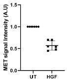
(3) Missing information on incubation times and inconsistencies in MET protein levels. The figure legends do not indicate how long the cells were incubated with HGF or the MET inhibitor PHA665752 before immunoblotting. This information is crucial, particularly because both HGF and PHA665752 cause a substantial decrease in the total MET protein level. Notably, such a decrease is absent in MDA-MB-231 cells treated with HGF in the presence of cycloheximide (Figure S2F). The authors should comment on these inconsistencies. Additionally, the MET bands in Figure S1J appear different from those in Figure S1C, and MET phosphorylation seems already high under basal conditions, with no further increase upon stimulation (Figure S1J). The authors should address these issues.
We apologise for the unintentional omission of experimental detailing about HGF or drug incubation time, which we have incorporated into the figure legend appropriately. Regarding the decreased MET level in the drug-treated condition: literature suggests that the MET inhibitor PHA665752 also promotes MET degradation, corroborating our result shown in Fig. S1J (PMID: 15788682, 18327775). Further in Fig. S1J, the relative phosphorylation of MET when compared to the total MET level in the HGF-treated condition is higher (~2-fold). Quantification of the blot has been added now.
Further, addition of HGF for 3 h leads to 40±15% reduction in the MET protein levels as seen in the Author response image 1 representing quantification of different immunoblot. The degradation of MET in the Fig S1J is 65% which nearly fall in the range for HGF-mediated MET degradation.
Author response image 1.
Quantification of immunoblots showing MET signal intensity in the presence or absence of HGF normalized with the loading control. N=6.
Next, in the fig. S1A, K the rabbit anti-MET (CST, D1C2 XP) antibody has been used, which binds to a C-terminal motif of MET and identifies both the 170kDa as well as 140kDa protein representing the uncleaved and cleaved form of MET. In Fig. S1J, the mouse antiMET (CST, L6E7) antibody has been used, which binds to an N-terminal motif of MET and recognizes only the 140kDa protein.
(4) Insufficient representation and randomization of microscopic data. For microscopy, only single representative cells are shown, rather than full fields containing multiple cells. This is particularly problematic for invadopodia analysis, as only a subset of cells forms these structures. The authors should explain how they ensured that image acquisition and quantification were randomized and unbiased. The graphs should also include the percentage of cells forming invadopodia, a standard metric in the field. Furthermore, some images include altered cells - for example, multinucleated cells - which do not accurately represent the general cell population.
We thank the reviewer for raising this point. The single-cell images are shown for clarity and to visualize the subcellular features; however, the conclusions are made based on the quantitative analysis of multiple cells collected from multiple fields of view (Frames). At least 30 such frames per condition having 4-7 cells/ frame has been acquired and analysed for the quantification throughout this manuscript. We would like to highlight that the image acquisition has been done over random fields on a coverslip. In the revised manuscript, for a better representation of the population of cell-forming invadopodia, a graph showing the percentage of cells forming invadopodia have been added (Fig S1G). Line no: 118-119. The percentage of cells forming invadopodia increased upon HGF stimulation in MDAMB-231.
(5) Use of a single siRNA/shRNA per target. As noted earlier, using only one siRNA or shRNA carries the risk of off-target effects. For every experiment involving gene silencing (MET, RAB4, RAB14, RCP, MT1-MMP), at least two independent siRNAs/shRNAs should be used to validate the phenotype.
We would like to clarify that we are using SMARTPool siRNA, which contains 4 individual siRNAs for the target gene. Literature suggests that using a pool of siRNA has reduced off-target effects compared to using single oligos for gene silencing (PMID: 14681580, 33584737, 24875475).
While SMARTpool siRNA minimizes the off-target effect, it does not eliminate the possibility of it. To confirm that the observed phenotypes are specifically attributable to the genes investigated in this study, we now performed functional experiments using two independent siRNAs targeting RCP and RAB14. The results have been added to figure S4K-L. Silencing of RCP or RAB14 using single oligos resulted in decrease in the degradation index comparable to SMARTpool siRNA, thus phenocopied the SMARTpool siRNA. Line no: 392-400.
Further, RAB4 is well established to be associated with MET trafficking and it served as a positive control in our study (PMID: 21664574, 30537020). Additionally, a recent study by Hey et al. have used individual oligos for KIF16B to demonstrate the effect of KIF16B silencing on gelatin degradation, which corroborate with our observation from the KIF16B silencing using the SMARTpool siRNA (PMID: 37696580).
For MET, we used siRNA, shRNA and an inhibitor to show the effect of MET inhibition/perturbation in the invadopodia-associated activity, which validates the observations of siRNA-mediated gene silencing (detailed in point 2).
We did not perform any experiments using single oligos targeting MT1-MMP, since in our MT1-MMP siRNA-based study, now we have taken an appropriate positive control to validate the efficacy of MT1-MMP silencing (Fig. S1I). In addition, we have shown the effect of MT1-MMP depletion on invadopodia formation using a CRISPR-based gene knock-out study, and another study from our group has shown a similar effect using siRNA (PMID: 31820782), which supports our MT1-MMP KO cell observation.
(6) Insufficient controls for antibody specificity. The specificity of MET, p-MET, and MT1-MMP staining should be demonstrated in cells with effective gene silencing. This is an essential control for immunofluorescence assays.
The anti-MET antibody (CST, D1C2 XP) has been used in several studied (PMID: 41166312, 41152910, 39748059). The CST L6E7 anti-MET antibody has been used in studied by Radke et al, Wang et el., Kong et al. (PMID: 36435874, 38262412, 32214092). In our study, immunoblots demonstrating depletion of MET in the siRNA or shRNA-treated cells has been provided in Fig. S1I, K respectively. Further, we have demonstrated MET silencing using immunofluorescence. We also have now added the entire field of view in Fig S1L of showing cells treated with control or shRNA against MET. In the shRNA-treated condition, the cell at the centre shows low MET fluorescence intensity indicating depletion of the RTK, while the surrounding cells have MET staining similar to control.
Tyr 1234/35 are present in the active site of MET kinase domain and upon binding of the ligand promotes their autophosphorylation (PMID: 17667909). Earlier studies have established the specificity of the phosphor-MET antibody by immunoblotting and immunofluorescence using MET inhibitors (PMID: 21973114, 41009793). In our study we have shown that the inhibition of MET kinase activity using PHA665752 abolished the MET phosphorylation at the Tyr 1234/35, as shown in Fig S1J which revalidates the specificity of the antibody.
Additionally, in a previous study Joffre et al. have shown that an oncogenic mutant form of MET, M1250T is highly phosphorylated at the Tyr 1234/1235 (PMID: 21642981). Using the phospho-MET antibody, we have shown in Fig 3C, S2I the increased Tyr phosphorylation of M1250T MET mutant as reported by Joffre et al.
The anti-MT1-MMP antibody is also a very well-established antibody reported in multiple studies (PMID:32479595, 31820782, 35762511). In our study, we have shown the specificity of the antibody using immunoblot analysis. Immunoblots showing significant depletion of MT1-MMP protein level following the SMARTpool siRNA and sgRNA-mediated gene silencing has been provided in Fig. S5I, M’, respectively. Further MT1MMP silencing has been also validated by immunofluorescence in the following studies. PMID: 22291036, 21571860, 20505159.
(7) Inadequate demonstration of MET recycling. MET recycling should be directly demonstrated using the same approaches applied to study MT1-MMP recycling. The current analysis - based solely on vesicles near the plasma membrane - is insufficient to conclude that MET is recycled back to the cell surface.
We appreciate the reviewer’s suggestion for an alternative approach to show MET trafficking. We have demonstrated MET trafficking using surface biotinylation, where we have shown that the RCP and KIF16B depletion affect the surface delivery of MET (Fig 5J). Line no: 382-391.
In addition, to study the surface delivery of cargo, TIRF is a widely used reliable approach and it is highly sensitive technique for detection of surface delivery events (PMID: 24344185, 20971701). We have also tried to investigate the trafficking of MET using antibody uptake approach; however, it could not be established as the binding of the antibody hindered the ligand binding and vice versa.
(8) Insufficient evidence for MET-MT1-MMP interaction. The interaction between MET and MT1-MMP should be validated by immunoprecipitation of endogenous proteins, particularly since both are endogenously expressed in the studied cell lines.
We thank the reviewer for pointing out the insufficient evidence for MET-MT1-MMP interaction at the endogenous level. We now carried out the immunoprecipitation of endogenous MET to validate the interaction with MT1-MMP (Fig S5H). A light (low intensity) band corresponding to MT1-MMP was detected in the anti-MT1-MMP immunoblot for the immunoprecipitated sample. We believe that the interaction between MT1-MMP and MET may be weak in nature, resulting in limited co-immunoprecipitation of the endogenous MT1-MMP by MET. The immunoblot is now added to the revised manuscript. Line no: 460-461.
(9) Inconsistent use of cell lines and lack of justification. The authors use two TNBC cell lines: MDA-MB-231 and BT-549, without providing a rationale for this choice. Some assays are performed in MDA-MB-231 and shown in the main figures, whereas others use BT-549, creating unnecessary inconsistency. A clearer, more coherent strategy is needed (e.g., present all main findings in MDA-MB-231 and confirm key results in BT549 in supplementary figures).
MDA-MB-231 and BT-549 are two well-characterized TNBC cell lines that readily form invadopodia. These cell lines have been extensively used to study invadopodia-associated breast cancer cell invasion (PMID: 32697977, 35915226, 31533971). These two cell lines also show overexpression of MET, making them suitable model cell lines for our study (PMID: 36139568, 20687930, 27502396).
Overall, most of the conclusions reported in this manuscript are derived from multiple experimental approaches using two TNBC cell lines for generalization.
We agree with the reviewer that showing the results from one type of cell line in the main figure would have been better, and wherever possible, we now provided the observations from a single cell line in the main figures and the data from the other cell line in the supplementary figures. However, some of the overexpression studies were performed in BT-549 cells to derive robust statistically meaningful conclusions. Therefore, we could not avoid adding results from both the cell lines in some of the figures. We would like to add that the legends for these figures have been edited to clearly mention the cell lines associated with each of the figure panels to avoid any confusion or inconsistency.
(10) Inconsistency in invadopodia numbers under identical conditions. The number of invadopodia formed in Figure 1E is markedly lower than in Figure 1C, despite identical conditions. The authors should explain this discrepancy.
We sincerely thank the reviewer for pointing out the inconsistency in invadopodia numbers across 2 experiments. Fig. 1C has 2 conditions: UT and the HGF-treated condition. The Untreated condition has the serum-free media without any stimulation. Whereas we have added vehicle (DMSO) in Fig. 1E, E’, since the drug is resuspended in DMSO. This difference in the treatment is likely to be responsible for the decreased numbers of invadopodia in Fig. 1E. In different studies it has been shown that DMSO is not biologically inert and can affect invasive properties of cells by perturbing actin dynamics and metalloprotease activity (PMID: 33552397, 22529897, 7188610).
(11) Questionable colocalization in some images. In some figures - for example, Figure 2G - the dots indicated by arrows do not convincingly show colocalization. The authors should clarify or reanalyze these data.
As suggested by the reviewer, we have now re-analyzed the data for figure 2G. The apparent visual lack of colocalization is likely due to the relatively lower fluorescence intensity of MET at these structures. We have now added the line intensity plots for the indicated puncta to show the intensity of both channels at the ‘dots’ in the figure 2G’ and they show correlation in their intensity distribution.
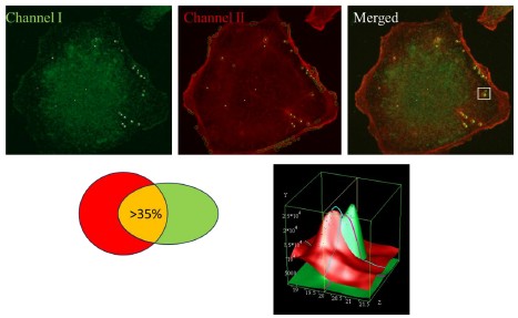
We would also like to elaborate that to quantify the colocalization of two channels, we have used the automated image analysis software Motiontracking (motiontracking.mpi-cbg.de) (PMID: 16143105), which has been detailed in the method section. Briefly, the algorithm works on object-based co-localization. If the fluorescence intensity distributions at a given object corresponding to any two different channels (fluorophores) show 35% or more overlap (Author response image 2), the object is considered as a multi-colour object and the overlapped area value is used to calculate the degree of co-localization. The calculation is carried out over all the objects in a given field of view (frame) and over all the field of views (frames) acquired for a given condition. Also, the apparent colocalization is corrected for random colocalization, which is the random permutation of object colocalization. This makes object-based colocalization more reliable than intensity-based colocalization.
Author response image 2.
Image showing the object identification and contour of the multicolour object identified by Motiontracking. The plot shows the intensity distribution of these two objects as analyzed by Motiontracking.
(12) Abstract, Introduction, and Discussion require substantial rewriting.
(a) The abstract should be accessible to a broader audience and should avoid using abbreviations and protein names without context.
(b) The introduction should better describe the cellular processes and proteins investigated in this study.
(c) The discussion currently reads more like an extended summary of results. It lacks deeper interpretation, comparison with existing literature, and consideration of the broader implications of the findings.
We thank the reviewer for this suggestion. We have substantially modified the abstract, and the introduction following the reviewer’s suggestion. The introduction has been edited to describe the cellular processes investigated in this study and some of the key associated molecular machineries. In the discussion section, we have avoided redundant descriptions of the results but retained some of them wherever necessary for interpretation and relevant discussion in the light of existing literature.
Reviewer #2 (Recommendations for the authors):
(1) Quality of charts. Several charts (e.g., Figure 1B, 1C, 1F) are of poor visual quality. The authors should provide higher-resolution graphs with clearer axis labels, consistent formatting, and properly scaled data.
We thank the reviewer for pointing out the insufficient visual quality of some of the charts. We believe the resolution of the charts/graphs have changed while converting to PDF, due to image compression. We will provide the uncompressed charts with much improved visual quality, provided they are not restricted by file size limitation.
(2) Full protein names on first mention. Whenever a protein appears for the first time in the manuscript, its full name should be provided, if possible, before using the abbreviation.
We have incorporated the full name of the protein while reporting for the first time in the manuscript.
(3) Correct use of "invadopodium" vs. "invadopodia." Invadopodia is the plural form; the singular is invadopodium. The sentence "Invadopodia, an actin-rich membrane protrusion decorated with proteases, is a tool for ECM and basement membrane degradation during cancer cell invasion" should be corrected accordingly.
We are thankful to the reviewer for pointing out the grammatical error. We corrected the error in the revised version. Line no: 44.
(4) Unclear sentence about resistance and invasion.
The sentence "However, often patients develop resistance to EGFR-targeted therapies due to overexpression of MET; yet, the mechanistic understanding of MET-dependent cancer invasion is unclear" is confusing because the shift from drug resistance to invasion is abrupt. The authors should revise this sentence for clarity and logical flow.
We are thankful to the reviewer for highlighting this sentence. We have rewritten the sentence as follows “Since one of the receptor tyrosine kinases (RTK), EGFR is often amplified in TNBC patients, they are usually targeted for its treatment. However, often patients develop resistance to EGFR-targeted therapies due to overexpression of another RTK MET”. Line no: 35-38.
(5) Incorrect figure reference. In the paragraph describing the results related to RAB proteins, there is an incorrect reference to Figure 3 instead of Figure 4. This should be corrected.
We sincerely apologize to the reviewer for the incorrect figure reference. We have corrected the reference to figures in the revised manuscript. Line no: 251, 261.
(6) Ambiguous sentence regarding MET activation. The sentence "MET, upon activation by HGF, triggers the activation of the RTK that induces cancer cell invasion" is unclear and should be rewritten for precision and clarity.
We are thankful to the reviewer for highlighting the unintentional mistake. We have now added a clearer sentence “MET-HGF signalling axis are reported to promotes invasion in gastric cancer cells and melanoma cells”. Line no: 96-97.
(7) Questionable wording of figure legend. The phrase "Immunoblotting of indicated cell lines with MET and Tubulin" is an informal shortcut. The authors should rephrase it.
We are thankful to the reviewer for highlighting this sentence. We have modified the figure legend with appropriate text. The modified text is as follows: Lysates of MDA-MB-231, BT-549 and MCF10A DCIS were separated by SDS-PAGE and analyzed by Western blot. Membranes were probed with anti-MET and anti-Vinculin antibody.
(8) Unnecessary capitalization. Terms such as invadopodia and cortactin should not be capitalized. The authors should correct capitalization throughout the manuscript.
We are thankful to the reviewer for raising this point. We have modified this accordingly in the revised version. Line no: 142, 159, 280, 436.



