Cerebellar Activation Bidirectionally Regulates Nucleus Accumbens Core and Medial Shell

  1. Center for Neuroscience, Davis CA 95616, United States
  2. Department of Neurobiology, Physiology and Behavior, University of California Davis, Davis CA 95616, United States

Editors

  • Reviewing Editor
    Jun Ding
    Stanford University, Stanford, United States of America
  • Senior Editor
    Lu Chen
    Stanford University, Stanford, United States of America

Reviewer #1 (Public Review):

This is a well-written manuscript, aiming to seek experimental evidence to establish anatomical and functional connectivity between the cerebellum and the nucleus accumbens (NAc). The authors combined anatomical, neural tracing, and electrophysiological approaches with electrical stimulation and optogenetics and provided a novel and solid set of data supporting the existence of disynaptic connections between the cerebellum and the NAc. The results are convincing and the main conclusion is supported by the data. Overall, this was a well-conceived project, and the experiments were conducted carefully, though some gaps remain to be filled. The knowledge generated from this manuscript will build a foundation for further research focusing on the interaction between cerebellum and limbic system as well as the role of such interaction in controlling motivated behavior.

Overall, this is a well-conceived project. The experiments were conducted carefully. The results support the conclusion of the existence of disynaptic circuits from the cerebellum to the NAc.

Reviewer #2 (Public Review):

It is increasingly recognized that the cerebellum is involved in a wide range of cognitive and behavioral processes beyond motor coordination and motor learning. This work contributes to the recent body of work showing functional connections between the cerebellum and many other brain regions. This study uses a combination of in vivo electrophysiology, viral tracing, and optogenetics to identify pathways from the deep cerebellar nuclei (DCN) to the nucleus accumbens (NA) core and medial shell running through "nodes" in the ventral tegmental area (VTA) and centromedial and parfascicular nuclei of the thalamus. The significance of this work is in providing function data and anatomical pathways that may underlie the role of the cerebellum in reward behavior.

This work makes two significant contributions to the field. First, the authors show that electrical stimulation in the DCN (the output of the cerebellar circuit) elicits (primarily excitatory) responses in neurons of the NA core and medial shell. Previous studies have shown that stimulation in the cerebellum increases dopamine in the NA, but this study is the first to use in vivo electrophysiology to measure changes in neuronal firing rates. Responses in NA neurons are primarily excitatory, with a small number of neurons showing inhibitory or mixed excitatory/inhibitory responses. The data here are clear and support the conclusions. The only caveat, acknowledged by the authors, is the use of ketamine/xylazine to anesthetize the mice may alter the firing properties of NA neurons and the balance of excitation and inhibition in neuronal responses. The specific mechanisms (neurotransmitters, synapses, or circuits) resulting in excitation or inhibition of NA neurons are not investigated here, though this may be an interesting avenue of future work.

The second significant contribution of this work is identifying anatomical pathways that connect DCN to the NA. The identification of these pathways is well supported by the viral injection data. The data using cre-expressing AAV in the DCN and floxed td-tomato AAV in the VTA or thalamus is particularly convincing. However, the inclusion of additional controls would strengthen the conclusions (see below).

In general, the conclusions are well-supported by the data. However, in a few places inadequate controls or description of the experiments weakens the conclusions.

1. In Figure 4, the authors injected a retrograde tracer in the NA and an anterograde tracer in DCN to find potential "nodes" of overlap. From this experiment, the authors identify the VTA and regions of the thalamus as potential areas of tracer overlap, but it is unclear how many other brain regions were examined. Did the authors jump straight to likely locations of overlap based on previous findings, or were large swaths of the brain examined systematically? If other brain regions were examined, which regions and how was this done? A table listing which brain regions were examined and the presence/intensity of ctb-Alexa568 and GFP fluorescence would be helpful.
2. In Figure 5, the authors inject AAV1-Cre in DCN and AAV-FLEX-tdTomato in VTA or thalamus. This is an interesting experiment, but controls are missing. An important control is to inject AAV-FLEX-tdTomato in the VTA or thalamus in the absence of AAV1-Cre injection in DCN. Cre-independent expression of tdTomato should be assessed in the VTA/thalamus and the NA.

Reviewer #3 (Public Review):

In this manuscript, D'Ambra and colleagues report the effects of stimulating the deep cerebellar nuclei (DCN) on neurons in the core and the medial shell of the nucleus accumbens (NAc). Electrical stimulation results in both excitation and inhibition, with excitation preceding the inhibition. In general, neurons that underwent excitation had lower baseline activity than neurons that underwent inhibition. They observed no relationship between the location of the stimulation site within the DCN, and the type of response observed in the NAc. In order to identify disynaptic connections between the two areas, the authors combined the injection of a retrograde tracer in the NAc with an anterograde tracer in the DCN. These experiments led them to describe co-localization of the anterograde and retrograde signals within two regions, the intralaminar thalamus (IL), and the ventral tegmental area (VTA). In order to confirm these results, they then used an anterograde transsynaptic viral tracing strategy to mark neurons in the IL and the VTA that project to the NAc. In addition, by injecting an excitatory opsin into the DCN, and stimulating these axons within the VTA and the IL, the authors were able to demonstrate increased activity in the NAc and describe the latency of these responses. Thus, using a series of rigorous and complementary experiments, the authors provide evidence for a disynaptic connection between the DCN and the NAc, via the VTA and the IL.

Novelty and relationship to previous studies: The presence of a disynaptic connection between the DCN and the NAc has previously been shown, as has the projection from the DCN to the parafascicular nucleus of the intralaminar thalamus (Fujita et al. 2020); however, the intermediary nodes of the disynaptic connection between the DCN and NAc have not previously been mapped. Some other pieces of these results have also been shown previously: DCN to VTA: Watabe-Uchida et al. 2012, DCN-VTA-NAc Beier et al. 2015, Xiao and Schieffele 2018) Interestingly, the Beier et al. paper suggests that the connection from DCN-VTA-NAc is an extremely small proportion of the total inputs to the NAc. In contrast to the Fujita et al. paper, here the authors also stimulate or trace projections from the two other deep cerebellar nuclei, the lateral and the interposed (this is relevant for a comment below). In addition, previous studies have shown a projection from the DCN to the IL and, separately, from the IL to the NAc. Thus, the existence of the pathways described here is in line with previous work. Moreover, this study expands on previous ones through its electrophysiological measurement and description of neural responses to stimulation of DCN and DCN projections.

Strengths: The strengths of this paper include the authors' use of multiple techniques to confirm the presence of the connections that they describe. Any one of the experiments using electrical stimulation, combining anterograde and retrograde tracing, transsynaptic tracing, or optical stimulation of DCN axons in the IL and VTA has its own caveats. However, the combination of these techniques nullifies many of these caveats.

Weaknesses: While this is an interesting and exciting paper, there are a few weaknesses, listed below:

- The novelty of this paper lies in the mapping of projections from the interposed and the lateral nuclei of the cerebellum, as the authors themselves mention. However, in some of the experiments the medial nucleus is also clearly injected (Fig. 4B and 6B). In those experiments, it is impossible to distinguish which nucleus these projections come from, and they could be the ones from the medial nucleus that were previously described (see above).
- A strength of the paper is the use of both electrical and optogenetic stimulation. However, the responses to the two in the NAc are very different - electrical stimulation results in both excitation and inhibition, whereas opto stimulation mostly results in only excitation.
- The stimulation frequency at which the electrical stimulation in Fig 1 is done to identify responses in the NAc is 200 Hz for 25 ms. Is this physiological? In addition, responses in the NAc are measured for 500 ms after, which is a very long response time.
- Previous studies have described how different cell types within the DCN have different downstream projections (Fujita et al. 2020). However, the experiments here bundle together all this known heterogeneity.
- Previous studies have also highlighted the importance of different cell types within the NAc and how input streams are differentially targeted to them. Here, that heterogeneity is also obscured.
- In Fig. 4C, E and F, the experiments on overlap between anterograde and retrograde tracers are not particularly convincing - it's hard to see the overlap.
Author Response

Reviewer #1 (Public Review):

We thank the Reviewer for their comments.

Reviewer #2 (Public Review):

  1. In Figure 4, the authors injected a retrograde tracer in the NA and an anterograde tracer in DCN to find potential "nodes" of overlap. From this experiment, the authors identify the VTA and regions of the thalamus as potential areas of tracer overlap, but it is unclear how many other brain regions were examined. Did the authors jump straight to likely locations of overlap based on previous findings, or were large swaths of the brain examined systematically? If other brain regions were examined, which regions and how was this done? A table listing which brain regions were examined and the presence/intensity of ctb-Alexa568 and GFP fluorescence would be helpful.

We thank the Reviewer for their comments. Exhaustive characterizations of inputs into nucleus accumbens (NAc) as well as of direct outputs of the deep cerebellar nuclei (DCN) have appeared elsewhere (e.g, Ma et al., 2020 doi: 10.3389/fnsys.2020.00015; Novello et al., 2022 doi: 10.1007/s12311-022-01499-w). Our anatomical investigations with retrograde and anterograde tracers were focused on putative intermediary nodal regions with robust inputs from the DCN, clear outputs to NAc, and limbic functionality. Only a handful of brain regions fulfill these criteria, and from those, we chose to target the VTA and intralaminar thalamus based on the observation that cerebellar activation induces dopamine release in the NAc medial shell and core (Holloway et al., 2019 doi: 10.1007/s12311-019-01074-w; Low et al., 2021 10.1038/s41586-021-04143-5) and on our previous work on DCN projections to the midline thalamus (Jung et al., 2022 doi: 10.3389/fnsys.2022.879634).

  1. In Figure 5, the authors inject AAV1-Cre in DCN and AAV-FLEX-tdTomato in VTA or thalamus. This is an interesting experiment, but controls are missing. An important control is to inject AAV-FLEX-tdTomato in the VTA or thalamus in the absence of AAV1-Cre injection in DCN. Cre-independent expression of tdTomato should be assessed in the VTA/thalamus and the NA.

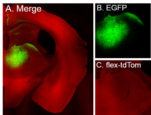
We thank the reviewer for bringing up this important control. We routinely perform this control experiment to test for any “leakiness” of floxed vectors prior to use but we did not include it in the paper. In response to the Reviewer’s comment, we show results from this control below. Briefly, we performed a large injection (300 nl) of AAV-FLEX-tdTomato in the thalamus area together with AAV-EGFP (to visualize the injection). No Cre-expressing virus was injected anywhere in the brain. PFA-fixed brain slices were obtained at 3 weeks post-injection and imaged for EGFP and tdTomato. Author Response Figure 1 shows images of the injected thalamus area. No tdTomato expression (Fig. 1C, red) was observed despite abundant EGFP expression (Fig. 1B, green), which confirms that in the absence of Cre the floxed construct does not “leak”.

Author Response Figure 1 (related to Fig. 5 of manuscript). Control experiment for “leakiness” of floxed tdTomato. A, Epifluorescence image of thalamus region in brain slice with EGFP (green) and tdTomato (red) channels merged. Gain settings in the red channel were increased intentionally, to ensure detection of any “leaky” cells. B, Cellular EGFP expression marks successful viral injection. C, No cellular expression of tdTomato without Cre. Note diffuse red signal from background fluorescence.

Reviewer #3 (Public Review):

  1. The novelty of this paper lies in the mapping of projections from the interposed and the lateral nuclei of the cerebellum, as the authors themselves mention. However, in some of the experiments the medial nucleus is also clearly injected (Fig. 4B and 6B). In those experiments, it is impossible to distinguish which nucleus these projections come from, and they could be the ones from the medial nucleus that were previously described (see above).

We thank the Reviewer for their comments. As stated in the Results and in the legend of Fig. 4, in addition to experiments with injections in all DCN (Fig. 4B-D), we also performed experiments with injections in only the lateral nucleus (Fig. 4E and F). The results of these experiments support the existence of lateral DCN projections that overlap with NAc-projecting neurons in VTA and thalamus. This finding is further corroborated by our transsynaptic experiments with lateral DCN-only injections (Fig. 5E,F). Regarding the optophysiological experiments, as mentioned in the Results, all DCN were injected (Fig. 6B) in order to maximize ChR2 expression and the chances of successful stimulation of projections.

  1. A strength of the paper is the use of both electrical and optogenetic stimulation. However, the responses to the two in the NAc are very different - electrical stimulation results in both excitation and inhibition, whereas opto stimulation mostly results in only excitation.

We thank the Reviewer for this comment. At least two different explanations could account for the observed differences in the prevalence of inhibitory responses elicited by optogenetic vs electrical stimulation: i) inhibitory response prevalence is sensitive to stimulation intensity (Table 1 and Fig. 2B). Because of inherent differences between optogenetic and electrical stimulation, it is not possible to directly compare intensities between the two modalities in order to determine at which intensities, if at all, the prevalence of responses should match. The observation that, at least in the VTA, the prevalence of inhibitory responses elicited by 1 mW light intensity (the lowest intensity that we tested) was comparable to the prevalence of inhibitory responses elicited by 100 µA electrical stimulation is in line with this explanation; ii) DCN electrical stimulation is not node-specific. To our knowledge, there is currently no evidence to support mesoscale topographic organization in lateral and interposed DCN that is node-specific. Consequently, electrical stimulation of DCN could, in principle, result in NAc responses through various polysynaptic pathways and/or nodes. This possibility would still exist even if electrical stimulation had limited spread of a few hundred microns (as in our experiments) and is at least partly supported by the observed long latencies of electrically-evoked inhibitory responses (NAcCore: 268 ± 25 ms (10th percentile: 42 ms), NAcMed: 259 ± 14 ms (10th percentile: 60 ms). Our recognition of this intrinsic limitation of DCN electrical stimulation was the impetus behind the node-specific optogenetic experiments.

  1. The stimulation frequency at which the electrical stimulation in Fig 1 is done to identify responses in the NAc is 200 Hz for 25 ms. Is this physiological? In addition, responses in the NAc are measured for 500 ms after, which is a very long response time.

Regarding stimulation frequency, DCN neurons readily reach firing rates between 100-200 Hz in vivo and ex vivo (e.g., Beekhof et al., 2021 doi.org/10.3390/cells10102686; Sarnaik & Raman, 2018 doi:10.7554/eLife.29546; Canto et al., 2016 doi:10.1371/journal.pone.0165887). Regarding the duration of the response window, at the outset of our experiments we were agnostic to the type of responses that our stimulation protocols would evoke in NAc. We therefore established a response time window that would allow us to capture both fast neurotransmitter-mediated responses as well as neuromodulatory (e.g., dopaminergic) responses, which are known to occur at hundred-millisecond latencies or longer (Wang et al., 2017 doi.org/10.1016/j.celrep.2017.02.062; Chuhma et al., 2014 doi:10.1016/j.neuron.2013.12.027; Gonon, 1997). A posteriori analysis indicated that even if we reduced the response time window by 50%, the ratio of DCN-evoked excitatory vs inhibitory responses in NAc would not change substantially (E/I500: 4.3 vs E/I250: 5).

  1. Previous studies have described how different cell types within the DCN have different downstream projections (Fujita et al. 2020). However, the experiments here bundle together all this known heterogeneity.

We agree with the Reviewer that dissecting the contributions of specific DCN cell types to NAc circuits is an important next step, which we are excited to undertake in future studies. Here we have broken new ground by identifying for the first time nodes and functional properties of DCN-NAc connectivity. Performing these studies with DCN cell type-specificity was not justified or feasible, given that the molecular identity of participating DCN neurons is currently unknown.

  1. Previous studies have also highlighted the importance of different cell types within the NAc and how input streams are differentially targeted to them. Here, that heterogeneity is also obscured.

Along the same lines as #4, we agree with the Reviewer about the importance of examining the cellular identity of NAc neurons that participate in DCN-NAc circuitry. We are excited to undertake these examinations using ex vivo approaches, which are well suited to dissect cellular and molecular mechanisms.

  1. In Fig. 4C, E and F, the experiments on overlap between anterograde and retrograde tracers are not particularly convincing - it's hard to see the overlap.

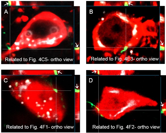
We thank the reviewer for this comment and have included revised figure panels 4C5, E3, Author Response Figure 1 and Figure 2 below. Single optical sections with altered color scheme and orthogonal confocal views are presented in order to show the overlap between DCN projections and NAc-projecting nodal neurons more clearly. The findings of these imaging experiments are corroborated by the results of our transsynaptic labeling approach (Fig. 5), which we have validated elsewhere (Jung et al., 2022 doi:10.3389/fnsys.2022.879634; and Author Response Figure 1).

Author Response Figure 2 (related to Fig. 4 of manuscript). Co-localization of NAc-projecting neurons with DCN axonal projections in VTA and thalamus. A-D, Single optical sections and orthogonal views are shown. Green: EGFP-expressing DCN axons; white: ctb- Alexa 568; red: anti-TH (A-B; VTA) or NeuN (C-D; thalamus). White arrows in orthogonal views point to regions of overlap.

  1. Howard Hughes Medical Institute
  2. Wellcome Trust
  3. Max-Planck-Gesellschaft
  4. Knut and Alice Wallenberg Foundation