
Peer review process
Revised: This Reviewed Preprint has been revised by the authors in response to the previous round of peer review; the eLife assessment and the public reviews have been updated where necessary by the editors and peer reviewers.
Read more about eLife’s peer review process.Editors
- Reviewing EditorOwen TamplinUniversity of Wisconsin-Madison, Madison, United States of America
- Senior EditorSofia AraújoUniversitat de Barcelona, Barcelona, Spain
Reviewer #1 (Public review):
This study by Alonso-Calleja and colleagues aimed to determine whether TGR5 regulates hematopoiesis and the bone marrow microenvironment under steady-state conditions and following transplantation. The revised manuscript substantially improves upon the original submission by providing additional characterization of TGR5 expression in hematopoietic and stromal populations, incorporating analyses in female mice, and expanding the investigation of bone marrow adipose tissue under aging and high-fat diet conditions. These additions more convincingly establish TGR5 as a regulator of bone marrow adipose tissue and stromal composition.
Major strengths of the study include the comprehensive characterization of the bone marrow adipose tissue phenotype across multiple experimental settings and the demonstration that TGR5 deficiency consistently alters the stromal compartment. The strongest and most convincing aspect of the work is the identification of TGR5 as a regulator of bone marrow adipose tissue and the bone marrow microenvironment. These findings provide useful insights into how metabolic signaling pathways influence the hematopoietic niche.
However, the evidence supporting a direct role for TGR5 in hematopoietic recovery following transplantation remains limited. Although reciprocal transplantation experiments and peripheral blood recovery analyses strengthen the manuscript, the conclusions regarding hematopoietic regeneration continue to rely largely on correlative observations. The study does not directly demonstrate that expansion of adipocyte progenitors is responsible for the enhanced recovery phenotype, nor does it establish improved regeneration of hematopoietic stem or progenitor cells within the bone marrow. Overall, the revised work addresses many of the concerns raised in the original review and provides useful new insights into the regulation of the bone marrow microenvironment by TGR5. Nevertheless, the conclusions regarding hematopoietic recovery should remain appropriately tempered, as the mechanistic basis linking the stromal phenotype to enhanced regeneration has not been directly demonstrated.
Reviewer #2 (Public review):
Summary:
The authors showed the expression of TGR5 in hematopoietic compartments and that loss of TGR5 doesn't impair steady-state hematopoiesis. Notably, TGR5 knockout significantly decreases BMAT, increase the APC population and accelerate the recovery upon bone marrow transplantation.
Strengths:
The role of TGR5 is interesting.
Weaknesses:
Additional mechanistic studies would further strengthen the work and provide deeper insight into how TGR5 regulates the bone marrow microenvironment.
Author response:
The following is the authors’ response to the original reviews
eLife assessment
This study investigates the role of the bile acid receptor TGR5 in adult hematopoiesis of the mouse model. The findings are potentially useful because the loss of TGR5 leads to dysregulation of bone marrow adipose tissue (BMAT) that has emerging regulatory functions. However, the study is still incomplete because the mechanism of TGR5 is not clear, the stromal cells expressing TGR5 have not been well defined, and there is not strong evidence for the role of TGR5 in recovery from transplant stress.
We thank the eLife editorial team for handling our manuscript. In our revised version, we took into consideration the suggestions of the reviewers, which we believe have significantly improved the quality of the current study. In summary, our new data provide further evidence that TGR5 is expressed in both hematopoietic cell lineage and stromal cells of the bone marrow (BM), including subpopulation analyses for both. While steady-state hematopoiesis remains intact in TGR5 knockout mice, we demonstrate that loss of TGR5 significantly impacts progenitor reconstitution under stress conditions, alters the BM adipose tissue (BMAT) homeostasis in a sex-specific manner and influences the balance of stromal cell differentiation. In particular, TGR5 deficiency resulted in reduced regulated BMAT and an accumulation of adipocyte progenitors, correlating with improved hematopoietic recovery following BM transplantation. The BMAT decrease is further observed in physiologically and pathophysiologically relevant contexts such as aging and obesity, where TGR5 deficiency is associated with a decrease in the myeloid bias. Collectively, our findings support a previously unrecognized role for TGR5 in maintaining BM niche integrity and highlight its potential as a modulator of hematopoietic support, although the precise molecular mechanisms still need to be elucidated.
Public Reviews:
Reviewer #1 (Public Review):
Summary:
Alonso-Calleja and colleagues explore the role of TGR5 in adult hematopoiesis at both steady state and post-transplantation. The authors utilize two different mouse models including a TGR5-GFP reporter mouse to analyze the expression of TGR5 in various hematopoietic cell subsets. Using germline Tgr5-/- mice it's reported that loss of Tgr5 has no significant impact on steady-state hematopoiesis, with a small decrease in trabecular bone fraction, associated with a reduction in proximal tibia adipose tissue, and an increase in marrow phenotypic adipocytic precursors. The authors further explored the role of stroma TGR5 expression in the hematopoietic recovery upon bone marrow transplantation of wild-type cells, although the studies supporting this claim are weak. Overall, while most of the hematopoietic phenotypes have negative results or small effects, the role of TGR5 in adipose tissue regulation is interesting to the field.
Strengths:
This is the first time the role of TGR5 has been examined in the bone marrow.
This paper supports further exploration of the role of bile acids in bone marrow transplantation and possible therapeutic strategies.
We thank the reviewer for pinpointing the strengths of our study.
Weaknesses:
(1) The authors fail to describe whether niche stroma cells or adipocyte progenitor cells (APCs) express TGR5.
Using the TGR5:GFP reporter model, we identified GFP+ cells in the stroma-enriched CD45-Ter119-CD31- population that contains the adipogenic progenitor cells (APC).
These data, along with the corresponding gating strategy are outline in Figure 6A and B.
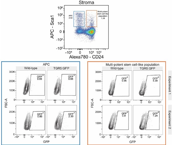
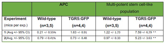
We found the subpopulation analyses within the stroma gate challenging at the individual mouse level given the limited cell numbers for these progenitor populations. We attempted to circumvent this by concatenating the individual files per genotype (WT and TGR5:GFP) for two independent experiments. We were surprised to find that there is little to no GFP expression in the APC population. Nevertheless, we found that the CD45-Ter119-CD31-CD24+Sca1+, multi-potent stem cell-like population (Ambrosi et al., 2017), reproducibly showed a TGR5:GFP positivity comparable to that of Ly6lo monocytes. We believe this would be compatible with our results showing an increase in CFU-F in Tgr5-/- mice, but we remain cautious about its interpretation. Results are shown in Author response image 1 for the concatenated flow plots and numerically in Error! Reference source not found. considering all individual mice analyzed (i.e., non pooled) for completeness. We kindly request the Reviewer’s opinion on whether these subpopulation analyses should be included in the main manuscript or solely here as part of the public review section.
Author response image 1.
Flow cytometry gating strategy used to identify stroma subpopulations in the stroma enriched CD45-Ter119-CD31- gate and their GFP signal for BM cells in TGR5:GFP mice. Subpopulations were immunophenotypically defined as CD45-Ter119-CD31-CD24+Sca1+ (adipogenic progenitor cells (APC)) and CD45Ter119-CD31-CD24+Sca1+ (multi-potent stem cell-like populations) (Ambrosi et al., 2017). Results are shown as the concatenated data of all mice in each phenotype for two independent experiments, as indicated.
Author response table 1.
Frequencies in the total live cell gate for adipogenic progenitor cells (APC) and CD45Ter119-CD31-CD24+Sca1+ (multi-potent stem cell-like populations, MPSC-like) (gating as in Ambrosi et al., 2017) in the experiments presented in Author Response Figure 1, expressed as average +/- 95% confidence interval for the two independent experiments. The number of mice per genotype and per experiment is indicated in parenthesis. **Paired, two-tailed Student’s t-test statistical analysis for the combined experiments (n=8 per group) indicates p < 0.01 for GFP expressing cells in APC versus MPSC-like populations.
(2) Although the authors note a significant reduction in bone marrow adipose tissue in Tgr5-/- mice, they do not address whether this is white or brown adipose tissue especially since BA-TGR5 signaling has been shown to play a role in beiging.
The nature of BMAT and how it relates to brown, white or brown/beige adipose tissue has been a persisting question in the field. Our understanding is that BMAT is currently considered as a distinct adipose depot that is neither white nor brown/beige (Sebo et al., 2019; Suchacki et al., 2020). BMAT does not express UCP1 to an appreciable extent, with reports showing that detectable expression possibly stems from contamination by tissues surrounding bone (Craft et al., 2019). Beyond this consideration, as the regulated BMAT in Tgr5-/- mice is almost absent, determination of the brown/beige vs white nature of the little regulated BMAT that remains would be technically extremely challenging.
(3) In Figure 1, the authors explore different progenitor subsets but stop short of describing whether TGR5 is expressed in hematopoietic stem cells (HSCs).
We have added these data to the manuscript as part of Figure 1C.
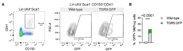
We have further expanded our data in Figure 1D and Figure 1–figure supplement 1A with the expression of TGR5:GFP in megakaryocyte progenitors (Lin-cKit+Sca1CD150+CD41+) as shown in Author response image 2.
Author response image 2.
A, representative flow cytometry gating strategy used to identify megakaryocyte progenitors (MkProg) and GFP positivity in TGR5:GFP mice and their wild-type controls. B, frequencies of GFP+ cells in MkProg population in the BM of 8-12-week-old male TGR5:GFP mice and their controls (n=3 for wild-type control mice, n=4 for TGR5:GFP mice). Results represent the mean ± s.e.m., n represents biologically independent replicates. Two-tailed Student’s t-test (B) was used for statistical analysis. p-values (exact value) are indicated.
Finally, we have completed the characterization of BM progenitor populations to include the erythroid lineage, thus covering the main hematopoietic populations. We have added these data to Figure 1E and Figure 1–figure supplement 1B.
(4) Are there more CD45+ cells in the BM because hematopoietic cells are proliferating more due to a direct effect of the loss of Tgr5 or is it because there is just more space due to less trabecular bone?
We observe an average 20% increase in CD45+ cell counts in baseline Tgr5-/- mice. The absolute volume of bone and BMAT lost in these animals does not account for 20% of the medullary cavity volume, so we speculate that the increase in CD45+ counts is not solely due to increased available volume for these cells.
(5) In Figure 4 no absolute cell counts are provided to support the increase in immunophenotypic APCs (CD45-Ter119-CD31-Sca1+CD24-) in the stroma of Tgr5-/- mice. Accordingly, the absolute number of total stromal cells and other stroma niche cells such as MSCs, ECs are missing.
These data are now included in the manuscript and in Author response image 3. Although we detect an increase in the relative frequency of APCs (Figure 7A), on a per-leg basis we did not detect an increase in APC numbers per leg (Author response image 3). We did however observe a decrease in total CD45-Ter119sup>-CD31sup>- stromal-enriched cells in Tgr5-/- mice. Nevertheless, given that only 2–5% of stromal cells are recovered in single-cell suspensions compared with native tissue (Coutu et al., 2017; Gomariz et al., 2018), we consider results for absolute stromal cell numbers prone to over interpretation. Our conclusion, therefore, remains one of relative enrichment of immunophenotypic APCs, supported by in vitro findings of increased adipogenesis and CFU-F formation after plating equal cell numbers.
Endothelial cells (CD45-Ter119-CD31+) were also quantified, with no differences observed between groups.
Author response image 3.
Absolute number of adipocyte progenitor cells (APC), stroma cells (CD45-Ter119CD31-) and endothelial cells (CD45-Ter119-CD31+) (n=5 for both Tgr5+/+ and Tgr5-/- mice). Results represent the mean ± s.e.m., n represents biologically independent replicates. Unpaired, two-tailed Student’s t-test was used for statistical analysis. p values (exact values) are shown.

(6) There are issues with the reciprocal transplantation design in Fig 4. Why did the authors choose such a low dose (250 000) of BM cells to transplant? If the effect is true and relevant, the early recovery would be observed independently of the setup and a more robust engraftment dataset would be observed without having lethality post-transplant. On the same note, it's surprising that the authors report ~70% lethality post-transplant from wild-type control mice (Fig 4E), according to the literature 200 000 BM cells should ensure the survival of the recipient post-TBI. Overall, the results even in such a stringent setup still show minimal differences and the study lacks further in-depth analyses to support the main claim.
We thank the reviewer for this comment. On the one hand, we respectfully disagree on the relevance of the effect size, as Tgr5-/- mice recover from low platelet and leukocyte counts significantly faster than wild-type controls. Tgr5-/- recipients recovered their platelet levels faster than the Tgr5+/+ recipients, showing values consistently above 200.000/µL one week sooner; platelet levels below this threshold are considered a risk factor for bleeding events (Morowski et al., 2013; Vannini et al., 2019). In addition, on day 15 post-irradiation, Tgr5-/- recipients had higher neutrophil levels, at over the 500 cells/µL-threshold value for infection risk (Morowski et al., 2013; Vannini et al., 2019), but this difference was no longer statistically significant upon stringent multiple-comparison correction (uncorrected p-value 0.028, multiplecomparison corrected p-value 0.108). Underlining the relevance, in a clinical setting, G-CSF is routinely administered to patients daily to enhance myeloid recovery even if the acceleration of recovery is by 1-2 days (Trivedi et al., 2009).
From the perspective of mortality, we agree that it is higher than expected and constitutes a limitation of our work. Note that we discovered a mistake in data plotting during the preparation of the revised version of the manuscript which renders the mortality curve no longer statistically significant, with a p value that changed from 0.0254 to 0.0676. We have duly changed our interpretation in the text to that of a trend towards higher survival.
Regarding the mortality in our experiments being higher than expected, we have unfortunately suffered from cases of “swollen muzzles syndrome” in our facilities that have greatly hampered our ability to perform myeloablation experiments (Garrett et al., 2018), as even sublethal doses have resulted in the appearance of side effects that were reasons for euthanasia under Swiss legislation. For example, a strong reduction in mobility requires immediate euthanasia. All experiments were performed blinded to genotype allocation, so we can reasonably exclude experimenter bias. Finally, it could be argued that mice with more marked symptomatology leading to euthanasia are more likely to have hematopoietic deficits, which in our case was mostly seen for WT animals. We have therefore chosen to report mortality alongside the longitudinal assessment of peripheral blood counts to ensure clarity of the coherent effect and full transparency. We strongly believe that this disclosure, when examined as a limitation in the discussion section, aligns with the open science efforts advocated by eLife. Unfortunately, it is beyond the scope of this manuscript and the authors' timeline to rederive the Tgr5-/- colony in our new facility and perform new bone marrow transplantation studies with titrating doses of BM.
Lastly, the choice of 250,000 BM cells serves as the control dose per recommended standards for competitive repopulation assays (Purton and Scadden, 2007). Quoting from this methodological reference paper: “In our experiments, each recipient mouse receives cell doses … together with 2x10⁵ competing congenic bone marrow. We have found these cell doses sufficient to detect both reductions (Purton et al., 2006) and increases (Janzen et al., 2006; Walkley et al., 2005) in HSC numbers in different mutant mice… Furthermore, caution should be used when designing competitive repopulation assays, as it has been shown that the reliability of this assay is critically dependent on the numbers of HSCs present in the populations being assessed: when too few or too many HSCs (recipients of <1 1x10⁵ or >2x10⁷ bone marrow cells each from donor and competing sources) are present, the data may not be meaningful (Harrison et al., 1993).”
Moreover, our lethal radiation rescue experiments with 2.5x10⁵ cells are designed to deliver a minimal hematopoietic source known to rescue the great majority of animals in standard conditions, and thus to best mimic the situations when enhancement of hematopoietic recovery would be clinically meaningful (as used by our group members in (Naveiras et al., 2009; Tratwal et al., 2020; Vannini et al., 2019)). In our hands and in the absence of “swollen muzzles syndrome”, this approach leads to 80-95% overall survival, which mirrors the clinical setting of autologous transplantation that our transplants are meant to mimic. In clinical practice strategies, enhancing hematopoietic recovery would be most useful to improve morbimortality in either alternative hematopoietic progenitor allotransplants, known associated with delayed engraftment, or in the case of febrile neutropenia associated to bacteriemia, which affects 11-15% of both hematopoietic autologous or allogeneic transplant patients (Gil et al., 2013; Gooley et al., 2010). In summary, septic neutropenia was unfortunately modelled by the rescue experiments presented in Figure 7 C-J with swollen muzzles syndrome concomitant to the reconstitution, but we strongly believe that this complication enhances the clinical relevance of our data.
(7) Mechanistically, how does the loss of Tgr5 impact hematopoietic regeneration following sublethal irradiation?
As delineated in the previous point, we have been seriously conditioned by cases of “swollen muzzles syndrome” (Garrett et al., 2018), which has stopped us from proceeding with more irradiation experiments for this particular study. Mechanistic studies are unfortunately beyond the scope of this manuscript, but the mechanistic basis for the relationship between BM adipocyte differentiation and hematopoiesis is now a focus for one of the involved laboratories and should produce follow-up manuscripts in the near future.
(8) Only male mice were used throughout this study. It would be beneficial to know whether female mice show similar results.
We thank Reviewer #1 for this question, as it led us to perform the characterization of steady-state hematopoiesis and morphological bone and BMAT evaluation of young female mice, yielding new findings that strengthen our manuscript. In summary, we have found that the decrease in BMAT is sexually dimorphic, with young females not showing reduced BMAT levels. Conversely, we did find a trend towards lower trabecular bone content in females. We present these data as part of Figure 3.
Reviewer #2 (Public Review):
Summary:
In this manuscript, the authors examined the role of the bile acid receptor TGR5 in the bone marrow under steady-state and stress hematopoiesis. They initially showed the expression of TGR5 in hematopoietic compartments and that loss of TGR5 doesn't impair steady-state hematopoiesis. They further demonstrated that TGR5 knockout significantly decreases BMAT, increases the APC population, and accelerates the recovery upon bone marrow transplantation.
Strengths:
The manuscript is well-structured and well-written.
We thank Reviewer #2 for this comment.
Weaknesses:
The mechanism is not clear, and additional studies need to be performed to support the authors' conclusion.
We agree with Reviewer #2 that more studies are needed to understand the role of TGR5 in the hematopoietic system. We have been hampered in our studies of stress hematopoiesis because of frequent cases of swollen muzzles syndrome (Garrett et al., 2018), which prevented us from conducting additional experiments involving myelosuppression. Furthermore, the identification of the mechanism that links changes in the adipocyte differentiation axis and hematopoietic support, which is more complex than initially thought, has become a top priority for one of the involved laboratories and should produce follow-up manuscripts in the near future.
Recommendations For The Authors:
Reviewer #2 (Recommendations For The Authors):
(1) Figure 1: the authors showed the presence of TGR5 in hematopoietic cells using a GFP report in mice. What's the expression pattern of TGR5 in the nonhematopoietic cells? For example, adipocytes, stromal cells, endothelial cells, etc. In addition to analyze the percentage of TGR5-GFP+ cells, the authors should also quantify the expression levels of TGR5 in various hematopoietic and niche components.
We thank Reviewer #2 for this question, which we have addressed as points 1 and 3 from Reviewer #1. The expression of TGR5:GFP signal in stromal cells, HSCs, and the various hematopoietic compartments is now presented respectively as new panels in Figure 6A-B, Figure 1C-D, Figure 1–Figure Supplement 1A-B, and Figure 1figure supplement 2A, as well as Author response image 1 for stromal progenitors. Please note that specifically for the stromal compartment, we kindly requested Reviewer #1’s opinion on whether these subpopulation analyses should be included in the main manuscript or solely here as part of the public review section, as we are concerned about over interpretation for these rare APC and multi-potent stem cell-like subpopulations.
Consistent with the typical low-abundance expression of G protein-coupled receptors, where ligand-mediated activation is more relevant than absolute receptor abundance, TGR5 is also lowly expressed in most cell types. Although technical limitations prevent us from directly quantifying TGR5 expression in adipocytes (due to the difficulty of isolating these populations from bone marrow), previous studies have reported TGR5 expression in human BMSC-derived adipocytes (Velazquez-Villegas et al., 2018) . For similar reasons, we could not quantify TGR5 expression levels by RT-qPCR in the bone marrow niche, as isolating enough cells from each compartment to reliably detect a low-abundance receptor is technically challenging.
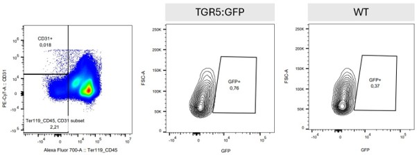
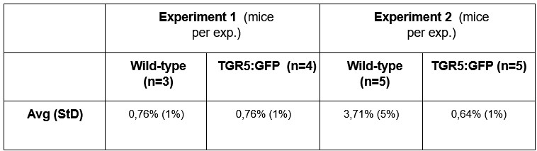
Regarding endothelial cells, previous studies have shown TGR5 expression in vascular endothelial cells (Kida et al., 2013) as well. In our dataset the number of endothelial cells recovered was limited, largely due to the lack of a dedicated endothelial isolation protocol for the BM. Even so, the data we collected are shown as Author response image 4 and Author response table 2 and suggest that TGR5:GFP level in endothelial cells is lower than in the stromal and hematopoietic compartments. As for Author response image 1 and Author response table 1, we remain cautious about the interpretation of GFP expression in these low frequency stromal and endothelial populations and we kindly request the Reviewer’s opinion on whether these subpopulation analyses should be included in the main manuscript or solely here as part of the public review section.
Author response image 4.
Representative flow cytometry gating strategy used to identify endothelial cells in BM, defined as CD45-Ter119-CD31+ and their GFP positivity in TGR5:GFP mice.
Author response table 2.
Frequency of GFP-expressing cells in the CD45-Ter119-CD31+ gate for endothelial cells presented in Author Response Figure 4, expressed as average (standard deviation). The number of mice per genotype and per experiment is indicated in parenthesis.
(2) Figure 2: Regarding the competitive transplantation, the authors should bleed the mice every 4 weeks and show the dynamics of donor chimerism up to 16 or 20 weeks. The difference at the 3-week time point is very tiny and is this change significant? There is no statistical significance shown in Supplemental Figure 2 Panel C at a 3-week time point.
The full data for repetitive monthly bleedings in primary, secondary and tertiary transplants is now shown in Figure 2J and Figure 2-figure supplement 1I-K. The statistically significant difference on the first month, which represents a 26% loss in short-time progenitor phenotype, is small but potentially clinically relevant as it is these progenitors that sustain early hematopoietic recovery from severe leucopenia and thrombocytopenia. Indeed, the hematopoietic phenotype described in Figure 7 C-J for accelerated rescue of Tgr5-/- recipients with wild-type bone marrow would be coherently associated to this short-term progenitor phenotype.
(3) Figure 3: in addition to the irradiation/transplantation model, the authors should also use alternative models for stress hematopoietic, for example, 5-FU, etc.
This is an excellent suggestion, but unfortunately beyond the scope of this manuscript due to the move of one of the co-senior authors to another institution. Follow-up studies are however planned with ablative chemotherapy models relevant to the hematopoietic transplant setting (non 5-FU).
(4) To get a better understanding of the role of TGR5 in the bone marrow niche, the authors should get and/or generate the TGR5 floxed mice and this will allow the authors to delete it from specific cell populations using cell-type specific Cre. In addition, it would be very interesting to investigate further the molecular mechanisms downstream of TGR5.
We much appreciate this comment and the interest it reflects in TGR5 BM biology. Mechanistic studies are unfortunately beyond the scope of this manuscript, but the mechanistic basis for the relationship between BM adipocyte differentiation and hematopoiesis are now a focus for one of the involved laboratories and should produce concrete follow-up manuscripts soon.
References
Ambrosi, T.H., Scialdone, A., Graja, A., Gohlke, S., Jank, A.-M., Bocian, C., Woelk, L., Fan, H., Logan, D.W., Schürmann, A., Saraiva, L.R., Schulz, T.J., 2017. Adipocyte Accumulation in the Bone Marrow during Obesity and Aging Impairs Stem Cell-Based Hematopoietic and Bone Regeneration. Cell Stem Cell 20, 771-784.e6. https://doi.org/10.1016/j.stem.2017.02.009
Coutu, D.L., Kokkaliaris, K.D., Kunz, L., Schroeder, T., 2017. Three-dimensional map of nonhematopoietic bone and bone-marrow cells and molecules. Nat Biotechnol 35, 1202–1210. https://doi.org/10.1038/nbt.4006
Craft, C.S., Robles, H., Lorenz, M.R., Hilker, E.D., Magee, K.L., Andersen, T.L., Cawthorn, W.P., MacDougald, O.A., Harris, C.A., Scheller, E.L., 2019. Bone marrow adipose tissue does not express UCP1 during development or adrenergic-induced remodeling. Sci Rep 9, 17427. https://doi.org/10.1038/s41598-019-54036-x
Garrett, J., Sampson, C.H., Plett, P.A., Crisler, R., Parker, J., Venezia, R., Chua, H.L., Hickman, D.L., Booth, C., MacVittie, T., Orschella, C.M., Dynlachta, J.R., 2018. Characterization and Etiology of Swollen Muzzles in Irradiated Mice. Radiation Research 191, 31. https://doi.org/10.1667/RR14724.1
Gil, L., Poplawski, D., Mol, A., Nowicki, A., Schneider, A., Komarnicki, M., 2013. Neutropenic enterocolitis after high-dose chemotherapy and autologous stem cell transplantation: incidence, risk factors, and outcome. Transpl Infect Dis 15, 1–7. https://doi.org/10.1111/j.1399-3062.2012.00777.x
Gomariz, A., Helbling, P.M., Isringhausen, S., Suessbier, U., Becker, A., Boss, A., Nagasawa, T., Paul, G., Goksel, O., Székely, G., Stoma, S., Nørrelykke, S.F., Manz, M.G., Nombela-Arrieta, C., 2018. Quantitative spatial analysis of haematopoiesis-regulating stromal cells in the bone marrow microenvironment by 3D microscopy. Nat Commun 9, 2532. https://doi.org/10.1038/s41467-018-04770-z
Gooley, T.A., Chien, J.W., Pergam, S.A., Hingorani, S., Sorror, M.L., Boeckh, M., Martin, P.J., Sandmaier, B.M., Marr, K.A., Appelbaum, F.R., Storb, R., McDonald, G.B., 2010. Reduced mortality after allogeneic hematopoietic-cell transplantation. N Engl J Med 363, 2091–2101. https://doi.org/10.1056/NEJMoa1004383
Harrison, D.E., Jordan, C.T., Zhong, R.K., Astle, C.M., 1993. Primitive hemopoietic stem cells: direct assay of most productive populations by competitive repopulation with simple binomial, correlation and covariance calculations. Exp Hematol 21, 206–219.
Janzen, V., Forkert, R., Fleming, H.E., Saito, Y., Waring, M.T., Dombkowski, D.M., Cheng, T., DePinho, R.A., Sharpless, N.E., Scadden, D.T., 2006. Stem-cell ageing modified by the cyclin-dependent kinase inhibitor p16INK4a. Nature 443, 421–426. https://doi.org/10.1038/nature05159
Kida, T., Tsubosaka, Y., Hori, M., Ozaki, H., Murata, T., 2013. Bile acid receptor TGR5 agonism induces NO production and reduces monocyte adhesion in vascular endothelial cells. Arterioscler Thromb Vasc Biol 33, 1663–1669. https://doi.org/10.1161/ATVBAHA.113.301565
Morowski, M., Vögtle, T., Kraft, P., Kleinschnitz, C., Stoll, G., Nieswandt, B., 2013. Only severe thrombocytopenia results in bleeding and defective thrombus formation in mice. Blood 121, 4938–4947. https://doi.org/10.1182/blood-2012-10-461459
Naveiras, O., Nardi, V., Wenzel, P.L., Hauschka, P.V., Fahey, F., Daley, G.Q., 2009. Bonemarrow adipocytes as negative regulators of the haematopoietic microenvironment. Nature 460, 259–263. https://doi.org/10.1038/nature08099
Purton, L.E., Dworkin, S., Olsen, G.H., Walkley, C.R., Fabb, S.A., Collins, S.J., Chambon, P., 2006. RARgamma is critical for maintaining a balance between hematopoietic stem cell self-renewal and differentiation. J Exp Med 203, 1283–1293. https://doi.org/10.1084/jem.20052105
Purton, L.E., Scadden, D.T., 2007. Limiting factors in murine hematopoietic stem cell assays. Cell Stem Cell 1, 263–270. https://doi.org/10.1016/j.stem.2007.08.016
Sebo, Z.L., Rendina-Ruedy, E., Ables, G.P., Lindskog, D.M., Rodeheffer, M.S., Fazeli, P.K., Horowitz, M.C., 2019. Bone Marrow Adiposity: Basic and Clinical Implications. Endocr Rev 40, 1187–1206. https://doi.org/10.1210/er.2018-00138
Suchacki, K.J., Tavares, A.A.S., Mattiucci, D., Scheller, E.L., Papanastasiou, G., Gray, C., Sinton, M.C., Ramage, L.E., McDougald, W.A., Lovdel, A., Sulston, R.J., Thomas, B.J., Nicholson, B.M., Drake, A.J., Alcaide-Corral, C.J., Said, D., Poloni, A., Cinti, S., Macpherson, G.J., Dweck, M.R., Andrews, J.P.M., Williams, M.C., Wallace, R.J., Van Beek, E.J.R., MacDougald, O.A., Morton, N.M., Stimson, R.H., Cawthorn, W.P., 2020. Bone marrow adipose tissue is a unique adipose subtype with distinct roles in glucose homeostasis. Nat Commun 11, 3097. https://doi.org/10.1038/s41467-020-16878-2
Tratwal, J., Bekri, D., Boussema, C., Sarkis, R., Kunz, N., Koliqi, T., Rojas-Sutterlin, S., Schyrr, F., Tavakol, D.N., Campos, V., Scheller, E.L., Sarro, R., Bárcena, C., Bisig, B., Nardi, V., de Leval, L., Burri, O., Naveiras, O., 2020. MarrowQuant Across Aging and Aplasia: A Digital Pathology Workflow for Quantification of Bone Marrow Compartments in Histological Sections. Front Endocrinol (Lausanne) 11, 480. https://doi.org/10.3389/fendo.2020.00480
Trivedi, M., Martinez, S., Corringham, S., Medley, K., Ball, E.D., 2009. Optimal use of G-CSF administration after hematopoietic SCT. Bone Marrow Transplant 43, 895–908. https://doi.org/10.1038/bmt.2009.75
Vannini, N., Campos, V., Girotra, M., Trachsel, V., Rojas-Sutterlin, S., Tratwal, J., Ragusa, S., Stefanidis, E., Ryu, D., Rainer, P.Y., Nikitin, G., Giger, S., Li, T.Y., Semilietof, A., Oggier, A., Yersin, Y., Tauzin, L., Pirinen, E., Cheng, W.-C., Ratajczak, J., Canto, C., Ehrbar, M., Sizzano, F., Petrova, T.V., Vanhecke, D., Zhang, L., Romero, P., Nahimana, A., Cherix, S., Duchosal, M.A., Ho, P.-C., Deplancke, B., Coukos, G., Auwerx, J., Lutolf, M.P., Naveiras, O., 2019. The NAD-Booster Nicotinamide Riboside Potently Stimulates Hematopoiesis through Increased Mitochondrial Clearance. Cell Stem Cell 24, 405-418.e7. https://doi.org/10.1016/j.stem.2019.02.012
Velazquez-Villegas, L.A., Perino, A., Lemos, V., Zietak, M., Nomura, M., Pols, T.W.H., Schoonjans, K., 2018. TGR5 signalling promotes mitochondrial fission and beige remodelling of white adipose tissue. Nat Commun 9, 245. https://doi.org/10.1038/s41467-017-02068-0
Walkley, C.R., Fero, M.L., Chien, W.-M., Purton, L.E., McArthur, G.A., 2005. Negative cellcycle regulators cooperatively control self-renewal and differentiation of haematopoietic stem cells. Nat Cell Biol 7, 172–178. https://doi.org/10.1038/ncb1214



