Author response:
The following is the authors’ response to the original reviews.
Reviewer #1 (Recommendations for The Authors):
Q1: Please replace lymphocytes with lymphatic endothelial cells throughout the manuscript.
A1: Thank you for your conscientious review. Per your suggestion, we have replaced “lymphocytes” with “lymphatic endothelial cells (LECs)” throughout the manuscript.
Q2: Please re-analyse lymphatics using LYVE1 and CD68 or another macrophage marker, as Lyve1 is NOT specific for lymphatics.
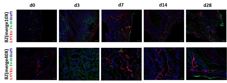
A2: Thank you for your suggestion. We completely agree with your opinion. Because both the CD68 (CST,97778S) and LYVE1 antibodies (Abcam,ab14917) are rabbit multiclonal antibodies and to more accurately label cardiac lymphatics, we performed immunofluorescence co-staining using LYVE1 and PDPN antibodies (Thermo,53-5381-82) and re-measured the lymphatic vessel area using the Image J software (version 1.53). The result is shown in Figure 1A and 1B. Further, we performed co-staining with PDPN and CD68 to observe the relationship between macrophage and cardiac lymphatic vessel distributions at different time points post-myocardial infarction (MI) (Figure1-figure supplement 1F). Per your comment, some LYVE1 markers are positive, whereas PDPN markers may be negative for macrophages in the heart tissue. We have added notes on the catalog numbers of anti-PDPN and anti-CD68 in the methods (Page 10, Lines 351‒352) and updated them in the KRT template and MDAR checklist.
Q3: Rephrase title 2.6, 2.7 to fit the results in these sections that are purely descriptive and do not add any insight into the functional relevance of the findings.
A3: Thank you for your suggestion. We have rephrased titles 2.6 and 2.7 as follows:
2.6 AQP1 in LEC is correlated with myocardial edema occurrence and resolution post-MI.
2.7 Gal9 secreted by LEC can affect macrophage migration.
Q4: Please refrain from extensive discussion of non-significant findings, such as Figures 6D, and 7A, B, and M (ifng vs ifng + antiGal9 is n.s).
A4: Thank you for your suggestions. Lymphatic endothelial cells (LECs) are a type of cell that exists in the myocardial tissue in small quantities. Owing to the extremely small number of LECs, elucidating their biological functions and regulation may be challenging during MI. To gain a deeper understanding of the role of the lymphatic system post-MI, we attempted to analyze the transcriptomic changes of LEC subsets at different time points after MI by combining single-cell sequencing and spatial transcriptomics data. We have selected relevant molecules with significant differences in transcription levels and conducted the validation analysis in LECs at different time points after MI. Among them, AQP1 and GAL9 showed significant differences. CD44, as a receptor for GAL9, showed significant differences in its expression in macrophages at different time points after MI. Therefore, we have added the relevant information to the discussion section (marked with yellow) on Page 9, Lines 299‒312.
Q5: Please explain the method used to calculate lymphatic areas in Figure 1.
A5: Thank you for your observation. The method we used is consistent with that described in previous studies[1,2]. (PMID: 30582443 and PMID: 32404007). The detailed methods have been described in the Methods as follows (Page 10, Lines 358‒363):
For quantification of vessel area, vessels with visible co-staining were measured using Image J software. First, we selected an image, turned it into 8-bit, and then applied a suitable threshold adjustment (present co-stained areas wherever possible). Second, five equally sized squares were selected in the respective zones (remote, infarct, and border zones) of each slice. ROI manager tools were used to analyze the automatic signal intensity quantification by the software in the area inside this square. Finally, the GraphPad software was used to plot the results as a bar graph.
Q6: In Figure 1 supp C, the upper and lower panels don't seem to have the same zoom factor.
A6: Thank you for pointing this out. The upper and lower images in Figure S1C have the same magnification. To facilitate your review, we have added a 1× image and re-labeled the position and scale information of the image. The revised Figure S1C was added to the manuscript and is shown as follows:
Q7: In Figure 2d please include aqp1 among displayed genes.
A7: Thank you for your suggestion. The Aqp1 gene is already displayed in the 11th, and we have labeled it.
Q8: In Figure 2f include markers of LECs such as Prox1, Flt4, Itga9, and also show Aqp1 here.
A8: Thank you for your valuable comment. We have updated Figure 2f.
Q9: Please indicate in Figure 3a what the y axis means? % of total LECs? % of total LECs at a given time point? The data is really not clear.
A9: Thank you for your suggestion. The y-axis represents the percentage of the total number of LECs at d1, d3, d7, d14, and d28 post-MI, relative to the number of LECs at d0, which is used as the reference value set at 100%. Meanwhile, different colors were applied to represent the proportion of different cell subtypes at different time points. We have updated Figure 3a.
Q10:Add n of LECs per time points in Figures 3a and b.
A10: Thank you for your suggestion. We have updated Figure 3b.
Q11: For Figure 3c please explain what marker genes were used to identify LEC enriched areas. What was the spatial resolution of the transcriptomic screens? How do these images relate to the localization of lymphatics in the heart?
A11: We appreciate your observation. We have added the required information to the Methods on Page 13, Lines 442‒448, as follows:
“We conducted spatial transcriptome data analysis using the deconvolution algorithm. The deconvolution algorithm refers to the application of feature genes to infer the full matrix information of single-cell transcriptome of cell subclusters. We then compared and anchored the matrix information of the single-cell transcriptome with the information of each SPOT in the spatial transcriptome, predicting cell types based on the similarity between the two sets of information.”
Q12:Figure 6 explains the y-axis in panel A, the timepoint in panel G, and absence of aqp1 staining in blood vessels in images d1 and d3 in panel D.
A12: Thank you for your suggestion. The y-axis in Figure 6A (Figure to reviewer 7A) shows Aqp1 expression in LECs at different time points from the sc-RNA sequence data. We have also added the timepoint in Figure 6G, which is for 24 hours. To clarify the expression trend of APQ1 more clearly, we performed immunofluorescence staining of APQ1 and LYVE1 at different time points after MI (d0, d1, d3, d7, and d14). The results are shown in Figure to reviewer 7C. APQ1 expression was found to be increased in the border zone of infarction at d3 post-MI adjacent to LYVE1 staining positive area.
Q13: Explain the y-axis unit in Figure 7a.
A13: Thank you for your comment. The y-axis in Figure 7A shows Lgals9 gene expression in LECs at different time points from the Sc-RNA sequence data.
Q14: In Figure 7c, d how was the induction of cell death excluded as a cause of IFNg-mediated effects in LECs?
A14: Thank you for your suggestion. To remove the interference of apoptosis on the results, we performed TUNEL staining of LECs after stimulation with different concentrations of IFN-r for 24 h. As shown in the Figure to reviewer 9, little apoptosis of LECs was observed in this concentration gradient range. Therefore, we can exclude the potential impact of IFN-r-induced cell apoptosis.
Author response image 1.
TUNEL staining of LECs after stimulation with different concentrations of IFN-r for 24 h.
Q15: Results with hypoxia in Figure 7 are mentioned but not shown.
A15: Thank you for your observation. In the revised article, we supplemented the detection of Gal9 expression after hypoxic stimulation. We conducted hypoxia intervention experiments using two methods. First, we applied 1% oxygen concentration stimulation to detect the expression of Gal9 at 0 h, 2 h, 4 h, 8 h, 12 h, and 24 hours. Second, we applied CoCl2 intervention to activate HIF1α expression and simulated cell hypoxia stimulation to detect Gal9 expression. Both results confirmed that hypoxia could not stimulate LECs to secrete galectin 9. The results are presented in Figure 7-figure Supplement 1 (A-D).
Reviewer #3 (Recommendations For The Authors):
Q1: In Figure 1, the so-called "LYVE1-labeled lymphatic capillaries with discontinuous walls" might be macrophages. The authors measured lymphatic area by measuring "vessels with visible lumens", which is unclear. This may underestimate the number of capillaries that expand after MI in the border zone of the infarct area. The authors need to use CD68 and Pdpn markers, as Lyve1 is not specific for lymphatics and also stains macrophages, and Pdpn is more reliable for assessing lymphatic identity.
A1: Thank you for your good suggestion. We totally agree with your opinion. Because both the CD68 (CST,97778S) and LYVE1 antibodies (Abcam,ab14917) are rabbit multiclonal antibodies and to more accurately label cardiac lymphatics, we performed immunofluorescence co-staining using LYVE1 and PDPN antibodies(Thermo,53-5381-82) and re-measured the lymphatic vessel area using the Image J software (version 1.53). The result is shown in Figure to reviewer 1 (Figure 1A and 1B in manuscript). Further, we performed co-staining with PDPN and CD68 to observe the relationship between macrophage and cardiac lymphatic vessel distributions at different time points post-myocardial infarction (Figure to reviewer 2,and Figure1-figure supplement 1F in manuscript). Per your comment, some LYVE1 markers are positive, whereas PDPN markers may be negative for macrophages in the heart tissue. We have added notes on the catalog numbers of anti-PDPN and anti-CD68 in the methods (Page 10, Lines 351‒352) and updated them in the KRT template and MDAR checklist.
Q2: It is not clear how they analyse the lymphatic area in Figure 1, please explain.
A2: Thank you for your observation. The method we used is consistent with that described in previous studies[1,2]. (PMID: 30582443 and PMID: 32404007). The detailed methods have been described in the Methods as follows (Page 10, Lines 347‒352):
For quantification of vessel area, vessels with visible co-staining were measured using Image J software. First, we selected an image, turned it into 8-bit, and then applied a suitable threshold adjustment (present co-stained areas wherever possible). Second, five equally sized squares were selected in the respective zones (remote, infarct, and border zones) of each slice. ROI manager tools were used to analyze the automatic signal intensity quantification by the software in the area inside this square. Finally, the GraphPad software was used to plot the results as a bar graph.
Q3: Figure 1-supplement 1D: The authors claim that the observed structure is a lymphatic valve, however in 2D sections, this shape might result from membrane destruction due to the cutting and staining process. To accurately identify valves, the authors should employ 3D imaging of the lymphatic network, such as using a clearing protocol followed by lightsheet microscopy.
A3: Thank you for your good suggestion. We performed a 3D scan using a confocal microscope on another slice. The results are shown in Figure 1-supplement 1D. We believe it is more like the lymphatic valve than chips from membrane destruction.
Q4: In Figure 2, the number of LECs is too little. Indeed, 242 LECs were identified over 44860 total cell numbers and 5688 endothelial cells cannot be representative and cannot afford to distinguish 4 different clusters.
A4: We further analyzed the percentage of LEC in the adult mouse heart in the physiological state on day d0 based on the results of single-cell nuclear sequencing from public databases (GSE214611). A total of 292 LEC cells were obtained from 26,779 cells captured on board in three samples, meaning that the percentage of LEC cells in the normal adult mouse heart is 1.09%. Cardiac LECs are really rare, and enrichment methods such as flow cytometry and magnetic beads separation for cardiac LECs are under marked probing, which might exhibit more irrefutable evidence in future studies.
Q5: The authors claimed that there is transcriptional heterogeneity in regenerated cardiac LECs post-MI, based on their over-clusterization. However, to substantiate this claim, they need to include a control comparison. Currently, the observed differences in cardiac LEC profiles lack a direct connection to the disease condition.
A5: Thank you for pointing this out. Because we could not download spatial transcriptome data for day d0 in the public database (GSE214611) or from the authors, we have used data of 1 h after IR as a reference for approximating the physiological state in Figure 3 and in Supplemental Figure 1.
Q6: Line 131, what is the regeneration ratio the authors cite here?
A6: Thank you for the comment. Regeneration ratio is an inappropriate use of the word, and we apologize for this confusion. We were actually referring to the regenerative potential of LECs.
Q7: Line 132, it is not clear what is the "normal myocardial tissue" in the graphs presented Figures 3A and B. Is it d0 time point?
A7: Thank you for your suggestion. The d0 time point means LECs in the normal adult mouse heart.
Q8: In Figure 2D, please add more lymphatic markers as Ccl21, Flt4, Itga9, FoxC2 and Aqp1.
A8: Thank you for your suggestion. We have added these markers (Except Ccl21, whose gene expression is too low to mark) in Figure 2D in the revised manuscript.
Q9: The authors must replace "lymphocyte" with "lymphatic" from 2.5, where they start to present interactions between lymphatic and immune cells.
A9: Thank you for your good comments. We have corrected these words.
Q10: In Figure 3, please indicate what the color scale means.
A10: Thank you for your suggestion. We have supplied a color scale label.
Q11: In Figures 3C and D, the authors distinguished the same LECs clusters in the spatial transcriptomic as in the scRNAseq analysis. This is not clear whether they used the same markers.
A11: We appreciate your observation. We have added the required information to the Methods on Page 12, Lines 429‒434, as follows:
“We conducted spatial transcriptome data analysis using the deconvolution algorithm. The deconvolution algorithm refers to the application of feature genes to infer the full matrix information of single-cell transcriptome of cell subclusters. We then compared and anchored the matrix information of the single-cell transcriptome with the information of each SPOT in the spatial transcriptome, predicting cell types based on the similarity between the two sets of information.”
Q12: In 2.5, it is not clear whether the main message is about macrophage interactions with lymphocytes or with lymphatics(LEC interact with others)
A12: Thank you for your suggestion. We have revised the title 2.5 as “Assessment of Cell-Cell Communication between LECs and immune cells,” which is clearer for the reader.
Q13: In 2.6, the authors claim that they reveal "that fluid retention occurs in LEC ca I and LEC co. They don't show any data supporting this.
A13: Thank you for your comment. “…that fluid retention occurs in LEC ca I and LEC co” is mainly supported by Figure 3D KEGG enrichment. LEC Ca I is related to vasopressin-regulated water reabsorption, and LEC co is related to renin secretion.
Q14: In Figure 6A, please add statistical values, as the authors claim a significant correlation. Please also add a figure to support the correlation between Aqp1 and edema score, as mentioned in 2.6.
A14: Thank you for pointing this out. We have presented the information on statistical values in Figure 6A. Moreover, we calculated the correlation between Aqp1 and edema score in Figure 6D (shown in Author response image 2).
Author response image 2.
Correlation between Aqp1 expression intensity and edema score.
Q15: In Figure 6B, myocardial edema assessment using H&E staining is not accurate. If the authors wish to analyse cardiac edema, they must use gravimetry or MRI techniques.
A15: Thank you for your comment. We totally agree with your opinion. However, owing to limitations in experimental conditions, we could not perform MRI detection of mouse myocardial injury. To evaluate whether edema occurred in the mouse heart tissue, we used classic pathological evaluation methods described in the literature (PMID: 30582443). This method has been described in detail as follows (Page 11, Lines 365‒370):
Four high-power (40×) representative images were chosen per animal under the H&E stained section; each image must have a clear border of the section visible. Images were blinded, and five visual fields per sample were evaluated. Subsequently, an edema score was determined for each sample (Score 1=no edema, 2=mild edema, 3=severe edema). Graphs represent the average score value per animal.
Q16: Line 227, please correct "LVEC" with "LEC".
A16: Thank you for your careful review. We have revised this in the manuscript.
Q17: In Figure 6D, IF co-staining of Aqp1 and lymphatic vessels is mentioned as "significantly reduced". However, we don't see any quantification data supporting this.
A17: Thank you for your comment. To clarify the expression trend of APQ1 more clearly, we performed immunofluorescence staining of APQ1 and LYVE1 at different time points post-MI (d0, d1, d3, d7, and d14). The results are shown in the corrected Figure 6-figure supplement 1A. The result showed that APQ1 expression increased in the border zone of infarction in d3 post-MI adjacent to LYVE1 staining positive area.
Q18: As Gal9 was not significantly impaired in LECs post. MI, Figure 7A does not support any real finding concerning the role of this molecule in monocytes/macrophages interaction with cardiac lymphatics.
A18: Thank you for your comment. The Lgals9 gene is significantly impaired in LEC post-MI, as well as the Cd44 gene in macrophage. We have updated them in Figures 7A and 7B.
Q19: In Figure 7, please correct INF by IFN.
A19: Thank you for your careful review. We have revised this in the manuscript.