Mother-daughter asymmetry of pH underlies aging and rejuvenation in yeast
- Fred Hutchinson Cancer Research Center, United States
Figures
Figure 1 with 1 supplement
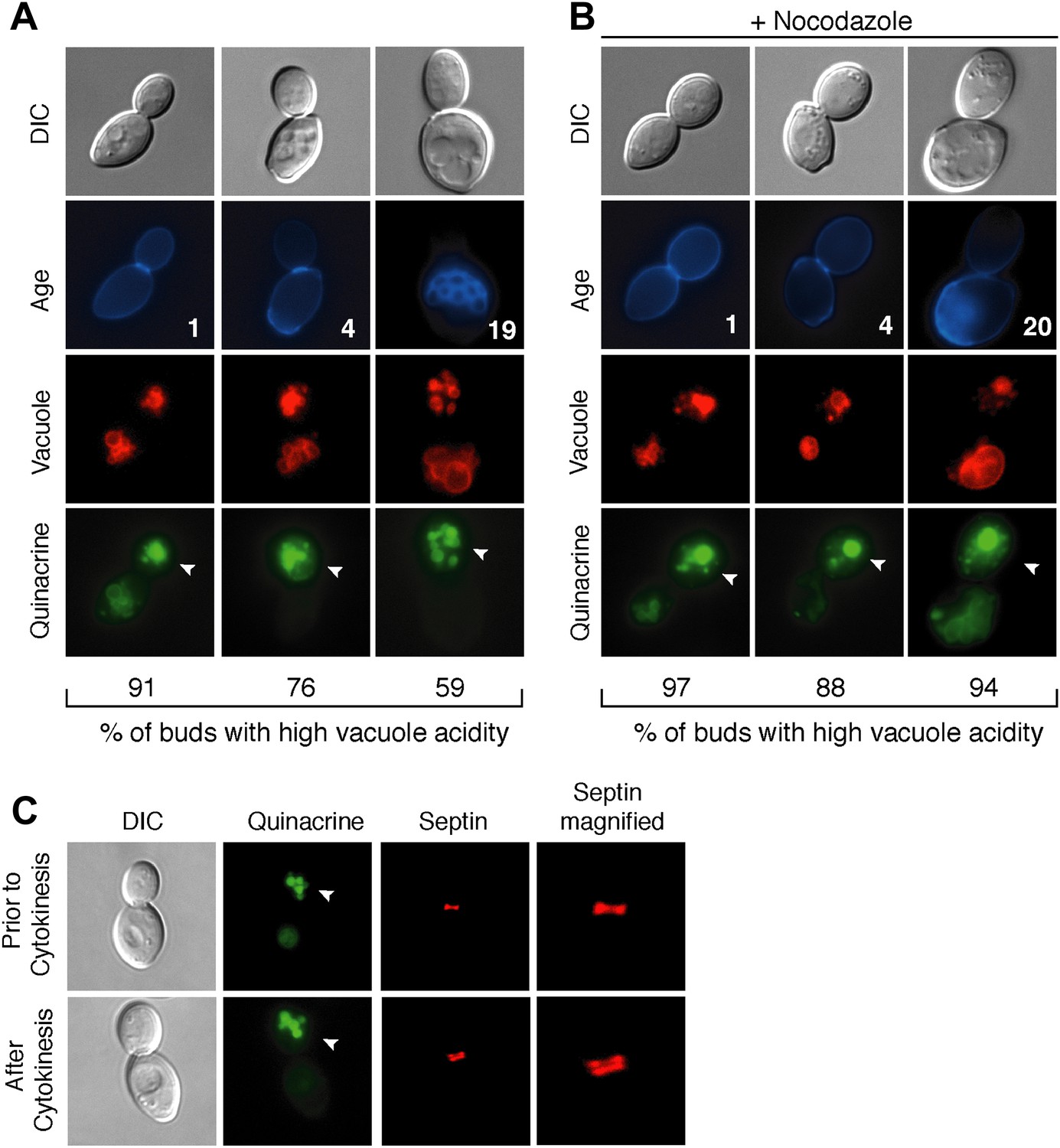
Vacuole acidity regenerates in daughter cells throughout mother cell aging and reacidification occurs prior to cytokinesis.
(A and B) Age (# of cell divisions) is shown in the second row and represents exact age determined by calcofluor staining of bud scars. Representative images are shown. n ≥30 cells per timepoint. Arrowheads indicate the daughter cell. DIC, differential interference contrast. (A) Vacuole acidity indicated by quinacrine staining of aged cells expressing Vph1-mCherry (vacuole membrane marker). (B) Vacuole acidity of cells expressing Vph1-mCherry and arrested prior to cytokinesis by nocodazole treatment. (C) Cells with septin morphology indicated by Cdc10-mCherry were quinacrine stained and vacuole acidity was examined before or after cytokinesis (one septin ring or two rings).
Figure 1—figure supplement 1

Subunits of the V-ATPase are not asymmetric between mother cells and buds.
(A) Cells in their first division expressing Vph1-GFP (V0 domain) or (B) Vma2-GFP (V1 domain).
Figure 2
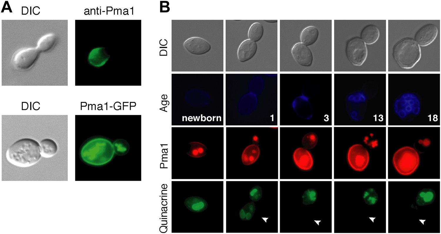
Plasma membrane Pma1 levels are asymmetric between mother cells and buds, accumulate with age, and are inversely correlated with vacuole acidity.
(A) Top panel: Indirect immunofluorescence imaging of Pma1 with anti-Pma1 antibody in untagged young cell. Bottom panel: Pma1-GFP localization in young cell. (B) Newborn daughter cells and aged mother cells expressing Pma1-mCherry were quinacrine stained. Arrowheads indicate the vacuoles of interest.
Figure 3 with 1 supplement
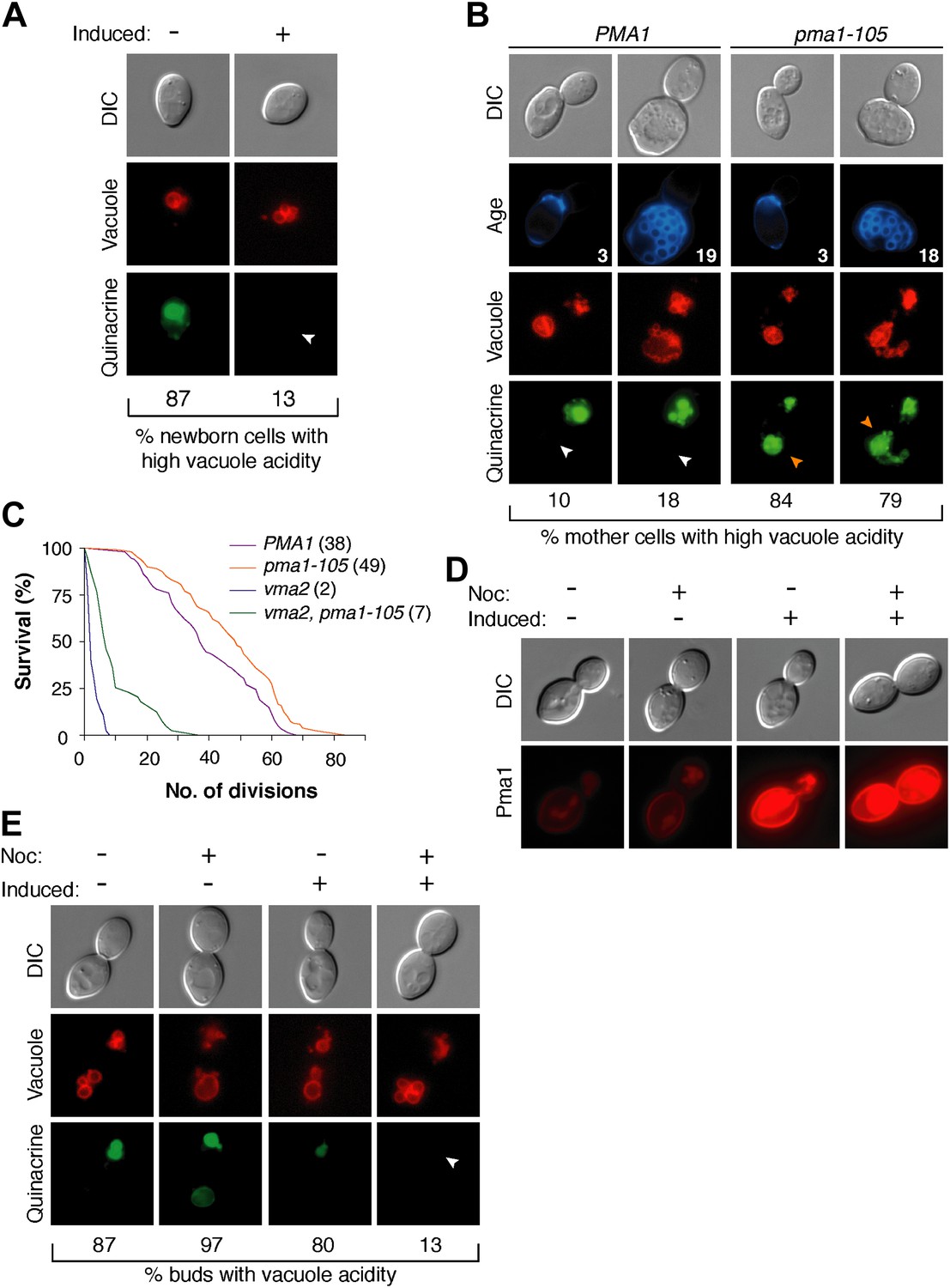
Pma1 antagonizes vacuole acidity and its absence facilitates regeneration of vacuole acidity in buds.
(A) PMA1 was overexpressed in newborn daughter cells expressing Vph1-mCherry using a β-estradiol inducible system where a GAL4-Estrogen binding domain-VP16 (GEV) fusion protein drives GAL1 promoter expression of an extra copy of PMA1. (n ≥ 30 cells per condition). (B) Wild-type and pma1-105 cells expressing Vph1-mCherry were aged and quinacrine stained (n ≥ 30 cells per timepoint). White arrowheads indicate mother cell vacuoles with reduced acidity. Orange arrowheads indicate acidic mother-cell vacuoles. (C) Replicative lifespan of wild-type, pma1-105, vma2, and vma2 pma1-105 cells by micromanipulation. Median lifespan is indicated. For the difference between wild-type and pma1-105, p < 0.0001, one-tailed logrank test. (n = 114 cells for PMA1, n = 119 for pma1-105, n = 36 for vma2, and n = 39 for vma2 pma1-105). (D) PMA1-mCherry was overexpressed in cells undergoing their first division that expressed endogenous Pma1-mCherry and that were treated with β-estradiol and then with β-estradiol plus nocodazole (Noc). (E) As in D, cells that expressed Vph1-mCherry were induced to overexpress PMA1 and were quinacrine stained. (n ≥ 30 cells per condition). Arrowheads indicate the vacuoles of interest.
Figure 3—figure supplement 1
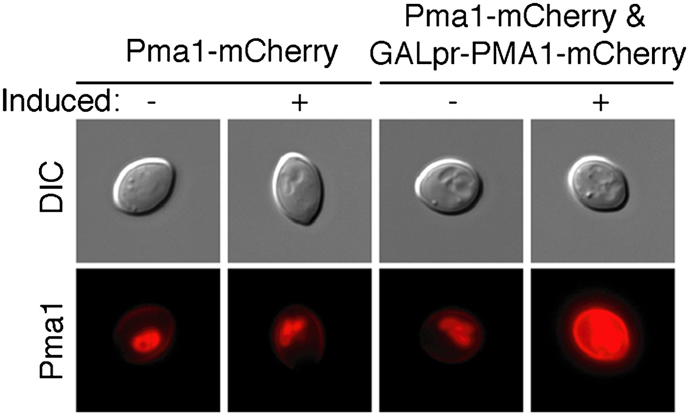
Overexpression increases Pma1 levels at the plasma membrane.
PMA1-mCherry was overexpressed in newborn daughter cells using a β-estradiol inducible system where a GAL4-Estrogen binding domain-VP16 (GEV) fusion protein drives GAL1 promoter expression of an extra copy of PMA1-mCherry in cells that also expressed endogenous Pma1-mCherry.
Figure 4 with 1 supplement
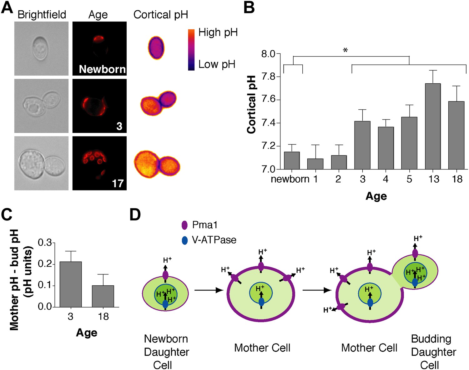
Cortex-proximal cytosolic pH increases with age and is asymmetric between mother cells and buds.
(A) Visualization of cortical pH of newborn and aged mother cells and their buds as indicated with a plasma membrane-anchored ratiometric pHluorin using its bimodal excitation spectrum. Age is indicated by wheatgerm agglutinin-Alexa 594 staining of budscars, which also detects birth scars on newborn cells. (B) As in A, measurement of cortical pH of newborn and aged mother cells was made at the plasma membrane. n ≥13 cells per timepoint. Mean cortical pH is significantly increased in mother cells undergoing their third division and thereafter compared to newborn cells (p ≤ 0.014, one-tailed unpaired t test). Error bars represent SEM. (C) Difference of the cortical pH of mother cells and their buds. Bud pH was lower (more acidic) than mother cell pH (p = 0.003, n = 17 cells at 3 divisions and p = 0.04, n = 16 cells at 18 divisions, one-tailed paired t tests) and was subtracted from mother cell pH. (D) Model of the effect of Pma1 asymmetry and increased Pma1 levels during aging on the magnitude of proton translocation out of the cytosol and into the vacuole.
Figure 4—figure supplement 1
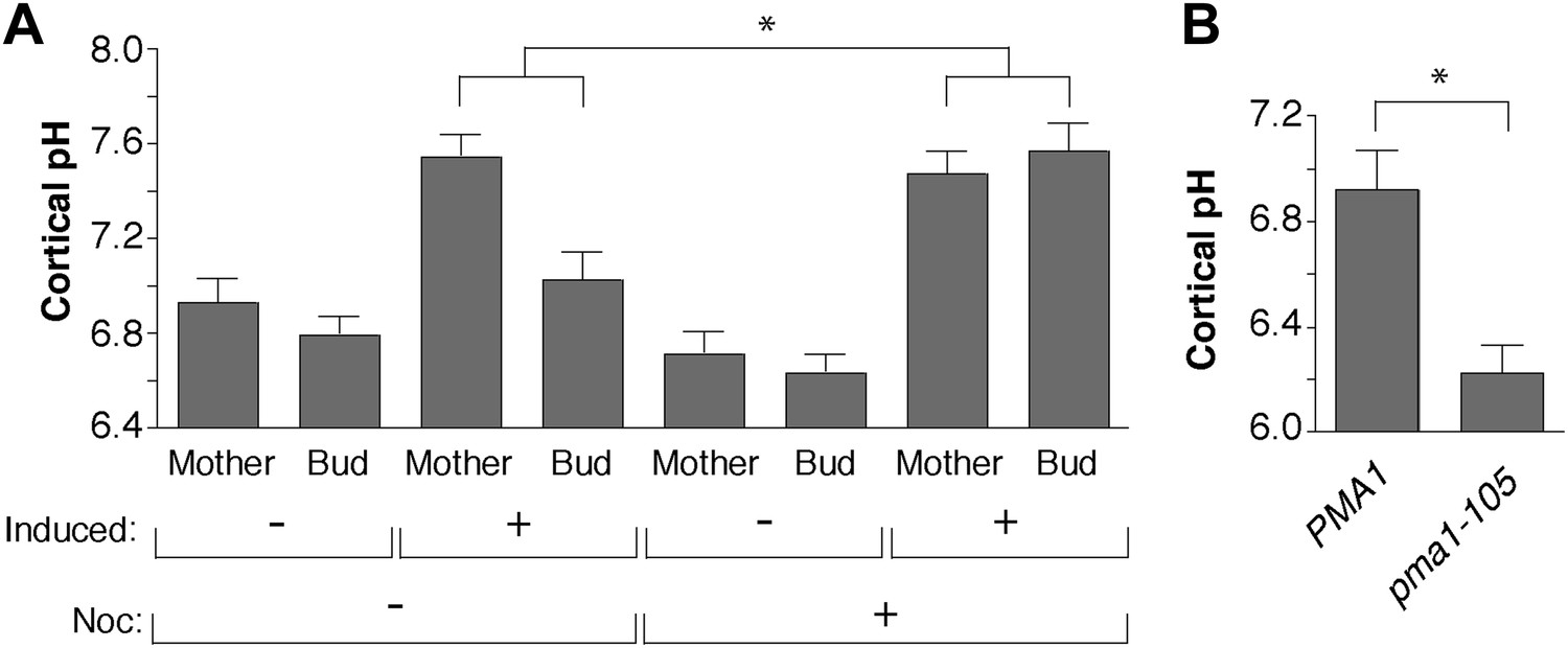
Pma1 asymmetry mediates cortical cytosolic pH asymmetry.
(A) PMA1 was overexpressed as in Figure 3 and cortical pH measured in mother cells undergoing their first division and their buds as in Figure 4. Cells were treated with β-estradiol for 2 hr to induce PMA1 and with Nocodazole (Noc) and β-estradiol for an additional 1.5 hr. For all mother-bud pairs, bud pH was significantly more acidic than mother pH (p < 0.05, one-tailed paired t test, n ≥ 14 cell per condition), except when Pma1 levels in buds were high due to the combination of PMA1 overexpression and nocodazole treatment. The difference between mother and bud pH was significantly greater when Pma1 was asymmetric (PMA1 induced without nocodazole) than when Pma1 was symmetric (PMA1 induced plus nocodazole) (p < 0.0001, one-tailed unpaired t test). Mean cortical pH is indicated, error bars represent SEM. (B) Cortical pH of wild-type and pma1-105 mother cells in their first cell division (p < 0.0005, t test, n = 12 cells for PMA1 and n = 13 for pma1-105).
Additional files
-
Supplementary file 1
(A) Yeast Strains. (B) Oligonucleotides.
- https://doi.org/10.7554/eLife.03504.010
Download links
A two-part list of links to download the article, or parts of the article, in various formats.
Downloads (link to download the article as PDF)
Open citations (links to open the citations from this article in various online reference manager services)
Cite this article (links to download the citations from this article in formats compatible with various reference manager tools)
Mother-daughter asymmetry of pH underlies aging and rejuvenation in yeast
eLife 3:e03504.
https://doi.org/10.7554/eLife.03504


