Hox genes control vertebrate body elongation by collinear Wnt repression
- University of Strasbourg, France
- Stowers Institute for Medical Research, United States
- Ehime University, Japan
- Howard Hughes Medical Institute, United States
- University of Kansas Medical Center, United States
- Brigham and Woman's Hospital, United States
- Harvard Medical School, United States
Peer review process
This article was accepted for publication as part of eLife's original publishing model.
History
- Version of Record published
- Accepted Manuscript published
- Accepted
- Received
Decision letter
-
Marianne E BronnerReviewing Editor; California Institute of Technology, United States
eLife posts the editorial decision letter and author response on a selection of the published articles (subject to the approval of the authors). An edited version of the letter sent to the authors after peer review is shown, indicating the substantive concerns or comments; minor concerns are not usually shown. Reviewers have the opportunity to discuss the decision before the letter is sent (see review process). Similarly, the author response typically shows only responses to the major concerns raised by the reviewers.
Thank you for sending your work entitled “Hox genes control vertebrate body elongation by collinear Wnt repression” for consideration at eLife. Your article has been favorably evaluated by Janet Rossant (Senior editor), Marianne Bronner (Reviewing editor), and three reviewers.
The Reviewing editor and the three reviewers discussed the comments before we reached this decision, and the Reviewing editor has assembled the following comments. It is the policy of eLife to only ask for revisions that can be accomplished in a reasonable time. Given the extensive revisions that would be required to satisfy the reviewers' concerns, we cannot accept your paper for publication at this time. However, we would be open to considering a new submission at a later time that addresses the reviewers’ comments after completion of further experiments and addition of extensive revisions to the text.
Summary:
The manuscript by Denans, Iimura and Pourquie presents evidence for a role of Hox genes in mesoderm ingression and body elongation in vertebrates. They test the interesting hypothesis that some posterior Hox genes repress Wnt and FGF signaling and so decrease expression of mesodermal gene T, which then leads to reduced ingression of cells through the primitive streak. This eventually leads to termination of the body elongation process by lack of available mesoderm. In this view, 'posterior Hox genes' regulate the extension of the body plan by indirectly controlling the amount of available mesoderm cells in the tail end of the embryo (via the regulation of cell ingression from the epiblast).
This is an interesting piece of work, which adds important arguments supporting the function of Hox genes during the elongation of the vertebrate body axis. However, the reviewers raised major concerns regarding technical approaches, the lack molecular explanation for the described phenomena, and appropriate reconciliation of the authors’ conclusions with previously published work. Additional control experiments and further discussion of the literature as suggested below are required to substantiate if the proposed mechanism indeed holds true.
1) One concern is the quantification of all the various phenomenon described in this work. There are several parameters, which can easily escape control in this context (mRNA stability, protein presence, electroporation success, copy number, saturation etc.). This criticism is not particular to this work and a similar question could be asked regarding the results by others in mice in mice, where the strong effect was not explicitly related to a 'physiological' gain of function (even though it may indeed be the case). Vertebral formula is very stable within the same species and thus must rely upon a robust mechanism. How the robustness of this process relates to the quantities of factors is a key issue (are these factors saturating?) At the time of the start of decreasing Wnt signaling due to increasing Hox amounts, a rather smooth gradient of T must be established. At these time points, genetic conditions in mice would be expected to have an effect perhaps stronger than what was reported by Capecchi for the Hoxb13 mutants? And if several Hox genes contribute to this phenomenon, then how to prevent intra-species variations, in particular at the time-points when the system would no longer be saturated?
2) From this model, one can make rather strong predictions, such as e.g. that the addition of yet another Hox cluster with posterior genes should make the body shorter? Is this the case in the literature?
3) There are important differences in effects between the Hoxa13 and Hoxd13 genes. In many developmental contexts, these two genes have very redundant functions and therefore, the difference reported here would suggest that Hoxa13 achieves its function in the tailbud by affecting a pathway or by using a peptide sequence that is not associated or existing in Hoxd13? This is particularly unexpected, as Hoxd13 (at least in mammals) seems to be expressed in the late tail bud more strongly than Hoxa13. This should be discussed.
4) A major claim of the authors is that there is delayed ingression, but this is not convincing. They report delayed expression of the Hox-Ires-Venus cassettes following electroporation into the chick epiblast as measured by Venus protein fluorescence. The authors interpret this effect such that Hox gene over-expression can delay ingression of the cells receiving the construct. However, the Hox constructs are driven by the ubiquitous Caggs promoter. Why is expression of the Hox constructs not observed at the same time as that of the control construct? A simple explanation might be that the RNA derived from these constructs is post-transcriptionally controlled, either by immediate degradation or by translational repression. This might assure position-adequate activity of the Hox genes. RNA in situ hybridization experiments testing for the time and position of transcription of the constructs are missing. However, they are required at least for some of the constructs. In addition, co-transfection of the control reporter construct with the Hox expression constructs should reveal if indeed ingression is delayed by Hox over-expression. Furthermore, RNAi of critical Hox genes (e.g. Hoxa13) would be a means of testing if the proposed effect of Hox genes can be opposed.
5) Critical missing data are the level of Brachyury expression along the body axis. If the authors are right that increasing Hox gene expression has a repressive effect on Brachyury expression resulting in decreasing mesoderm formation and motility along the body axis, this should be reflected by decreasing Brachyury transcription in the wild-type embryo as it elongates. Is this the case? Of course this would mean measuring expression levels per mesoderm cell (e.g. FPKM or qPCR values determined on FAC sorted cells) at different embryonic stages but at the same axial position (e.g. close to the node), not just overall expression in the embryo. The authors have all the tools at hand for doing the test.
6) It is not clear if the data from chick can be extrapolated to vertebrates in general. If the proposed mechanism were generally true, vertebrates with tails should not exist. All Hox genes are already activated during trunk development and the tail does not gain extra Hox activity grinding Brachyury expression eventually down to a standstill. Thus in tailed animals a different mechanism is more likely. The authors therefore should present their conclusions with respect to the chick and possibly other vertebrates with tiny tails (humans, some monkeys?).
7) The accuracy of the experimental approaches is difficult to assess, in particular, the accurate and reproducible targeting of the epiblast layer of the primitive streak by electroporation is not documented. It seems likely that there is some variability between embryos and that in some cases cells that are already ingressing are targeted. Furthermore, it seems possible that control cells are sometimes affected by Hox mis-expressing cells in the primitive streak, making this assay difficult to interpret with confidence, particularly when only a handful of embryos are analyzed for each gene. A data set demonstrating localized epiblast targeting reproducibility would strengthen the claims of the authors that they are assessing regulation of the ability of mis-expressing cells to ingress. It is also concerning that the arrow indicating the most rostral red fluorescent label in Figure 3B is mis-placed caudally.
8) The time difference between “successive” electroporations should be clarified; if the first construct labels cells earlier then these will progress to a more rostral position than cells electroporated slightly later, which could result in a misleading conclusion that the second construct used delays ingression. It seems the control construct is always electroporated first; can the authors invert the order in which the constructs that induce a delay are electroporated and obtain the same result?
9) There is no explanation at a molecular level for differences in the ability of posterior Hox genes to delay ingression nor for the effect of posterior dominance within the subset of posterior Hox genes that appear to affect ingression. How does collinear onset of posterior Hox genes lead to progressive loss of Wnt/FGF and T if this does not involve protein accumulation? Identifying these molecular mechanisms is needed to take the paper beyond the discovery of an interesting phenomenon.
10) We are left at the end of the paper without a molecular explanation for how Hox genes influence the ingression of cells. It is surprising that a gene that leads to reduced Wnt and FGF signaling in primitive streak epiblast does not affect cell adhesion and E-cadherin expression (data for which is not shown) nor the balance of Sox2/T expression in this cell population which would lead to cells adopting a neural over a mesodermal fate. Previous work has shown that FGF signaling is upstream of E-cadherin downregulation, prompts EMT and formation of T expressing mesoderm at the mouse primitive streak (Ciruna and Rossant, 2001, Dev. Cell), while more recent work has demonstrated that high Wnt signaling promotes mesoderm differentiation from neuro-mesodermal (Sox2/T co-expressing) progenitors and that reduced Wnt signaling in this context leads to neural differentiation (Tsakiridis et al, 2014, Dev; Gouti et al, 2014, PLOS Biology). Other work, in chick, analyzing the expression of Sox2 and T transcription throughout body axis elongation shows that strong T persists in the tailbud long after the 25 somite stage, declining only just prior to cessation of elongation at ∼ 44 somite after which T begins to decline and Sox2 is up regulated in tailbud cells, in a step that involves decreased FGF signaling (Olivera-Martinez et al, 2012, PLOS Biology).
Indeed, Tenin et al, 2010, BMC Dev. Biol. show that elongation cessation takes place when the presomitic mesoderm is still present at HH24 (rather than having been used up by somites forming right to the tail tip), suggesting that the body axis does not end due to a lack of ingressed cells. This observation contradicts the main conclusion of this current paper that it is a lack of ingressed presomitic cells that underlies elongation arrest.
11) The authors present only Sox2 in situ data (Figure 2–figure supplement 2) in whole mount embryos to support their claim that there is not a cell fate change when Hoxa13 is mis-expressed. Sections of such embryos in which the Hoxa13 GFP construct is visible with the Sox2 in situ signal on a cell by cell basis is required for convincing evidence on this point. A further possibility is that Hoxa13 expressing cells simply down regulate T but remain Sox2 positive and so adopt a neural fate and therefore do not ingress. Again comparing control and Hoxa13 GFP positive cells for levels of T and Sox2 on a cell-by-cell basis may provide evidence for the decline in T and maintenance Sox2.
12) It is not clear why the authors provide in situ images for T and Fgf8 at HH14 (Figure 5) using an intronic probe; other genes are not assessed in this way and no comment or explanation is provided in the text or figure legend. It is not clear how this data compares with published work showing strong and extensive T transcripts until the end of axis elongation. The authors need to address this difference with previous work, explain the significance of loss of active T transcription and consider how T transcripts continue to be regulated by FGF in the tailbud at these late stages (Olivera-Martinez et al, 2012) if the gene is not actively transcribed. One logic may be that the effects of endogenous posterior Hox genes are very small and slow acting, however, the authors appear to argue that there is a step change as the tailbud forms and the image presented in Figure 5F implies that T transcripts are lost in the tailbud at this time.
[Editors' note: further revisions were requested prior to acceptance, as described below.]
Thank you for resubmitting your work entitled “Hox genes control vertebrate body elongation by collinear Wnt repression” for further consideration at eLife. Your revised article has been favorably evaluated by Janet Rossant (Senior editor), Marianne Bronner (Reviewing editor), and the original three reviewers. The manuscript has been greatly improved and is acceptable pending changes that can easily be accomplished by revisions in writing of the manuscript without requiring further experiments. However, the reviewers feel that being more cautious in interpretation is important so we ask you to carefully attend to these remaining issues that need to be addressed before acceptance, as outlined below:
Overall, this paper presents a striking phenomenon in the regulation of cell ingression through the primitive streak by posterior Hox genes and some very interesting data that explain to some extent the molecular mechanisms at play here. The authors still need to place the work in an appropriate context both in terms of the literature and the significance of this mechanism during body axis elongation; it may be that collinear activation of posterior Hox genes regulates tailbud morphogenesis half way through axis elongation, but does not directly regulate PSM length.
1) The role of RhoA is not sufficiently well investigated and documented for giving it a prominent role in the Abstract. If the authors find this point so important they should provide some more experimental support. Alternatively they may want to rewrite the Abstract.
2) Last line of the Abstract (end of the Introduction and elsewhere,): “this mechanism leads to progressive reduction of PSM size associated to the arrest of somitogenesis” does not follow logically. What does this mean if it is clear that, at least in the chick, body axis elongation ceases when the PSM is still present and there is room for more somites to be generated? How is ingression then associated with arrest of somitogenesis? Somitogenesis arrest is more likely linked to loss of Notch pathway oscillations in the remaining PSM (Tenin et al, 2010).
3) In the Introduction the authors should mention the evidence that explanted chick tailbuds (i.e. without somites) are a demonstrated source of RA (Tenin et al, 2010). This is an important finding, which bears on their hypothesis that shortening of the PSM is a major step in that brings retinoic acid to tailbud and so curtails body axis elongation. It may be that both mechanisms operate and I think this should be made clear upfront, rather than appearing later in the Discussion.
4) In the Introduction and elsewhere it is confusing to state that “Hoxb genes are only expressed in anterior regions of the embryo precluding their playing a role in the control of axis extension”, when Hoxb13 is known to regulate axis elongation. They need to be more precise, Hoxb1-9.
5) It would be informative to explain in the paper that the 25 somite stage is ∼when the tailbud forms and that this is when when cell ingression through the primitive streak ceases—refer to work from Susan Mackem's lab in chick showing cessation at HH stage16 (∼ 24-28 somites, Knezevic et al, 1998 and see Catala et al, 1995, and also Wilson and Beddington, 1996, in the mouse). It seems likely that the mechanism identified by the authors is most functionally significant at this mid-way stage; from then on the PSM is added to from an “internalized” set of precursor cells. McGrew et al, 2009, and Olivera-Martinez et al, 2012 identify these cell populations by lineage analysis in the chick tail bud as the NMps (Sox/bra expressing cells) and also a transit amplifying cell population of mesodermal progenitors. I think the authors should explain this in the Discussion (and in the Introduction). As the paper currently reads it might be thought that ingression through the primitive streak continues until the end of axis elongation, when in fact in both chick and mouse embryos this ceases at somite 30 of ∼50-53 (chick, HH16) and 33 of 65 (mouse, E10.5). The cessation of ingression thus fits more closely with the establishment of the tailbud about half way through body axis elongation. As Hoxb and d13 begin to be expressed just after tailbud formation, they may contribute to conclusion of the ingression process, but this early role also leaves room for other actions for such Hox genes, including the regulation of cell proliferation in the tailbud mesoderm, described by Economides et al, 2003. I think these considerations are important to clarify that the regulation of ingression that they uncover is distinct from the model they originally proposed in which this was directly linked to “exhaustion” of PSM precursors and elongation arrest.
6) Related to this the authors suggest in the Discussion that posterior Hox genes might act in place of retinoic acid as a mechanism for arresting axis elongation in mouse. However, as ingression has ceased and continued extension is due to the activity of internalized precursors it is difficult to see Hox regulation of cell ingression as a molecular explanation for axis arrest.
7) The new data localize the region of Hox genes involved, but shows that this does not contain a conserved sequence that correlates with repressive activity. We are left with further speculation that this must be due to local protein structure. Similarly, the authors may have identified a further part of the molecular mechanism that regulates cell ingression, by rescuing Hox gene induced delay by co-expression with the DN-RhoA construct. However, this simply shows that de-stabilizing microtubules can force ingression and there is no molecular link made here from Brachyury to regulation of RhoA activity. The authors have therefore made a little progress on uncovering molecular explanations for the phenomena observed when posterior Hox genes are mis-expressed.
8) Finally, the authors have attempted to determine whether Sox2 expression is present in epiblast cells that are delayed in their ingression through the primitive streak. Unfortunately, they have been unable to improve the in situ hybridization for Sox2 transcripts (even the control embryo does not reveal the normal caudal domain of Sox2 expression, which extends into the tailbud (e.g. see Uchikawa et al, 2011, Dev. Growth & Diff; Olivera-Martinez et al, 2012). The authors have also been unable to detect Sox2 protein using immunocytochemistry, and it is not clear whether this reflects a problem with antibody batches or another technical difficulty. The net result is that they do not provide any new data to address the possibility that cells fail to ingress because having reduced Wnt signaling and T expression they have lost mesodermal identity in favor of a neural fate. Given that they are unable to reproduce the Sox2 expression pattern published by many other groups, they should be cautious in the interpretation of their lower power in situ data, which leaves open the possibility that accumulating non-ingressing primitive streak epiblast cells continue to express Sox2.
[Editors' note: a further round of revisions was requested prior to acceptance, as described below.]
Thank you for resubmitting your work entitled “Hox genes control vertebrate body elongation by collinear Wnt repression” for further consideration at eLife. Your revised article has been favorably evaluated by Janet Rossant (Senior editor), a Reviewing editor, and two of the original reviewers. The manuscript has been improved but there are some relatively minor but still important issues that need to be addressed before acceptance, as outlined below:
1) The authors have responded with some discussion of the literature on when ingression ceases, but this is not convincing. It is certainly debatable whether movement of cells from the late chordo-neural-hinge (CNH) to the PSM is considered an ingression; the CNH is the already internalized structure derived from the primitive streak. They should clarify their interpretation.
2) The response to comment 8 is confused; expression of Sox2 in epiblast cells that fail to ingress does not automatically lead to their contribution to the neural tube. Given the poor quality data (low magnification wholemount in situs which do not recapitulate published caudal expression of Sox2 even in the controls), they should be cautious with the interpretation.
https://doi.org/10.7554/eLife.04379.030Author response
1) One concern is the quantification of all the various phenomenon described in this work. There are several parameters, which can easily escape control in this context (mRNA stability, protein presence, electroporation success, copy number, saturation etc.). This criticism is not particular to this work and a similar question could be asked regarding the results by others in mice in mice, where the strong effect was not explicitly related to a 'physiological' gain of function (even though it may indeed be the case). Vertebral formula is very stable within the same species and thus must rely upon a robust mechanism. How the robustness of this process relates to the quantities of factors is a key issue (are these factors saturating?)
We agree that the robustness of the data generated by our electroporation protocol is striking. However, even though the technique does not allow us to precisely control the number of plasmid copies per cell or the number of cells electroporated, we have well established its reliability in the lab over the past 15 years. Successful electroporations like microsurgical experiments require a thorough training, and Nicolas Denans has been practicing these experiments on a regular basis for the past 6 years.
The quantitative analysis of the different phenotypes shown in the paper clearly demonstrates that these experiments give highly consistent results with most embryos overexpressing a given construct showing a similar phenotype which is specific for the construct (see graphs in Figure 3C, Figure 3–figure supplement 1B, 6C, 9B). Regarding mRNA stability, we overexpress only the coding sequences (without UTRs) and use the same SV40 polyA signal for all of our constructs. Also in most cases, Hox genes are expressed from an IRES-YFP vector and thus Hox expression can be indirectly monitored by examining YFP fluorescence. All Hox genes are expressed from the CAGGS promoter which is a strong promoter and which is expected to generate expression levels higher than the endogenous ones. Moreover, we clearly show in Figure 6F-H that the quantity of Hox protein overexpressed does not affect the phenotype observed. So our conditions are saturating, abolishing any effect of gene dosage of the overexpressed Hox genes and leaving the differences only due to the nature of the Hox genes overexpressed. These observations are consistent with the analysis of paralog knock-out experiments from Deneen Wellik (Wellik et al, Science, 2003; McIntyre et al; Development, 2007)) showing that leaving only one single wild type allele leads to a much milder phenotype than the deletion of an entire paralog group. These experiments also confirm previous results published in Iimura and Pourquie (Nature, 2006) showing that Hoxb genes expression driven by promoters of different strength (CMV, TK and CAGGS) leads to similar ingression phenotypes. Our data suggest that the quantitative effects on ingression result from the progressive expression of more posterior genes. The fact that 8 of the 16 posterior Hox genes from all posterior paralogs groups except Hox12 show an effect in the ingression, elongation and Wnt signaling assays argue for an extreme redundancy of the system that could explain the intraspecific robustness of the vertebral formula. We now discuss these ideas in more detail in the revised version of the text.
At the time of the start of decreasing Wnt signaling due to increasing Hox amounts, a rather smooth gradient of T must be established.
We have now analyzed by qPCR the expression of the Wnt targets T/Brachyury, Axin2 and Fgf8 in the tail bud from the 10 to 25-somite stage (Figure8 C-E). These new data show that repression of Wnt targets parallels activation of posterior Hox genes in vivo starting with a smooth gradient until the 20-somite stage. A significant drop in expression is then observed at the 25-somite stage, which corresponds to the time at which axis elongation drops abruptly when Hox13 genes are first expressed. This data has been added to the text and is shown in Figure 8C-E.
At these time points, genetic conditions in mice would be expected to have an effect perhaps stronger than what was reported by Capecchi for the Hoxb13 mutants?
In null mutant mice embryos for Wnt3a or T, the first 7 to 9 somites form normally (Takada et al, 1994, Genes and Dev; Wilson et al, Development, 1993), and the body axis is truncated posterior to this level. This phenotype is much stronger than that reported for Hoxb13 in which only the tail part of the axis is affected. In the experiments performed by Deschamps and Mallo where they overexpress Hox13 paralog genes in the paraxial mesoderm along the entire AP axis, these genetic conditions result in axis truncations, occurring posterior to the thoracic level (Young et al, Dev Cell, 2009). Strikingly no effect in the anterior regions is seen in these transgenic animals. The phenotype of these animals resembles hypomorphic Wnt3A or T mutants suggesting that the Wnt repression by posterior Hox genes is not an on-off mechanism but rather a quantitative effect. This notion is further supported by the different truncation levels reported for Hoxa13, Hoxb13, and Hoxc13 expressed from the same promoter in mouse (Young et al, Dev Cell, 2009). These experiments are consistent with the notion that Wnt and T play an important role in the control of axis elongation from the cervical to the caudal level and that posterior Hox genes quantitatively modulate Wnt activation in the paraxial mesoderm precursors of these regions. We now discuss these points in the revised version of the manuscript.
And if several Hox genes contribute to this phenomenon, then how to prevent intra-species variations, in particular at the time-points when the system would no longer be saturated?
Our experiments reported here (Figure 6) and in show that concentrations of Hox proteins exhibit a similar effect over a large dilution range (Figure 6) suggesting that low levels of Hox proteins are saturating. This property is expected to confer significant robustness to the system in line with the limited intra-species variations observed.
2) From this model, one can make rather strong predictions, such as e.g. that the addition of yet another Hox cluster with posterior genes should make the body shorter? Is this the case in the literature?
It is true that zebrafish which has extra Hox clusters exhibits a shorter axis when compared to mouse or chicken while snakes which contain less posterior Hox genes expressed in the tailbud region (Di-Poi N. et al, Nature, 2010) make a much longer axis. However, whether this can be generalized will require further studies. The Duboule lab has generated transgenic mice with a human HoxD cluster or an extra mouse HoxD cluster (Spitz et al, Genes and Dev, 2001). Since the effect of Hox proteins is saturating, duplicating an existing cluster is expected to act on expression levels (rather than timing) of the Hox proteins and thus is not expected to interfere with axis elongation. Accordingly, they did not observe any change in the vertebral counts in these transgenic animals. They also have performed many duplications and deletions within the HoxD cluster and shown that these manipulations can lead to heterochronic expression of HoxD genes in the tail bud. However, they do not report any significant variations in vertebral numbers in these cases either (Tarchini et al, Dev Cell, 2006; Tschopp et al, PLOS Genet, 2009, Denis Duboule, personal communication). Importantly, all these genetic manipulations have been performed on the HoxD cluster and in our hands, neither Hoxd12 nor Hoxd13 have an effect on elongation/Wnt, and the effect of Hoxd10 and Hoxd11 is weaker than that of other Hox genes thus providing a possible explanation for the lack of significant effect of this heterochronic expression of posterior HoxD genes. In contrast, heterochronic expression of other posterior Hox genes such as Hoxa13, Hoxb13 or Hoxc13 (but not Hoxd13) genes in the tail bud precursors as described in Young et al (Dev Cell, 2010; and Jacqueline Deschamps, personal communication) was shown to make the body shorter consistent with our hypothesis. We now present our results in the light of these papers in the Discussion.
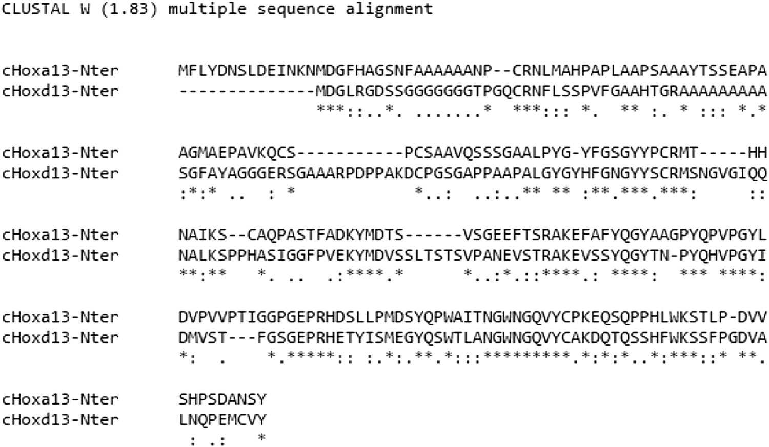
3) There are important differences in effects between the Hoxa13 and Hoxd13 genes. In many developmental contexts, these two genes have very redundant functions and therefore, the difference reported here would suggest that Hoxa13 achieves its function in the tailbud by affecting a pathway or by using a peptide sequence that is not associated or existing in Hoxd13? This is particularly unexpected, as Hoxd13 (at least in mammals) seems to be expressed in the late tail bud more strongly than Hoxa13. This should be discussed.
In mouse, a13 is the only Hox13 required for embryonic survival because its mutation blocks allantois growth is required for formation of the placenta (Shaut et al, PLOS Genet, 2008). Also whereas transgenic mice overexpressing Hoxa13 from the cdx2 promoter exhibit axis truncation (Young et al, Dev Cell, 2009), overexpressing Hoxd13 from the same promoter has no effect (Jacqueline Deschamps, personal communication). We now show that the effect of posterior Hox genes on T repression is controlled by the N-terminal domain of posterior Hox proteins (see text and new Figure 10). While we have not mapped precisely the sequences responsible for the effects described in this paper, there are very significant differences in the N-terminal regions of Hoxa13 and Hoxd13 that could explain why in the control of ingression, of elongation and of Wnt signaling, these 2 proteins show different phenotypes (see alignment in Author response image 1).
Author response image 1
4) A major claim of the authors is that there is delayed ingression, but this is not convincing. They report delayed expression of the Hox-Ires-Venus cassettes following electroporation into the chick epiblast as measured by Venus protein fluorescence. The authors interpret this effect such that Hox gene over-expression can delay ingression of the cells receiving the construct. However, the Hox constructs are driven by the ubiquitous Caggs promoter. Why is expression of the Hox constructs not observed at the same time as that of the control construct? A simple explanation might be that the RNA derived from these constructs is post-transcriptionally controlled, either by immediate degradation or by translational repression. This might assure position-adequate activity of the Hox genes. RNA in situ hybridization experiments testing for the time and position of transcription of the constructs are missing. However, they are required at least for some of the constructs. In addition, co-transfection of the control reporter construct with the Hox expression constructs should reveal if indeed ingression is delayed by Hox over-expression. Furthermore, RNAi of critical Hox genes (e.g. Hoxa13) would be a means of testing if the proposed effect of Hox genes can be opposed.
We apologize if the description of our experiments was not clear enough leading the reviewer to think that there is a delayed expression of the Hox-Ires-Venus cassettes following electroporation into the chick epiblast as measured by Venus protein fluorescence. We do not claim that expression is delayed but that ingression is delayed. The two constructs (Hox-green and control-red) are electroporated one after the other (see detailed description for consecutive electroporations below) and then we routinely examine electroporated embryos 3 hours after electroporation to make sure that both control and Hox constructs are expressed at the same time in the precursors of the paraxial mesoderm in the anterior streak region. Then whereas control cells expressing Cherry start to ingress in the posterior PSM, cells expressing the Hox-IRES-GFP constructs remain in the epiblast of the anterior streak region ingressing only later. Only a subset of posterior Hox genes shows a phenotype in these experiments. This experimental paradigm was originally introduced in , see Figure 2K-O, using grafts of epiblast expressing different Hox genes. These experiments results in a gap separating the position of the anterior boundary of red control cells from the position of the anterior boundary of green Hox-expressing cells reflecting the fact that cells ingressing later become located more posteriorly. This gap is proportional to the ingression delay experienced by Hox-expressing cells. To quantitatively estimate the variation in the ingression delay, we measure the ratio between the red and the green domains in the paraxial mesoderm (Figure 3A-C). We have tried to improve the description of these experiments in the main text and in Material and methods and provide a more detailed illustrative scheme (Figure 3A).
Concerning RNAi, we have attempted to block the function of several posterior Hox genes such as Hoxa13 by electroporating RNAi and in no case could we observe a phenotype. This is not unexpected, given that RNAi only achieve partial inhibition and that Hox proteins appear to be saturating even at low levels as shown in Figure 6. This led us to turn to the dominant-negative approach described in the paper.
5) Critical missing data are the level of Brachyury expression along the body axis. If the authors are right that increasing Hox gene expression has a repressive effect on Brachyury expression resulting in decreasing mesoderm formation and motility along the body axis, this should be reflected by decreasing Brachyury transcription in the wild-type embryo as it elongates. Is this the case? Of course this would mean measuring expression levels per mesoderm cell (e.g. FPKM or qPCR values determined on FAC sorted cells) at different embryonic stages but at the same axial position (e.g. close to the node), not just overall expression in the embryo. The authors have all the tools at hand for doing the test.
We agree with the referee and now provide this data. We have performed Quantitative-PCR on microdissected tailbuds from 10 somite stage (ss), 15ss, 20ss, 25ss for T, Axin2 and Fgf8 that clearly show a down-regulation of the expression of these 3 genes between the 10-25ss stage. This decrease parallels the slowing-down of axis elongation observed during those stages. We now include these results in Fig 8C-E and discuss them in the main text.
6) It is not clear if the data from chick can be extrapolated to vertebrates in general. If the proposed mechanism were generally true, vertebrates with tails should not exist. All Hox genes are already activated during trunk development and the tail does not gain extra Hox activity grinding Brachyury expression eventually down to a standstill. Thus in tailed animals a different mechanism is more likely. The authors therefore should present their conclusions with respect to the chick and possibly other vertebrates with tiny tails (humans, some monkeys?).
This is a very interesting point and an additional mechanism distinct from the one reported here is possibly involved in tail growth. In chicken which lack a tail, retinoic acid (RA) was shown to play an important role in the termination of axis elongation and somitogenesis (Tenin et al, 2010; Olivera-Martinez et al, 2011). This increase in RA level is accompanied by Raldh2 reexpression in the tail bud and it triggers differentiation and death of the paraxial mesoderm precursors. Remarkably, no such increase in RA levels is observed in mouse embryos. Furthermore, a normal tail can form even in the absence of RA in the raldh2 mutant (Cunningham et al, 2011). Therefore, it could be that in mouse, termination of the axis and hence tail formation, does not result from an active RA-dependent process but simply from exhaustion of paraxial mesoderm due to the shrinking of the PSM resulting from posterior Hox genes effect on elongation. Whether this RA-dependent mode of axis termination can be more broadly generalized to species lacking a tail remains to be explored. What causes this difference is currently unknown but it could be that in chicken, specific genetic alterations of posterior Hox genes can trigger the late expression of radlh2 in the tail bud thus leading to premature termination of axis elongation. We now present these hypotheses in the Discussion of the revised version.
7) The accuracy of the experimental approaches is difficult to assess, in particular, the accurate and reproducible targeting of the epiblast layer of the primitive streak by electroporation is not documented. It seems likely that there is some variability between embryos and that in some cases cells that are already ingressing are targeted. Furthermore, it seems possible that control cells are sometimes affected by Hox mis-expressing cells in the primitive streak, making this assay difficult to interpret with confidence, particularly when only a handful of embryos are analyzed for each gene. A data set demonstrating localized epiblast targeting reproducibility would strengthen the claims of the authors that they are assessing regulation of the ability of mis-expressing cells to ingress. It is also concerning that the arrow indicating the most rostral red fluorescent label in Figure 3B is mis-placed caudally.
We understand that this assay appears difficult and challenging and like microsurgical experiments, these electroporations require a significant training to position the electrode at the desired location appropriately. My lab has developed a significant expertise in the electroporation of paraxial mesoderm precursors which we pioneered (Dubrulle et al, Cell, 2001) and we have already published several papers based on this technique (Iimura an Pourquie, Nature, 2006; Iimura et al, PNAS, 2007; Benazeraf et al, Nature, 2010). Nicolas Denans has performed these electroporations on a regular basis for the last 6 years now and he can obtain very reproducible results as shown on the graphs in Figure 3C, Figure 3–figure supplement 1B, 6C, 9B. The electroporation success is first monitored 3 hours after by examining the embryos under a fluorescent stereomicroscope. Only embryos successfully electroporated in the PSM progenitors at the exact same antero-posterior level and the same medio-lateral level relative to the midline of the primitive streak are reincubated for about 20 more hours. About 10 to 20% of the embryos fail to develop because of the 2 consecutive injections/electroporations. The embryos that develop successfully are then examined for GFP expression and routinely more than 90% show restricted YFP expression in the paraxial mesoderm. Such a high level of success rate is obtained only after a long period of training. As can be seen on the embryos shown in Figure 3, Figure 6, and Figure 9 or in the movies, virtually no cells are seen outside of the paraxial mesoderm meaning that the electroporation accurately targeted the anterior streak epiblast. Since expression of the constructs is driven by the ubiquitous CAGGS promoter, even a slight inaccuracy in positioning the electrode would result in expression in the neural tube or the lateral plate. To better illustrate the accuracy of the electroporation performed, we now present a movie showing expression of a control construct electroporated in the anterior epiblast and showing the ingressing PSM cells (Video 3).
Our experiments have been carefully quantified as shown on the DotPlot graphs. As can be seen in Figure 3B-C and Figure 7K, when we overexpress a cherry control followed by a venus control, cells always end up at the same AP position (with very minimal variations) which shows that this assay is particularly robust. We electroporate one side of the PS with one construct and the other side of the PS with the other construct which largely prevents one cell to receive both plasmids. We agree that in some rare cases we could electroporate some ingressing cells. These rare cells are escapers and are found located much more anteriorly than the electroporated epiblast cells. We discard those rare cells in our quantifications and Figure 3B is a good example of it. We have now introduced a more thorough discussion of these technical aspects in the text and in the Material and methods section.
8) The time difference between “successive” electroporations should be clarified; if the first construct labels cells earlier then these will progress to a more rostral position than cells electroporated slightly later, which could result in a misleading conclusion that the second construct used delays ingression. It seems the control construct is always electroporated first; can the authors invert the order in which the constructs that induce a delay are electroporated and obtain the same result?
We now better describe our successive electroporation procedure (which we renamed consecutive electroporation for clarity) in the Methods section. We first harvest and culture the embryos on filter paper on an agar plate at room temperature (EC culture) which “pauses” the development of the embryos. We then process to two consecutive electroporations. We microinject the first construct and immediately electroporate it and right after the first electroporation we proceed the same way for the second construct. The time delay between the 2 electroporations is about 10 seconds. Moreover as the experiments are carried out at room temperature, the embryo is paused and cells do not ingress until the embryo is reincubated at 37 C. The fact that when we overexpress 2 control constructs we find the cells at the same anterior position clearly shows that the sequence of electroporation does not affect the timing of ingression (Figure 3B, 7K). In all cases we always invert the order of electroporations in the same batch of embryos (but not the side of electroporation for consistency) and do not observe any bias in the timing of ingression). We now provide a better description of the consecutive electroporation protocol in the text and Material and methods.
9) There is no explanation at a molecular level for differences in the ability of posterior Hox genes to delay ingression nor for the effect of posterior dominance within the subset of posterior Hox genes that appear to affect ingression. How does collinear onset of posterior Hox genes lead to progressive loss of Wnt/FGF and T if this does not involve protein accumulation? Identifying these molecular mechanisms is needed to take the paper beyond the discovery of an interesting phenomenon.
We agree that it is indeed a very interesting question. We designed chimeras of Hox proteins to determine which domain contains the region responsible for the repressive effect of posterior Hox genes on Brachyury using the cTprLuc assay as readout. These experiments show that the N-ter region of Hoxa13 contain the repressive domain (see new Figure 10 in the revised version of the manuscript). Sequence alignment of the N-ter region of Hoxa9, d10, c11 and a13 shows limited conservation at the amino acid level suggesting that it is not a conserved amino acids domain but rather a structural domain that is responsible for the repression activity of these proteins (see Figure 10–figure supplement1). The situation we describe is reminiscent of the recently described structural colinearity observed at the Hox protein level by Richard Mann’s group (Slattery et al, 2011). A more thorough structure function analysis of the protein sequences for all these genes must be conducted but we feel like it falls beyond the scope of the present paper.
10) We are left at the end of the paper without a molecular explanation for how Hox genes influence the ingression of cells. It is surprising that a gene that leads to reduced Wnt and FGF signaling in primitive streak epiblast does not affect cell adhesion and E-cadherin expression (data for which is not shown) nor the balance of Sox2/T expression in this cell population which would lead to cells adopting a neural over a mesodermal fate. Previous work has shown that FGF signaling is upstream of E-cadherin downregulation, prompts EMT and formation of T expressing mesoderm at the mouse primitive streak (Ciruna and Rossant, 2001, Dev. Cell), while more recent work has demonstrated that high Wnt signaling promotes mesoderm differentiation from neuro-mesodermal (Sox2/T co-expressing) progenitors and that reduced Wnt signaling in this context leads to neural differentiation (Tsakiridis et al, 2014, Dev; Gouti et al, 2014, PLOS Biology).
Ciruna and Rossant nicely showed that FGF positively regulates T and thus FGFR1 mutation leads to T down-regulation consequently preventing ingression, which correlates perfectly with our observations. Our data suggest that a subset of posterior Hox genes trigger some down-regulation of Wnt/T and FGF signaling which does not completely prevent ingression but delays it. We do not claim that there is no overexpression of E-cadherin but only that we do not see ectopic E-cadherin in cells overexpressing the Hox constructs. Our model, proposes that Hox genes down-regulate T expression, delaying its accumulation which is required for triggering ingression of epiblast cells by breaking down the basement membrane and undergoing EMT (see Figure 4). However, when cells overexpressing posterior Hox genes accumulate enough T protein they eventually ingress normally (like WT cells). As stated above, we do not see cells overexpressing posterior Hox genes activating Sox2 (Figure 4I-J) nor entering massively the neural tube (see Figure 3, Figure 6, and Figure 9 or supplementary videos) arguing against a conversion of epiblast cells to a neural fate by posterior Hox genes. Ingression of cells during gastrulation requires disruption of a RhoA-dependent mechanism required for stabilization of basal microtubules in epiblast cells (Nakaya Y et al, NCB, 2008). This ultimately leads to loss of cell basement membrane interaction and breakdown of the basement membrane allowing cell ingression. We now include experiments showing that co-overexpression of a Dominant-negative-RhoA construct rescues the ingression phenotype caused by Hoxa13 overexpression (see Figure 4K). This suggests that posterior Hox genes control the timing of ingression by stabilizing the connection between microtubules and the ECM rather than diverting epiblast cells to a neural fate. We now discuss these arguments in more detail in the revised text.
Other work, in chick, analyzing the expression of Sox2 and T transcription throughout body axis elongation shows that strong T persists in the tailbud long after the 25 somite stage, declining only just prior to cessation of elongation at ∼ 44 somite after which T begins to decline and Sox2 is up regulated in tailbud cells, in a step that involves decreased FGF signaling (Olivera-Martinez et al, 2012, PLOS Biology).
Indeed, Tenin et al, 2010, BMC Dev. Biol. show that elongation cessation takes place when the presomitic mesoderm is still present at HH24 (rather than having been used up by somites forming right to the tail tip), suggesting that the body axis does not end due to a lack of ingressed cells. This observation contradicts the main conclusion of this current paper that it is a lack of ingressed presomitic cells that underlies elongation arrest.
We do not claim that we have a complete downregulation of T but a slight decrease in expression over time which is not incompatible with the papers cited by the reviewers. The cited data has been obtained by ISH which is not a quantitative method. We now provide a quantitative PCR analysis of T expression between 10 and 25 somites showing that T mRNA is decreasing during this period (Figure 8C-E).
Our study emphasizes the role of Hox genes in regulating axis elongation but does not address the arrest of somitogenesis per se even though our data suggest that both are linked. This reviewer is right in saying that it is unlikely to result from exhaustion of PSM precursors in the chicken embryo as there is still a small amount of PSM remaining when axis elongation stops. This might however be the case in mouse and we have amended our text accordingly. We have proposed that axis termination is caused by exposure of the tail bud to retinoic acid produced by the somitic region, which is made possible due to the shrinking of the PSM (Gomez et al, Nature, 2008). RA has been shown to down-regulate FGF signaling and to induce differentiation and death of the tail bud precursors leading to the arrest of elongation (Tenin et al, 2010, BMC Biol; Olivera Martinez et al, 2011, PLOS Biol). In line with this hypothesis, we found that Cyp26A1 which is involved in RA degradation is downregulated by Hoxa13, which was also observed by Deschamps’s group in mouse (Young et al, Dev Cell, 2009). Whether the raise in retinoids levels observed in the chicken embryo is caused by the shrinking of the PSM, which brings the RA-producing somitic region next to the tail bud or by another mechanism remains to be investigated. We now provide a more detailed description of these arguments in the new version of the manuscript.
11) The authors present only Sox2 in situ data (Figure 2–figure supplement 2) in whole mount embryos to support their claim that there is not a cell fate change when Hoxa13 is mis-expressed. Sections of such embryos in which the Hoxa13 GFP construct is visible with the Sox2 in situ signal on a cell by cell basis is required for convincing evidence on this point. A further possibility is that Hoxa13 expressing cells simply down regulate T but remain Sox2 positive and so adopt a neural fate and therefore do not ingress. Again comparing control and Hoxa13 GFP positive cells for levels of T and Sox2 on a cell-by-cell basis may provide evidence for the decline in T and maintenance Sox2.
We performed sections on embryos electroporated with control or Hoxa13 vectors and processed them for ISH with a Sox2 probe but the signal was too weak to be convincingly detected in sections. We also tried to perform immuno-histochemistry for Sox2 on tailbud sections but failed to find an antibody that faithfully labels the Sox2 population in the CNH. We tried the antibody published in (Olivera-Martinez et al, 2012, PLOS Biology) but in our hands it only gives a strong background even if we dilute the antibody to 1/2000 (in the paper they use it at 1/200). It is a polyclonal antibody so it might be related to batches issues (we tried 2 batches). Importantly, the descendants of the epiblast cells overexpressing posterior Hox genes are always found in the paraxial mesoderm and rarely in the neural tube (see Figure 3, Figure 6, and Figure 9 or supplementary videos). This further indicates that Hox overexpressing cells do not convert to a Sox2 positive neural fate. We have added this important argument to the text of the revised version.
12) It is not clear why the authors provide in situ images for T and Fgf8 at HH14 (Figure 5) using an intronic probe; other genes are not assessed in this way and no comment or explanation is provided in the text or figure legend. It is not clear how this data compares with published work showing strong and extensive T transcripts until the end of axis elongation. The authors need to address this difference with previous work, explain the significance of loss of active T transcription and consider how T transcripts continue to be regulated by FGF in the tailbud at these late stages (Olivera-Martinez et al, 2012) if the gene is not actively transcribed. One logic may be that the effects of endogenous posterior Hox genes are very small and slow acting, however, the authors appear to argue that there is a step change as the tailbud forms and the image presented in Figure 5F implies that T transcripts are lost in the tailbud at this time.
T and Fgf8 are highly expressed genes and the exonic probes for these genes saturate extremely rapidly, even when we develop the embryos at 4 degrees, which makes it extremely difficult to quantify a slight change in gene expression. This is why we decided to use intronic probes which showed a downregulation of the signal which parallels the slowing down of axis elongation. We now also provide a quantitative PCR analysis for these genes from microdissected tail buds in Fig 8C-E that confirms our ISH data. This new data confirms small progressive downregulation of T with a significant drop at the 25 somite stage when the first Hox13 genes are activated.
[Editors' note: further revisions were requested prior to acceptance, as described below.]
[…] Overall, this paper presents a striking phenomenon in the regulation of cell ingression through the primitive streak by posterior Hox genes and some very interesting data that explain to some extent the molecular mechanisms at play here. The authors still need to place the work in an appropriate context both in terms of the literature and the significance of this mechanism during body axis elongation; it may be that collinear activation of posterior Hox genes regulates tailbud morphogenesis half way through axis elongation, but does not directly regulate PSM length.
1) The role of RhoA is not sufficiently well investigated and documented for giving it a prominent role in the Abstract. If the authors find this point so important they should provide some more experimental support. Alternatively they may want to rewrite the Abstract.
We have deleted the sentence referring to RhoA in the Abstract.
2) Last line of the Abstract (end of the Introduction and elsewhere,): “this mechanism leads to progressive reduction of PSM size associated to the arrest of somitogenesis” does not follow logically. What does this mean if it is clear that, at least in the chick, body axis elongation ceases when the PSM is still present and there is room for more somites to be generated? How is ingression then associated with arrest of somitogenesis? Somitogenesis arrest is more likely linked to loss of Notch pathway oscillations in the remaining PSM (Tenin et al, 2010).
We agree that the formulation was unclear and we have changed the last sentence of the Abstract into: “Due to the continuation of somite formation, this mechanism leads to the progressive reduction of PSM size. This ultimately brings the retinoic acid (RA)-producing segmented region in close vicinity to the tail bud, potentially accounting for the termination of segmentation and axis elongation”. We now expand on these notions in the Introduction and Discussion as discussed in point 3 below.
3) In the Introduction the authors should mention the evidence that explanted chick tailbuds (i.e. without somites) are a demonstrated source of RA (Tenin et al, 2010). This is an important finding, which bears on their hypothesis that shortening of the PSM is a major step in that brings retinoic acid to tailbud and so curtails body axis elongation. It may be that both mechanisms operate and I think this should be made clear upfront, rather than appearing later in the Discussion.
We have added these findings and discuss them more extensively in the Introduction. The following text has been added to the same part of the text:.
“In chicken and fish embryos, the arrest of axis elongation has been linked to the inhibition of FGF and Wnt signalling in the tail-bud which leads to the down-regulation of the transcription factor T/Brachyury and of the Retinoic Acid (RA)-degrading enzyme Cyp26A1 (Young et al, 2009; Martin and Kimelman, 2010; Tenin et al, 2010; Olivera-Martinez et al, 2012). […] The shrinking of the PSM which brings the segmented region producing RA in the vicinity of the tail bud might also contribute to the raise in RA levels in the tail bud and possibly to the late Raldh2 activation in the tail bud.”
We have also added the following text to the revised Discussion:
“Furthermore, the inhibition of FGF and Wnt signaling which are required for the segmentation clock oscillations […] is also responsible for Raldh2 activation in the late tail bud remains to be explored.”
4) In the Introduction and elsewhere it is confusing to state that “Hoxb genes are only expressed in anterior regions of the embryo precluding their playing a role in the control of axis extension”, when Hoxb13 is known to regulate axis elongation. They need to be more precise, Hoxb1-9.
We agree and have modified the text accordingly.
5) It would be informative to explain in the paper that the 25 somite stage is ∼when the tailbud forms and that this is when when cell ingression through the primitive streak ceases—refer to work from Susan Mackem's lab in chick showing cessation at HH stage16 (∼ 24-28 somites, Knezevic et al, 1998, and see Catala et al, 1995, and also Wilson and Beddington, 1996, in the mouse). It seems likely that the mechanism identified by the authors is most functionally significant at this mid-way stage; from then on the PSM is added to from an “internalized” set of precursor cells. McGrew et al 2009 and Olivera-Martinez et al, 2012 identify these cell populations by lineage analysis in the chick tail bud as the NMps (Sox/bra expressing cells) and also a transit amplifying cell population of mesodermal progenitors. I think the authors should explain this in the Discussion (and in the Introduction). As the paper currently reads it might be thought that ingression through the primitive streak continues until the end of axis elongation, when in fact in both chick and mouse embryos this ceases at somite 30 of ∼50-53 (chick, HH16) and 33 of 65 (mouse, E10.5). The cessation of ingression thus fits more closely with the establishment of the tailbud about half way through body axis elongation. As Hoxb and d13 begin to be expressed just after tailbud formation, they may contribute to conclusion of the ingression process, but this early role also leaves room for other actions for such Hox genes, including the regulation of cell proliferation in the tailbud mesoderm, described by Economides et al 2003. I think these considerations are important to clarify that the regulation of ingression that they uncover is distinct from the model they originally proposed in which this was directly linked to “exhaustion” of PSM precursors and elongation arrest.
Our model argues that posterior Hox genes regulate axis elongation by controlling cell ingression in the PSM (and also by regulating cell motility, independently of ingression) and thus makes the implicit assumption that this ingression process continues during axis elongation. Whether this is the case or not as raised by this comment is an excellent point. However, we do not agree with the assertion that in the chicken embryo, cell ingression ceases at stage 16HH (25-somite stage). While this has been published in the Knezevic paper in 98, since then Ohta et al (Gen Yamada’s lab) have published in 2007 in Development that ingression continues at the level of the Ventral Ectodermal Ridge (the remnant of the primitive streak which forms around the 25-somite stage) at least up to stage 20 HH (40-43 somites). More recent data from Kate Storey’s lab (Olivera-Martinez et al, PLOS Biol, 2012) also show ingression from the late chordo-neural hinge as late as stages 21-22HH (>45 somites). Overall, very little is known about the movement of cells in the tail-bud after the 25-somite stage in the chicken embryo.
We now provide an extended discussion of the literature concerning ingression and paraxial mesoderm precursors. The text below has been added to the Discussion of the revised version:
“In the chicken embryo, PM precursors originate initially from the lateral epiblast which migrate toward the midline during formation of the primitive streak ( Selleck and Stern, 1991; Hatada and Stern, 1994). […] Whether Hox13 genes might regulate the late ingression of PM at the level of the VER as shown by Ohta or at the level of the CNH as proposed by Olivera-Martinez remains to be established. In both cases, however, the PSM is expected to shrink in response to Hox13 genes.”
6) Related to this the authors suggest in the discussion that posterior Hox genes might act in place of retinoic acid as a mechanism for arresting axis elongation in mouse. However, as ingression has ceased and continued extension is due to the activity of internalized precursors it is difficult to see Hox regulation of cell ingression as a molecular explanation for axis arrest.
In mouse Wilson and Beddington (1996) initially reported an arrest of ingression when the posterior neuropore closes at the 30-somite stage, but subsequently Wilson and Cambray (2002) provided evidence for mesoderm ingression after neuropore closure. In addition, we also show that posterior Hox genes act on cell motility in the PSM, which by itself might be sufficient to explain some slowing down of elongation.
To clarify this point, we have added the following text to the revised Discussion:
“In mouse embryos, Wilson and Beddington (1996) initially reported an arrest of ingression when the posterior neuropore closes at the 30-somite stage (Wilson and Beddington, 1996), but Wilson and Cambray (2002) subsequently provided evidence for continued ingression of cells in the PM after this stage (Cambray and Wilson, 2002). Thus, in mouse embryos, termination of axis elongation could simply result from exhaustion of PM progenitors caused by the slowing of axis elongation triggered by posterior Hox genes acting on cell ingression and motility.”
7) The new data localize the region of Hox genes involved, but shows that this does not contain a conserved sequence that correlates with repressive activity. We are left with further speculation that this must be due to local protein structure. Similarly, the authors may have identified a further part of the molecular mechanism that regulates cell ingression, by rescuing Hox gene induced delay by co-expression with the DN-RhoA construct. However, this simply shows that de-stabilizing microtubules can force ingression and there is no molecular link made here from Brachyury to regulation of RhoA activity. The authors have therefore made a little progress on uncovering molecular explanations for the phenomena observed when posterior Hox genes are mis-expressed.
We feel that there is already a very significant amount of data in the paper characterizing the properties of a set of Hox genes on axis elongation. While it will be interesting to perform structure function analysis, we feel this is beyond the scope of the paper. Moreover, Hox genes have been first cloned in the 80’s and to date, very little is still understood of their function as transcription factors.
8) Finally, the authors have attempted to determine whether Sox2 expression is present in epiblast cells that are delayed in their ingression through the primitive streak. Unfortunately, they have been unable to improve the in situ hybridization for Sox2 transcripts (even the control embryo does not reveal the normal caudal domain of Sox2 expression, which extends into the tailbud (e.g. see Uchikawa et al, 2011, Dev. Growth & Diff; Olivera-Martinez et al, 2012). The authors have also been unable to detect Sox2 protein using immunocytochemistry, and it is not clear whether this reflects a problem with antibody batches or another technical difficulty. The net result is that they do not provide any new data to address the possibility that cells fail to ingress because having reduced Wnt signalling and T expression they have lost mesodermal identity in favor of a neural fate. Given that they are unable to reproduce the Sox2 expression pattern published by many other groups, they should be cautious in the interpretation of their lower power in situ data, which leaves open the possibility that accumulating non-ingressing primitive streak epiblast cells continue to express Sox2.
If the cells overexpressing posterior Hox genes were retained in the epiblast because of a switch to a neural identity, then one would expect to see the descendents of these cells in the neural tube. This is absolutely not the case as the overexpressing cells massively enter the PSM. Together with our low magnification Sox2 in situs that do not show upregulation of Sox2 in overexpressing cells, we believe that this strongly argue against Hox genes maintaining cells in the epiblast by converting them to a neural fate. Furthermore as shown on the movies, we clearly see ingression of Hox expressing cells in the PSM.
[Editors' note: a further round of revisions was requested prior to acceptance, as described below.]
The manuscript has been improved but there are some relatively minor but still important issues that need to be addressed before acceptance, as outlined below:
1) The authors have responded with some discussion of the literature on when ingression ceases, but this is not convincing. It is certainly debatable whether movement of cells from the late chordo-neural-hinge (CNH) to the PSM is considered an ingression; the CNH is the already internalized structure derived from the primitive streak. They should clarify their interpretation.
When it comes to ingression passed the primitive streak stage, the situation is extremely unclear. We have nevertheless attempted to clarify the situation as suggested by modifying the original conclusion paragraph as shown below:
“There is also some lineage continuity at the level of the PM precursors of the Node/primitive streak border […]. Overall, very little is known about the movements of cells in the tail-bud after the 25-somite stage in chicken and mouse embryos.”
2) The response to comment 8 is confused; expression of Sox2 in epiblast cells that fail to ingress does not automatically lead to their contribution to the neural tube. Given the poor quality data (low magnification wholemount in situs which do not recapitulate published caudal expression of Sox2 even in the controls), they should be cautious with the interpretation.
We have now modified the text in conclusion as shown below to add to more arguments against the fact that posterior Hox genes induce cells towards a Sox2-positive neural plate, hence preventing their ingression, namely the fact that Sox2 is not detected in the microarray analysis of Hoxa13 overexpressing cells and the fact that Hox-ingressing cells are clearly observed as shown in Figure 4. As suggested, we have nevertheless added a sentence of caution at the end of the paragraph:
“Thus the Wnt repression experienced by epiblast cells in response to posterior Hox genes overexpression could induce these cells toward a neural fate hence preventing them to ingress. However, Hoxa13 overexpression does not lead to up-regulation of the neural marker Sox2 in electroporated cells as detected by in situ hybridization and in our microarray analysis. Furthermore, electroporated cells are seen to enter the PM and do not enter the neural tube (Figure 4 and supplementary movies). This therefore suggests that posterior Hox genes are unlikely to control cell ingression by promoting acquisition of a neural fate in epiblast cells. While we cannot completely rule out that a subpopulation of these cells remains in the tail-bud as Sox2-positive cells, it is unlikely that this contributes to the dramatic axis elongation slow-down observed after posterior Hox genes overexpression.”
https://doi.org/10.7554/eLife.04379.031Download links
A two-part list of links to download the article, or parts of the article, in various formats.
Downloads (link to download the article as PDF)
Open citations (links to open the citations from this article in various online reference manager services)
Cite this article (links to download the citations from this article in formats compatible with various reference manager tools)
Hox genes control vertebrate body elongation by collinear Wnt repression
eLife 4:e04379.
https://doi.org/10.7554/eLife.04379


