Mechanism of Na+-dependent citrate transport from the structure of an asymmetrical CitS dimer

  1. David Wöhlert
  2. Maria J Grötzinger
  3. Werner Kühlbrandt  Is a corresponding author
  4. Özkan Yildiz  Is a corresponding author
  1. Max Planck Institute of Biophysics, Germany

Abstract

The common human pathogen Salmonella enterica takes up citrate as a nutrient via the sodium symporter SeCitS. Uniquely, our 2.5 Å x-ray structure of the SeCitS dimer shows three different conformations of the active protomer. One protomer is in the outside-facing state. Two are in different inside-facing states. All three states resolve the substrates in their respective binding environments. Together with comprehensive functional studies on reconstituted proteoliposomes, the structures explain the transport mechanism in detail. Our results indicate a six-step process, with a rigid-body 31° rotation of a helix bundle that translocates the bound substrates by 16 Å across the membrane. Similar transport mechanisms may apply to a wide variety of related and unrelated secondary transporters, including important drug targets.

https://doi.org/10.7554/eLife.09375.001

eLife digest

Cells have specialized proteins known as transporters in their surface membranes that move molecules into or out of the cell. Transporters pass their cargo through the membrane by changing shape. This process requires energy and is sometimes driven by simultaneously transporting a charged ion such as sodium. There are different classes of transporters and researchers have described a range of structural changes, and therefore transport mechanisms, that different transporters use.

Citrate transporters are found in a wide range of organisms. In bacteria, they bring the citrate substrate molecule into the cell to be used as a nutrient. In humans, citrate transporters are important in metabolism, and are of interest as targets for drugs that could potentially treat obesity and diabetes. This requires an understanding of the atomic structure and the transport mechanisms used by citrate transporters, which were not known.

Wöhlert et al. now use a technique called X-ray crystallography to uncover the structure of a citrate transporter called SeCitS in high detail. This transporter is found in a bacterium called Salmonella enterica, a well-known human pathogen that causes typhoid. The crystallized protein simultaneously showed three different structural states – one where the citrate binding site faces the outside of the cell, and two where the binding site faces the inside of the cell. The simultaneous occurrence of different functional states in one and the same crystal structure of a membrane transporter is so far unique.

Combining the detailed structures of SeCitS with biochemical studies allowed Wöhlert et al. to deduce that citrate is transported in a six-step process. Sodium ions attach to SeCitS, and then citrate binds to the transporter from outside the cell. This binding causes part of the protein to undergo a substantial rotation, shifting it to an inward-facing state and moving the citrate and sodium ions inside the cell. The release of the citrate and sodium ions then triggers the reverse rotation of the transporter, bringing the empty binding site back to the outside of the cell for a repeat of the cycle.

After working out the mechanisms of a bacterial citrate transporter, the next challenge is to extend the analysis to the structure of similar transporters in more complex organisms, including human cells. This could provide an accurate basis for drug development.

https://doi.org/10.7554/eLife.09375.002

Introduction

Citrate transporters are found in a wide range of bacteria, archaea and eukaryotes. Bacteria use specific transporters (Sobczak and Lolkema, 2005) to take up di- and tricarboxylates as a carbon source (Mulligan et al., 2014; Pos et al., 1998). The human citrate transporter NaCT plays a central role in fatty acid synthesis and glycolysis (Gopal et al., 2007), and is a potential drug target against obesity and diabetes (Liang et al., 2015). The Drosophila INDY gene encodes a related dicarboxylate transporter implicated in fat storage (Rogina et al., 2000). The x-ray structure of VcINDY, a homologous dicarboxylate transporter from Vibrio cholerae is known in the inward-facing state (Mancusso et al., 2012). Unexpectedly, a recent cryo-EM structure of the citrate transporter KpCitS from Klebsiella pneumoniae (Kebbel et al., 2013) revealed a similar overall domain architecture to VcINDY (Mancusso et al., 2012) and to archaeal Na+/H+ antiporters of the NhaP family (Goswami et al., 2011; Paulino et al., 2014; Wöhlert et al., 2014), in both cases without detectable sequence homology. CitS, VcINDY and the NhaP antiporters all form homodimers of two protomers, each organized in a helix bundle and a dimer contact domain (Kebbel et al., 2013; Mancusso et al., 2012; Vinothkumar et al., 2005), which suggests similar transport mechanisms.

Results and discussion

In membrane vesicles (Lolkema et al., 1994; van der Rest et al., 1992) and proteoliposomes (Pos and Dimroth, 1996), CitS from Klebsiella pneumoniae (KpCitS) was previously shown to transport citrate as HCit2- in a sodium-dependent manner. We observed similar transport properties for CitS from Salmonella enterica (SeCitS), which is closely related to KpCitS. The two homologues share a remarkably high sequence identity of 92% (Figure 1), indicating that their transport mechanisms must be very similar. Iso-citrate and, to a lesser extent, malate inhibit Na+-dependent 14C-citrate uptake by SeCitS into proteoliposomes. Succinate, α-ketoglutarate, and glutaric acid reduce uptake slightly, whereas tricarballylic acid, which lacks the citrate hydroxyl group, has no effect (Figure 2A). This demonstrates the specificity of the CitS binding site for 2-hydroxycarboxylates. Malate, which is smaller than citrate, inhibits citrate uptake by SeCitS but is not transported (Figure 2B). Citrate symport is driven by Na+ but not by K+ or Li+ (Figure 2C, D), demonstrating the exquisite specificity of SeCitS for Na+ ions. Sodium transport is cooperative with a Hill coefficient of 1.89, whereas citrate is not, suggesting that citrate transport is coupled to at least two Na+ ions (Figure 3). SeCitS is active between pH5 and pH8 with an optimum at pH7, resulting in a roughly bell-shaped pH profile (Figure 3—figure supplement 1). Down-regulation of transport at low pH can be attributed to a limitation in sodium binding, while at elevated pH the availability of the HCit2- citrate species is limiting. Citrate uptake is enhanced at lower outside pH (Figure 3—figure supplement 2A). Under these conditions, transport by SeCitS is electroneutral, since valinomycin has no effect (Figure 3—figure supplement 2B). A lower outside pH would shift the citrate buffer equilibrium towards HCit2-. Therefore, a low outside pH increases the local substrate concentration, while a high inside pH tends to deprotonate the HCit2- substrate and thus removes it from the transport equilibrium. We conclude that protons do not participate directly in the transport mechanism. This conclusion is substantiated by the observation that an increase in the internal Na+ concentration does not stimulate citrate uptake (Figure 3—figure supplement 2C), which argues against a previously proposed citrate/proton symport (or citrate/hydroxide antiport) in exchange for internal sodium (Pos and Dimroth, 1996).

Sequence alignment.

Sequence alignment of 2-hydroxycarboxylate transporters. The secondary structure of SeCitS is shown above the alignment. R402 and R428 of the citrate-binding site are outlined in red. Symbols above the sequence indicate residues involved in sodium binding. A hashtag (#) marks the residues that form the Na1 site. Residues with sidechains coordinating Na2 are marked with a diamond (♦), and those that coordinate Na2 with backbone carbonyls with an open circle (○). Most of the conserved residues (*) are found in the two helix hairpins H6 and H12, and in transmembrane helix H13.

SeCitS: Citrate/sodium symporter from Salmonella Enterica (WP_024797394.1)

KpCitS: Citrate/sodium symporter from Klebsiella pneumoniae (WP_025860623.1)

VcCitS: Citrate/sodium symporter from Vibrio_cholerae (WP_001003397.1)

BsCimH: Citrate/malate transporter from Bacillus_subtillis, (P94363.1)

KpCitW: Citrate/acetate transporter from Klebsiella_pneumoniae, (AF411142.1)

LmCitP: Citrate transporter from Leuconostoc_mesenteroides (AAA60396.1)

LlMleP: Malate transporter from Lactococcus lactis, (CAA53590.1)

BsMaeN: Malate/sodium symporter from Bacillus_subtilis, (AFQ59004.1)

https://doi.org/10.7554/eLife.09375.003
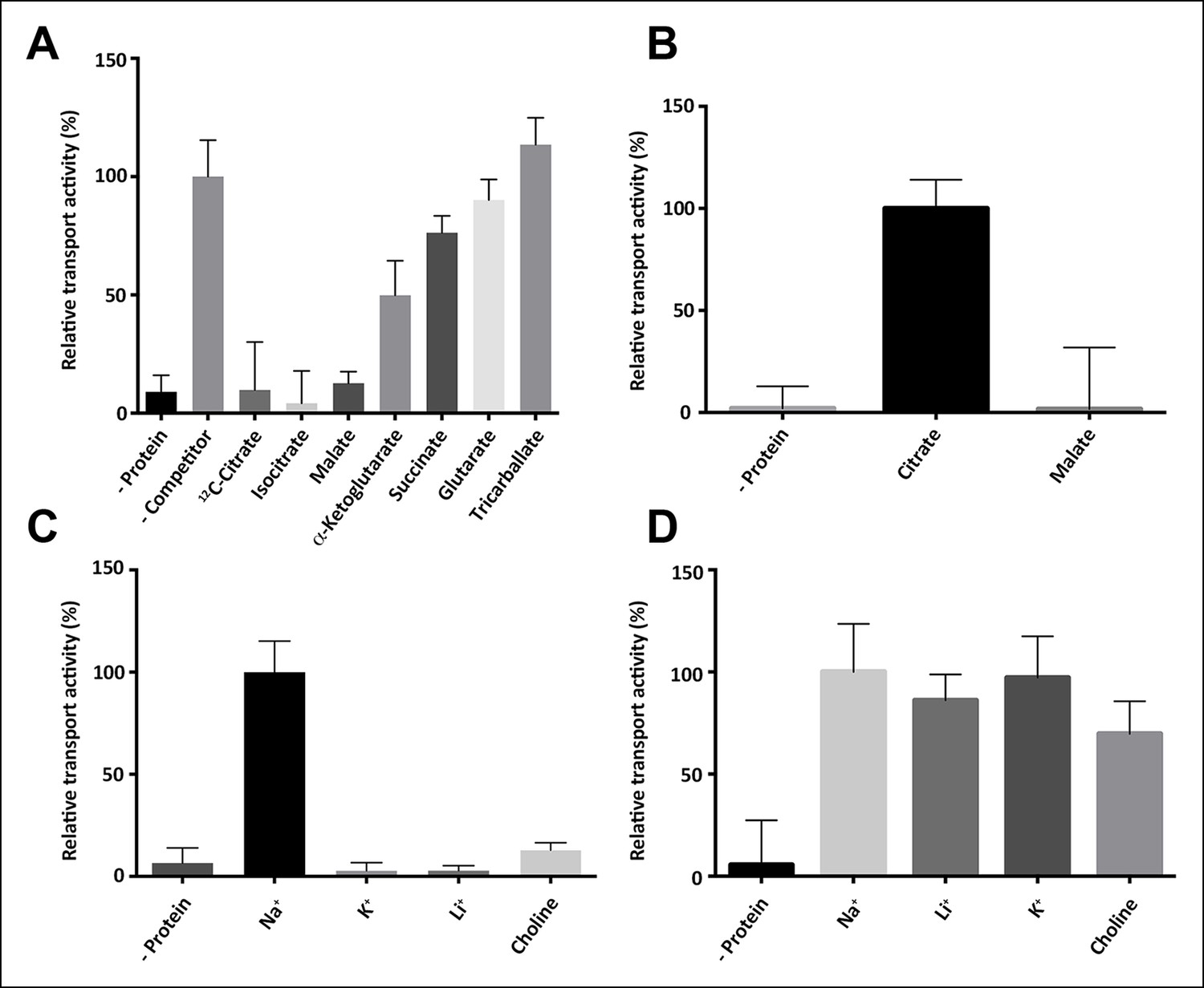
Substrate specificity of SeCitS.

(A) The substrate specificity of SeCitS was established by a proteoliposome uptake inhibition assay. Potential substrates or competitors were added in thousandfold excess of 14C-citrate (5 µM) and transport was measured. The 2-hydroxycarboxylates malate and iso-citrate inhibit 14C-citrate uptake completely. α-Ketoglutarate, which has a carbonyl instead of the citrate hydroxyl group, inhibits less strongly. Succinate and glutarate inhibit transport only slightly. Tricarballate has no effect. (B) While malate inhibits citrate uptake, it is not a substrate for SeCitS, as uptake of 14C-malate (43 µM) is not detectable. (C,D) SeCitS is highly specific for Na+. Neither Li+ nor K+ drive (C) or inhibit (D) citrate uptake. Choline was used as a negative control in both assays. Initial uptake rates were plotted relative to (A) absence of competitor, (B) citrate transport or (C,D) sodium-driven transport.

https://doi.org/10.7554/eLife.09375.004
Figure 3 with 2 supplements see all
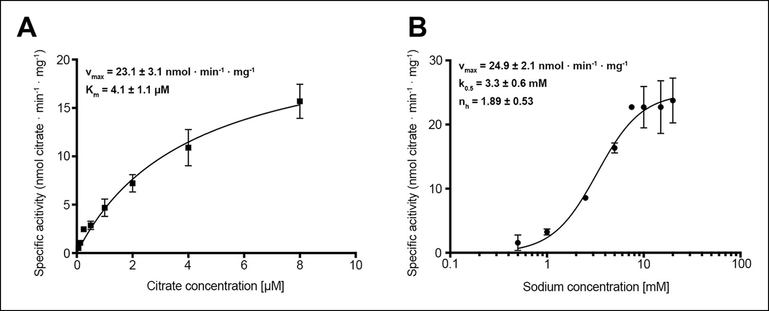
Citrate and sodium transport kinetics.

(A) Citrate uptake by SeCitS containing proteoliposomes in presence of 25 mM Na+ is non-cooperative and follows Michealis-Menten kinetics with a Km of 4.1 µM and a vmax of 23.1 nmol · min-1 · mg-1. (B) Na+ transport in presence of 5 µM citrate is cooperative, with a Hill coefficient of 1.89. The affinity of SeCitS for Na+ is lower than for citrate, as demonstrated by a Km of 3.3 mM. The vmax of 24.9 nmol · min-1 · mg-1 indicates a turnover rate of 1.2 citrate molecules per protomer per minute at room temperature.

https://doi.org/10.7554/eLife.09375.005

To understand the mechanism in detail we determined the structure of CitS from the human pathogen Salmonella enterica (Figure 4) by single-wavelength anomalous dispersion with crystals of seleno-methionine derivatized protein (Figure 4—figure supplement 1). Phases were extended to the 2.5 Å diffraction limit of native crystals (Table 1). The asymmetric unit contains two homodimers of two protomers in different conformations (Figure 4A, B). Each protomer has 13 helix elements (H1–H13), including eleven transmembrane helices (TMH) and two helix hairpins (H6, H12), with the N-terminus on the cytoplasmic side and the C-terminus on the outside. Helices H2-–7 and H8–13 are organized in two repeats with inverted topology (Figure 4C), connected by a flexible cytoplasmic loop (Figure 4B). Together, helices H5–7 of repeat 1 and H11–13 of repeat 2 form a bundle on either side of the central contact domains, which hold the dimer together through extensive hydrophobic interactions of H2, 4, 8 and 10. A 16 Å-deep hydrophobic cavity on the cytoplasmic side of the dimer interface contains the hydrophobic tail of a detergent or lipid molecule (Figure 5A).

Figure 4 with 1 supplement see all
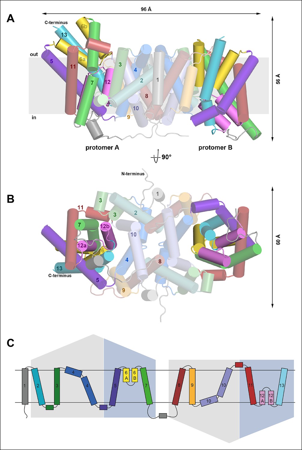
Overall structure of SeCitS and topology.

Side view (A) and cytoplasmic view (B) of the SeCitS homodimer. The dimer is oval, with a long axis of 96 Å and a short axis of 60 Å. Each protomer consists of eleven transmembrane helices and two helix hairpins (yellow and pink). (C) SeCitS consists of two inverted 5-TMH repeats connected by a long cytoplasmic loop plus an additional N-terminal helix. Each repeat contains one hairpin. Helices belonging to the helix bundle are shown on blue background, while helices of the dimer contact domain are shown on grey background. The extended flexible link between the two inverted repeats is completely resolved in protomer A (A).

https://doi.org/10.7554/eLife.09375.008
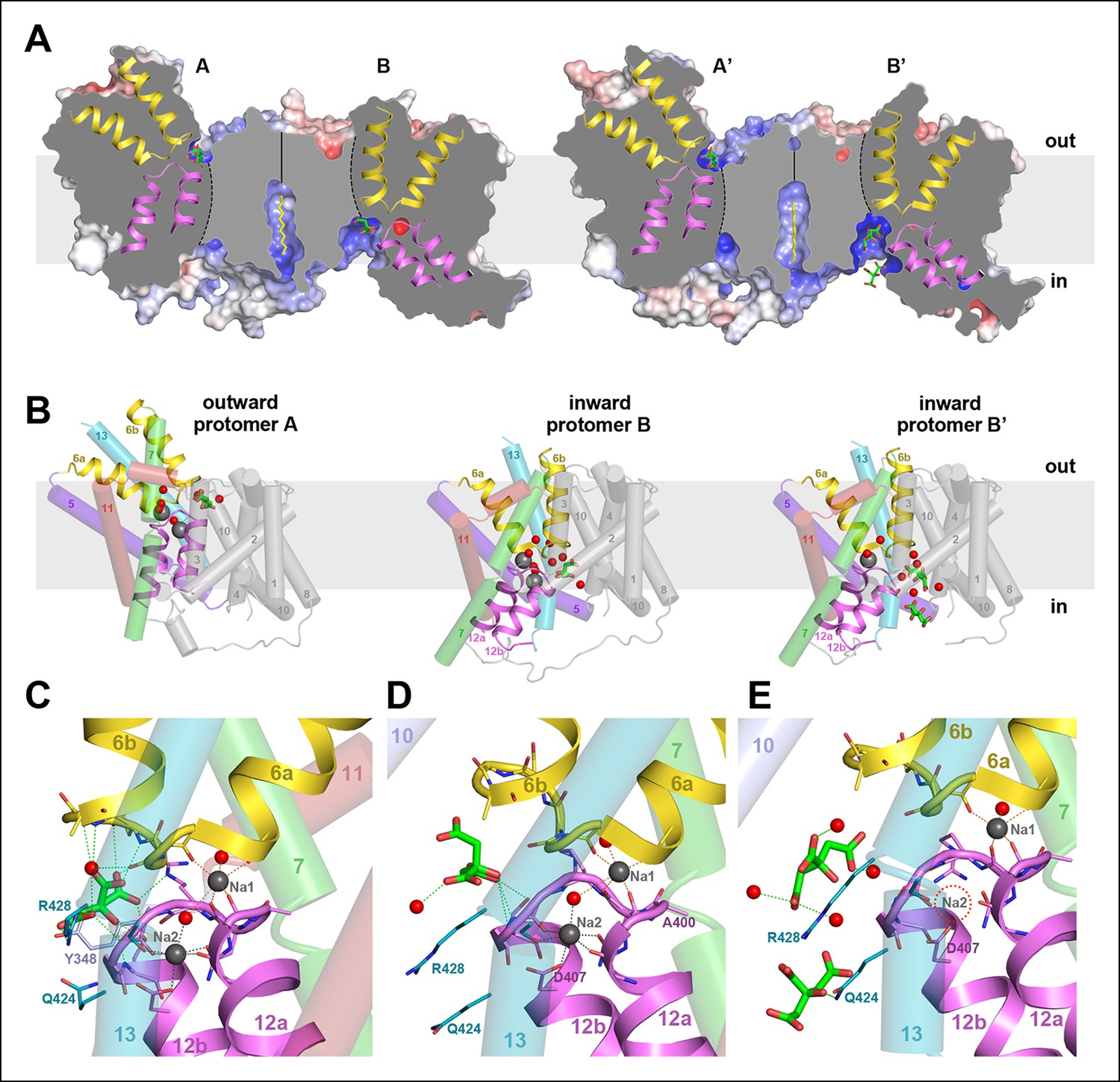
Two different states of the asymmetrical SeCitS dimer.

(A) The outward-facing protomers A and A' bind citrate in a shallow, positively charged cavity between the helix bundle and dimer contact domain. In the inward-facing protomers B and B', citrate binds in a deep cytoplasmic cavity. In B', two citrate molecules are resolved. (B) In protomers A, A’ and B, two Na+are occluded in the helix bundle, while in B' only one Na+ is present. The substrates are translocated 16 Å across the membrane by a 31° rotation of the helix bundle relative to the static dimer contact domain. (C) In the outward-facing protomers, citrate is closely coordinated by sidechains of both hairpins and H13. Neither Na+ participates directly in citrate coordination. (D) In the inward-facing protomer B, citrate is hydrated and attached weakly to the glycine-rich loop of H12. The Na1 and Na2 sites in (C) and (D) are virtually identical, indicating that the transition from the outward-facing to the inward-facing state does not affect Na+-coordination geometry. (E) In protomer B', only the Na1 site is occupied. Two citrate molecules are resolved, outlining a likely trajectory for citrate release (Video 1).

https://doi.org/10.7554/eLife.09375.010
Video 1
Schematic representation of SeCitS transport.

The movie shows a morph from the outward-facing to the inward-facing conformation for one protomer of the SeCitS dimer. Arg402, Arg428 and Tyr348, which coordinate citrate in the outward-facing conformation, are drawn as stick models, while the Na+ ions are represented as grey spheres. Na+ ions bind to their respective sites in the helix bundle, followed by citrate binding between helix bundle and dimer contact domain. Subsequently, the substrates are translocated by a rotation of the bundle. Citrate release is independent from the release of either Na+ ion. Due to the empty Na2 binding site in protomer B’ we assume that this ion is released immediately after the citrate. After substrate release the empty transporter changes its conformation back to the outward-facing state to repeat the cycle.

https://doi.org/10.7554/eLife.09375.011

The two dimers in the asymmetric unit are similar, with an overall rmsd of 0.5 Å, whereas the protomers within one dimer differ substantially by an rmsd of 8.4 Å. The most conspicuous differences are manifest in the vertical positions of the two hairpins H6 and H12 in the helix bundle (Figure 5A). Comparing the two protomers of dimer 1, the C-terminal end of H6 projects 16 Å above the outer membrane surface in protomer A, while it hardly protrudes in protomer B. Conversely, the cytoplasmic H12 ends roughly at the inner membrane surface in protomer A, but extends 13 Å above it in protomer B. The relative position of helices and hairpins within each bundle is unchanged. Evidently, the whole bundle moves as a rigid body from its position in protomer A to that in protomer B, while the central dimer contact domain remains static. The crystal contacts of both dimers in the asymmetric unit are different. Since the polyptide structures of the two dimers are almost identical, the observed asymmetry cannot be attributed to crystal packing. Dimer asymmetry is equally striking with respect to surface structure and electrostatic potential distribution. Protomer A has more positive charges on the periplasmic side than protomer B (Figure 6A). On the cytoplasmic side, positive charges predominate on the surface of protomer B, while positive and negative charges are roughly evenly distributed on protomer A (Figure 6B). Overall, positive charges dominate on the cytoplasmic side of the dimer (Figure 6C, 6D), in line with the positive-inside rule for membrane proteins (Nilsson and von Heijne, 1990; von Heijne, 1992).

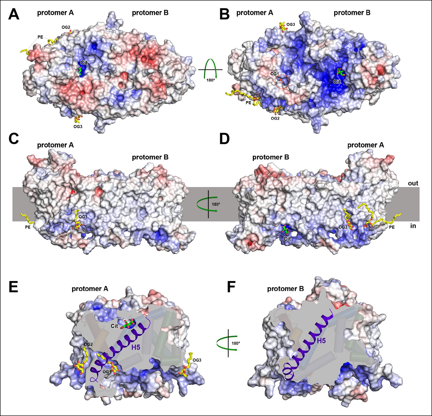
Electrostatic surface potential and bound detergent/lipid molecules.

Exterior (A) and cytoplasmic views (B) of the electrostatic surface potential of SeCitS accentuates the dimer asymmetry. The binding sites for the citrate di-anion (green) on the exterior surface of protomer A and the cytoplasmic side of protomer B are strongly positively charged (dark blue). (C, D) Positions of bound detergent and lipid molecules (yellow) are shown in the side view of the electrostatic surface. Apart from the aliphatic chain in the hydrophobic cavity of the dimer interface (Figure 5), they are positioned close to the helix bundle. (E) In the outward-facing protomers, a hydrophobic cavity between H5, H13 and the dimer contact domain is filled by a detergent molecule. This cavity is closed in the inward-facing protomers (F).

https://doi.org/10.7554/eLife.09375.012

Short stretches of glycine-rich unwound polypeptide link the two halves of hairpins H6 and H12. Together they define the substrate-binding site (Figure 5C–E) at the interface between the helix bundle and the dimer contact domain. On the extracellular surface, the binding site is found in a ∼6 Å deep cavity of protomer A (Figure 6A), while in protomer B it is located at the bottom of a ∼13 Å-deep channel on the cytoplasmic side (Figure 6B). We conclude that protomer A is outward-facing and that protomer B faces inward. The binding sites in both protomers are strongly positively charged (Figure 6A, B). Two detergents and one lipid molecule were identified on the periphery of the dimer. A further detergent molecule was situated in a hydrophobic cavity between the central dimer contact domain and the six-helix bundle of the outward-facing protomer A (Figure 6C–E). H5 in this bundle is straight in protomer A but kinked near its cytoplasmic end in protomer B, to accommodate the movement of the helix bundle (Figure 6E, F).

All four protomers show clear electron density for citrate in the binding site (Figure 7). In the outward-facing protomers, the citrate is closely coordinated by two arginines (Arg402, Arg428), two polar sidechains (Asn186, Ser405) and the protein backbone of both hairpins (Figure 5 and 7A). The only residue in the static contact domain involved in substrate coordination is Tyr348 in H10, which forms a π-π-interaction with a citrate carboxyl. One ordered water molecule participates directly in citrate binding. Its trigonal-bipyramidal coordination geometry (Figure 7) might suggest a Na+ ion rather than water, which would imply that the transported entity is NaCit2- rather than HCit2-. Because the electron density is weak and the coordination distance of >2.8 Å is longer than would be expected for Na+, we interpret this density as a water molecule. In both outward-facing protomers, two Na+ ions are clearly resolved next to the citrate (Figures 5C and 7A). In the Na1 site, four backbone carbonyls in the unwound hairpin stretches coordinate one Na+. In the Na2 site, the carboxyl group of Asp407, the polar sidechains of Asn401, Ser427 and the backbone carbonyls of Cys398 and Gly403 coordinate the ion. Two ordered water molecules participate in Na+binding, one of them suspended between the two Na+ ions (Figures 5C and 7A), accounting for the observed cooperativity of Na+ transport (Figure 3B). Asn401, which coordinates Na1 with its backbone carbonyl and Na2 via its side chain, may contribute to this effect.

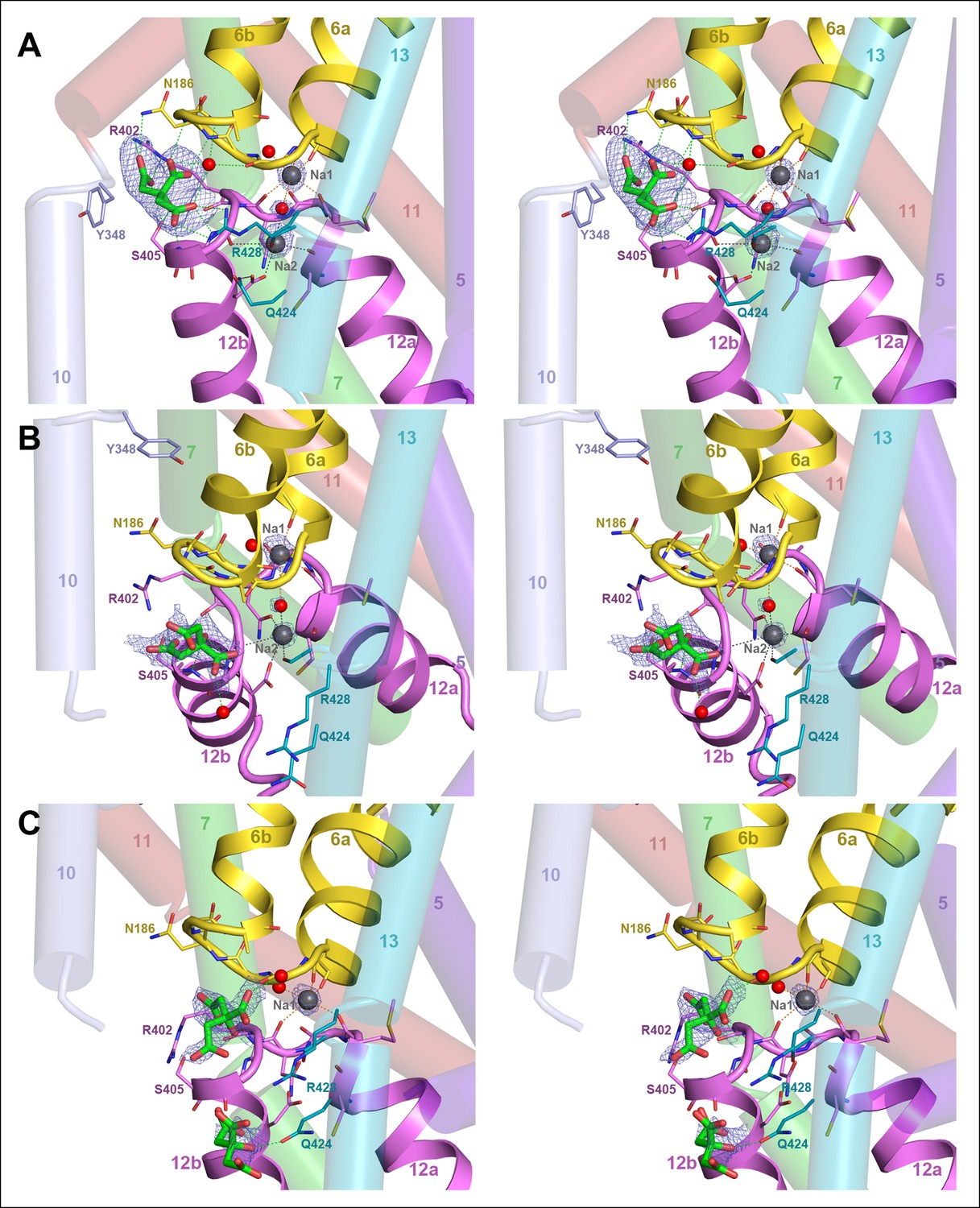
Binding sites and Fo-Fc ligand density.

(A) Stereo view of the outward-facing substrate-binding site of protomer A with an extensively coordinated citrate molecule. (B) In the inward-facing binding site of protomer B the citrate is attached less strongly. In (A) and (B) the Fo-Fc density (blue mesh) is contoured at 3σ for citrate and at 5σ for the two bound Na+ ions and the water molecule between them. (C) In the inward-facing protomer B’, the Fo-Fc map contoured at 4σ shows an occupied Na1 site, while the Na2 site is empty. The Fo-Fc omit map contoured at 2.5 clearly shows two citrate molecules.

https://doi.org/10.7554/eLife.09375.013

There is no difference in substrate coordination or in main-chain conformation between the two outward-facing protomers A and A’ (rmsd 0.5 Å). A and A’ can therefore be considered as identical. Interestingly, the main chain conformations of the two inward-facing protomers B and B’ are likewise practically identical (rmsd 0.6 Å), but the citrate and Na+ coordination in B and B’ is clearly different. In protomer B, citrate is partially hydrated and coordinated by the hydroxyl of Ser405 and the backbone carbonyl of Gly404 in the conserved GGXG motif of H12 (Figures 1, 5D and 7B). Both Na+ sites are occupied and take up the same position relative to the citrate as in the outward-facing state. In protomer B', the Na2 site is empty, even though the structure of the ion-coordinating hairpin hardly changes (Figures 5E and 7C). The citrate is fully hydrated and not directly attached to a sidechain, and a second citrate is present near Gln424 in H7.

The rigid-body movement of the helix bundle from its position in protomers A and A’ to that in protomers B or B' can be described as a 31° arc-like rotation around an axis roughly parallel to the membrane and perpendicular to the long dimer axis (Figures 5, 7 and Video 1). The rotation is facilitated by the greasy interface between the helix bundle and the static dimer contact domain. The greasy interface consists almost entirely of small hydrophobic sidechains and a bound detergent molecule that may take the place of a membrane lipid alkyl chain (Figures 5 and 8 A-D). During the bundle rotation the detergent molecule is displaced by H13. As the helix bundle reaches the inward-facing position, the straight, hydrophobic helix H5 kinks at Gly143, thus preventing its partial exposure to the cytoplasm, and an ion bridge forms between Asp112 and Arg205 in H7 (Figure 8E, F; Video 2). As a result of the helix bundle rotation, the binding site with the bound citrate moves by 16 Å from the external membrane surface in the outward-facing state to a position where it is accessible from the cytoplasmic membrane surface in the inward-facing state (Figure 5). Since transport is non-cooperative with respect to citrate (Figure 3A), we conclude that the two binding sites in the dimer act independently of one another.

Hydrophobic interface between helix bundle and dimer contact domain.

(A, C) In the outward-facing protomers A and A’, a hydrophobic pocket between helix H5, H13 and the dimer contact domain harbors a detergent molecule that apparently replaces a membrane lipid. (B, D) In the inward-facing protomers B and B' H5 kinks at Gly143 and shifts towards the cytoplasm. We assume that H13 fills this hydrophobic cavity in the inward-facing state. (E) In the outward-facing protomers, Tyr348 coordinates the citrate by π-π-interactions. As a result of the arc-like helix bundle rotation, an ion bridge forms between Asp112 and Arg205 (H7) in the inward-facing protomers (F). Arg205 moves by more than 20 Å from its position in the outward-facing conformation (E). The sidechain of Tyr348 rotates by 90°, blocking the entrance to the substrate binding site (F).

https://doi.org/10.7554/eLife.09375.014

Unlike the Na+ ions, the citrate di-anion is not occluded by the hairpin loops in SeCitS. Similarly, the dicarboxylate substrate is not occluded in VcINDY (Mancusso et al., 2012), whereas the corresponding substrate is occluded within the helix bundle of GltPh (Boudker et al., 2007). SeCitS may lack a well-defined substrate-occluded state, but the citrate would effectively be occluded during the transition from the outward-facing to the inward-facing state, while the occupied binding site rotates past the hydrophobic surface of the dimer interface domain (Video 2).

Video 2
Schematic representation of domain, helix and sidechain movements.

Three synchronized movies show different views of one SeCitS protomer during the transport cycle: (A) from the membrane plane, (B) in the perpendicular direction from the cell exterior and (C) a detailed view of the substrate-binding site and the detergent/lipid binding pocket. Helices of the rotating bundle domain are coloured, while helices in the static dimer contact domain are shown in grey behind their corresponding transparent electrostatic surface. The negatively charged periplasmic surface of SeCitS (transparent red) attracts Arg205 of H7 (green), which, in the inward-facing state, forms an ion bridge to Asp112 in H4 and a hydrogen bond to Tyr348 (lavender) in H10 of the dimer contact domain. In the outward-facing state, Asp112 interacts with Tyr348, which rotates to block access to the substrate-binding site in the inward-facing state. A detergent molecule (yellow) in the hydrophobic pocket between H5 (purple), H13 (cyan) and the dimer contact domain, is displaced in the inward-facing state by the movement of H13. H5, which is straight in the outward-facing state, kinks during the bundle rotation to prevent its partial exposure to the cytoplasm.

https://doi.org/10.7554/eLife.09375.015

In the outward-facing state, strong polar and ionic interactions facilitate citrate binding at low ambient substrate concentrations (Figure 3A and 5C). In the inward-facing state, the binding affinity for the substrate is reduced (Pos and Dimroth, 1996), so the citrate can detach. We propose that the three citrate positions we observe in the two inward-facing protomers mark the path of the substrate during its release from the binding site past the highly conserved Arg428 (Figures 1, 5 and 7), along a trajectory that guides the negatively charged substrate towards the cytoplasm, where it is metabolized. Partial release of the citrate di-anion would weaken Na+ binding, which explains why only one Na site is occupied in B’. Since transport is electroneutral, both Na+ ions must dissociate from the inward-facing state. MD simulations suggest that in other Na-dependent transporters such as LeuT (Grouleff et al., 2015), GltPh (Zomot and Bahar, 2013), vSGLT (Watanabe et al., 2010), at least one of the Na+ ions is released before the main substrate. In the case of SeCitS, comparison of the inward-facing protomers B and B’ indicates unambiguously that citrate is released before Na+, and that Na2 is released before Na1 (Figure 5D, E). Once the citrate has left the binding site, the helix hairpins or H13 would need to rearrange to release Na1, while a minor reorientation of the Asp407 or Ser427 sidechains is sufficient to release Na2.

Comparison of the binding sites in the outward-facing protomers indicates that both Na+ ions have to be in place before citrate can bind. A cryo-EM structure from 2D crystals of the closely related KpCitS from Klebsiella pneumoniae found that sodium citrate induced a major conformational change in the helix bundle, whereas potassium citrate did not (Kebbel et al., 2013), supporting the proposed binding order. Therefore the complete transport mechanism entails the following six steps: (1) The Na sites are occupied by Na+ in the outward-facing state; (2) a citrate binds from the external medium; (3) citrate binding triggers the arc-like rotation of the helix bundle in the transition from the outward-facing to the inward-facing state; (4) in the inward-facing state, the citrate becomes hydrated and diffuses into the cytoplasm; (5) the sodium ions come off; (6) the release of all substrates enables the reverse arc-like rotation of the helix bundle to expose the empty binding site again to the cell exterior, and the cycle repeats (Figure 9; Videos 1 and 2).

Table 1

Data collection and refinement statistics

https://doi.org/10.7554/eLife.09375.016
Native SeCitSSeMet SeCitS
Data collection SLS PXII
Wavelength (Å)0.9790.980
Space groupP1P21
Cell dimensions
a, b, c (Å)86.4, 89.9, 91.890.9, 168.8, 97.9
α, β, γ (°)90.4, 113.8, 99.590.0, 91.0, 90.0
Resolution (Å)47.98 – 2.5 (2.6 – 2.5)48.95 – 3.9 (4.0 -– 3.9)
Rpim0.052 (0.872)0.038 (0.539)
I / σI 8.9 (1.3)16.8 (2.2)
CC*0.999 (0.828)1.000 (0.944)
Completeness (%)98.8 (98.1)100 (100)
Multiplicity8.2 (8.1)41.4 (40.9)
Refinement
Resolution (Å)47.98 – 2.5 (2.6 – 2.5)
Unique reflections84765
Reflections in test set4193
Rwork/Rfree (%)21.0/24.8 (33.6/36.3)
CC(work)/CC(free)0.848/0.742 (0.796/0.773)
Average B-Factor (Å2)70
No. atoms in AU13270
Protein12916
Ligands285
Water69
r.m.s. deviations:
Bond lengths (Å)0.003
Bond angles (°)0.762
  1. Values for the highest resolution shell are shown in parentheses

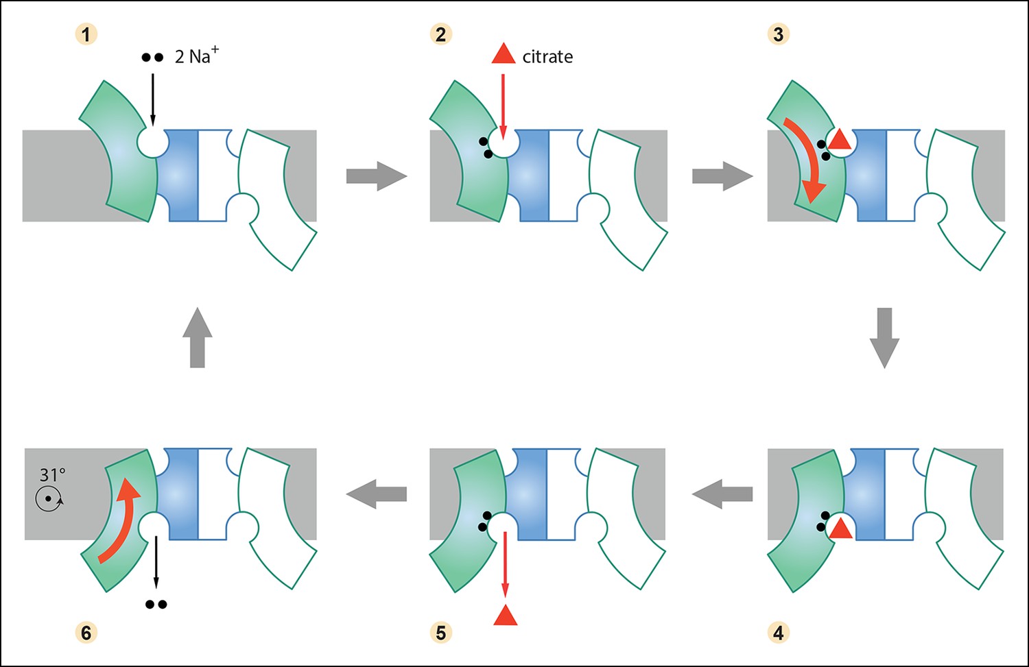
Six-step mechanism of Na+-dependent citrate uptake by SeCitS.

(1) Two Na+ bind to the empty transporter; (2) citrate from the external medium attaches to the binding site; (3) the substrates are translocated across the membrane through a rigid-body 31° rotation of the helix bundle domain; (4) first the citrate and then (5) the Na+ ions are released to the cytoplasm; (6) the unloaded protomer changes its conformation back to the outward-facing state and the cycle restarts. In the cell, the inward-directed Na+gradient drives citrate uptake, but all steps are in principle reversible. The approximate position of the rotation axis parallel to the membrane and perpendicular to the long dimer axis is indicated in (6).

https://doi.org/10.7554/eLife.09375.017

Notwithstanding the large domain movements associated with substrate translocation, citrate exchange rates are high, with a turnover of up to 137 s-1 reported for the closely related KpCitS (Pos and Dimroth, 1996). Citrate uptake by SeCitS is substantially slower at 1.2 molecules per minute (Figure 3). Therefore, the arc-like rotation of the helix bundle that translocates the bound substrate across the membrane is not rate-limiting. The same seems to hold true for GltPh, which shows a slow substrate uptake rate of 0.29 molecules per minute (Ryan et al., 2009), while crosslinking experiments show that the conformational change happens within seconds (Reyes et al., 2009). Assuming that the Na+ concentration does not limit substrate binding or release under physiological conditions, the rate-limiting step in SeCitS is most likely the reverse rotation of the helix bundle with the binding site empty.

An influence of lipids on the conformational dynamics in GltPh by inserting a lipid molecule between both domains was recently proposed by MD simulations (Akyuz et al., 2015). The structure of SeCitS offers experimental evidence for the existence of a hydrophobic pocket at the interface of both domains and highlights the importance of the bilayer for the activity of membrane transporters. It remains to be seen whether any of the lipid-binding sites in these transporters are structurally conserved.

The arc-like rotation of the helix hairpins in SeCitS is reminiscent of the recently proposed conformational change for substrate translocation in GltPh (Crisman et al., 2009; Reyes et al., 2009; Verdon et al., 2014). In the Na+/H+ antiporters (Lee et al., 2013; Paulino et al., 2014) or the bile acid transporter ASBT (Zhou et al., 2014), where the binding site is defined by unwound stretches of two trans-membrane helices in a structurally homologous bundle, this process also involves a rotation of the bundle around a similar axis as in SeCitS, although the movement is significantly smaller. The domain structure of the unrelated transporter YdaH (Bolla et al., 2015) bears a striking resemblance to that of SeCitS, suggesting that it may work in the same way. The rotating arc mechanism described here for SeCitS thus seems to apply to a large class of secondary membrane transporters with unwound helix elements or hairpins that were previously thought to be unrelated.

Materials and methods

Protein expression and purification

Request a detailed protocol

A gene coding for CitS from Salmonella enterica (WP_000183608) was cloned into a pET21d plasmid harboring an N-terminal His10-Tag and a thrombin cleavage site between tag and target protein. The resulting plasmid was used to transform E. coli C41-(DE3) cells. After expression for 10 h at 37°C in ZYM-5052 autoinduction medium (Studier, 2005) cells were harvested, resuspended in 20 mM Tris/HCl pH7.4, 150 mM NaCl, 5 mM EDTA, 5 mM β-mercaptoethanol (β-ME) and broken using a microfluidizer (M-110L, Microfluidics). Unbroken cells and cell debris were removed by centrifugation at 18,000 g for 30 min. Membranes were isolated by centrifugation at 100,000 g for 1 h and resuspended at a total protein concentration of 15 mg/ml in 20 mM Tris/HCl, 140 mM choline chloride, 250 mM sucrose, 1 mM Na-citrate, 5 mM β-ME. SeCitS was solubilized by 1:1 dilution of membranes with 20 mM Tris/HCl pH7.4, 150 mM NaCl, 3% n-decyl-β-D-maltopyranoside (DM), 1 mM Na-citrate, 5 mM β-ME. Unsolubilized material was removed by ultracentrifugation at 100,000 g for 1h. The supernatant was supplemented with 45 mM imidazole and incubated with Ni-NTA beads equilibrated with 20 mM Tris/HCl pH7.4, 300 mM NaCl, 45 mM imidazole, 1 mM Na-citrate, 0.15% DM, 5 mM β-ME for 2h at 4°C. The mixture was loaded on a column and washed with equilibration buffer to remove unspecifically bound protein. For on-column cleavage the buffer was changed to 10 mM Tris/HCl pH8.2, 150 mM NaCl, 2.5 mM CaCl2, 1 mM Na-citrate, 0.15% DM. Thrombin was added to the beads to a concentration of 1 U/mg protein and incubated overnight under constant mixing. The beads were washed with exchange buffer to recover tag-free SeCitS and the protein was concentrated to 5 mg/ml (50 kDa cut-off). The concentrated protein was applied to a Superdex-200 size exclusion column equilibrated with 20 mM Tris/HCl pH8.2, 150 mM NaCl, 1 mM Na-citrate, 0.15% DM, 1 mM TCEP (Tris-(2-carboxyethyl)phosphine). Fractions containing SeCitS were pooled, concentrated as above, frozen in liquid nitrogen and stored at -80°C.

Selenomethionine (SeMet)-substituted protein was expressed in a defined medium by methionine biosynthesis inhibition (Doublie, 2007). Expression cultures were directly inoculated with pre-cultures grown in non-inducing PA-0.5G medium (Studier, 2005). The main culture was grown at 37°C, induced at an OD600 of 0.5 and harvested after 4 h. Purification of SeMet SeCitS was performed as described for the native protein.

Crystallization and data collection

Request a detailed protocol

For crystallization, native SeCitS was supplemented with n-octyl-β-D-glucopyranoside (OG) to a concentration of 1%. The protein was mixed 1:1 with reservoir solution (100 mM MES pH6.5, 200 mM NaCl, 29% PEG400) and crystallized in 24-well hanging drop plates. Rhombic crystals appeared within 3 days and grew to a size of 150 µm within a week. Crystals were harvested and vitrified in liquid nitrogen using Al´’s oil (D'arcy et al., 2003) as cryo-protectant.

SeMet-derivatized SeCitS was supplemented with 2% n-heptyl-β-D-glucopyranoside (HG) and mixed 1:1 with reservoir solution (100 mM MES pH6.5, 250 mM NaCl, 30% PEG400). Thin needle-like crystals grew to 400 µm within a week and were vitrified in liquid nitrogen directly. All datasets were collected on beamline X10SA (PXII) at the SLS (Villigen, Switzerland).

Data processing and structure solution

Request a detailed protocol

All datasets were processed with XDS (Kabsch, 1993) and scaled with AIMLESS (Evans, 2006) from the CCP4 package (Winn et al., 2011). Resolution limits were based on I/σ(I)-values, completeness and cross correlation of half datasets (Karplus and Diederichs, 2012) in the high-resolution shells. PHENIX (Adams et al., 2010) and Coot (Emsley et al., 2010) were used for refinement and model building, respectively. Experimental phases were obtained by single-wavelength anomalous dispersion (SAD) from SeMet-derivatized SeCitS. Initial SeMet positions were determined by SHELXD (Schneider and Sheldrick, 2002) through the HKL2MAP (Pape and Schneider, 2004) interface and fed into Crank2 (Skubak and Pannu, 2013) for substructure refinement, phasing with Refmac (Murshudov et al., 1997), hand determination, initial density modification with Parrot (Zhang et al., 1997) and model building using Buccaneer (Cowtan, 2006). An initial backbone model of SeCitS was created for phasing of the native high-resolution data by molecular replacement with PHASER (McCoy et al., 2007). Model building was performed by PHENIX autobuild (Terwilliger et al., 2008), followed by cycles of manual model building and refinement. Superimpositions were performed with GESAMT (Winn et al., 2011). Figures were drawn and rmsd values were calculated with PyMOL (DeLano and Lam, 2005). Electrostatic surfaces were calculated with PDB2PQR (Dolinsky et al., 2004) and APBS (Baker et al., 2001).

Reconstitution into liposomes

Request a detailed protocol

E. coli polar lipids in chloroform (Avanti Polar Lipids) were dried under nitrogen and resuspended in reconstitution buffer (20 mM Tris/BisTris/Acetate pH 4-–8, 50 mM choline chloride), supplemented with 15 mM β-ME. Unilamellar ∼400 nm vesicles were prepared using polycarbonate filters in an extruder (Avestin). Preformed liposomes were diluted to 5 mg/ml in reconstitution buffer and destabilized by addition of 1% OG. SeCitS was added at a lipid-to-protein ratio of 50 and incubated for 1 h. The protein/lipid mixture was filled into dialysis bags (14 kDa cutoff) and dialyzed against detergent-free reconstitution buffer overnight. Biobeads were added to the dialysis buffer to facilitate complete detergent removal. The proteoliposomes were centrifuged for 25 min at 300,000 g and resuspended in fresh reconstitution buffer.

Transport measurements

Request a detailed protocol

Transport activity was measured with [1,5]14C-citrate or 1,4(2,3)-14C-malate as a reporter molecule. Measurements were started by dilution of 2 µl freshly prepared proteoliposomes into 200 µl reaction buffer (20 mM Tris/BisTris/acetate pH 5–8, 50 mM NaCl, 5 µM [1,5]14C-citrate or 43 µM 1,4(2,3)-14C-malate). Within the linear range of uptake, 200 µl samples were transferred on 0.2 µm nitrocellulose filters that were subsequently washed with 3 ml of reaction buffer. Filters were transferred into counting tubes and filled with 4 ml liquid scintillation cocktail (Rotiszint) before evaluation. All measurements were performed in triplicates. In all experiments initial rates within the linear range of uptake were recorded over a total of 4 time points.

Kinetic measurements were performed at pH6 by varying the concentration of one substrate while keeping the other constant. Ion specificity of SeCitS was determined by changing the co-substrate to LiCl, KCl or choline chloride, which is not transported. Specificity for citrate was established with a competition assay. Potential substrates were added to the reaction buffer at a concentration of 5 mM (1000x excess) to compete with 14C-citrate uptake. The effect of ΔpH on the transport activity was measured by changing the pH of the reaction buffer while keeping the inside pH constant.

Data availability

The following data sets were generated

References

    1. Delano WL
    2. Lam JW
    (2005)
    PyMOL: A communications tool for computational models
    Abstracts of Papers of the American Chemical Society pp. U1371–U1372.
    1. Emsley P
    2. Lohkamp B
    3. Scott WG
    4. Cowtan K
    (2010) Features and development of Coot
    Acta Crystallographica. Section D, Biological Crystallography 66:486–501.
    https://doi.org/10.1107/S0907444910007493
    1. Evans P
    (2006) Scaling and assessment of data quality
    Acta Crystallographica. Section D, Biological Crystallography 62:72–82.
    https://doi.org/10.1107/S0907444905036693
    1. Pape T
    2. Schneider TR
    (2004)
    HKL2MAP: a graphical user interface for macromolecular phasing with SHELX programs
    Journal of Applied Crystallography 37:843–844.
    1. Pos KM
    2. Dimroth P
    3. Bott M
    (1998)
    The Escherichia coli citrate carrier CitT: a member of a novel eubacterial transporter family related to the 2-oxoglutarate/malate translocator from spinach chloroplasts
    Journal of Bacteriology 180:4160–4165.
    1. Schneider TR
    2. Sheldrick GM
    (2002)
    Substructure solution with SHELXD
    Acta Crystallogr  58:1772–1779.
    1. van der Rest ME
    2. Molenaar D
    3. Konings WN
    (1992)
    Mechanism of Na(+)-dependent citrate transport in Klebsiella pneumoniae
    Journal of Bacteriology 174:4893–4898.

Decision letter

  1. John Kuriyan
    Reviewing Editor; Howard Hughes Medical Institute, University of California, Berkeley, United States

eLife posts the editorial decision letter and author response on a selection of the published articles (subject to the approval of the authors). An edited version of the letter sent to the authors after peer review is shown, indicating the substantive concerns or comments; minor concerns are not usually shown. Reviewers have the opportunity to discuss the decision before the letter is sent (see review process). Similarly, the author response typically shows only responses to the major concerns raised by the reviewers.

Thank you for submitting your work entitled "Complete mechanism of Na+ -dependent citrate transport from the structure of an asymmetrical CitS dimer" for peer review at eLife. Your submission has been favorably evaluated by John Kuriyan (Senior Editor) and three reviewers.

This paper has received a very positive response, and all three reviewers are in agreement that this paper presents important structural results that are suitable for publication in eLife. The reviewers, in their individual reviews and in the online discussion, had many suggestions as to how to improve the manuscript and to properly balance the structural work with the functional studies. Rather than attempt to condense this discussion, the editor has provided the reviews in full with many of the points raised in reviewer discussion embedded within. The authors should use their own judgment in how best to respond to these comments in preparing a revised manuscript. Note that no new experimental work is called for.

Reviews and discussion comments:

Reviewer 1: The manuscript by Wöhlert et al. presents X-ray crystal structures at high resolution for a secondary transporter from Salmonella enterica. The crystallographic unit reveals four distinct protomers arranged into two dimers, with two distinct conformational states and several different substrate binding states. The structures provide extensive insights into the details of the binding sites, and the conformational change required to transition from outward-facing substrate-bound to inward-facing substrate-bound states for this transporter. Several biochemical measurements are also included using proteoliposomes under various conditions to demonstrate that the transporter can transport citrate in the presence of sodium, ruling out other di- and tri-carboxylic substrates, and testing the effect of pH, from which they conclude that the transporter does not translocate H+. Their measurements also indicate that malate and isocitrate are competitive inhibitors. The manuscript is clearly written and the structure analyzed in detail.

My main concerns with the manuscript relate to overinterpretation of the structural data (particularly with respect to the substrate binding/release order, and role of lipids), which in light of the extensive findings, does not seem to be necessary. In this regard, the title and Abstract should be toned down, as I do not think that the structures are sufficient to explain the entire mechanism, as there are no structures of the apo states. I also would welcome some discussion of the similarities and differences between other folds in the literature, especially VcINDY and GltPh. Finally, some issues need to be clarified with respect to the substrates being transported, particularly the species of citrate. My concerns are laid out by topic below.

1) Sodium stoichiometry: the authors state that a Hill coefficient of 1.89 suggests that coupling requires two Na+ (Results and Discussion, first paragraph). This is not correct, a Hill coefficient > 1 means that more than one Na+ ion is involved; higher stoichiometries of 3 or more cannot be ruled out from the Hill coefficient. Although the structures show evidence for two sodium ions, neither the Hill coefficient, nor the 2.5 Angstrom structure, can rule out co-transport of a third (or fourth) Na+ ion. Since related proteins couple to three sodium ions, this is an important point.

2) Role of protons and citrate protonation state (Results and Discussion, first paragraph onwards): I found the arguments relating to the effect of protons and the citrate protonation state needed to be better argued and supported. First, "van der Rest" et al. 1992 is cited as evidence that CitS transports HCit2-. There seem to be two problems with this citation: the van der Rest paper studies Klebsiella pneumoniae CitS, not Salmonella enteric, and it is entirely possible that they transport different species. At the least, the reasons to make that assumption should be explored and described, including mentioning the pKa's of the different protonation states of citrate. In addition, van der Rest et al. concluded that the stoichiometry is one sodium and two protons, whereas the current work concludes that sodium is cotransported but not protons. The roles of the different species are interconnected, so these two different scenarios could lead to different conclusions about the citrate protonation state in order that the process is electroneutral. It is therefore important that the arguments or uncertainties relating to the transport stoichiometry and species be laid out more clearly.

On a similar note, regarding the pH dependence, the authors invoke a role for sodium at low pH; first, I'm not sure I understand the argument. Moreover, a recent work by Mulligan et al. (PMID: 24821967) follows the pH-dependence of succinate transport by a related protein VcINDY, and shows that a role for sodium need not be invoked to explain complex pH-dependence profiles, at least for succinate.

3) Detergent molecule in the interface between H5/H13 and dimerization domain (legend for Figure 5E; Results and Discussion, seventh paragraph; legend for Figure 7; and legend for Video 2): The fact that a detergent is present in the outward-facing conformation does not indicate that a membrane lipid would "replace it" and lead to "participation of a membrane lipid in the transport mechanism". The authors may have been lucky that OG binds there, as it may be stabilizing the outward-facing state in the crystal, but OG is not the same as a physiologically relevant phospholipid. Indeed, in a different secondary transporter called LeuT, an OG detergent molecule binds to the extracellular cavity and stabilizes the outward-facing conformation (Nissen and coworkers). No-one would argue that this site is required for a lipid interaction during transport. These references (including to H13 "dislodging" the detergent molecule") should be removed. Any speculation with respect to the role of this detergent should be clearly labeled as speculation.

4) Ligand binding site: please replace the word "tightly" with "closely" when describing the ligand interactions. Close proximity in a crystal structure, while suggestive, does not equate to strength. Similarly, the reference to Tyr348 being "held in place" by Asp112, is speculative. Biochemical evidence, and molecular simulations would be useful to support such assertions, but even then, it is difficult to state conclusively that such interactions are maintained outside of the crystal lattice, and/or are important for function. Indeed, D112 is not conserved across other homologues shown in Figure 3–figure supplement 2, and nor is R205 or Y348. Moreover, for a "strong" interaction it is surprising to see a difference in the orientation of one of the citrate carboxylates groups, as seen when comparing protomer A with protomer A' in Figure 4C.

The density assigned to citrate in the inward-facing protomer B, and the one assigned to the second citrate in the inward-facing protomer B', is significantly less well resolved, and it is not clear that they fit citrate as well as in the other site. The authors should discuss the possibility that some other (crystallization buffer) molecule occupies those regions.

5) The presence of a state with one sodium ion and one citrate, but one of the sodium sites empty, leads to over-interpretation of the substrate release steps (Results and Discussion, ninth paragraph). Since the citrate is not completely removed from the pathway and presumably blocks it, it is not clear how the missing sodium is supposed to have "released". This issue becomes further contradictory when visualizing the movie (s), which are shown as one citrate and one sodium coming off together (see below), and then when looking at the schematic of the transport cycle (Figure 8), when the substrate, and then the sodiums, come off one after another. I strongly recommend that these descriptions of the steps of unbinding be mentioned only in passing and clearly described as speculation. Such detail is not necessary as these are already, in many other ways, very informative structures.

6) The authors do not mention what the crystal packing is like. Are any of the contacts potentially relevant?

7) The videos should show only the states that are known. At most, it is reasonable to show the morphed transition between the bound forms. The binding steps are too speculative. First, the sodiums bind essentially to the same state of the protein as the substrate-bound form (or one in which some side chains have been selected to move out of the way). There is no known structure of the sodium-free or substrate free forms of SeCitS, and evidence from other proteins shows that the protein backbone changes when those substrates are missing; therefore it is misleading to suggest that the structure remains the same. Similarly in the inward-facing states, it is implied by the movie that one sodium and citrate come off before the second ion; however none of those intermediates are known; in fact, they contradict the occupancy shown in protomers B and B'.

Reviewer 2:Yildiz and colleagues describes a remarkable structure of a bacterial citrate transporter, CitS. The dimeric transporter is captured in the crystal in an asymmetric state such that one domain occupies an outward position and the other is inward. Thus, this single structure reveals the key aspects of CitS transport mechanism. Specifically, the structure shows that CitS belongs to a growing class of secondary active transporters that function by an elevator-like mechanism. In these transporters, one domain carrying the bound substrate and coupled ions moves across the bilayer along a proteinous track provided by another, stationary domain, which is involved in subunit association. Despite some concerns, I feel that overall the work is technically sound and most conclusions are well founded. I do not have major concerns that would require extensive rebuttal. However, I feel that the manuscript could be substantially improved through editorial changes, and I have a number of questions and suggestions. These are only minor comments. Overall, these fall into three categories: (A) concerns regarding the presentation and discussion of the functional data, including certain vagueness of the methods; (B) information content of the structural figures and corresponding structural discussions, which I think can be significantly strengthened; (C) putting results into overall perspective, which I feel could also be improved.

A) Concerns regarding functional data:

1) In all functional figures, it is unclear what does "Relative transport activity" constitute. Is it vmax, initial rate at a sub-saturating substrate concentration, a measurement at a single time point? If the latter, it would be great to see how does typical time dependence look like? Similarly, when Km values were determined, was the initial rate measured for each concentration of the substrate/ion? The methods are not very clear on these aspects.

Related comments raised during the consultation between the reviewers:

The Materials and methods describes the "Transport measurements". There, they report that they counted total radioactivity of captured C14, measuring how much radioactive citrate was imported into liposomes. Perhaps move this up to main text to avoid confusion.

My comment regarding the ambiguity of the term "activity" stems from the following consideration. Ideally, when comparing activity under different conditions (substrate concentration, pH etc.) one would want to plot the initial rates. This is why I would have liked to see how does the time dependence of the uptake look like: for how long the uptake is linear and when does it plateau. Such plot together with the note on which time point was used to measure the activity in the remaining experiments would have provided a clearer more rigorous description of the data.

2) In Figure 2, I am surprised by the substantial variability of background uptake in the absence of the protein. For example, it is particularly high in panel C. What is the origin of this variability?

Related comments raised during the consultation between the reviewers:

Figure 1A, B, D show uptake and its inhibition, Figure 1C shows how the substrate drives the uptake.

According to one of the reviewers, in Figure 1A, B, D, it seems that the authors measured how much C14 was able to enter liposomes, when different buffers were on the outside. In (D), they show that C14-citrate can always enter, as long as Na and Citrate are outside, even in the presence of additional Li+ or K+ . In (C) they show that C14-citrate can only enter if Na+ is present on the outside, but not if Na+ is absent and K+ or Li+ are present instead. The referees agree that here the high C14 uptake in the absence of protein is strange and needs further explanation. Did the authors temporarily break the liposomes when adding salts, due to osmotic imbalances, and that is how C14 could enter the liposomes?

Very high background in Figure 1C (uptake by naked liposomes is ~40% of that in the presence of Na+ gradient) is worrying, but not crucial.

3) It would be helpful if the authors commented whether the turnover rate of 1.2 per minute is slower than expected. Is such rate physiologically relevant? Could it be that citrate is not the physiological substrate?

4) In the legend for Figure 2, it would be helpful if the authors included the concentration of Na+ ions used in panel A and citrate in panel B.

5) In the first paragraph of the Results and Discussion, "hill" should be capitalized. Hill coefficient above 1 suggests that at least 2 Na+ ions are required for substrate binding, but does not exclude the possibility that more than 2 Na+ ions are involved. Hence the sentence should read "coupled to at least two Na+ ions".

6) Was background (uptake in the absence of Na+ gradient) subtracted in Figure 2–figure supplement 1 and supplement 2? If not, what fraction of the observed uptake at pH 5 and 8 or in the presence of internal monovalent cations is due to background? How does background uptake depend on external pH in Figure 2–figure supplement 2A? Can that dependence explain increased uptake in the lower external pH buffer?

7) I find the observation that the symmetric low pH inhibits transport while asymmetric external low pH stimulates uptake very interesting. However, the authors' explanation of this phenomenon seems unsatisfying. They suggest that inhibition in symmetric low pH could be due to limiting Na+ binding. If so, it should also manifest in asymmetric conditions. In contrast, the suggestion that low pH increases concentration of transportable doubly protonated citrate species explaining activation in asymmetric conditions seems reasonable. Could it be that low internal pH inhibits the transporter because some protonatable protein groups with a near neutral pKa need to be deprotonated to allow the return into the outward facing state?

Related comments raised during the consultation between the reviewers:

How are the authors sure that the liposomes stayed intact under all experiments?

8) The inhibition by internal monovalent cations is striking. What is a possible explanation of the observed inhibition by all monovalent cations?

Related comments raised during the consultation between the reviewers:

How are the authors sure that the liposomes stayed intact under all experiments?

One of the referee’s concerns is that the authors over-interpret the steps of substrate release and binding, given the lack of an apo structure. Moreover, their assertions do not seem to be necessary given that the overall conformational change and the binding sites are clearly shown and are already informative.

Another reviewer considered that redoing all functional experiments in a time-dependent manner might be too much. Just clarifying how the experiments were done (showing that the measurements were taken during linear phase of uptake) is sufficient. The authors’ interpretation needs to be phrased in more speculative terms. Similarly for the mechanistic discussion based on the structure; it has to be clearly stated that the proposed mechanism is speculative.

B) Concerns regarding structural figures and discussion:

9) It would be helpful if authors referenced Figure 3–figure supplement 2 early, when describing the overall structure, to make it easier for the reader to follow the discussion.

10) In the fifth paragraph of the Results and Discussion the authors say that citrate is coordinated by "backbone carbonyls of both hairpins". They should probably add that backbone amino groups also contribute to coordination of the substrate.

11) Panels C–E in Figure 4 seem to be redundant with Figure 6. On the other hand, I feel that it would be very helpful to show the close-ups of individual substrate and Na + binding sites with all coordinating residues and how they change in protomer A, B and B'. Some superpositions might be helpful to show subtle changes. Also, is it clear from how citrate is coordinated which carboxyls are protonated and which is not?

12) Some of the side chains involved in substrate/ion coordination are not labeled in Figure 4C–E and/or Figure 6, making it difficult to follow the discussion in the main text.

13) Figure 5 is underused. It is only referred to briefly and does not seem to contain information pertinent to the discussion.

14) I am not sure what is meant by "pi stacking with a citrate carboxyl". I might be wrong, but I thought that "stacking" generally referred to stacking interactions between aromatic rings. Also from the figure it does not look like the aromatic ring of Y348 interacts with citrate carboxyl. Instead, it looks like there is an interaction between the ring and the carbons of the substrate.

15) For the domain movement in GltPh (Results and Discussion, last paragraph), the authors should cite Reyes et al., Nature 2009 paper.

16) It seems to me that the authors could further capitalize on their structures to explain mechanistic features. For example, the bundle domain has to be able to rotate when it is fully bound but not when it is bound to only Na+ ion. Does it transpire from the structures why fully bound bundle can move (protomers A) but only Na+ -bound (protomer B') cannot? It would be also interesting to see the surface of the bundle domain that faces the scaffold domain colored by local electrostatics. Is it hydrophobic or is it polar? Are there exposed charged groups that have to move across the interface? Does the property of the surface change in citrate bound (A) and citrate free (B') structures?

Related comments raised during the consultation between the reviewers:

This might be beyond the scope of this paper. This manuscript would be a good base for molecular dynamics simulations and follow-up interpretations.

C) Discussion and perspective:

17) Discussion of inverted repeats in relation to the mechanism could be helpful here. Also comparison with other "elevator" proteins could perhaps be improved and maybe accompanied by a figure that would emphasize common and/or distinct features.

18) I did not understand the kinetic argument in the eleventh paragraph of the Results and Discussion. The fact that KpCitS is fast and SeCitS is slow in proteoliposomes does not seem to necessarily imply that rotation movement of the substrate-loaded domains is not rate limiting. That rate could, in principle, be protein-dependent. In contrast to the remarks regarding GltPh, in that protein the movements of the loaded domain do appear to be rate limiting based on Akyuz et al. (Nature, 2013 and Nature 2015). I also do not understand the sentence "Part of the energy to drive this slow step may be stored in the kinked helix H5, which straightens like a spring in the transition to the outward-facing state." The helix would only function as a "spring" if its kinked conformation were energetically unfavorable. But are there any reasons to suggest that it is? Also the following sentence seems vague.

Reviewer 3: Wöhlert et al. present the structure of the Citrate-Sodium Symporter CitS from Salmolenna enterica. They have determined the crystallographic structure of SeCitS to 2.5Å resolution, with the asymmetric crystal unit cell containing two homodimers in different conformations, i.e., four monomers. This allowed them to observe the monomer in three different conformations. The homo-dimeric CitS thereby was found to have drastically different conformations for the two monomers, one in the outwards open state and one in the inwards open state. Slightly different third conformations were found for the inwards open states, giving three conformations in total. This allowed the authors to draw conclusions about the mechanism of symport, which is in detail described.

The manuscript is excellently written and convincing. It includes extensive functional activity tests, and is well agreeing with earlier work by 2D crystals on a related CitS. The mechanism of translocation and conformational changes described are new, however.

https://doi.org/10.7554/eLife.09375.022

Author response

Reviewer 1:

My main concerns with the manuscript relate to overinterpretation of the structural data (particularly with respect to the substrate binding/release order, and role of lipids), which in light of the extensive findings, does not seem to be necessary. In this regard, the title and Abstract should be toned down, as I do not think that the structures are sufficient to explain the entire mechanism, as there are no structures of the apo states. I also would welcome some discussion of the similarities and differences between other folds in the literature, especially VcINDY and GltPh. Finally, some issues need to be clarified with respect to the substrates being transported, particularly the species of citrate. My concerns are laid out by topic below.

We removed "Complete" from the title, which now reads "Mechanism of Na+ -dependent citrate transport from the structure of an asymmetrical CitS dimer".

1) Sodium stoichiometry: the authors state that a Hill coefficient of 1.89 suggests that coupling requires two Na + (Results and Discussion, first paragraph). This is not correct, a Hill coefficient > 1 means that more than one Na+ ion is involved; higher stoichiometries of 3 or more cannot be ruled out from the Hill coefficient. Although the structures show evidence for two sodium ions, neither the Hill coefficient, nor the 2.5 Angstrom structure, can rule out co-transport of a third (or fourth) Na+ ion. Since related proteins couple to three sodium ions, this is an important point. We changed the statement "… citrate transport is obligatory coupled to two Na+ ions" to "… citrate transport is coupled to at least two Na+ ions".

2) Role of protons and citrate protonation state (Results and Discussion, first paragraph onwards):I found the arguments relating to the effect of protons and the citrate protonation state needed to be better argued and supported. First, "van der Rest" et al. 1992 is cited as evidence that CitS transports HCit2-. There seem to be two problems with this citation: the van der Rest paper studies Klebsiella pneumoniae CitS, not Salmonella enteric, and it is entirely possible that they transport different species. At the least, the reasons to make that assumption should be explored and described, including mentioning the pKa's of the different protonation states of citrate.

Although it is in principle not impossible that KpCitS and SeCitS transport differently charged citrate ions, we consider this extremely unlikely. One reason is the high level of sequence identity of 92% between SeCitS and KpCitS. Identical residues include in particular those that form the substrate-binding pocket. Such a high level of sequence identity indicates that the structures of the proteins, and hence their mechanisms, must be essentially the same.

As requested by referee #2 (comment 9), the sequence alignment (Figure 3–figure supplement 2 in the original manuscript) is now Figure 1 in the revised manuscript. All subsequent figure numbers have gone up by 1. We have added a short passage on the sequence homology and what we conclude from it (Results and Discussion, first paragraph).

In addition, van der Rest et al. concluded that the stoichiometry is one sodium and two protons, whereas the current work concludes that sodium is cotransported but not protons. The roles of the different species are interconnected, so these two different scenarios could lead to different conclusions about the citrate protonation state in order that the process is electroneutral. It is therefore important that the arguments or uncertainties relating to the transport stoichiometry and species be laid out more clearly.

Initially van der Rest et al. proposed that CitS co-transports HCit2- together with one Na+ and two H+ (van der Rest et al. 1992). Later the same group demonstrated that HCit2- is co-transported with two Na+ ions (Lolkema 1994). Both Lolkema et al. and van der Rest et al. used whole-cell measurements to determine transport activity, which could have resulted in a high background from other membrane transporters. In 1996 Pos et al. verified the coupling of citrate transport to at least two sodium ions and demonstrated electroneutral transport by KpCitS, both in proteoliposomes, as we now do for SeCitS. In addition, Pos et al. demonstrated a stimulating effect of ΔpH on citrate transport, which was interpreted in the context of the antiport mechanism (OH- antiport/ proton symport). All these studies agree on the fact that KpCitS and SeCitS take up divalent citrate. This is now explained briefly in of the revised manuscript.

On a similar note, regarding the pH dependence, the authors invoke a role for sodium at low pH; first, I'm not sure I understand the argument. Moreover, a recent work by Mulligan et al. (PMID: 24821967) follows the pH-dependence of succinate transport by a related protein VcINDY, and shows that a role for sodium need not be invoked to explain complex pH-dependence profiles, at least for succinate.

The pKa values for citrate and succinate ions are:

Succinate: 1. pH 4.2 2. pH 5.6

Citrate: 1. pH 3.1 2. pH 4.8 3. pH 6.4

In our measurements transport drops to background level at pH 5. Using the pKa values above we calculate a theoretical fractional composition of divalent citrate at various pH values as follows:

Cit2-

pH 5.5: 76.4%

pH 6.0: 68.7%

pH 6.5: 43.9%

pH 7.0: 20.1%

While the amount of potential HCit2- substrate increases substantially at lower pH, the transport activity of SeCitS drops. It is therefore clear that the pH profile of SeCitS is not simply explained by the pKa of citrate alone. Instead, we conclude that at low pH, sodium binding becomes limiting. Our conclusion is substantiated by Lolkema et al. (1994), who report a competition of sodium ions and protons at the sodium-binding site. Lolkema et al. also demonstrated that protons cannot replace sodium during transport.

A detailed discussion of this point can be found in the Results and Discussion.

3) Detergent molecule in the interface between H5/H13 and dimerization domain (legend for Figure 5E; Results and Discussion, seventh paragraph; legend for Figure 7; and legend for Video 2): The fact that a detergent is present in the outward-facing conformation does not indicate that a membrane lipid would "replace it" and lead to "participation of a membrane lipid in the transport mechanism". The authors may have been lucky that OG binds there, as it may be stabilizing the outward-facing state in the crystal, but OG is not the same as a physiologically relevant phospholipid. Indeed, in a different secondary transporter called LeuT, an OG detergent molecule binds to the extracellular cavity and stabilizes the outward-facing conformation (Nissen and coworkers). No-one would argue that this site is required for a lipid interaction during transport. These references (including to H13 "dislodging" the detergent molecule") should be removed. Any speculation with respect to the role of this detergent should be clearly labeled as speculation.

We deleted ’’taking the place of a membrane lipid “(legend for Video 2). We are still convinced that this is the case, because it is hard to see why else there would be a detergent in this position. It is however difficult to prove that it does replace a lipid, and therefore we replaced "… apparently replacing a membrane lipid"

for "… that may take the place of a membrane lipid alkyl chain."

We refer the reviewer to the study by Quick et al. (2009), “Binding of an octylglucoside detergent (OG) molecule in the second substrate (S2) site of LeuT establishes an inhibitor-bound conformation”. In LeuT the OG molecule has an inhibitory effect, blocking the S2 site from the outside. In SeCitS, OG binds to an intracellular cavity, which rules out a similar effect as in LeuT. This conclusion is further supported by the fact that our structure shows both an inward- and an outward-facing protomer, which are present simultaneously under identical conditions. If the bound detergent were to stabilize the outward-facing conformation, we would expect both protomers in the dimer to be in this conformation. Nevertheless we cannot exclude that a detergent molecule in this position stabilizes the outward-facing conformation. We conclude that lipids are likely to be present at this position, especially in the context of the recent publication by Akyuz (2015), who performed MD simulations to show that lipids can stably insert between both domains of GltPh. This paper was cited in the original manuscript, now in the Results and Discussion.

These references (including to H13 "dislodging" the detergent molecule" should be removed; any speculation with respect to the role of this detergent should be clearly labeled as speculation.

We have modified the revised manuscript as follows (legend for Figure 8): “We assume that H13 fills this hydrophobic cavity in the inward-facing state”.

4) Ligand binding site: please replace the word "tightly" with "closely" when describing the ligand interactions. Close proximity in a crystal structure, while suggestive, does not equate to strength. Similarly, the reference to Tyr348 being "held in place" by Asp112, is speculative. Biochemical evidence, and molecular simulations would be useful to support such assertions, but even then, it is difficult to state conclusively that such interactions are maintained outside of the crystal lattice, and/or are important for function. Indeed, D112 is not conserved across other homologues shown in Figure 3–figure supplement 2, and nor is R205 or Y348.

We replaced “tightly” for “closely” and removed “held in place”.

Moreover, for a "strong" interaction it is surprising to see a difference in the orientation of one of the citrate carboxylates groups, as seen when comparing protomer A with protomer A' in Figure 4C.

Figure 4C shows protomer A. The only figure that includes protomer A’ is 4A. The coordination of citrate in protomers A and A’ is identical. We have added a short passage in the Results and Discussion to better explain the four protomers A, A’, B, B’ and the differences and similarities between them.

The density assigned to citrate in the inward-facing protomer B, and the one assigned to the second citrate in the inward-facing protomer B', is significantly less well resolved, and it is not clear that they fit citrate as well as in the other site. The authors should discuss the possibility that some other (crystallization buffer) molecule occupies those regions.

The figures show omit maps that avoid model bias. It is true that the electron densities for citrate in the inward-facing protomers are less well defined than in the outward-facing state. We assume that this reflects either a lower occupancy or a higher degree of flexibility. Both explanations are reasonable, as citrate is only loosely attached to the protein. The only molecules in the crystallization buffer that might account for the observed electron density are citrate, TRIS or TCEP. Refinement against each of these indicated that only citrate explains the data well. Therefore the omit densities in Figure 6 are best described by citrate molecules.

5) The presence of a state with one sodium ion and one citrate, but one of the sodium sites empty, leads to over-interpretation of the substrate release steps (Results and Discussion, ninth paragraph). Since the citrate is not completely removed from the pathway and presumably blocks it, it is not clear how the missing sodium is supposed to have "released". This issue becomes further contradictory when visualizing the movie (s), which are shown as one citrate and one sodium coming off together (see below), and then when looking at the schematic of the transport cycle (Figure 8), when the substrate, and then the sodiums, come off one after another. I strongly recommend that these descriptions of the steps of unbinding be mentioned only in passing and clearly described as speculation. Such detail is not necessary as these are already, in many other ways, very informative structures.

Our aim in the manuscript was to discuss requirements for substrate release, rather than the exact physiological order in which substrates are released, which would be highly dependent on substrate concentrations. Citrate is only loosely attached to the inward-facing protomer B, while both sodium ions are still bound. It is therefore reasonable to conclude that citrate release is independent from the release of either sodium ion. We further mentioned in the original manuscript (now Results and Discussion) that in this conformation the Na2 site would be easily accessible upon a slight movement of D407. As this site is empty in protomer B’, it seems inescapable that Na2 is released before Na1. Whether citrate and Na2 are released simultaneously or sequentially is impossible to tell from the structure alone. Nevertheless the release of Na2 immediately after citrate would be plausible with our structures, as the ion may attach to one of the carboxyl groups. In order to prevent any misunderstanding we state in the legend for Video 1 that the order in which substrates are released is speculative.

Due to the location of the sodium binding sites, which are covered by the bound citrate in the outward-facing protomer, we are convinced that the sodium ions bind first in the outward conformation, while in the inward-facing conformation, Na1 has to come off last, after citrate and Na2 have left. This enabled us to come up with a basic scheme of the complete transport cycle, which is necessarily speculative. This was mentioned explicitly in the legend for Video 1.

The point of Figure 8 is to illustrate substrate translocation and binding/unbinding events schematically. We did not go into details of the release order of the ions here, because we wanted to keep this schematic as simple as possible. Figure 8 and the movies do not contradict one another, as both show the same events. Of course the movies are more detailed.

6) The authors do not mention what the crystal packing is like. Are any of the contacts potentially relevant?

Author response image 1
shows the two dimers in the asymmetric unit (multi-coloured) surrounded by 12 symmetry-related dimers.
https://doi.org/10.7554/eLife.09375.018

Both the interface and the helix bundle domains are involved in crystal contacts. The backbone geometry of the two dimers in the asymmetric unit is very similar, even though the crystal contacts for each dimer are different. Therefore the impact of crystal contacts on the conformation of the helix bundle is negligible. This is why we did not discuss the crystal packing in the original manuscript. We have now added a short statement to say that the observed conformational differences are not due to crystal packing (Results and Discussion).

7) The videos should show only the states that are known. At most, it is reasonable to show the morphed transition between the bound forms. The binding steps are too speculative. First, the sodiums bind essentially to the same state of the protein as the substrate-bound form (or one in which some side chains have been selected to move out of the way). There is no known structure of the sodium-free or substrate free forms of SeCitS, and evidence from other proteins shows that the protein backbone changes when those substrates are missing; therefore it is misleading to suggest that the structure remains the same. Similarly in the inward-facing states, it is implied by the movie that one sodium and citrate come off before the second ion; however none of those intermediates are known; in fact, they contradict the occupancy shown in protomers B and B'.

We do not share the reviewer’s point of view. The quality of our results entitles us to some mild speculation, as is generally accepted in the best structural biology papers. We stated that Video 1 is a schematic, and thus necessarily simplified representation of the transport cycle. To avoid any misunderstanding we have now included the same note in the legend of Video 2. To show substrate binding and release in a movie will be useful information for most readers at this stage, since otherwise our description would be limited to text information or Figure 8. We agree that structures of the apo-state are desirable, but it is not reasonable to imply, as the reviewer does, that this state would look completely different from the three conformations in hand, which is more than most transporters can muster.

The only protein of a similar fold with evidence for backbone changes during substrate binding is GltPh. The main substrate of SeCitS is bound between both transporter domains, while in GltPh it is occluded within the helix bundle. Therefore larger rearrangements during substrate binding in GltPh are expected, as the hairpins need to reorient to give access to this site. In SeCitS substrate binding requires only sidechain (Arg402 & Arg48) and slight main chain rearrangements in the loop regions of the helix hairpins. To us it seems evident that protomers B and B’ demonstrate the release order of sodium. In protomer B’ citrate is only loosely attached to SeCitS and hydrated, indicating that basically it has been released. On the other hand the empty Na2 site in protomer B’ indicates without doubt that Na1 is released last. Both findings together offer strong support to the order of substrate release presented in our movie.

Reviewer 2:

Yildiz and colleagues describes a remarkable structure of a bacterial citrate transporter, CitS. The dimeric transporter is captured in the crystal in an asymmetric state such that one domain occupies an outward position and the other is inward. Thus, this single structure reveals the key aspects of CitS transport mechanism. Specifically, the structure shows that CitS belongs to a growing class of secondary active transporters that function by an elevator-like mechanism. In these transporters, one domain carrying the bound substrate and coupled ions moves across the bilayer along a proteinous track provided by another, stationary domain, which is involved in subunit association. Despite some concerns, I feel that overall the work is technically sound and most conclusions are well founded. I do not have major concerns that would require extensive rebuttal. However, I feel that the manuscript could be substantially improved through editorial changes, and I have a number of questions and suggestions. Overall, these fall into three categories: (A) concerns regarding the presentation and discussion of the functional data, including certain vagueness of the methods; (B) information content of the structural figures and corresponding structural discussions, which I think can be significantly strengthened; (C) putting results into overall perspective, which I feel could also be improved.

We thank reviewer 2 for her or his overall constructive comments. Given that (1) we have already dealt with the criticisms of reviewer 1 in considerable detail, (2) reviewer 3 finds the manuscript excellently written and convincing, and (3) reviewer 2 has no major concerns, we feel that we can keep our response to the minor comments of this reviewer to a minimum.

A) Concerns regarding functional data:1) In all functional figures, it is unclear what does "Relative transport activity" constitute. Is it vmax, initial rate at a sub-saturating substrate concentration, a measurement at a single time point? If the latter, it would be great to see how does typical time dependence look like? Similarly, when Km values were determined, was the initial rate measured for each concentration of the substrate/ion? The methods are not very clear on these aspects.

In all experiments initial rates within the linear range of uptake were recorded over a total of 4 time points. This information is now included in the Materials and methods. Furthermore we noted in the figure legends which rates were set to 100%.

Related comments raised during the consultation between the reviewers:

The Materials and methods describes the "Transport measurements". There, they report that they counted total radioactivity of captured C14, measuring how much radioactive citrate was imported into liposomes. Perhaps move this up to main text to avoid confusion.

The section "Transport measurements" only describes the methods.

My comment regarding the ambiguity of the term "activity" stems from the following consideration. Ideally, when comparing activity under different conditions (substrate concentration, pH etc.) one would want to plot the initial rates. This is why I would have liked to see how does the time dependence of the uptake look like: for how long the uptake is linear and when does it plateau. Such plot together with the note on which time point was used to measure the activity in the remaining experiments would have provided a clearer more rigorous description of the data.

We added a note that initial uptake rates were measured over 4 time points during the linear range of uptake (subsection “Transport measurements”).

2) In Figure 2, I am surprised by the substantial variability of background uptake in the absence of the protein. For example, it is particularly high in panel C. What is the origin of this variability?

Related comments raised during the consultation between the reviewers:

Figure 1A, B, D show uptake and its inhibition, Figure 1C shows how the substrate drives the uptake.

According to one of the reviewers, in Figure 1A, B, D, it seems that the authors measured how much C14 was able to enter liposomes, when different buffers were on the outside. In (D), they show that C14-citrate can always enter, as long as Na and Citrate are outside, even in the presence of additional Li+ or K+ . In (C) they show that C14-citrate can only enter if Na+ is present on the outside, but not if Na+ is absent and K+ or Li+ are present instead. The referees agree that here the high C14 uptake in the absence of protein is strange and needs further explanation. Did the authors temporarily break the liposomes when adding salts, due to osmotic imbalances, and that is how C14 could enter the liposomes?

Very high background in Figure 1C (uptake by naked liposomes is ~40% of that in the presence of Na+ gradient) is worrying, but not crucial.

In the revised manuscript this is Figure 2C. We repeated the measurements with freshly prepared proteoliposomes, resulting in a substantially lower background.

3) It would be helpful if the authors commented whether the turnover rate of 1.2 per minute is slower than expected. Is such rate physiologically relevant? Could it be that citrate is not the physiological substrate?

A turnover of 1.2 per minute is not uncommon. VcINDY, which has a similar domain architecture, has a turnover of 1.6 succinate molecules per minute (Mulligan et al., 2014). For GltPh a turnover time of ~3 minutes was reported (Reyes et al., 2009) and similar turnover rates were also observed for LeuT, even though conformational changes in this transporter are smaller (Piscitelli et al., 2012). As demonstrated by Figure 2–figure supplement 2, the transport rate more than doubles when a ΔpH is applied, which may be relevant under physiological conditions. Given that the turnover rate of SeCitS is within the range of other transporters, we are convinced of its physiological relevance. Citrate is likely to be the main substrate as KpCitS is a key player of the anaerobic citrate metabolism in Klebsiella pneumoniae (Bott et al., 1995).

4) In the legend for Figure 2, it would be helpful if the authors included the concentration of Na+ ions used in panel A and citrate in panel B.

In the revised manuscript this is Figure 3. "25 mM Na+ " is added to the legend of Figure 3A. "5 µM citrate" is added to the legend of Figure 3B.

5) In the first paragraph of the Results and Discussion, "hill" should be capitalized. Hill coefficient above 1 suggests that at least 2 Na + ions are required for substrate binding, but does not exclude the possibility that more than 2 Na + ions are involved. Hence the sentence should read "coupled to at least two Na + ions".

Done.

6) Was background (uptake in the absence of Na+ gradient) subtracted in Figure 2–figure supplement 1 and supplement 2? If not, what fraction of the observed uptake at pH 5 and 8 or in the presence of internal monovalent cations is due to background? How does background uptake depend on external pH in Figure 2–figure supplement 2A? Can that dependence explain increased uptake in the lower external pH buffer?

In the revised manuscript these are Figure 3–figure supplements 1 and 2.

Background of uptake was not subtracted either in Figure 3–figure supplement 1 nor supplement 2. We repeated the measurements with freshly prepared proteoliposomes (new Figure 2C), resulting in a substantially lower background. Uptake in the absence of protein is clearly lower than 10% in all of the measurements now (Figure 2). We are therefore convinced that a subtraction of background has no influence on any of the results shown in the supplementary figures for Figure 3.

A systematically higher background in the experiment of Figure 3–figure supplement 2A would not increase the rate of initial uptake, which was measured over 4 time points. Instead it would increase the overall number of counts in all time points. This does not lead to a higher uptake, especially as the negative control demonstrates that background in these measurements is negligible.

7) I find the observation that the symmetric low pH inhibits transport while asymmetric external low pH stimulates uptake very interesting. However, the authors' explanation of this phenomenon seems unsatisfying. They suggest that inhibition in symmetric low pH could be due to limiting Na+ binding. If so, it should also manifest in asymmetric conditions. In contrast, the suggestion that low pH increases concentration of transportable doubly protonated citrate species explaining activation in asymmetric conditions seems reasonable. Could it be that low internal pH inhibits the transporter because some protonatable protein groups with a near neutral pKa need to be deprotonated to allow the return into the outward facing state?

Our best guess for a protonable residue would be D407, which is present in the Na2 binding site. Protonation of this residue may leave an uncompensated charge close to domain interface, thereby preventing the translocation of the empty carrier. Lolkema et al., 1994 showed that protons can compete for sodium binding sites in CitS, but cannot replace them, adding further substance to this notion.

Related comments raised during the consultation between the reviewers:

How are the authors sure that the liposomes stayed intact under all experiments?

We kept salt concentrations inside and outside constant in order to prevent osmotic pressure, by replacing a part of choline chloride in the reconstitution buffer with sodium.

8) The inhibition by internal monovalent cations is striking. What is a possible explanation of the observed inhibition by all monovalent cations? And how are the authors sure that the liposomes stayed intact under all experiments?

We have shown in Figure 2C that none of the other ions are able to drive transport, which does of course not exclude that they may specifically inhibit transport on one side of the membrane. The point of showing this figure is to demonstrate that internal sodium does not increase the transport rate.

Related comments raised during the consultation between the reviewers:

How are the authors sure that the liposomes stayed intact under all experiments?

As explained above, we kept salt concentrations inside and outside constant in order to avoid osmotic effects, by replacing a part of choline chloride in the reconstitution buffer with sodium.

One of the referee’s concerns is that the authors over-interpret the steps of substrate release and binding, given the lack of an apo structure. Moreover, their assertions do not seem to be necessary given that the overall conformational change and the binding sites are clearly shown and are already informative.

Another reviewer considered that redoing all functional experiments in a time-dependent manner might be too much. Just clarifying how the experiments were done (showing that the measurements were taken during linear phase of uptake) is sufficient. The authors’ interpretation needs to be phrased in more speculative terms. Similarly for the mechanistic discussion based on the structure; it has to be clearly stated that the proposed mechanism is speculative. B) Concerns regarding structural figures and discussion:9) It would be helpful if authors referenced Figure 3–figure supplement 2 early, when describing the overall structure, to make it easier for the reader to follow the discussion.

Thanks, done. In the revised manuscript this is Figure 1.

10) In the fifth paragraph of the Results and Discussion the authors say that citrate is coordinated by "backbone carbonyls of both hairpins". They should probably add that backbone amino groups also contribute to coordination of the substrate.

This passage has been rewritten: "In the outward-facing protomers, the citrate is closely coordinated by two arginines (Arg402, Arg428), two polar sidechains (Asn186, Ser405) and the protein backbone of both hairpins (Figure 5 and 7A)."

11) Panels C–E in Figure 4 seem to be redundant with Figure 6. On the other hand, I feel that it would be very helpful to show the close-ups of individual substrate and Na + binding sites with all coordinating residues and how they change in protomer A, B and B'. Some superpositions might be helpful to show subtle changes. Also, is it clear from how citrate is coordinated which carboxyls are protonated and which is not?

The first paragraph of the Results and Discussion section discusses the protonation state of citrate. Furthermore, in the fifth and sixth paragraphs we describe the citrate coordination in great detail. We assume that the terminal carboxyls are deprotonated.

12) Some of the side chains involved in substrate/ion coordination are not labeled in Figure 4C–E and/or Figure 6, making it difficult to follow the discussion in the main text.

Introducing more labels into the figures would make them difficult to read and unclear, which would be counterproductive. We prefer not to add extra labels.

13) Figure 5 is underused. It is only referred to briefly and does not seem to contain information pertinent to the discussion.

This figure is now discussed in more detail in the Results and Discussion section.

14) I am not sure what is meant by "pi stacking with a citrate carboxyl". I might be wrong, but I thought that "stacking" generally referred to stacking interactions between aromatic rings. Also from the figure it does not look like the aromatic ring of Y348 interacts with citrate carboxyl. Instead, it looks like there is an interaction between the ring and the carbons of the substrate.

We replaced the term “pi stacking” in with “pi interactions” (fifth paragraph of the Results and Discussion).

In the citrate carboxyl group, pi electrons of the carbon and oxygens form a de-localized electron orbital. Similar to the well-known pi–pi stacking of aromatic rings, pi–pi interactions do occur between aromatic and non-aromatic systems (e.g. pi–pi interactions of Tyr and Arg sidechains (PMID:24438169, PMID:8196060). Therefore, there is no reason why the same sort of interaction between pi orbitals should not occur between the aromatic ring of Tyr348 and citrate. Please refer to Author response image 2, which shows electron density between the two pi systems, indicating a binding interaction.

15) For the domain movement in GltPh (Results and Discussion, last paragraph), the authors should cite Reyes et al., Nature 2009 paper.

Done.

16) It seems to me that the authors could further capitalize on their structures to explain mechanistic features. For example, the bundle domain has to be able to rotate when it is fully bound but not when it is bound to only Na+ ion. Does it transpire from the structures why fully bound bundle can move (protomers A) but only Na+ -bound (protomer B') cannot? It would be also interesting to see the surface of the bundle domain that faces the scaffold domain colored by local electrostatics. Is it hydrophobic or is it polar? Are there exposed charged groups that have to move across the interface? Does the property of the surface change in citrate bound (A) and citrate free (B') structures?

Related comments raised during the consultation between the reviewers:

This might be beyond the scope of this paper. This manuscript would be a good base for molecular dynamics simulations and follow-up interpretations.

Charges in a helix bundle that is only partially loaded with substrate would be uncompensated, which would likely inhibit the substrate translocation movement. Any further insight into this would require a structure of the apo state.

C) Discussion and perspective:17) Discussion of inverted repeats in relation to the mechanism could be helpful here. Also comparison with other "elevator" proteins could perhaps be improved and maybe accompanied by a figure that would emphasize common and/or distinct features.

We agree that these points are interesting to discuss. However we are concerned that a detailed discussion of these topics within this manuscript be more of a distraction rather than being informative, given the wealth of data that would need to be discussed.

18) I did not understand the kinetic argument in the eleventh paragraph of the Results and Discussion. The fact that KpCitS is fast and SeCitS is slow in proteoliposomes does not seem to necessarily imply that rotation movement of the substrate-loaded domains is not rate limiting. That rate could, in principle, be protein-dependent. In contrast to the remarks regarding GltPh, in that protein the movements of the loaded domain do appear to be rate limiting based on Akyuz et al. (Nature, 2013 and Nature 2015). I also do not understand the sentence "Part of the energy to drive this slow step may be stored in the kinked helix H5, which straightens like a spring in the transition to the outward-facing state." The helix would only function as a "spring" if its kinked conformation were energetically unfavorable. But are there any reasons to suggest that it is? Also the following sentence seems vague.

In the Results and Discussion section, we state that citrate uptake of SeCitS is substantially slower than citrate exchange in KpCitS. In uptake measurements the transporter completes the complete transport cycle, while in exchange measurements the sodium-bound transporter shuttles citrate from one side to the other. A discrepancy between uptake and exchange rates was also reported in Pos et al., 1996 for KpCitS alone, even though an exact uptake rate was not reported.

The sequence identity between KpCitS and SeCitS of 92.2%, indicating that the two structures, and therefore the transport mechanisms are essentially identical. Comparison of the uptake rate of SeCitS with the exchange rate of KpCitS implies that the movement of the substrate-loaded transporter does not limit the transport rate. We removed the spring statement and the subsequent sentence in the revised manuscript.

https://doi.org/10.7554/eLife.09375.023

Article and author information

Author details

  1. David Wöhlert

    Department of Structural Biology, Max Planck Institute of Biophysics, Frankfurt am Main, Germany
    Contribution
    DW, Acquisition of data, Analysis and interpretation of data, Drafting or revising the article
    Competing interests
    No competing interests declared.
  2. Maria J Grötzinger

    Department of Structural Biology, Max Planck Institute of Biophysics, Frankfurt am Main, Germany
    Contribution
    MJG, Acquisition of data, Analysis and interpretation of data
    Competing interests
    No competing interests declared.
  3. Werner Kühlbrandt

    Department of Structural Biology, Max Planck Institute of Biophysics, Frankfurt am Main, Germany
    Contribution
    WK, Conception and design, Analysis and interpretation of data, Drafting or revising the article
    For correspondence
    werner.kuehlbrandt@biophys.mpg.de
    Competing interests
    WK: Reviewing editor, eLife.
  4. Özkan Yildiz

    Department of Structural Biology, Max Planck Institute of Biophysics, Frankfurt am Main, Germany
    Contribution
    ÖY, Conception and design, Acquisition of data, Analysis and interpretation of data, Drafting or revising the article
    For correspondence
    Oezkan.Yildiz@biophys.mpg.de
    Competing interests
    No competing interests declared.
    ORCID icon "This ORCID iD identifies the author of this article:" 0000-0003-3659-2805

Funding

Max-Planck-Gesellschaft (Dept Struct Biol)

  • David Wöhlert
  • Maria J Grötzinger
  • Werner Kühlbrandt
  • Özkan Yildiz

Deutsche Forschungsgemeinschaft (SFB807)

  • David Wöhlert
  • Maria J Grötzinger
  • Werner Kühlbrandt

The funders had no role in study design, data collection and interpretation, or the decision to submit the work for publication.

Acknowledgements

We thank Sabine Häder and Heidi Betz for technical assistance, Klaas Martinus Pos, Gerhard Hummer and Christine Ziegler for discussion, and Pavol Skubak for the Crank2 software. Crystals were screened at beamlines id23.1 and id29 of the European Synchrotron Radiation Facility (ESRF Grenoble) and data were collected at the Max Planck beamline PXII of the Swiss Light Source (SLS). This work was funded by the Max Planck Society; the Frankfurt International Max Planck Research School; and SFB 807 “Transport and communication across biological membranes”.

Reviewing Editor

  1. John Kuriyan, Howard Hughes Medical Institute, University of California, Berkeley, United States

Version history

  1. Received: June 12, 2015
  2. Accepted: September 28, 2015
  3. Version of Record published: December 4, 2015 (version 1)

Copyright

© 2015, Wöhlert et al.

This article is distributed under the terms of the Creative Commons Attribution License, which permits unrestricted use and redistribution provided that the original author and source are credited.

Metrics

  • 2,355
    Page views
  • 415
    Downloads
  • 47
    Citations

Article citation count generated by polling the highest count across the following sources: Crossref, PubMed Central, Scopus.

Download links

A two-part list of links to download the article, or parts of the article, in various formats.

Downloads (link to download the article as PDF)

Open citations (links to open the citations from this article in various online reference manager services)

Cite this article (links to download the citations from this article in formats compatible with various reference manager tools)

  1. David Wöhlert
  2. Maria J Grötzinger
  3. Werner Kühlbrandt
  4. Özkan Yildiz
(2015)
Mechanism of Na+-dependent citrate transport from the structure of an asymmetrical CitS dimer
eLife 4:e09375.
https://doi.org/10.7554/eLife.09375

Further reading

    1. Biochemistry and Chemical Biology
    2. Cell Biology
    Sevim Kahraman, Kimitaka Shibue ... Rohit N Kulkarni
    Tools and Resources

    Pancreatic a-cells secrete glucagon, an insulin counter-regulatory peptide hormone critical for the maintenance of glucose homeostasis. Investigation of the function of human a-cells remains a challenge due to the lack of cost-effective purification methods to isolate high-quality a-cells from islets. Here, we use the reaction-based probe diacetylated Zinpyr1 (DA-ZP1) to introduce a novel and simple method for enriching live a-cells from dissociated human islet cells with ~ 95% purity. The a-cells, confirmed by sorting and immunostaining for glucagon, were cultured up to 10 days to form a-pseudoislets. The a-pseudoislets could be maintained in culture without significant loss of viability, and responded to glucose challenge by secreting appropriate levels of glucagon. RNA-sequencing analyses (RNA-seq) revealed that expression levels of key a-cell identity genes were sustained in culture while some of the genes such as DLK1, GSN, SMIM24 were altered in a-pseudoislets in a time-dependent manner. In conclusion, we report a method to sort human primary a-cells with high purity that can be used for downstream analyses such as functional and transcriptional studies.

    1. Biochemistry and Chemical Biology
    2. Cell Biology
    Valentin Chabert, Geun-Don Kim ... Andreas Mayer
    Research Article

    Eukaryotic cells control inorganic phosphate to balance its role as essential macronutrient with its negative bioenergetic impact on reactions liberating phosphate. Phosphate homeostasis depends on the conserved INPHORS signaling pathway that utilizes inositol pyrophosphates and SPX receptor domains. Since cells synthesize various inositol pyrophosphates and SPX domains bind them promiscuously, it is unclear whether a specific inositol pyrophosphate regulates SPX domains in vivo, or whether multiple inositol pyrophosphates act as a pool. In contrast to previous models, which postulated that phosphate starvation is signaled by increased production of the inositol pyrophosphate 1-IP7, we now show that the levels of all detectable inositol pyrophosphates of yeast, 1-IP7, 5-IP7, and 1,5-IP8, strongly decline upon phosphate starvation. Among these, specifically the decline of 1,5-IP8 triggers the transcriptional phosphate starvation response, the PHO pathway. 1,5-IP8 inactivates the cyclin-dependent kinase inhibitor Pho81 through its SPX domain. This stimulates the cyclin-dependent kinase Pho85-Pho80 to phosphorylate the transcription factor Pho4 and repress the PHO pathway. Combining our results with observations from other systems, we propose a unified model where 1,5-IP8 signals cytosolic phosphate abundance to SPX proteins in fungi, plants, and mammals. Its absence triggers starvation responses.