Evolution of the head-trunk interface in tetrapod vertebrates
- Harvard University, United States
- University of Chicago, United States
- Yale University, United States
Figures
Figure 1 with 8 supplements

Cranial muscle evolution based on contrast-stained CT scans and an MRI scan (coelacanth adult).
(A−F) Left lateral views of gill-levator musculature and the cucullaris (or its homologue) in representative gnathostomes, showing its insertion on the pectoral girdle (except in caecilians, where it inserts on ventral fascia). (G, H) Left lateral views of gill levators and the cucullaris in relation to the branchial skeleton in a coelacanth. The cucullaris attaches to the posteriormost gill arch. Box in G is enlarged in H. (I) Left dorsolateral view of the cucullaris in a caecilian. The gill-levator musculature is shaded green, the cucullaris blue, and the pectoral girdle white. In the lower panels, the fifth ceratobranchial is in pink and the anterior branchial skeleton in yellow.
Figure 1—figure supplement 1
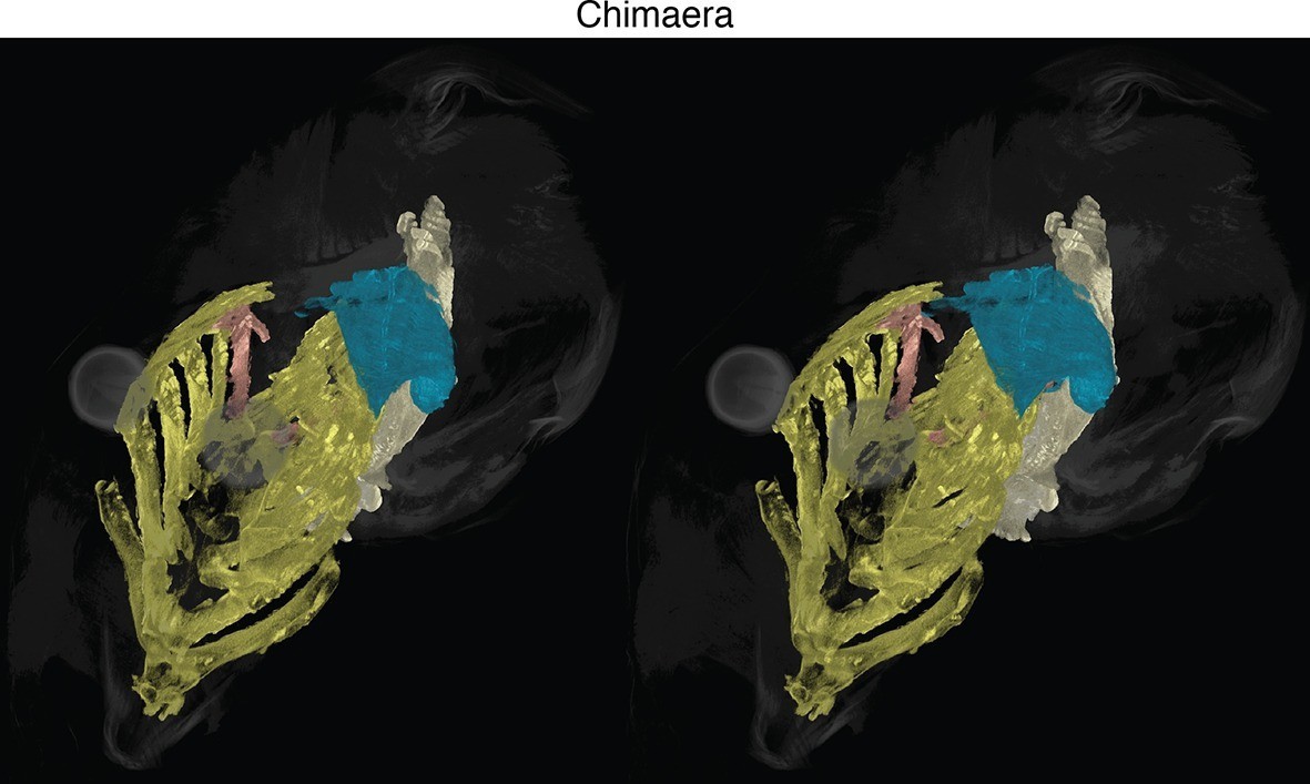
Stereo image of chimaera from Figure 1 with skeletal elements and muscles segmented.
See Figure 1 for color guide. Stereos created in VGStudio Max v2.2.
Figure 1—figure supplement 2
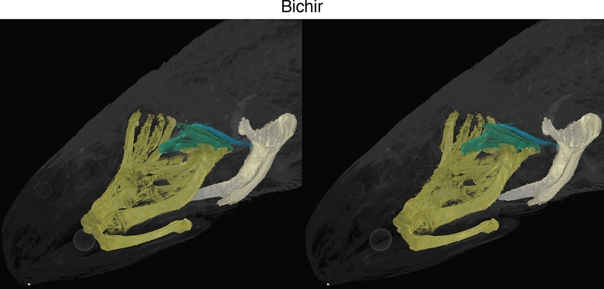
Stereo image of bichir in dorsolateral view.
https://doi.org/10.7554/eLife.09972.005
Figure 1—figure supplement 3
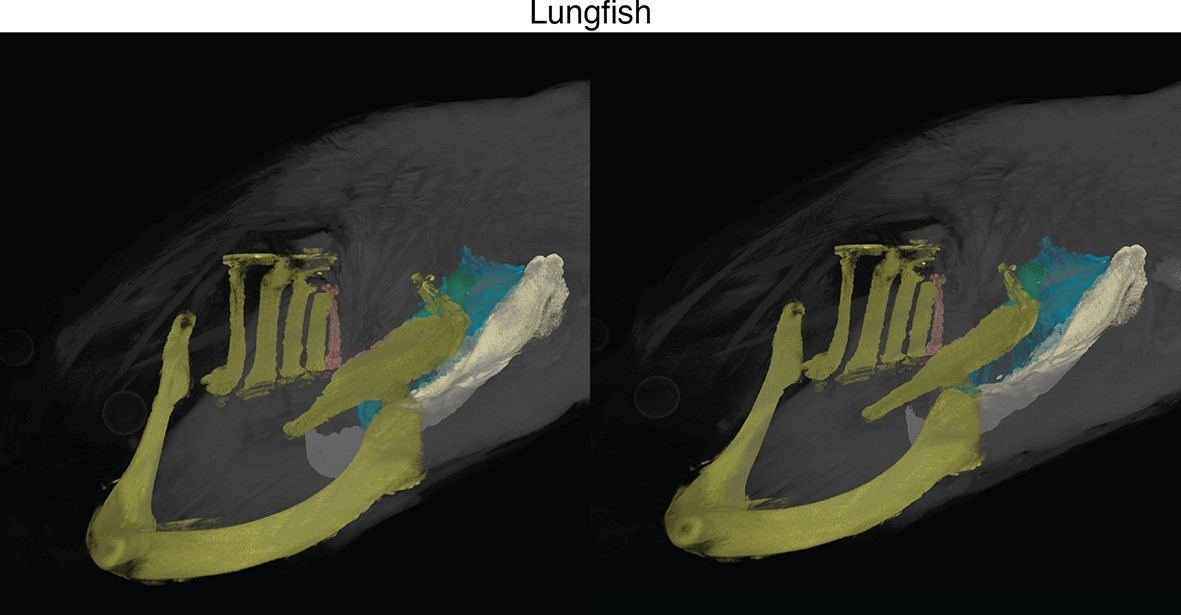
Stereo image of lungfish in dorsolateral view.
https://doi.org/10.7554/eLife.09972.006
Figure 1—figure supplement 4
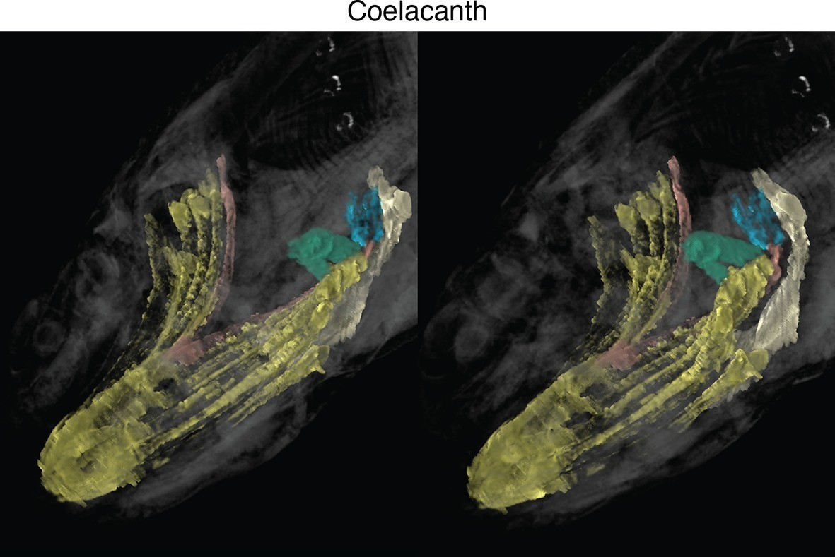
Stereo image of coelacanth in dorsolateral view.
https://doi.org/10.7554/eLife.09972.007
Figure 1—figure supplement 5
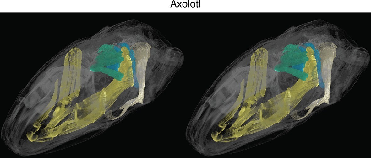
Stereo image of axolotl in dorsolateral view.
https://doi.org/10.7554/eLife.09972.008
Figure 1—figure supplement 6
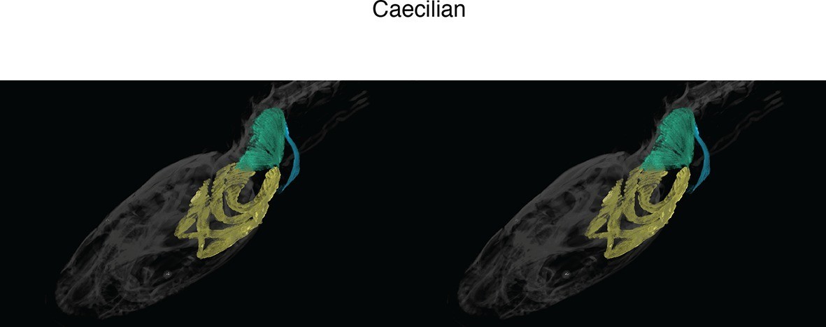
Stereo image of caecilian in dorsolateral view.
https://doi.org/10.7554/eLife.09972.009
Figure 1—figure supplement 7
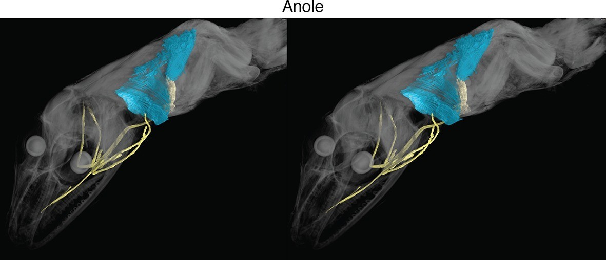
Stereo image of anole in dorsolateral view.
https://doi.org/10.7554/eLife.09972.010
Figure 1—figure supplement 8

Stereo image of opossum in lateral view.
https://doi.org/10.7554/eLife.09972.011
Figure 2 with 3 supplements
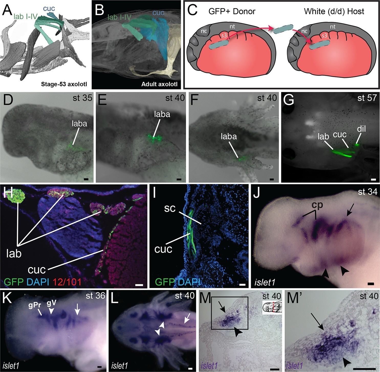
Development of the cucullaris muscle in the axolotl.
(A, B) Morphology of the developing cucullaris, with the four gill-levator muscles (lab I–IV) shaded light green and the cucullaris (cuc) blue. More-posterior muscles are shaded dark green. Anterior is to the left. (A) Dorsolateral view of an OPT scan of a juvenile axolotl stained with the 12/101 muscle antibody. (B) Contrast-stained CT scan of an adult axolotl in lateral view. The cucullaris is expanded into a broad sheet that inserts on the scapula. (C) Schematic depiction of an orthotopic transplantation of unsegmented mesoderm lateral to somites 1−3 at stage 21. Lateral views; anterior is to the left. nc, neural crest; nt, neural tube; s3, somite 3. (D−I) GFP labeling following stage-21 transplantation of unsegmented mesoderm lateral to somites 1−3. (D−F) Labeling of the levator arcuum branchiarum anlagen (laba) dorsal to the developing gills is visible in lateral (D, E) and dorsal (F) views. Anterior is to the left. (G) Gill-levator muscles (levator arcuum branchiarum, lab) of arches 3 and 4, the cucullaris (cuc) and the dilatator laryngis (dil) are labeled in a juvenile axolotl. Dorsal view; anterior is to the left. (H, I) Transverse sections through the posterior occipital region (H) and anterior trunk (I) of a juvenile axolotl. GFP labeling is visible in the gill levators and anterior cucullaris (H) and in the posterior cucullaris near its attachment with the scapula (I; sc). Lateral is to the left; dorsal is to the top. (J−M’) isl1 expression in albino embryos. (J) At stage 34, isl1 is expressed in ventral mesoderm, in the developing heart region (arrowheads) and around the dorsal cranial placodes (cp). Arrow indicates several stripes of expression dorsal to the developing gills. (K) At stage 36, isl1 marks the profundal (gPr)/trigeminal (gV) placode region and earlier expression is maintained dorsal to the gills (arrow). (L) At stage 40, isl1 is expressed in neurons within the dorsal spinal cord (arrow) and in the gill-levator region (arrowheads). Dorsal view. (M, M’) Transverse section of a stage-40 embryo with isl1 expression in the dorsal gill levator region (arrows) and ganglia (arrowheads). Box in M is enlarged in M’. Scale bars, 100 μm, except G, 500 μm.
Figure 2—figure supplement 1
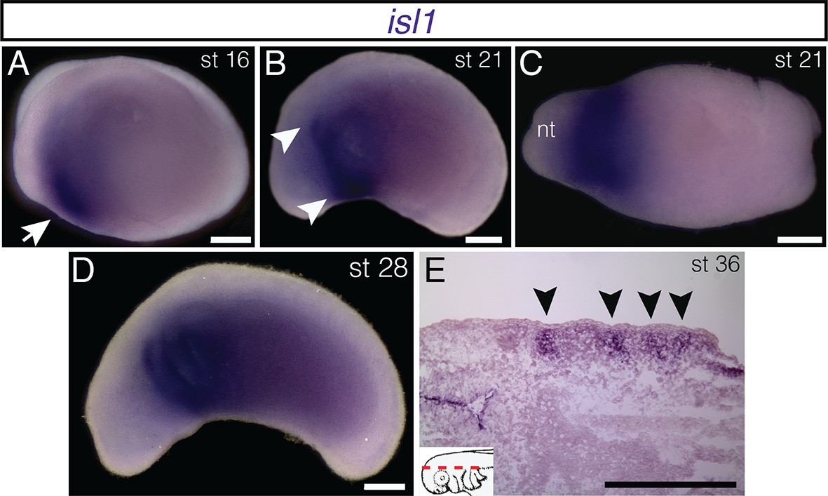
Additional stages of embryonic isl1 expression in A. mexicanum.
(A) At stage 16, isl1 is expressed in the region of the developing heart field (arrow). (B) By stage 21, expression has expanded dorsally (arrowheads). (C) Ventral region of isl1 at stage 21. nt, neural tube. (D) isl1 expression at stage 28, including the branchial arches. (E) Frontal section dorsal to the developing gill arches at stage 36. Inset panel indicates plane of section (dashed red line). Lateral is to the top. A, B and D, lateral views; C, ventral view. Anterior is to the left in all panels. Scale bars, 500 μm.
Figure 2—figure supplement 2
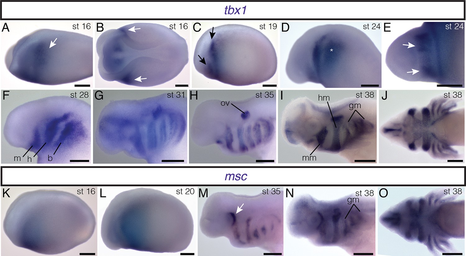
Embryonic expression of tbx1 and msc in A. mexicanum.
All embryos are depicted in lateral view except E, J and O, which are ventral views; anterior is to the left. (A, B) Bilateral stripes of tbx1 expression (arrows) are present in mid-neurula stages. (C) Two bilateral stripes of tbx1 expression are visible (arrows). (D) tbx1 is expressed in the region of the developing branchial arches (asterisk). (E) Patches of tbx1 expression in the mandibular arch (arrows). (F, G) tbx1 is expressed in mandibular (m), hyoid (h) and branchial (b) regions. (H) tbx1 is expressed in the otic vesicle (ov). (I, J) At stage 38, tbx1 is expressed in developing muscle groups. mm mandibular arch muscle; hm, hyoid arch muscle; gm, gill musculature. (K, L) msc is expressed anteriorly at neurula stages. (M) Patch of msc expression just posterior to the eye (arrow). (N) msc is expressed in the gill arch muscles (gm). (O) msc expression at stage 38. Scale bars, 500 μm.
Figure 2—figure supplement 3

Mesoderm fate mapping in A. mexicanum embryos.
Boundaries between regions 1 and 2 and regions 2 and 3 are approximate. LPM s, lateral plate mesoderm adjacent to somite; CM, cranial mesoderm; lme, levator mandibulae externus; lma, levator mandibulae anterior; im, intermandibularis posterior; bhe, branchiohyoideus externus; dm, depressor mandibulae; ldb, levatores et depressores branchiarum; lab, levatores arcus branchiarum; cuc, cucullaris; dil, dilatator laryngis s3, somite 3; nc, neural crest, nt, neural tube. In region key anterior is to the left; dorsal is to the top.
Figure 3
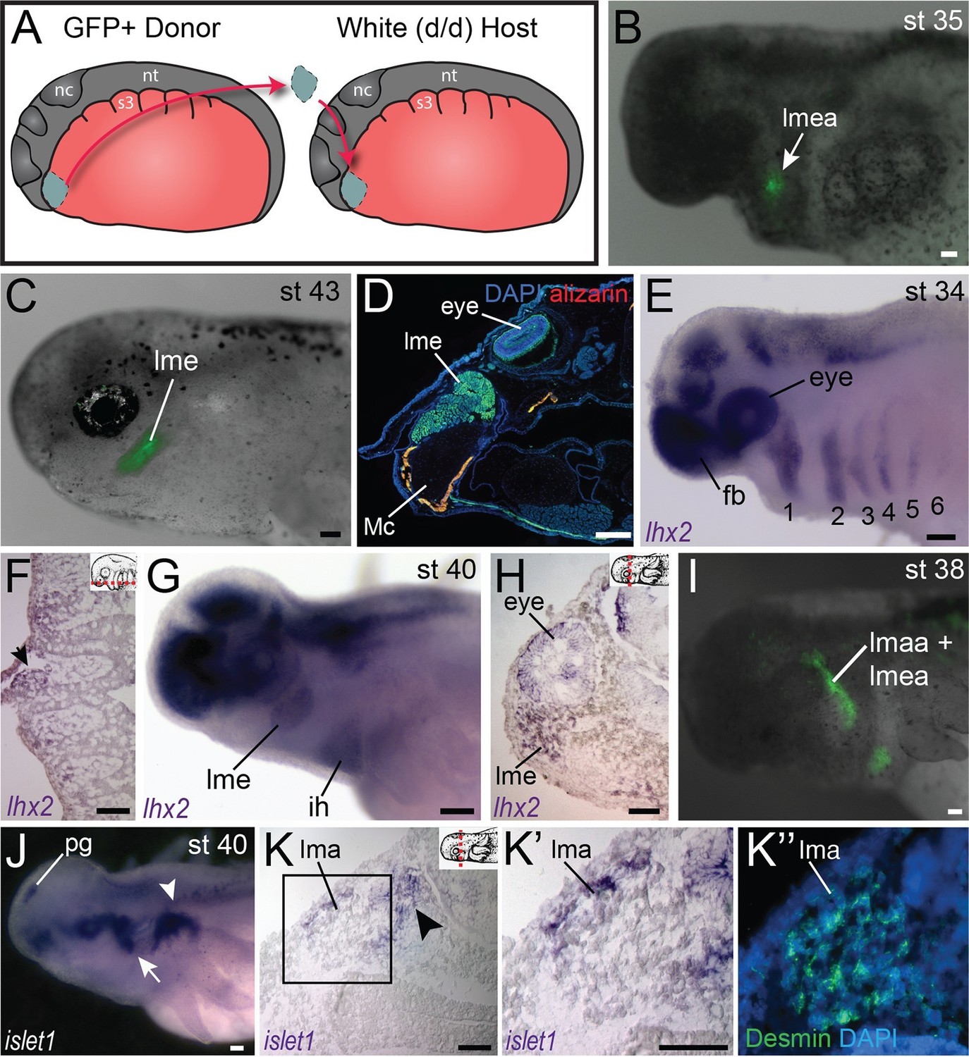
Fate-mapping and gene expression in mandibular adductor muscles.
(A) Schematic depiction of orthotopic transplantations of anterior cranial mesoderm. nc, neural crest; nt, neural tube; s3, somite 3. (B) Labeling of mandibular arch mesoderm at stage 35 following transplantation at stage 19. Arrow points to the anlage of the levator mandibulae externus (lmea). (C) Specimen in (B) at stage 43, with labeling of the levator mandibulae externus (lme). (D) Transverse section through the eye region of a stage-55 axolotl following transplantation at stage 20. The levator mandibulae externus is labeled ventral to the eye. Mc, Meckel’s cartilage. Lateral is to the left; dorsal is to the top. (E) At stage 34, lhx2 is expressed in the pharyngeal mesoderm of all six arches (1–6) as well as the forebrain (fb) and eye. (F) Frontal section through the head at stage 36 showing lhx2 expression in the mesodermal core of the third pharyngeal arch (arrow). Anterior is to the top. Inset panel depicts plane of section (dashed red line). (G) At stage 40, lhx2 is expressed in the levator mandibulae externus (lme) and in the interhyoideus (ih), a ventral cranial muscle. (H) Transverse section at the level of the eye at stage 40, showing lhx2 expression in the levator mandibulae externus. Dorsal is to the top. (I) Labeling of mandibular and hyoid arch mesoderm in a stage-38 embryo, including the anlage of the levator mandibulae anterior (lmaa), an anterior jaw adductor. Lateral view; anterior is to the left. (J−K”) isl1 expression in albino embryos. (J) At stage 40, isl1 is expressed dorsal to the gills (arrowhead) and in the pineal gland (pg). Expression posterior to the eye (arrow) overlaps with the region forming the levator mandibulae anterior. (K−K’’) Transverse sections of a stage-40 embryo. Box in K is enlarged in K’ and K”. (K) isl1 is expressed in the lateral portion of the developing levator mandibulae anterior (lma) and in the trigeminal nerve (arrowhead). (K’’) Desmin staining of muscle cells, including those expressing isl1. Scale bars, 100 μm.
Figure 4 with 1 supplement
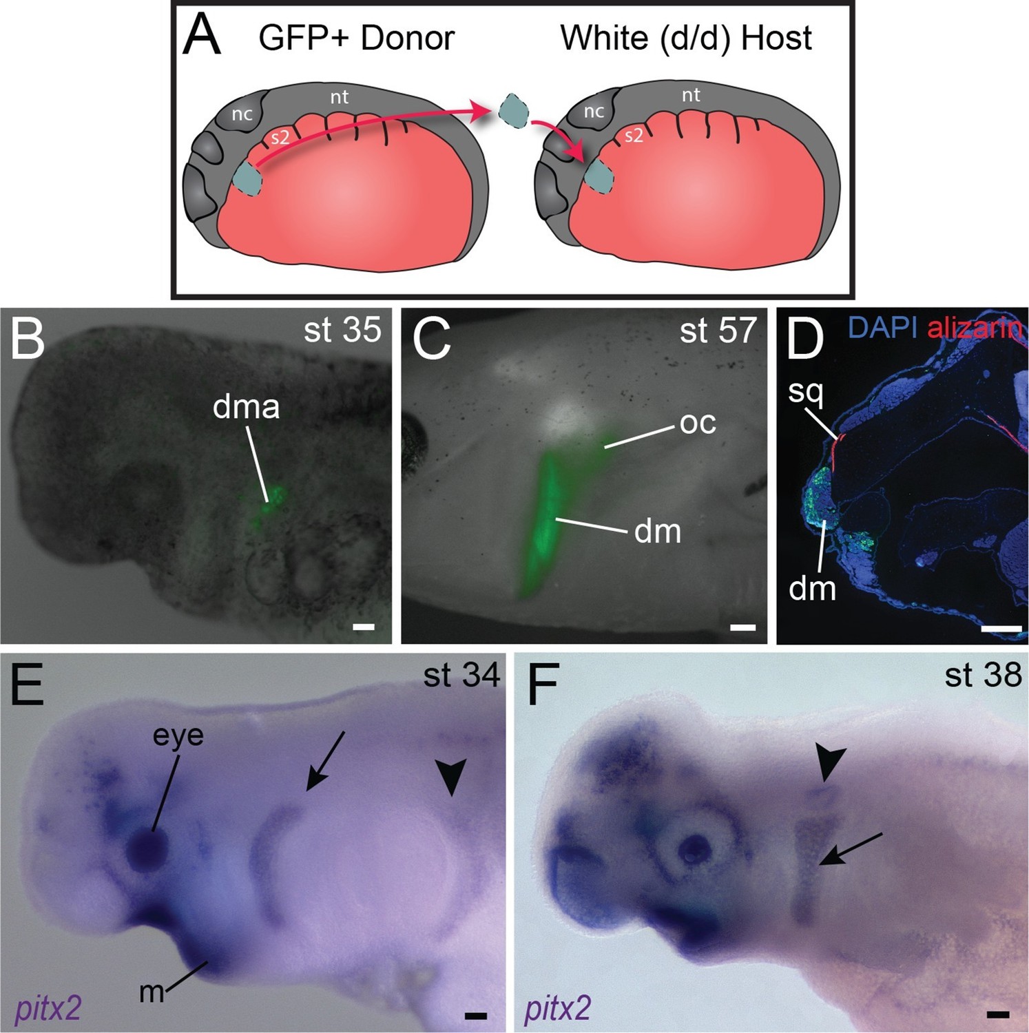
Origin of the mandibular depressor muscle and expression of pitx2 in the hyoid arch.
(A) Schematic depiction of orthotopic transplantation of cranial mesoderm. nc, neural crest; nt, neural tube; s2, somite 2. Somite 1 is small and triangular in shape. (B) GFP labeling of dorsal hyoid-arch mesoderm at stage 35 following transplantation at stage 20 includes the anlage of the depressor mandibulae (dma). (C) Specimen in (B) at stage 57, with labeling of the depressor mandibulae (dm) and otic capsule (oc). (D) Labeling of the depressor mandibulae in a transverse section through the jaw region of a stage-57 juvenile axolotl. sq, squamosal bone. Dorsal is to the top; lateral is to the left. (E) At stage 34, pitx2 is expressed in the eye, the ventral mandibular arch (m), the hyoid arch (arrow) and more faintly in migrating somitic cells (arrowhead). (F) At stage 38, pitx2 expression is maintained in the hyoid arch (arrow) and is also present in the otic vesicle (arrowhead). Scale bars, 100 μm, except C, 500 μm.
Figure 4—figure supplement 1
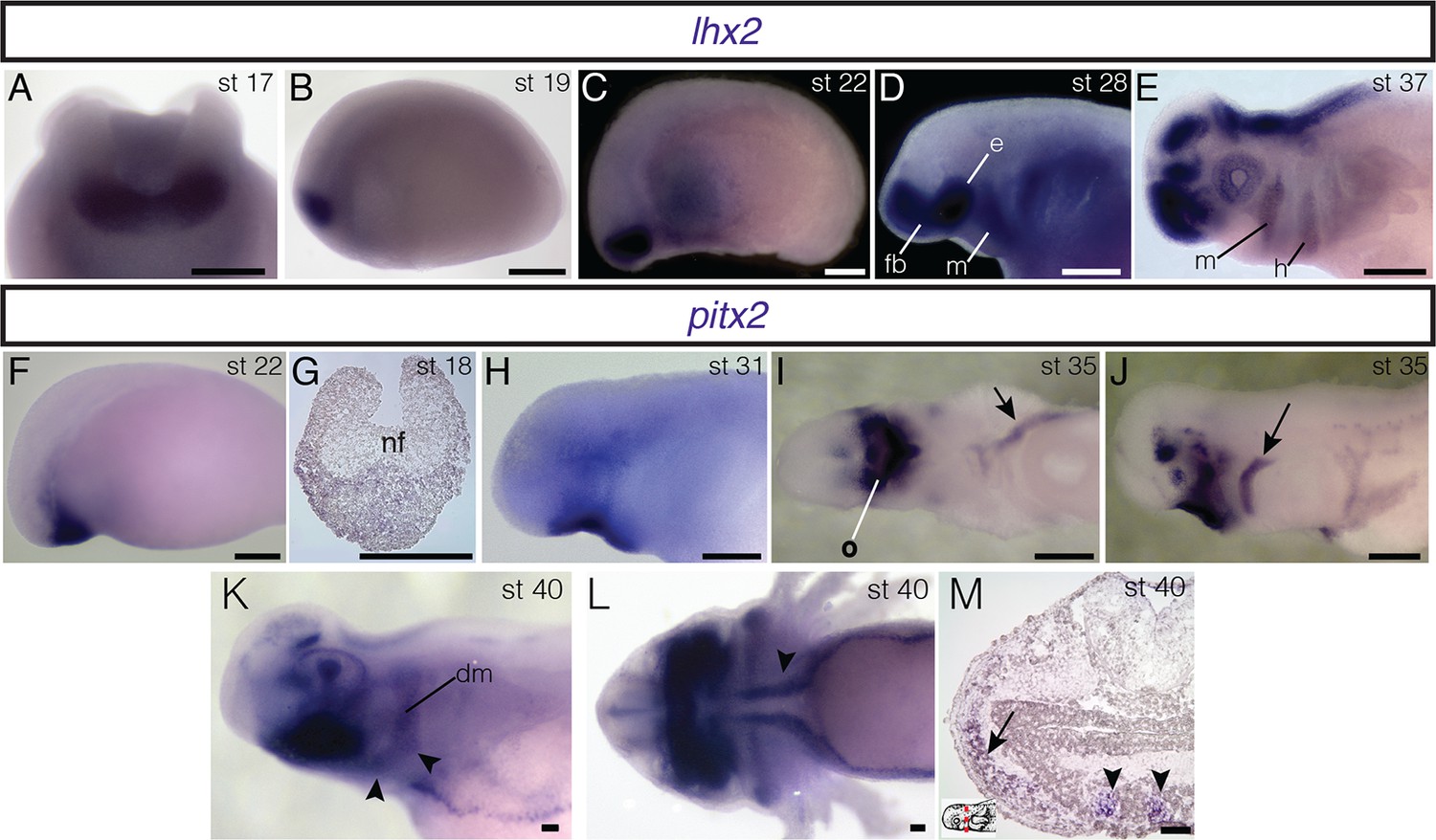
Embryonic expression of lhx2 and pitx2 in A. mexicanum.
(A, B) In neurula stages, lhx2 is expressed in anterior neuroectoderm. A: anterior view, dorsal is to the top. B: lateral view, anterior is to the left. (C) lhx2 is expressed in cranial mesoderm at stage 22. (D) During middle-tailbud stage, lhx2 is expressed in the mandibular-arch (m) and branchial-arch mesoderm as well as the eye field (e) and forebrain (fb). (E) lhx2 expression is maintained in the eye and brain, as well as in mandibular and hyoid (h) arches. (F) pitx2 is expressed in anterior cranial mesoderm. (G) Transverse section through the anterior neural folds (nf) reveals expression through the ectoderm and mesoderm at neurula stages. (H) At stage 31, pitx2 is expressed in oral ectoderm. (I) At stage 35, pitx2 is expressed asymmetrically in the left lateral-plate mesoderm (arrow) and oral region (o). Ventral view. (J) pitx2 is also expressed in hyoid arch mesoderm (arrow) at the same stage. (K) At stage 40, pitx2 is strongly expressed in the oral epithelium. It is also present in two hyoid arch muscles, the branchiohyoideus externus (bhe; arrowheads) and the depressor mandibulae (dm). (L) pitx2 is expressed in the tongue muscles (arrowhead). Ventral view; anterior is to the left. (M) Transverse section at stage 40 shows pitx2 expression in the tongue muscles (arrowheads) and hyoid arch musculature (arrow). Dorsal is to the top; lateral is to the left. Inset panel indicates plane of section (red dashed line). C−F, H, J, K, lateral views; anterior is to the left. Scale bars A−J, 500 μm; K−M, 100 μm.
Figure 5
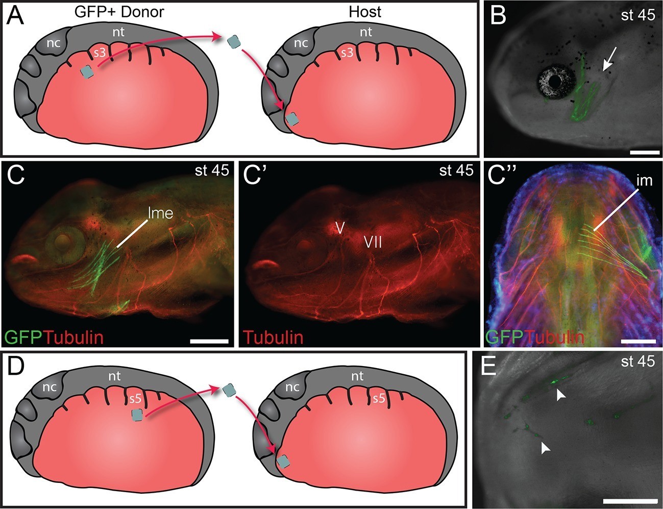
Heterotopic transplantation of lateral plate mesoderm.
(A) Schematic depiction of a caudal-to-cranial heterotopic transplantation of lateral plate mesoderm from somite level 2 (donor) to mandibular arch mesoderm (host). Lateral views; anterior is to the left. nc, neural crest; nt, neural tube; s3, somite 3. (B−C’’) Stage-45 larva following the heterotopic transplantation shown in (A). (B) GFP+ cells contribute to mandibular arch muscles (arrow). Lateral view; anterior is to the left. (C) Lateral plate mesoderm contributes to the levator mandibulae externus (lme). (C’) Innervation of the levator mandibulae externus by the mandibular branch of the trigeminal nerve (V) is normal. VII, facial nerve. (C’’) The intermandibularis (im), a ventral mandibular muscle, is also labeled. Ventral view; anterior is to the top. (D) Schematic depiction of a caudal-to-cranial heterotopic transplantation of lateral plate mesoderm from somite level 5 to mandibular arch mesoderm. (E) Stage-45 larva following the heterotopic transplantation shown in (D). Ventral view; anterior is to the left. No muscle fibers are formed, but labeled cells contribute to cranial vasculature (arrowheads). Scale bars, 100 μm.
Figure 6
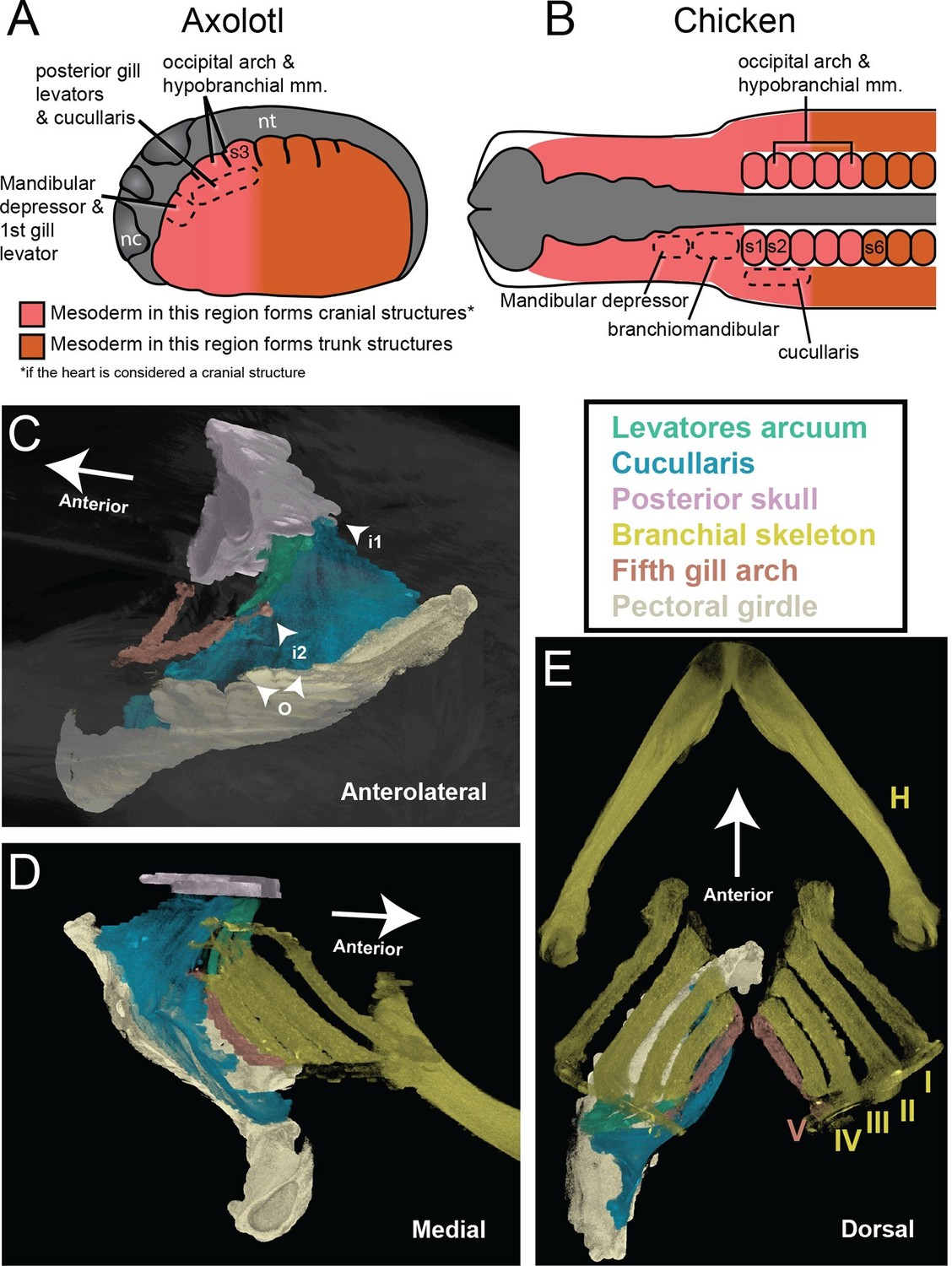
The cucullaris and the transition zone between the head and trunk.
(A) In the axolotl embryo, the head-trunk boundary in unsegmented mesoderm is closely congruent with that in the somites. Paraxial and lateral mesoderm anterior to somite 3 form cranial structures (including the heart). The illustration depicts a stage-21 embryo with epidermis removed; anterior is to the left. Somite fate-mapping data are from Piekarski and Olsson (2007); Piekarski and Olsson (2014). (B) In the chicken, the axial level of the head-trunk boundary in somitic mesoderm is posterior to the border in unsegmented mesoderm. Somite fate-mapping data are from Couly et al. (1993) and Huang et al. (1999); cucullaris data are from Theis et al. (2010); mandibular depressors and branchiomandibular data are from Noden (1983) and Evans and Noden (2006). (C−E) Contrast-stained CT images of the lungfish branchial skeleton, pectoral girdle, posterior skull, gill levators and cucullaris. All structures except the branchial skeleton are segmented on the left side only. The anterolateral view depicts only the fifth gill arch, with its attachment to the cucullaris; the body is rendered transparent. The lungfish cucullaris retains the ancestral tripartite attachment: origin from the pectoral girdle (o) and insertions on the posterior skull (i1) and fifth ceratobranchial (i2).
Videos
Video 1
Video of representative gnathostome cranial and pectoral regions spinning around their long axes with skeletal elements and muscles segmented.
See Figure 1 for color guide. Videos rendered in VGStudio Max v2.2 at 2048 X 1536 resolution, 25 frames per second (20 s), Windows AVI format, 85% quality. Sides segmented represent original specimens, whereas some images in figure panels were reversed so that all specimens are oriented in the same direction.
Tables
Table 1
Primer sequences.
| Transcript | Forward (5’ to 3’) | Reverse (5’ to 3’) | Product Size |
|---|---|---|---|
| lhx2 | AACAGTGACGCAAACAGTGG | TTGAAGCAGTTAGCGCAGAA | 755 bp |
| msc | ACCAGCAGACACCAAGCTCT | TGTGTCCTCCTCTGATGTGAA | 708 bp |
| pitx2 | AGATCGCCGTGTGGACTAAC | GGTGGTAGCGAGTTTTGGAA | 809 bp |
| tbx1 | GGAGTACGACCGAGATGGAA | ATGAAGCGCTGATGACAGTG | 688 bp |
Download links
A two-part list of links to download the article, or parts of the article, in various formats.
Downloads (link to download the article as PDF)
Open citations (links to open the citations from this article in various online reference manager services)
Cite this article (links to download the citations from this article in formats compatible with various reference manager tools)
Evolution of the head-trunk interface in tetrapod vertebrates
eLife 5:e09972.
https://doi.org/10.7554/eLife.09972


