Coordinated Tbx3/Tbx5 transcriptional control of the adult ventricular conduction system
- Departments of Pediatrics, Pathology, and Human Genetics, University of Chicago, United States
- Departments of Biomedical Engineering, Northwestern University, United States
- Department of Medicine, Section of Cardiology, University of Chicago, United States
eLife Assessment
The work presented is important for our understanding of the development of the cardiac conduction system and its regulation by T-box transcription factors. The conclusions are supported by convincing data. Overall this is an excellent study that advances our understanding of cardiac biology and has implications beyond the immediate field of study.
https://doi.org/10.7554/eLife.102027.3.sa0Significance of the findings:
Important: Findings that have theoretical or practical implications beyond a single subfield
- Landmark
- Fundamental
- Important
- Valuable
- Useful
Strength of evidence:
Convincing: Appropriate and validated methodology in line with current state-of-the-art
- Exceptional
- Compelling
- Convincing
- Solid
- Incomplete
- Inadequate
During the peer-review process the editor and reviewers write an eLife Assessment that summarises the significance of the findings reported in the article (on a scale ranging from landmark to useful) and the strength of the evidence (on a scale ranging from exceptional to inadequate). Learn more about eLife Assessments
Abstract
The cardiac conduction system (CCS) orchestrates the electrical impulses that enable coordinated contraction of the cardiac chambers. The T-box transcription factors TBX3 and TBX5 are required for CCS development and associated with overlapping and distinct human CCS diseases. We evaluated the coordinated role of Tbx3 and Tbx5 in the murine ventricular conduction system (VCS). We engineered a compound Tbx3:Tbx5 conditional knockout allele for both genes located in cis on mouse chromosome 5. Conditional deletion of both T-box transcriptional factors in the VCS, using the VCS-specific MinKCreERT2, caused loss of VCS function and molecular identity. Combined Tbx3 and Tbx5 deficiency in the adult VCS led to conduction defects, including prolonged PR and QRS intervals and elevated susceptibility to ventricular tachycardia. These electrophysiological defects occurred prior to detectable alterations in cardiac contractility or histologic morphology, indicative of a primary conduction system defect. Tbx3:Tbx5 double-knockout VCS cardiomyocytes revealed a transcriptional shift toward non-CCS-specialized working myocardium, indicating a change to their cellular identity. Furthermore, optical mapping revealed a loss of VCS-specific conduction system propagation. Collectively, these findings indicate that Tbx3 and Tbx5 coordinate to control VCS molecular fate and function, with implications for understanding cardiac conduction disorders in humans.
Introduction
The cardiac conduction system (CCS) constitutes a highly specialized network of cardiomyocytes that initiate and propagate the electrical impulses required for synchronized contractions of the heart. In the mature mammalian heart, the functional components of the CCS can be broadly divided into the slowly propagating atrial nodes (~5 cm/s), containing the sinoatrial node (SAN) and atrioventricular node (AVN), and the rapidly propagating ventricular conduction system (VCS) (~200 cm/s), including the AV (His) bundle and the right and left bundle branches (BBs). The VCS is responsible for rapid propagation of the electrical impulse from the AVN to the ventricular apex to enable synchronous ventricular contraction and effective ejection of blood from the ventricles (Arnolds et al., 2012; Park and Fishman, 2011; Moskowitz et al., 2007). Defects of CCS can occur in normally formed hearts as well as in patients with structural congenital heart disease and are a major source of morbidity and mortality (Arnolds et al., 2012; Moskowitz et al., 2007; Munshi, 2012; Rubart and Zipes, 2005; Huikuri et al., 2001). The VCS specifically has been recognized as a substrate for life-threatening ventricular arrhythmias, including bundle branch reentry tachycardia, idiopathic fascicular tachycardia, short-coupled torsade de pointes, and ventricular fibrillation (Arnolds et al., 2012; Huikuri et al., 2001; van Duijvenboden et al., 2014; Arnolds and Moskowitz, 2011b; Scheinman, 2009). Despite the severe clinical consequences of CCS disorders, the molecular mechanisms that establish and maintain regional functionality of the mature CCS domains require further study.
Human genetic studies have identified numerous loci associated with adult human CCS function, including the developmentally important factors Tbx3 and Tbx5 (reviewed in Arnolds et al., 2012; Arnolds et al., 2011a). Tbx3 and Tbx5 play crucial roles in adult CCS development and function (Arnolds et al., 2012; Moskowitz et al., 2007; Burnicka-Turek et al., 2020; Moskowitz et al., 2004; van Weerd and Christoffels, 2016; van den Boogaard et al., 2012; Bakker et al., 2012; Hatcher and Basson, 2009; Bakker et al., 2008; Hoogaars et al., 2007b; Mori et al., 2006; Bruneau et al., 2001). Tbx5 encodes a T-box transcriptional activator required for structural and conduction system cardiac development (Moskowitz et al., 2007; Moskowitz et al., 2004; Bruneau et al., 2001; Hoogaars et al., 2007a). Dominant mutations in human TBX5 cause Holt–Oram syndrome (HOS, OMIM:142900), an autosomal dominant disorder characterized by upper limb malformations, congenital heart defects, and CCS abnormalities (Basson et al., 1997; Li et al., 1997; Basson et al., 1994). The cardiac phenotype of HOS, including atrioventricular conduction delay, has been recapitulated in the Tbx5 heterozygous mice (Bruneau et al., 2001). Moreover, VCS-specific Tbx5 knockout caused slowed VCS function and ventricular tachycardia (VT) resulting in sudden death in mice (Arnolds et al., 2012), emphasizing the importance of Tbx5 in VCS conduction. Tbx5 is strongly expressed in the atria and VCS (Arnolds et al., 2012; Moskowitz et al., 2007; Moskowitz et al., 2004; Bakker et al., 2008), and directly regulates several targets required for VCS function (Moskowitz et al., 2007; Bruneau et al., 2001; Hiroi et al., 2001), including Gja5 (Cx40) (Bruneau et al., 2001) and Scn5a (Nav1.5) (Arnolds et al., 2012). Tbx3 encodes a T-box transcriptional repressor which is critical for cardiac development (Bakker et al., 2008; Frank et al., 2011). Dominant mutations in human TBX3 cause Ulnar–Mammary syndrome (OMIM:181450), a developmental disorder (Meneghini et al., 2006; Bamshad et al., 1997), that includes functional conduction system defects (Linden et al., 2009). In the heart, Tbx3 is specifically expressed within CCS (Bakker et al., 2008; Hoogaars et al., 2004), and its deficiency below critical level leads to lethal arrhythmias (Frank et al., 2011). Furthermore, Tbx3 is required for the molecular identity but not the function of the VCS (Bakker et al., 2008). In contrast, Tbx3 in SAN and AVN is required for their proper function (Hoogaars et al., 2004), emphasizing its critical role in maintaining proper cardiac rhythm.
A model for regional CCS specialization suggests that the adult CCS is organized entirely as a slow conduction system ground state by Tbx3 with a T-box-dependent, physiologically dominant fast conduction system network driven specifically in the VCS by Tbx5 (Burnicka-Turek et al., 2020). The adult VCS-specific removal of TBX5 or overexpression of TBX3 shifted the fast VCS into a slow nodal-like system, indicating that the Tbx3/Tbx5 ratio determines nodal versus VCS function (Burnicka-Turek et al., 2020). However, a comprehensive assessment of the coordinated requirements for Tbx3 and Tbx5 has been hindered by the inability to achieve their compound deletion due to their genomic proximity. Tbx3 and Tbx5 are situated in cis within 0.6 Mb on chromosome 5 in mice, rendering their simultaneous deletion unattainable with the available single allele conditional alleles. To investigate the consequences of Tbx3 and Tbx5 compound removal from the mature VCS, we generated a novel compound Tbx3:Tbx5 double-conditional allele. We found that VCS-specific genetic removal of both TBX3 and TBX5 transformed fast-conducting, adult VCS into working myocardium-like cardiomyocytes, shifting them from conduction to non-conduction myocytes. These results demonstrated the coordinated requirements of both Tbx3 and Tbx5 for maintained specification of the mature VCS.
Results
We generated a novel Tbx3:Tbx5 double-floxed allele to enable the simultaneous conditional deletion of Tbx3 and Tbx5 genes specifically from the adult VCS. Tbx3 and Tbx5 reside in cis on mouse chromosome 5 (Tbx3 mm39 chr5:119808734–119822789; Tbx5 mm39 chr5:119970733–120023284). Therefore, to generate a double-conditional knockout, we targeted Tbx5 in the background of a previously validated Tbx3 floxed allele (Frank et al., 2011) using the CRISPR–Cas9 system (Gurumurthy et al., 2021; Dow et al., 2015; Figure 1A). We engineered a Tbx5 floxed allele mirroring a previously published allele (Bruneau et al., 2001). This design enabled us to utilize the previously published individual Tbx3 floxed allele (Frank et al., 2011) and individual Tbx5 floxed allele (Bruneau et al., 2001) to serve as controls (Figure 1A, Figure 1—figure supplements 1 and 2).
Figure 1 with 5 supplements see all
Generation of VCS-specific Tbx3:Tbx5 double-conditional knockout mice.
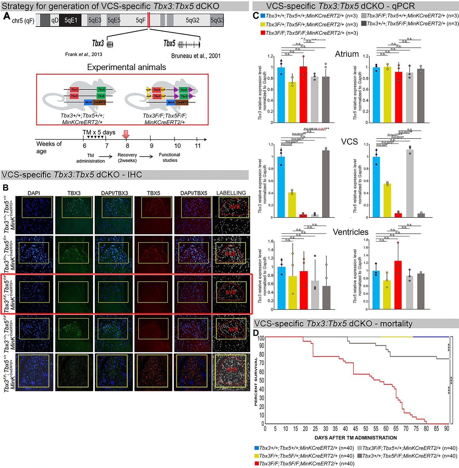
(A) Strategy to generate VCS-specific Tbx3:Tbx5 double-conditional knockout mouse line. A new Tbx3:Tbx5 double-conditional knockout mouse line (Tbx3fl/fl;Tbx5fl/fl) was generated using the CRISPR–Cas9 system (Gurumurthy et al., 2021; Dow et al., 2015) which allowed for the targeting of Tbx5 in the background of the previously validated Tbx3 floxed allele (Frank et al., 2011). A newly engineered Tbx5 floxed allele has been developed to mirror a previously published allele (Bruneau et al., 2001). This design has enabled the utilization of the previously published individual Tbx3 floxed allele (Frank et al., 2011) and individual Tbx5 floxed allele (Bruneau et al., 2001) as controls. To conditionally delete Tbx3 and Tbx5 genes specifically from the adult VCS and generate the experimental animals, the Tbx3:Tbx5 double-floxed mouse line (Tbx3fl/fl;Tbx5fl/fl) was combined with a VCS-specific tamoxifen inducible Cre transgenic mouse line (MinKCreERT2 [Tg(RP23-276I20-MinKCreERT2) Arnolds and Moskowitz, 2011b]). All allelic combinations were generated and evaluated as littermates in a mixed genetic background. The experimental mice employed in all studies were administered tamoxifen at 6 weeks of age and subsequently evaluated at 9 weeks of age (3 weeks post-tamoxifen administration). The loss of Tbx3 and Tbx5 expression, on both the protein and mRNA levels, assessed by immunohistochemistry (B) and qRT-PCR (C), respectively, was observed in the VCS of adult Tbx3:Tbx5 double-conditional mutant mice (Tbx3fl/fl;Tbx5fl/fl;R26ReYFP/+;MinKCreERT2/+), but not in their littermate controls (Tbx3+/+;Tbx5+/+;R26ReYFP/+;MinKCreERT2/+) (B, C). (C) qRT-PCR analysis showed a partial loss of Tbx3 and Tbx5 expression in the adult VCS of Tbx3:Tbx5 double-conditional heterozygous mice (Tbx3fl/+;Tbx5fl/+;R26ReYFP/+;MinKCreERT2/+) compared to their littermate controls (Tbx3+/+;Tbx5+/+;R26ReYFP/+;MinKCreERT2/+). Additionally, qRT-PCR analysis confirmed the specificity of the Tbx3:Tbx5 double knockout for the VCS by assessing Tbx3 and Tbx5 expression levels in the atria and ventricles of tamoxifen-treated experimental mice. Consistent with the VCS selectivity of Cre activity in the MinKCreERT2 mice (Arnolds and Moskowitz, 2011b), Tbx3 and Tbx5 expression remained similar in the atrial and ventricular myocardium across all allelic combinations, including Tbx3:Tbx5 double-conditional knockout mice (Tbx3fl/fl;Tbx5fl/fl;R26ReYFP/+;MinKCreERT2/+). (D) Conducted longitudinal studies revealed a significantly increased mortality rate in VCS-specific Tbx3:Tbx5-deficient mice compared to their littermate controls (***p < 0.0001, log-rank test, GraphPad Prism), suggesting a requirement for both Tbx3 and Tbx5 in the mature VCS. All allelic combinations of experimental and control mice (n = 40 biological replicates/genotype) were followed longitudinally after tamoxifen administration at 6 weeks of age. Tbx3:Tbx5 double-conditional knockout mice began to die suddenly at 3–4 weeks post-tamoxifen administration. Within the 3 months post-tamoxifen administration, all tamoxifen-treated Tbx3fl/fl;Tbx5fl/fl;R26ReYFP/+;MinKCreERT2/+ mice had died suddenly (n = 40) without previous signs of illness. In contrast, no mortality was observed among the tamoxifen-treated Tbx3+/+;Tbx5+/+;R26ReYFP/+;MinKCreERT2/+ and Tbx3fl/+;Tbx5fl/+;R26ReYFP/+;MinKCreERT2/+ littermates (each cohort n = 40) during this period. TBX3 and TBX5 protein expression was evaluated by immunohistochemistry (green and red signals, respectively) on serial sections from hearts of all allelic combinations (n = 3 biological replicates/genotype). Nuclei were stained with DAPI (blue signal). IHC original magnification: ×40. qRT-PCR data are presented as mean ± SD normalized to Gapdh and relative to Tbx3+/+;Tbx5+/+;R26ReYFP/+;MinKCreERT2/+ mice (set as 1). N = 3 biological replicates/genotype (VCS cardiomyocytes pooled from 30 mice per biological replicate); multiple testing correction using Benjamini and Hochberg procedure. Significance was assessed by Welch t-test (*FDR <0.05; **FDR <0.005; ***FDR <0.001) and confirmed by one-tailed Wilcoxon test (p value in parentheses) when normally distribution was rejected. Abbreviations: AVB, atrioventricular bundle (also known as bundle of His); FDR, false discovery rate; VCS, ventricular conduction system.
We assessed the impact of removing Tbx3 and Tbx5 from the mature VCS by combining the Tbx3:Tbx5 double-floxed mouse line (Tbx3fl/fl;Tbx5fl/fl) with a VCS-specific, tamoxifen (TM)-inducible Cre transgenic mouse line, Kcne1CreERT2 [Tg(RP23-276I20-MinKCreERT2)] – hereafter referred to as MinKCreERT2, in accordance with existing literature (Figure 1A; Arnolds et al., 2012; Arnolds and Moskowitz, 2011b; Burnicka-Turek et al., 2020). Individual Tbx3 floxed and Tbx5 floxed mouse lines combined with MinKCreERT2 transgenic mouse lines (Tbx3fl/fl;Tbx5+/+; R26ReYFP/+;MinKCreERT2/+ and Tbx3+/+;Tbx5fl/fl;R26ReYFP/+;MinKCreERT2/+, respectively) were generated as controls, and all allelic combinations were evaluated in a mixed genetic background. We compared VCS-specific Tbx3:Tbx5 double-conditional mutants (Tbx3fl/fl;Tbx5fl/fl;R26ReYFP/+;MinKCreERT2/+) with control littermates (Tbx3+/+;Tbx5+/+; R26ReYFP/+;MinKCreERT2/+) and VCS-specific Tbx3:Tbx5 double-conditional heterozygous littermates (Tbx3fl/+;Tbx5fl/+;R26ReYFP/+;MinKCreERT2/+). Additionally, we validated the newly created Tbx5 floxed allele demonstrating that it is efficiently converted to the Tbx5 null allele through Cre recombinase, causing a phenotype consistent with that observed from conversion of the previously published Tbx5 floxed allele (Arnolds et al., 2012; Bruneau et al., 2001; Figure 1—figure supplements 1 and 2, Methods section).
We assessed experimental mice at 8–9 weeks of age following tamoxifen administration at 6 weeks of age (Figure 1A, Methods section). We observed loss of both Tbx3 and Tbx5 expression, on both the mRNA and protein levels, in the VCS of adult Tbx3:Tbx5 double-conditional mutant mice (Tbx3fl/fl;Tbx5fl/fl;R26ReYFP/+;MinKCreERT2/+) but not in their littermate controls (Tbx3+/+;Tbx5+/+;R26ReYFP/+;MinKCreERT2/+) (Figure 1B, C). Partial loss of Tbx3 and Tbx5 expression in the adult VCS of Tbx3:Tbx5 double-conditional heterozygous mice (Tbx3fl/+;Tbx5fl/+;R26ReYFP/+;MinKCreERT2/+) was observed compared to littermate controls (Tbx3+/+;Tbx5+/+;R26ReYFP/+;MinKCreERT2/+) (Figure 1C). We confirmed the specificity of the Tbx3:Tbx5 double knockout for the VCS by assessing Tbx3 and Tbx5 expression levels in the atria and ventricles of tamoxifen-treated experimental mice. Consistent with the VCS selectivity of Cre activity in the MinKCreERT2 mice (Arnolds and Moskowitz, 2011b), Tbx3 and Tbx5 expression remained similar in the atrial and ventricular myocardium of all allelic combinations, including Tbx3:Tbx5 double-conditional knockout mice (Tbx3fl/fl;Tbx5fl/fl;R26ReYFP/+;MinKCreERT2/+) (Figure 1C).
Tbx3:Tbx5 double-conditional knockout mice (Tbx3fl/fl;Tbx5fl/fl;R26ReYFP/+; MinKCreERT2/+) appeared morphologically and functionally normal and indistinguishable from control littermates (Tbx3+/+;Tbx5+/+;R26ReYFP/+;MinKCreERT2/+) at 2 weeks post-tamoxifen. However, longitudinal analysis demonstrated sudden death of VCS-specific double-knockout mice beginning 3 weeks post-tamoxifen administration (Figure 1D). Within the 3 months post-tamoxifen administration, all tamoxifen-treated Tbx3fl/fl;Tbx5fl/fl; R26ReYFP/+;MinKCreERT2/+ mice had died suddenly (n = 40). In contrast, no mortality was observed among the tamoxifen-treated Tbx3+/+;Tbx5+/+;R26ReYFP/+;MinKCreERT2/+ or Tbx3fl/+;Tbx5fl/+;R26ReYFP/+;MinKCreERT2/+ littermates (each cohort n = 40) during this period (Tbx3:Tbx5 double-conditional knockout mice vs. control mice p < 0.0001; Tbx3:Tbx5 double-conditional knockout mice vs. Tbx3:Tbx5 double-conditional heterozygous mice p < 0.0001, log-rank test; Figure 1D). These results revealed that double deletion of Tbx3 and Tbx5 from the adult VCS causes lethality beginning at 3 weeks post-tamoxifen.
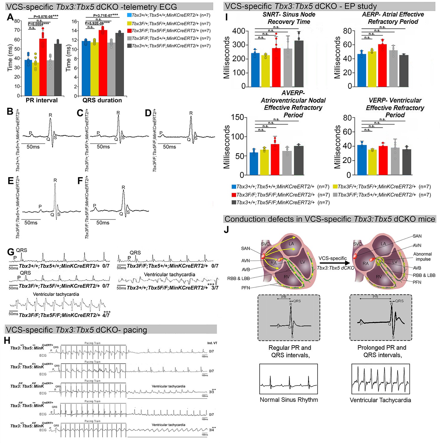
The onset of mortality observed in VCS-specific Tbx3:Tbx5-deficient mice starting at 3 weeks post-tamoxifen prompted us to investigate the electrophysiologic consequences of VCS-specific Tbx3:Tbx5 double-knockout at 2–3 weeks post-tamoxifen, prior to the onset of lethality (Figure 2). VCS-specific Tbx3:Tbx5 deficiency caused profound conduction slowing in Tbx3fl/fl;Tbx5fl/fl;R26ReYFP/+;MinKCreERT2/+ mice by ambulatory telemetry electrocardiography (ECG) analysis compared to Tbx3:Tbx5 double-conditional heterozygous mice (Tbx3fl/+;Tbx5fl/+;R26ReYFP/+;MinKCreERT2/+) and littermate controls (Tbx3+/+;Tbx5+/+;R26ReYFP/+;MinKCreERT2/+) (Figure 2A–F). Specifically, the PR interval, representing the period between atrial and ventricular depolarization, and the QRS duration, indicating the length of ventricular depolarization and early repolarization in mice, were both significantly prolonged (Figure 2A, B, D; PR: Tbx3:Tbx5 double-conditional knockout mice vs. control mice p < 0.05, n = 7, Welch t-test; QRS: Tbx3:Tbx5 double-conditional knockout mice vs. control mice p < 0.05, n = 7, Welch t-test). Removal of both Tbx3 and Tbx5 from the adult VCS resulted in increased episodes of spontaneous VT. Ambulatory studies revealed episodes of spontaneous VT in four out of seven Tbx3fl/fl;Tbx5fl/fl;R26ReYFP/+;MinKCreERT2/+ mice, in contrast to none observed in seven littermate controls (Figure 2G, p < 0.05, n = 7, Welch t-test). Furthermore, Tbx3:Tbx5 double-conditional knockout mice (Tbx3fl/fl;Tbx5fl/fl;R26ReYFP/+;MinKCreERT2/+) showed significantly increased susceptibility to VT following burst stimulation in invasive electrophysiology (EP) studies (three of three Tbx3fl/fl;Tbx5fl/fl;R26ReYFP/+;MinKCreERT2/+ mice vs. zero of seven littermate controls; Figure 2H, p < 0.05, Welch t-test). In contrast, VCS-specific Tbx3:Tbx5 double-conditional heterozygous mice (n = 7) showed neither conduction nor electrophysiological defects (Figure 2). Consistent with the use of a VCS-specific Cre (MinKCreERT2), no changes in the refractory/recovery periods of atrium, ventricle, or nodes (atrial effective refractory period, ventricular effective refractory period (VERP), atrioventricular nodal effective refractory period, or sinus node recovery time) were detected by intracardiac EP conducted on experimental and control mice (Figure 2I, p < 0.05, Welch t-test).
Figure 2
Arrhythmias and conduction abnormalities in mice with VCS-specific Tbx3:Tbx5 double-conditional knockout.
(A–F) VCS-specific Tbx3:Tbx5 double-conditional knockout causes significant VCS conduction slowing in adult Tbx3fl/fl;Tbx5fl/fl;R26ReYFP/+;MinKCreERT2/+ mice. (A) PR (left graph) and QRS (right graph) intervals calculated from ambulatory telemetry electrocardiography (ECG) recordings in (B–F). Tbx3:Tbx5 double-conditional adult mice (Tbx3fl/fl;Tbx5fl/fl;R26ReYFP/+;MinKCreERT2/+) displayed significant PR and QRS intervals prolongation compared to littermate controls (Tbx3+/+;Tbx5+/+;R26ReYFP/+;MinKCreERT2/+) (A left and right graphs, respectively). Data are presented as mean ± SD. N = 7 biological replicates/genotype, multiple testing correction using Benjamini and Hochberg procedure; *Welch t-test p < 0.05 and FDR <0.05; **Welch t-test p < 0.005 and FDR <0.05; ***Welch t-test p < 0.001 and FDR <0.05. Representative ambulatory telemetry ECG of Tbx3+/+;Tbx5+/+; R26ReYFP/+;MinKCreERT2/+ (B), Tbx3fl/+;Tbx5fl/+;R26ReYFP/+;MinKCreERT2/+ (C), Tbx3fl/fl;Tbx5fl/fl; R26ReYFP/+;MinKCreERT2/+ (D), Tbx3fl/fl;Tbx5+/+;R26ReYFP/+;MinKCreERT2/+ (E), Tbx3+/+;Tbx5fl/fl;R26ReYFP/+;MinKCreERT2/+ (F) mice. (G) Simultaneous genetic removal of Tbx3 and Tbx5 from the adult VCS resulted in significantly increased episodes of spontaneous ventricular tachycardia. Episodes of spontaneous ventricular tachycardia were observed in four of seven Tbx3fl/fl;Tbx5fl/fl;R26ReYFP/+; MinKCreERT2/+ mice versus zero of seven littermate controls (Tbx3+/+;Tbx5+/+;R26ReYFP/+; MinKCreERT2/+) in ambulatory studies. N = 7 biological replicates/genotype; multiple testing correction using Benjamini and Hochberg procedure; *FDR of Welch t-test ≤0.05. (H) Tbx3:Tbx5 double-conditional knockout mice (Tbx3fl/fl;Tbx5fl/fl;R26ReYFP/+;MinKCreERT2/+) showed significantly increased susceptibility to ventricular tachycardia following burst stimulation in invasive electrophysiology studies (three of three Tbx3fl/fl;Tbx5fl/fl;R26ReYFP/+; MinKCreERT2/+ mice vs. zero of seven control Tbx3+/+;Tbx5+/+;R26ReYFP/+;MinKCreERT2/+ mice. Fisher’s exact test: *p < 0.05; n = 7 biological replicates/genotype). (I) Intracardiac electrophysiology detected no significant changes in SNRT, AERP, AVERP, and VERP recorded from experimental and control animals (n = 7 biological replicates/genotype; multiple testing correction using Benjamini and Hochberg procedure; *FDR of Welch t-test ≤0.05). (J) Graphical summary of conduction defects observed in adult, VCS-specific Tbx3:Tbx5-deficient mice. Simultaneous genetic deletion of Tbx3 and Tbx5 from the mature VCS results in conduction slowing, prolonged PR and QRS intervals, as well as ventricular tachycardia. Abbreviations: AVB, atrioventricular bundle (also known as bundle of His); AVN, atrioventricular node; FDR, false discovery rate; LA, left atrium; LBB, left bundle branches; LV, left ventricle; PFN, Purkinje fiber network; RA, right atrium; RBB, right bundle branches; RV, right ventricle; SAN, sinoatrial node; SVC, superior vena cava; VCS, ventricular conduction system.
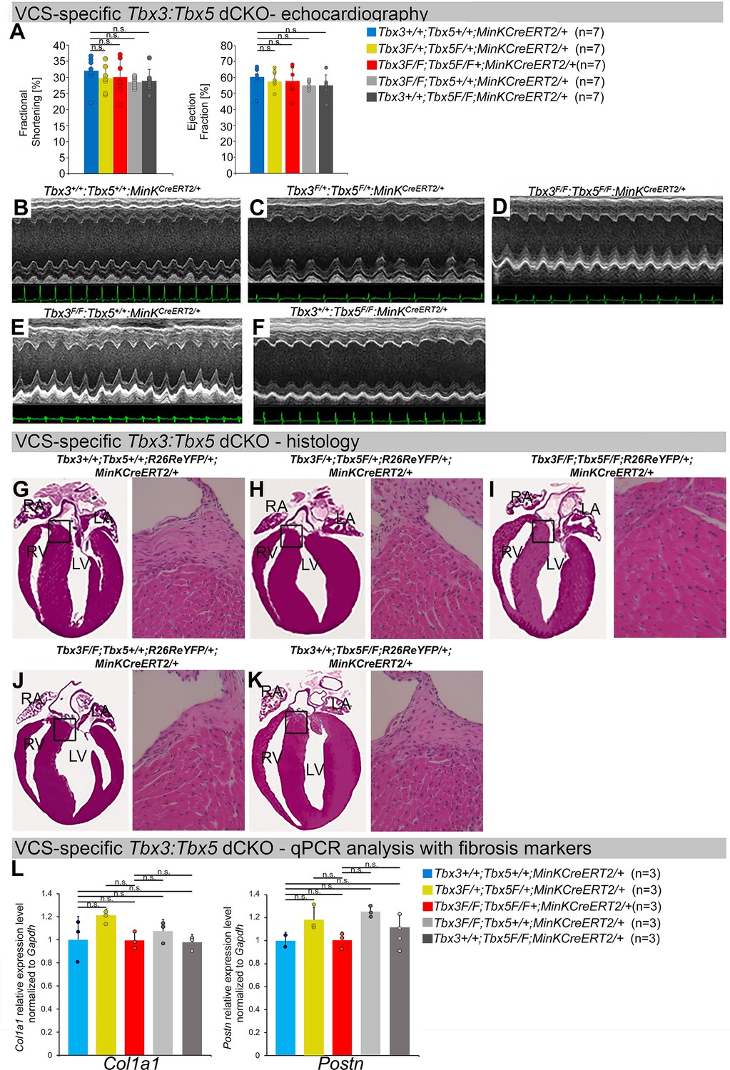
To distinguish a primary conduction system abnormality from a secondary conduction abnormality resulting from cardiac dysfunction or remodeling, we evaluated cardiac form and function at the time of arrhythmia assessment, 2–3 weeks post-tamoxifen treatment (Figure 3). Transthoracic echocardiography revealed no significant differences in left ventricular ejection fraction (LVEF) and fractional shortening (FS) between VCS-specific Tbx3:Tbx5-deficient (Tbx3fl/fl;Tbx5fl/fl;R26ReYFP/+;MinKCreERT2/+) and control (Tbx3+/+;Tbx5+/+;R26ReYFP/+;MinKCreERT2/+) mice (LVEF: Tbx3:Tbx5 double-conditional knockout mice vs. control mice p < 0.05, n = 7, Welch t-test; and FS: Tbx3:Tbx5 double-conditional knockout mice vs. control mice p > 0.05, n = 7, Welch t-test; Figure 3A, B, D). Histological examination of all four chambers demonstrated no discernible differences between VCS-specific Tbx3:Tbx5 double-knockout (Tbx3fl/fl;Tbx5fl/fl; R26ReYFP/+;MinKCreERT2/+) and control (Tbx3+/+;Tbx5+/+;R26ReYFP/+; MinKCreERT2/+) mice, nor between the double-knockout (Tbx3fl/fl;Tbx5fl/fl;R26ReYFP/+; MinKCreERT2/+) and single-knockout models for either Tbx3 (Tbx3fl/fl;Tbx5+/+;R26ReYFP/+;MinKCreERT2/+) or Tbx5 (Tbx3+/+;Tbx5fl/fl;R26ReYFP/+;MinKCreERT2/+). Ventricular muscle appeared normal without hypertrophy or myofibrillar disarray, and no fibrosis was present (Figure 3G, I–K, respectively). qRT-PCR analysis for fibrosis genes Col1a1 (Pan et al., 2022; Hua et al., 2020; Zhao et al., 2018) and Postn (Zhao et al., 2018; Ackerman et al., 2024; Wu et al., 2024; Oka et al., 2007) further confirmed no fibrosis in VCS of Tbx3:Tbx5-deficient mice (Figure 3L). No contractile dysfunction, histological abnormalities, or increased expression of fibrosis genes were observed in VCS-specific Tbx3:Tbx5 double-conditional heterozygous mice (Tbx3fl/+;Tbx5fl/+;R26ReYFP/+; MinKCreERT2/+) (Figure 3A, C, H, L). Taken together, these data indicate that the conduction defect and VT observed in mice with VCS-specific Tbx3:Tbx5 deletion (Figure 2) occur prior to the onset of left ventricular (LV) dysfunction or evidence of remodeling (Figure 3), implying a primary origin.
Figure 3
Cardiac function is preserved following double-conditional loss of Tbx3 and Tbx5 in the adult ventricular conduction system (VCS).
(A) Left ventricular (LV) fractional shortening (left graph) and LV ejection fraction (right graph) calculated from the M-mode electrocardiographies (ECGs) in (B–F) revealed no contractile dysfunction in VCS-specific Tbx3:Tbx5 double-conditional mutant mice (Tbx3fl/fl;Tbx5fl/fl;R26ReYFP/+; MinKCreERT2/+). Data are presented as mean ± SD. Welch t-test: ns, not significant (p > 0.05) ; n = 7 biological replicates/genotype. (B–F) Cardiac function, assessed by M-mode echocardiography from Tbx3+/+;Tbx5+/+;R26ReYFP/+;MinKCreERT2/+ (B), Tbx3fl/+;Tbx5fl/+; R26ReYFP/+;MinKCreERT2/+ (C), Tbx3fl/fl;Tbx5fl/fl;R26ReYFP/+;MinKCreERT2/+ (D), Tbx3fl/fl;Tbx5+/+; R26ReYFP/+;MinKCreERT2/+ (E), Tbx3+/+;Tbx5fl/fl;R26ReYFP/+;MinKCreERT2/+ (F) mice shown above surface ECGs. No functional differences between mutant and control mice were detected. The most representative images for each genotype were utilized in the figure. n = 7 biological replicates/genotype. (G–K) Histological examination of all four chambers from Tbx3+/+;Tbx5+/+;R26ReYFP/+;MinKCreERT2/+ (G), Tbx3fl/+;Tbx5fl/+;R26ReYFP/+; MinKCreERT2/+ (H), Tbx3fl/fl;Tbx5fl/fl;R26ReYFP/+;MinKCreERT2/+ (I), Tbx3fl/fl;Tbx5+/+;R26ReYFP/+; MinKCreERT2/+ (J), Tbx3+/+;Tbx5fl/fl;R26ReYFP/+;MinKCreERT2/+ (K) mice showed no histological abnormalities. The most representative images for each genotype were utilized in the figure. n = 3–4 biological replicates/genotype. Boxed areas in (G–K) have been shown at higher magnification at their right sides. (L) qRT-PCR analysis for fibrosis genes Col1a1 and Postn confirmed that there was no increase in expression of fibrosis markers in the VCS of Tbx3:Tbx5-deficient mice. Data are presented as mean ± SD normalized to Gapdh and relative to Tbx3+/+;Tbx5+/+;R26ReYFP/+;MinKCreERT2/+ mice (set as 1). ns: FDR >0.05 in both Welch t-test and Wilcoxon test; n = 2–3 biological replicates/genotype (VCS cardiomyocytes pooled from 30 mice per biological replicate). Histological examination original magnification: ×2.5, boxed area showed at the higher magnification: ×40. Abbreviations: FDR, false discovery rate; LA, left atrium; RA, right atrium; LV, left ventricle; RV, right ventricle.
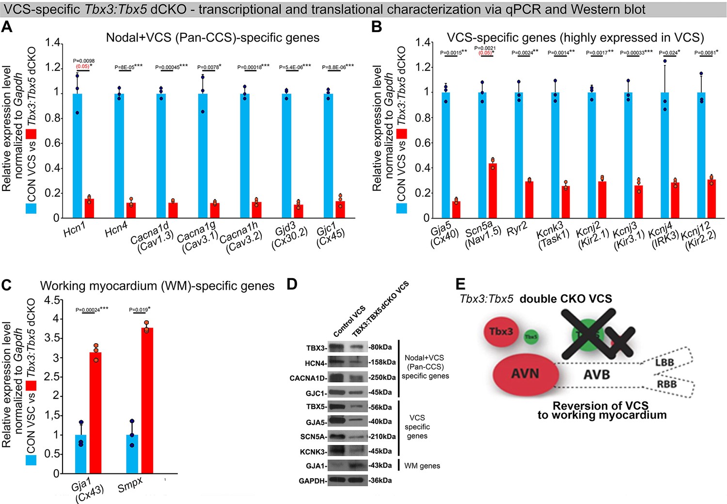
To assess the hypothesis that Tbx3 and Tbx5 collectively promote VCS versus working myocardium phenotype, we conducted a transcriptional characterization of the adult VCS in Tbx3fl/fl;Tbx5fl/fl;R26ReYFP/+;MinKCreERT2/+ mutant mice compared to their Tbx3+/+;Tbx5+/+;R26ReYFP/+;MinKCreERT2/+ control littermates using three distinct sets of molecular markers by qRT-PCR (Figure 4A–C). The first set encompassed genes expressed throughout the entire conduction system (Pan-CCS) and implicated in slow-conducting nodal phenotype, such as Hcn1, Hcn4, Cacna1d (Cav1.3), Cacna1g (Cav3.1d), Cacna1h (Cav3.2), Gjd3 (Cx30.2), and Gjc1 (Cx45) (Bakker et al., 2012; Hoogaars et al., 2004; van Eif et al., 2020; Verheule and Kaese, 2013; Liang et al., 2013; Greener et al., 2011; Mangoni et al., 2006; Marionneau et al., 2005; Garcia-Frigola et al., 2003; Schram et al., 2002; Figure 4A). The second set included genes highly expressed in the fast-conducting VCS and important for VCS function, including Gja5 (Cx40), Scn5a (Nav1.5), Ryr2, Kcnk3 (Task-1), Kcnj2 (Kir2.1), Kcnj3 (Kir3.1), Kcnj4 (IRK3), and Kcnj12 (Kir2.2) (Figure 4B; van Eif et al., 2020; Verheule and Kaese, 2013; Schram et al., 2002; Donner et al., 2011; Miquerol et al., 2010; Remme et al., 2009; Graham et al., 2006). The third set contained markers specifically present in the working myocardium but absent in the CCS, such as Gja1 (Cx43) and Smpx (Figure 4C; Hoogaars et al., 2004; van Eif et al., 2020; Alcoléa et al., 1999; Kempen et al., 1996). VCS-specific Tbx3:Tbx5-deficient mice lost VCS expression profile of genes required for the fast ventricular conduction (Figure 4B) as well as genes normally expressed in whole CCS (Pan-CCS genes) (Figure 4A). In contrast, these mice obtained VCS expression of working myocardium-specific molecular markers (Figure 4C). Immunoblotting analysis confirmed transcriptional changes observed by qRT-PCR (Figure 4D). This molecular characterization indicated that the Tbx3:Tbx5 double mutant VCS adopted a gene expression profile similar to wild-type working myocardial-like cells (Figure 4).
Figure 4
In the adult murine heart, Tbx3 and Tbx5 collectively promote ventricular conduction system (VCS) versus working myocardium (WM) phenotype.
(A–C) qRT-PCR analysis of molecular changes driven by VCS-specific Tbx3:Tbx5 double-conditional knockout in adult mice. Transcriptional characterization of the adult VCS in Tbx3fl/fl;Tbx5fl/fl;R26ReYFP/+;MinKCreERT2/+ mutant mice, compared to their Tbx3+/+;Tbx5+/+;R26ReYFP/+;MinKCreERT2/+ control littermates, was conducted using three distinct sets of molecular markers. (A) Genes expressed throughout the entire conduction system (Pan-CCS), implicated in the slow-conducting nodal phenotype. (B) Genes highly expressed in the fast-conducting VCS, critical for VCS function. (C) Markers specifically present in the working myocardium but absent in the CCS. VCS-specific Tbx3:Tbx5-deficient mice lost the VCS expression profile, including genes necessary for fast ventricular conduction (B) and those typically expressed in the entire CCS (Pan-CCS genes), essential for the slow-conducting nodal phenotype (A). In contrast, they acquired VCS expression of working myocardium-specific molecular markers important for working myocardial function (C). (D) Immunoblotting analysis confirmed transcriptional changes indicated by qRT-PCR analysis (A–C) in VCS-specific Tbx3:Tbx5 double-conditional knockout in adult mice. (E) Graphical summary of transcriptional changes observed in VCS of VCS-specific Tbx3:Tbx5-deficient mice. Simultaneous genetic deletion of Tbx3 and Tbx5 from the mature VCS resulted in a transcriptional profile resembling that of ventricular working myocardium. qRT-PCR data are presented as mean ± SD normalized to Gapdh and relative to Tbx3+/+;Tbx5+/+;R26ReYFP/+;MinKCreERT2/+ mice (set as 1). N = 3 biological replicates/genotype (VCS cardiomyocytes pooled from 30 mice per biological replicate); multiple testing correction using Benjamini and Hochberg procedure. Significance was assessed by Welch t-test (*FDR <0.05; **FDR <0.005; ***FDR <0.001) and confirmed by one-tailed Wilcoxon test (p value in parentheses) when normal distribution was rejected. Western blotting, n=2 biological replicates/ genotype (VCS cardiomyocytes pooled from 50 mice per each biological replicate). Abbreviations: AVB, atrioventricular bundle (also known as bundle of His); AVN, atrioventricular node; dCKO, double-conditional knockout; FDR, false discovery rate; LBB, left bundle branches; RBB, right bundle branches; VCS, ventricular conduction system.
-
Figure 4—source data 1
File containing original western blots corresponding to Figure 4D.
The black rectangle indicates the region included in the final figure panel.
- https://cdn.elifesciences.org/articles/102027/elife-102027-fig4-data1-v1.pdf
-
Figure 4—source data 2
Zipped folder containing original western blots files corresponding to Figure 4D.
- https://cdn.elifesciences.org/articles/102027/elife-102027-fig4-data2-v1.zip
The impact of the Tbx3:Tbx5 double-conditional knockout on electrical impulse propagation in the VCS of the heart was assessed with optical mapping of the anterior epicardial surface of the ventricles and right septal preparations where VCS function should be observed (Figure 5). Optical mapping records changes in transmembrane potential from multiple cells in tissue preparations, where ventricular septal optical action potentials (OAPs) have two distinct action potential upstrokes. The first peak is a result of depolarization of the specialized fast VCS, followed by depolarization of ventricular working myocardium.
Figure 5 with 1 supplement see all
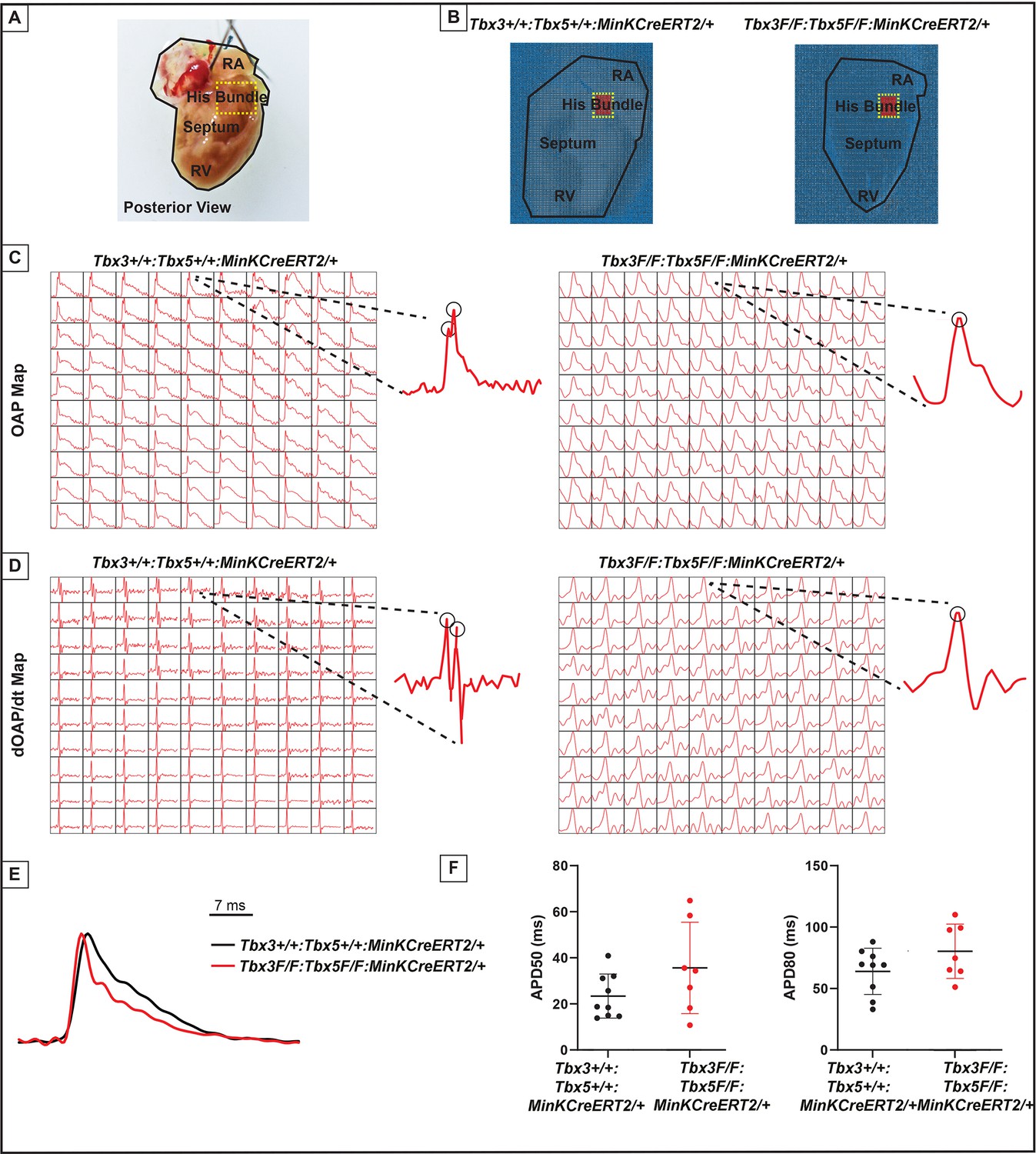
Loss of VCS optical action potential (OAP) morphology in Tbx3:Tbx5 double-conditional knockout mice.
(A) Schematic of the posterior view of a mouse heart with right ventricle (RV) free wall removed, highlighting the RV, septum, His bundle, and right atria (RA). (B) Representative 100 × 100 OAP map of OAP recorded during sinus rhythm from Tbx3:Tbx5 double-conditional knockout mice and control littermates with the free wall removed. The region of the His bundle is highlighted in red. (C) Representative 10 × 10 OAP map from the region of the His bundle. (D) Representative 10 × 10 map of the first derivative of the OAP from the region of the His bundle. (E) Representative ventricular OAP from whole heart intact preparation from Tbx3:Tbx5 double-conditional knockout mice (red) and control littermates (black). (F) Quantification of APD50 and APD80 at a basic cycle length of 125 ms. For (C-F), n=10 or 8 animals/ genotype.
To visualize electrical impulse propagation in Tbx3:Tbx5 double-conditional knockout mice, a 100 × 100 pixel data frame was plotted for the entire field of view of the right septal preparation (Figure 5A, B). For enhanced analysis of the VCS, the region encompassing the His bundle was distinguished in a red 10 × 10 area (Figure 5B). To specifically assess electrical impulse propagation within the His bundle, the same 10 × 10 pixel area representing this region was isolated from adult hearts of both control and Tbx3:Tbx5 double-conditional knockout mice (Figure 5C). We observed only one OAP upstroke in Tbx3:Tbx5 double-conditional knockout mice in contrast to control mice which showed two OAP upstrokes. The first derivative of the OAP (dOAP/dt) from the region of the His bundle was calculated and plotted in a dOAP/dt map further highlighting the number of temporally distinct upstrokes within the OAP (Figure 5D). In control mice, we observed two distinct depolarization upstrokes, one pertaining to the VCS and the second for working ventricular myocardium. However, in Tbx3:Tbx5 double-conditional knockout mice, only one maximum dOAP/dt was observed (Figure 5D). The OAP morphology observed in Tbx3:Tbx5 double-conditional knockout mice suggested a loss or reduction of the specialized fast VCS conduction (Figure 5C, D).
OAPs from the anterior epicardial surface of the ventricles were compared between control littermates and Tbx3:Tbx5 double-conditional knockout mice to assess changes in electrical impulse propagation in the ventricular working myocardium. Paced at a basic cycle length of 125 ms, control littermates and Tbx3:Tbx5 double-conditional knockout mice did not exhibit significant differences in action potential duration at 50% (APD50) or 80% (APD80) repolarization (Figure 5E, F). These observations suggested that the electrical activity of the ventricular working myocardium was not appreciably altered in double-knockout mice compared to controls. This finding is consistent with the lack of changes in the VERP observed in invasive EP studies of both control and Tbx3:Tbx5 double-conditional knockout mice (Figure 3I).
We further predicted that the double knockout would not affect action potentials or conduction properties distal to the VCS (Figure 6). OAP and dOAP/dt maps were created to observe the effects on electrical impulse propagation distal to the His bundle on the right septal preparation (Figure 6), similar to the methods applied to create OAP and dOAP/dt maps in the region of the His bundle (Figure 5), with the key difference being that the signals were recorded from the red 10 × 10 pixel regions plotted in the working ventricular myocardium distal to the His bundle instead of in the area of the His bundle (Figure 6A, B vs. Figure 5A, B, respectively). In both control littermates and Tbx3:Tbx5 double-conditional knockout mice, only one action potential upstroke and one dOAP/dt maximum are observed, which indicates that most of this region consists of the ventricular working myocardium (Figure 6C and D, respectively). In summary, the significant remodeling of electrical impulse propagation due to Tbx3:Tbx5 double-conditional knockout was observed by the loss of a distinct fast VCS impulse in the region of the His bundle, but not on the anterior epicardial surface of the ventricles or distal to the His bundle on the right ventricular (RV) septum (Figures 5 and 6).
Figure 6
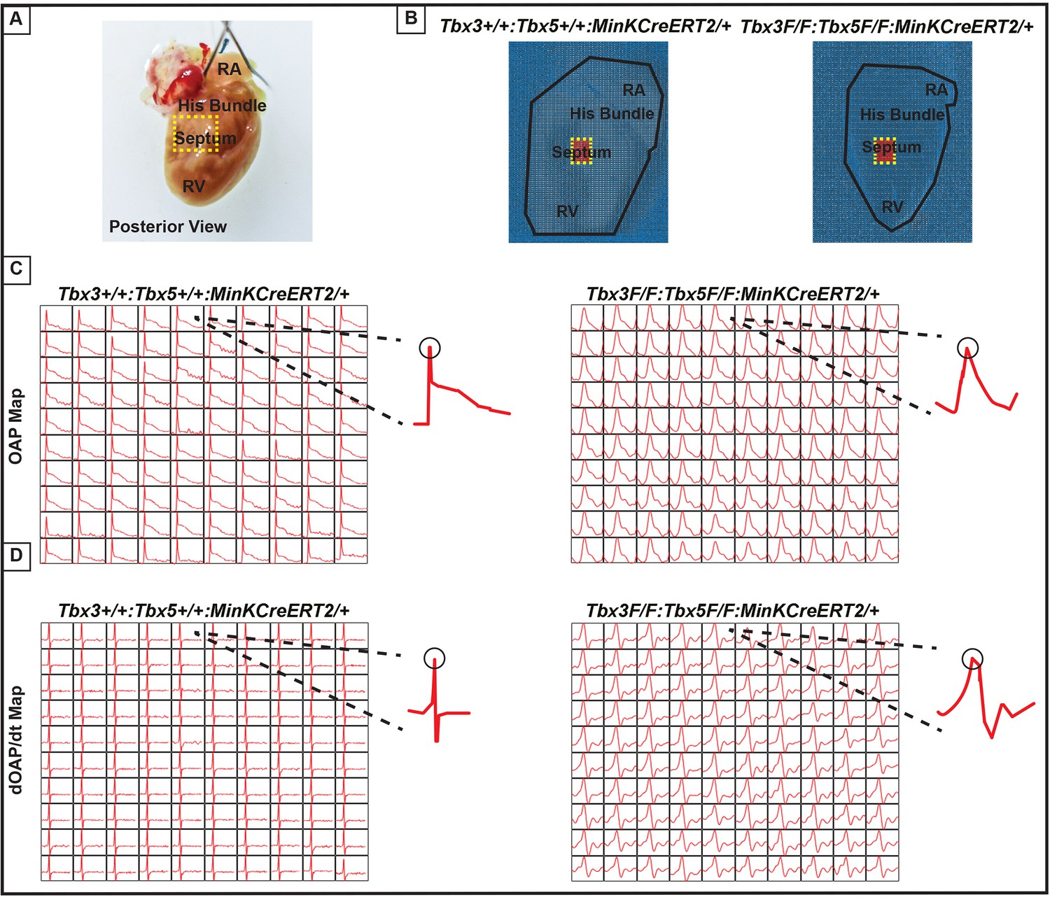
Ventricular optical action potentials (OAPs) distal from His bundle have only one OAP upstroke.
(A) Schematic of the posterior view of mouse heart with right ventricle (RV) free wall removed. (B) Representative 100 × 100 pixel OAP map recorded during sinus rhythm from Tbx3:Tbx5 double-conditional knockout mice and control littermates with RV free wall removed. The region of the working ventricular myocardium distal from the His bundle is highlighted in red. (C) Representative 10 × 10 pixel OAP map from the region distal to the His bundle. (D) Representative 10 × 10 dOAP/dt map from the region distal to the His bundle. For (C and D), n=10 or 8 animals/ genotype.
Discussion
Our study investigated the impact of compound Tbx3:Tbx5 deficiency on mature VCS function and molecular identity. Using a double-conditional knockout strategy, both genes were targeted specifically in the adult VCS. Loss of Tbx3 and Tbx5 expression in the mature VCS led to profound conduction defects, characterized by prolonged PR interval and QRS duration, increased susceptibility to VT, and sudden death. These alterations were observed in the absence of discernible changes in cardiac contractility or histological morphology, indicating a primary conduction system defect. Molecular characterization of the adult VCS unveiled an altered gene expression profile in Tbx3:Tbx5 double-conditional knockout mice, suggesting a transition from the distinctive fast VCS transcriptional profile to that resembling ventricular working myocardium. Optical mapping demonstrated loss of the specialized fast VCS function in Tbx3:Tbx5 double-conditional knockout mice, further suggesting that this region acquired an electrophysiological phenotype similar to the ventricular working myocardium.
Our previous research (Arnolds et al., 2012; Moskowitz et al., 2007; Burnicka-Turek et al., 2020; Moskowitz et al., 2004) and published literature van Duijvenboden et al., 2014; Bakker et al., 2012; Bruneau et al., 2001; Frank et al., 2011; Mohan et al., 2020; van Eif et al., 2018 have suggested that the balance between Tbx3 and Tbx5 expression determines the regional specialization of the mature central CCS (Figure 7). Tbx3 expression dominates in nodal myocardium, imparting nodal physiological characteristics. Tbx5 expression dominates in fast VCS myocardium, where a T-box-dependent fast conduction system network drives physiologically dominant fast conduction physiology, overriding nodal physiology (Burnicka-Turek et al., 2020; Figure 7). This model accurately predicts the outcome of targeted manipulations, such as adult VCS-specific removal of TBX5 or overexpression of TBX3, which convert the fast VCS into a slow nodal-like system (Burnicka-Turek et al., 2020; Figure 7).
Figure 7
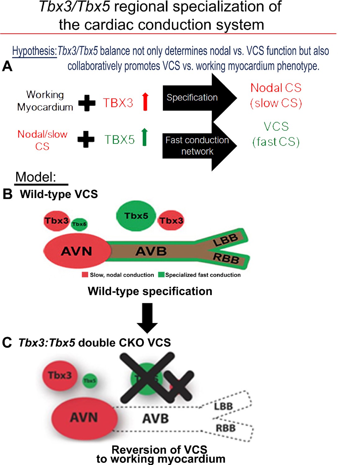
Tbx3 and Tbx5 play distinct roles in the adult VCS while collectively promoting CCS regional identity – a model elucidating our hypothesis for Tbx3/Tbx5 dose-dependent CCS regional specialization.
(A) The Tbx3/Tbx5 balance not only governs nodal versus ventricular conduction system (VCS) function but also collaboratively promotes the VCS versus working myocardium (WM) phenotype. Specifically, a high level of Tbx3 is linked to the specialization to the nodal conduction system, while an elevated Tbx5 level in nodal cells activates local expression of the Tbx5-dependent fast conduction network, resulting in the generation of VCS. (B) CCS regional specialization is driven by local expression of Tbx5-dependent fast conduction network in the VCS, which overlaps underlying Pan-CCS expression of nodal, slow conduction network. (C) VCS-specific simultaneous genetic removal of both the Tbx3 and Tbx5 transcription factors transforms the fast-conducting, adult VCS into cells resembling working myocardium, thereby shifting them from conduction to non-conduction myocytes. Therefore, within the adult CCS, the Tbx3 and Tbx5 expression levels are crucial not only for normal fast versus slow conduction system identity but also for maintaining the conduction versus contraction specialization of the VCS. AVB, atrioventricular bundle; AVN, atrioventricular node; CCS, cardiac conduction system; CKO, conditional knockout; LBB, left bundle branch; RBB, right bundle branch; VCS, ventricular conduction system.
The adult CCS organization model posits that Tbx3 and Tbx5 coordinately establish CCS characteristics and suggests their compound necessity to maintain specialized VCS identity (Figure 7), a hypothesis that remained untested. This model predicted that VCS-specific genetic ablation of both the TBX3 and TBX5 transcription factors would transform fast-conducting adult VCS into cells resembling working myocardium, eliminating specialized CCS identity (Figure 7). Testing this model necessitated the generation of a Tbx3:Tbx5 double-conditional knockout allele due to their proximal chromosomal location in cis on mouse Chr5. VCS-specific double-knockout mice showed a notable deceleration in VCS conduction, manifested by prolonged PR and QRS intervals, along with increased susceptibility to VT. A similar functional phenotype was observed in single deletion of Tbx5 from the adult VCS, leading to the transformation of the fast VCS into a nodal-like phenotype (Arnolds et al., 2012; Burnicka-Turek et al., 2020). However, we predicted that the nodal-like characteristics of the Tbx5-ablated VCS were due to retained expression of Tbx3. In fact, the autonomous beating and impulse initiation observed in the Tbx5-mutant VCS (Arnolds et al., 2012; Burnicka-Turek et al., 2020) were absent from the double Tbx3:Tbx5 mutant VCS, further suggesting the transformation from a nodal-like to an inert myocardial functionally and a shift from conduction toward simple working myocardium.
Molecular studies support a transformation from conduction to working myocardium in the Tbx3:Tbx5 double VCS knockout. Previous studies investigating the roles of Tbx3 and Tbx5 in maintaining adult VCS identity demonstrated that Tbx3 deletion resulted in the silencing of Pan-CCS gene expression in the atrioventricular conduction system (van Duijvenboden et al., 2014; Bakker et al., 2012; Bruneau et al., 2001; Frank et al., 2011; Mohan et al., 2020). Alternately, specific deletion of Tbx5 from the adult VCS led to the decreased expression of VCS-specific markers while Pan-CCS markers remained unchanged, indicating a transformation toward a nodal-like transcriptional phenotype in the absence of Tbx5 (Arnolds et al., 2012; Burnicka-Turek et al., 2020). Consistently, a similar transformation was induced by Tbx3 overexpression in the adult VCS. These findings underscored the significance of the Tbx3:Tbx5 ratio in preserving the molecular and functional characteristics of the fast versus slow conduction system. In Tbx3:Tbx5 double VCS knockout, we observed a reduction in the expression of both fast VCS markers and Pan-CCS markers transcribed throughout the entire CCS. As expected, not all VCS markers were ablated in VCS-specific Tbx3:Tbx5 mutants. A significant portion of VCS fast conduction markers are also transcribed in ventricular working myocardium, albeit at lower levels than in the VCS, and are crucial for normal working myocardial function, for example, Ryr2, Scn5a, and Kcnj2 (van Eif et al., 2020; Schram et al., 2002; Remme et al., 2009). Consistent with a shift from fast VCS to working myocardium, their expression levels are present, but at significantly lower levels in the double-knockout mutants than in normal VCS. Furthermore, the expression of markers specific for working myocardium, which are normally excluded in the VCS, emerged in the VCS of Tbx3:Tbx5 double mutant. These observations are consistent with a transcriptional shift from VCS conduction cardiomyocytes to working myocardium-like characteristics in the absence of both Tbx3 and Tbx5.
Our study highlights the critical role of Tbx3 and Tbx5 in maintaining the specialized electrical properties of the VCS. Comparative analysis of the phenotypes observed in single Tbx3 or Tbx5 knockouts and the Tbx3:Tbx5 double-conditional knockout supports a coordinated function for these transcription factors in preserving VCS functionality. In a normal mammalian heart, electrical or optical recordings from the proximal part of the VCS typically display two distinct electrical excitations, reflected as two spikes in electrograms or two upstrokes in OAPs. These signals correspond to the sequential activation of the VCS, followed by ventricular excitation, a phenomenon well documented in both basic and clinical studies across species, including mice and humans. Previous whole-heart EP and optical mapping studies demonstrated that the single conditional knockout of Tbx5 in the adult VCS caused significant ventricular conduction slowing (Arnolds et al., 2012), accompanied by a phenotypic shift of VCS cells toward a pacemaker-like cell, evidenced by ectopic beats originating in the ventricles, retrograde activation, and inappropriate automaticity (Burnicka-Turek et al., 2020). Whole-cell patch clamp recordings of Tbx5-deficient VCS cells further supported these findings, revealing action potential characteristics resembling pacemaker cells, including a slower upstroke (phase 0), prolonged plateau (phase 2), delayed repolarization (phase 3), and enhanced phase 4 depolarization (Burnicka-Turek et al., 2020). In contrast, reduced expression of Tbx3 in the Tbx3 haploinsufficiency model resulted in AV bundle hypoplasia, PR interval shortening, and prolonged QRS duration (Mohan et al., 2020). Optical mapping in these mice showed delayed apical activation with multiple small breakthroughs separated by regions of delayed activation. This fragmented activation pattern suggests that the reduced Tbx3 expression impaired the homogeneity and efficiency of ventricular electrical impulse propagation due to AV bundle and bundle branch hypoplasia (Mohan et al., 2020). In the Tbx3:Tbx5 double-conditional knockout mice, we observed a complete loss of fast VCS conduction, similar to the phenotype of the Tbx5 single conditional knockouts. However, unlike in the Tbx5 single CKO, the VCS in Tbx3:Tbx5 double mutants did not acquire pacemaker-like activity. Instead, optical mapping revealed a single upstroke in the His bundle region of Tbx3:Tbx5 double-conditional knockout mice, indicating the absence of rapid AV bundle electrical impulse propagation. Functionally, this transformation resulted in significantly slowed ventricular activation, without the retrograde activation observed in the Tbx5 single conditional knockouts or the rapid electrical impulse propagation typically associated with an intact VCS. The homogenous morphological and molecular transformation of His bundle and VCS cells in the Tbx3:Tbx5 double-conditional knockout mice rendered the electrical properties of the VCS-located cells functionally indistinguishable from those of ventricular working myocardium. Our data, including gene expression analysis of the double mutant VCS, are most consistent with the transition of double mutant VCS cells toward a working myocardium phenotype.
These findings emphasize the interdependent roles of Tbx3 and Tbx5 in maintaining the specialized electrical properties and identity of the VCS, distinguishing it from non-specialized ventricular working myocardium (Figure 7). While Tbx5 alone is critical for suppressing pacemaker-like characteristics and preserving fast conduction properties, Tbx3 plays a pivotal role in ensuring the structural and functional integrity of the AV and BB conduction pathways. The simultaneous deletion of both Tbx3 and Tbx5 disrupts these regulatory mechanisms, leading to a loss of VCS identity and a functional shift toward a working myocardium phenotype. Our study combined with prior literature (Arnolds et al., 2012; van Duijvenboden et al., 2014; Burnicka-Turek et al., 2020; Bakker et al., 2012; Frank et al., 2011; Mohan et al., 2020; van Eif et al., 2018) indicates that the concurrent presence of both Tbx3 and Tbx5 is necessary for maintaining VCS identity in the adult heart, ensuring efficient electrical conduction and preventing the transition of VCS cells into a working myocardium-like state (Figure 7).
Methods
Experimental animals
All animal experiments were performed under the University of Chicago Institutional Animal Care and Use Committee (IACUC) approved protocol (ACUP no. 71737) and in compliance with the USA Public Health Service Policy on Humane Care and Use of Laboratory Animals. MinKCreERT2 [Tg(RP23-276I20-MinKCreERT2)] and Tbx3fl/fl mice have been reported previously (Arnolds and Moskowitz, 2011b; Frank et al., 2011).
Tbx3:Tbx5 double-floxed mouse line was generated by University of Utah Core Research Facility using CRISPR/Cas9. Guide RNA (sgRNA) constructs were designed with software tools (ZiFiT Targeter Sander et al., 2010 and crispr.genome-engineering.org) predicting unique target sites throughout the mouse genome (Figure 1—figure supplement 3–5). The sgRNA constructs were transcribed in vitro using MEGAshortscript T7 (Invitrogen AM1354) and mMessage Machine T7 transcription kit (Invitrogen AM1344) according to the manufacturer’s instructions. The strategy to generate mouse founders involved a single-step microinjection into one-cell Tbx3 floxed zygotes (on a mixed background) with 10 ng/µl of each sgRNA (Tbx5-I2-S22 and Tbx5-I3-S31, Figure 1—figure supplements 3–5), 30 ng/µl of Cas9 protein, and 10 ng/µl of a long ssDNA donor. The donor contained two lox2272 sites in cis, spanning partial intron 2, entire exon 3, and partial intron 3 of the mouse Tbx5 gene, along with 100/150 bp 5′/3′ homology arms (Figure 1—figure supplement 4). Founders were validated by PCR, restriction enzyme digestion, and Sanger sequencing (Figure 1—figure supplement 1B–D). Founders were backcrossed with wild-type CD1 IGS mice (Charles River Lab, USA) to confirm germline transmission of the CRISPR/Cas9-generated compound Tbx3:Tbx5 double-floxed allele and obtain the F1 generation. F1 mice were then interbred to establish a stable Tbx3:Tbx5 double-floxed mouse line. Downstream experiments were performed on F4–F6 mice.
To simultaneously conditionally delete the Tbx3 and Tbx5 genes specifically from the adult VCS, we crossed our Tbx3:Tbx5 double-floxed mouse line (Tbx3fl/fl;Tbx5fl/fl) with a VCS-specific tamoxifen (TM)-inducible Cre transgenic mouse line (MinKCreERT2 [Tg(RP23-276I20-MinKCreERT2); Arnolds and Moskowitz, 2011b; Figure 1A]). All mice were maintained on a mixed genetic background. Tamoxifen (MP Biomedical) was administered at a dose of 0.167 mg/g body weight for 5 consecutive days by oral gavage at 6 weeks of age and then mice were evaluated at 9 weeks of age, as previously described (Arnolds et al., 2012; Arnolds and Moskowitz, 2011b; Burnicka-Turek et al., 2020). Age-, gender-, and genetic strain-matched controls were used in all experiments to account for any variations in datasets across experiments. Mice were bred and housed in specific pathogen-free conditions in a 12-hr light/12-hr dark cycle and allowed ad libitum access to standard mouse chow and water. Mice requiring medical attention were provided with appropriate veterinary care by a licensed veterinarian and were excluded from the experiments described. No other exclusion criteria were applied.
All experiments and subsequent analysis were conducted in a blinded fashion, with animals randomly assigned to experimental groups. Following genotyping, mice were randomly allocated to the studies based on their genotypes. Subsequently, their identities were anonymized using a numerical code to ensure that all experiments and analyses were performed in a blinded manner. Both male and female animals have been used in our studies in the ratio of 41%/59%, based on availability of relatively rare compound genotypes, respectively.
Validation of the newly generated Tbx5 floxed allele
The Tbx3:Tbx5 double-floxed mouse line (Tbx3fl/fl:Tbx5fl/fl) was engineered by generating a novel Tbx5 floxed allele in the background of a previously published Tbx3 floxed allele (Frank et al., 2011). We utilized single Tbx3 (Frank et al., 2011) and the novel Tbx5 floxed mouse lines (Tbx3fl/fl and Tbx5fl/fl, respectively) to serve as controls in our studies (Figure 1A, Figure 1—figure supplements 1 and 2). Although the newly engineered Tbx5 floxed allele was designed to mirror a previously published allele (Bruneau et al., 2001), the location of the flox sites was altered slightly, requiring validation.
To verify the ability of the newly generated Tbx5 floxed allele to recombine into the Tbx5 KO (null) allele, we conducted three rounds of breeding and utilized a PCR assay to distinguish the recombined Tbx5 KO (null) allele (Tbx5−) from the unrecombined floxed allele (Tbx5fl) (Figure 1—figure supplement 2A, B). First, we bred newly generated homozygous Tbx5 floxed males (Tbx5fl/fl) with homozygous Mef2CCre/Cre females, which express Cre recombinase at the zygote stage (Hoffmann et al., 2014; Xie et al., 2012; Verzi et al., 2005), to produce germline Tbx5+/−;Mef2Cre/+ double heterozygous mice. PCR analysis showed that 100% of the generated offspring were Tbx5+/−;Mef2CCre/+ double heterozygous mice (Figure 1—figure supplement 2B). This result confirmed complete recombination of the new Tbx5 floxed allele into the Tbx5 null allele in the presence of the Mef2CCre allele. Next, we backcrossed Tbx5+/−;Mef2CCre/− double heterozygote mice with wild-type CD1 IGS mice (Charles River Lab, USA) to obtain a germline Tbx5+/− heterozygous mouse line (Figure 1—figure supplement 2B). Subsequently, we bred Tbx5+/− mice to generate germline Tbx5 null mice (Tbx5−/−). Among 230 offspring (generated in 50 litters), we observed 113 wild-type pups (Tbx5+/+) and 116 Tbx5 heterozygous pups (Tbx5+/−), but no null newborn pups (Tbx5−/−) were obtained (Figure 1—figure supplement 2B). These results align with previous breeding outcomes indicating complete embryonic lethality of Tbx5 null mice (Bruneau et al., 2001).
To further confirm the ability of the newly generated Tbx5 floxed allele to produce germline Tbx5 null embryos (Tbx5−/−), we bred Tbx5 heterozygous mice and collected embryos at E9–E10. Genotyping analysis of 89 collected embryos from 10 timed pregnancies revealed the presence of 24 wild-type (Tbx5+/+), 42 heterozygous (Tbx5+/−), and 22 null (Tbx5−/−) embryos (Figure 1—figure supplement 2C). Supported by Chi-square analysis, these results collectively verified that the newly generated Tbx5 floxed allele could recombine to form the null allele, mimicking the embryonic lethality observed with the previously generated allele (Bruneau et al., 2001).
To confirm that the newly engineered Tbx5 conditional allele can generate VCS-specific Tbx5-deficient mice with the same phenotype as VCS-specific Tbx5 knockout mice obtained from the previously generated allele (Arnolds et al., 2012; Burnicka-Turek et al., 2020; Bruneau et al., 2001), we crossed the newly generated Tbx5 floxed mouse line (Tbx5fl/fl) with a VCS-specific tamoxifen-inducible Cre transgenic mouse line (MinKCreERT2 [Tg(RP23-276I20-MinKCreERT2)]) (Arnolds and Moskowitz, 2011b; Figure 1A). The same tamoxifen regimen was administered as in the previous study (Arnolds et al., 2012; Burnicka-Turek et al., 2020). We confirmed VCS-specific genetic deletion of Tbx5 via immunofluorescence (IF) (Figure 1B) and qPCR (Figure 1C). The resulting VCS-specific Tbx5-deficient mice exhibited slowed conduction, indicated by increased PR and QRS intervals (Figure 2A, and E vs. B), as well as episodes of VT resulting in sudden death (Figure 2G, H), consistent with the previously reported phenotype (Arnolds et al., 2012; Burnicka-Turek et al., 2020). Additionally, we confirmed the absence of histological and structural abnormalities in these mice, aligning with previous findings (Figure 3A, F vs. B, and K vs. G, respectively) (Arnolds et al., 2012; Burnicka-Turek et al., 2020).
Echocardiography studies
Transthoracic echocardiography in mice was conducted under inhaled isoflurane anesthesia administered through a nose cone. Prior to imaging, chest hairs were removed using a topical depilatory agent. Limb leads were affixed for electrocardiogram gating, and animals were imaged in the left lateral decubitus position with a VisualSonics Vevo 770 machine using a 30-MHz high-frequency transducer. To ensure stability, body temperature was carefully maintained using a heated imaging platform and warming lamps. Two-dimensional images were meticulously recorded in parasternal long- and short-axis projections, accompanied by guided M-mode recordings at the midventricular level in both views. LV cavity size and percent FS were measured in at least three beats from each projection and averaged. M-mode measurements were employed to ascertain LV chamber dimensions and percent LV FS, calculated as ([LVIDd – LVIDs]/LVIDd), where LVIDd and LVIDs represent LV internal diameter in diastole and systole, respectively.
Surface ECG
Nine weeks old, tamoxifen-treated control and mutant mice were anesthetized with a mixture of 2–3% isoflurane in 100% oxygen. Anesthetized mice were secured in a supine position on a regulated heat pad while lead I and lead II ECGs were recorded using platinum subdermal needle electrodes in a three-limb configuration. Core temperature was continuously monitored using a rectal probe and maintained at 36–37°C throughout the procedure. ECG data were collected and analyzed using Ponemah Physiology Platform (DSI) software and an ACQ-7700 acquisition interface unit (Gould Instruments, Valley View, OH, USA). Key parameters derived from the ECG measurements included: heart rate (HR), PR interval (from the beginning of the P wave to the beginning of the QRS complex), and QRS complex duration.
Telemetry ECG analysis
Nine weeks old, tamoxifen-treated control and mutant mice were anesthetized with 2–3% isoflurane in 100% oxygen, and wireless telemetry transmitters (ETA-F10; DSI) were surgically implanted in the back with leads tunneled to the right upper and left lower thorax, as previously described (Arnolds et al., 2012; Wheeler et al., 2004). Following a 24-hr recovery period after surgical instrumentation, heart rate and PR and QRS intervals were calculated using Ponemah Physiology Platform (DSI) from 48-hr recordings.
Catheter-based intracardiac EP
Detailed protocols for invasive EP studies have been previously described (Nadadur et al., 2016; Liu et al., 2008). Briefly, 9 weeks old, tamoxifen-treated control and mutant mice were anesthetized using 2–3% isoflurane in 100% oxygen. Then, a 1.1-Fr octapolar electrode catheter (EPR-800; Millar Instruments) was advanced via a right jugular venous cut-down to record right atrial, His bundle, and RV potentials, as well as to perform programmed electrical stimulation. Signals were identified through alignment with simultaneous surface ECG using subcutaneous needle electrodes in a lead II configuration. ‘Near-Field’ and ‘Far-Field’ signals were identified based on ECG alignment, signal deflection upstroke speed, and total signal duration. Standard tachycardia induction protocols included an eight-beat drive train with beats 80–120 ms apart (S1), followed by five beats (S2) at 50 ms apart (penta-extrastimulus, PES). Two attempts at this PES protocol were carried out. Mice also underwent single extrastimulus testing (SES) with eight beats 80–120 ms apart (S1) followed by a single S2 at 50 ms. This SES protocol was carried out five separate times per mouse. If these two protocols in the right atrium and RV (separately) failed to initiate their respective tachycardias, the study was deemed negative. S1 drive train intervals varied slightly due to the presence of the AV Wenckebach block at faster pacing rates in some mice; the S1 interval was lengthened to prevent this during the drive train.
Additional atrial and ventricular pacing protocols were carried out to obtain atrial, atrio-ventricular, and ventricular effective refractory periods (AERP, AVERP, and VERP) as well as sinus node recovery time, as described previously (Nadadur et al., 2016; Liu et al., 2008; Patel et al., 2003; Gehrmann and Berul, 2000). Effective refractory periods were measured using eight-beat S1 drive trains of 100 ms followed by SES.
ECG and optical mapping
ECG acquisition and analysis
ECGs were recorded in conscious Tbx3:Tbx5 double-conditional knockout mice and control littermates using the ecgTUNNEL device (emka Technologies) 2 weeks after tamoxifen treatment. Mice were positioned in the tunnel and ECGs were recorded for 5 min at a sampling rate of 1 kHz using lead I. ECGs were analyzed using a custom MATLAB program to measure P and QRS durations, as well as PR, QT, and RR intervals (George et al., 2020; Warhol et al., 2021; Figure 5—figure supplement 1).
Langendorff perfusion
Tbx3:Tbx5 double-conditional knockout mice (n = 8) and control littermates (n = 10) were deeply anesthetized with isoflurane. The heart was quickly excised following cervical dislocation and thoracotomy. The aorta was cannulated, and the heart was retrogradely perfused with warmed (37°C) and oxygenated (95% O2 and 5% CO2) modified Tyrode’s solution (in mM, NaCl 130, NaHCO3 24, NaH2PO4 1.2, MgCl2 1, glucose 5.6, KCl 4, and CaCl2 1.8) at a pH of 7.4. The heart was placed in a constant-flow (1.0–2.0 ml/min) Langendorff perfusion system, laying horizontally in a tissue bath.
Ventricular optical mapping
The Langendorff perfused heart was electromechanically uncoupled by 15 μM of blebbistatin (Cayman Chemicals 13186) perfusion. A voltage-sensitive fluorescent dye, 80 µM di-4-ANEPPs (Thermo Fisher Scientific D1199), was administered through the dye injection port. The heart was illuminated using a 520 ± 5 nm (Prizmatix, UHP-Mic-LED-520) wavelength light source to excite di-4-ANEPPs. Emitted photons were captured using complementary metal–oxide semiconductor cameras (MiCAM, SciMedia).
The stimulation threshold, where the heart would capture the stimuli and action potential 1:1, was determined using a point source platinum electrode placed on the anterior surface of the heart on the anterior epicardial surface of the ventricles. Pacing was applied at 1.5× the threshold amplitude to maintain 1:1 capture over the duration of the experiment. Optical recordings were analyzed using Rhythm 1.2 to analyze transmembrane potential (Cathey et al., 2019). Action potential duration at 50% and 80% repolarization was calculated for the ventricles of the intact whole heart preparation.
Right septal preparation optical mapping experiments
Following intact whole heart ex vivo optical mapping, the heart was removed from the tissue bath for the dissection of the RV free wall to expose the RV septal surface (Tamaddon et al., 2000). The heart was quickly returned to the tissue bath and the RV septal surface was focused into the field of view. Sinus rhythm optical recordings were acquired.
A custom MATLAB program was written to plot individual pixels in a 100 × 100 pixel image stack. A Butterworth filter of order 5 was applied to provide temporal filtering (Laughner et al., 2012). The location of the His bundle was identified as the region at the base of the interventricular septum (a red 10 × 10 pixel area in Figure 5B). Signals from the working ventricular myocardium were recorded from the 10x10 pixel region plotted in the working ventricular myocardium distal to the His bundle (Figure 6B).
Isolation of adult VCS cardiomyocytes and cell sorting
Adult mouse EYFP-positive VCS cardiomyocytes were isolated using the method described by Mitra and Morad, 1985. Briefly, 9 weeks old tamoxifen-treated control and Tbx3:Tbx5 double-conditional mutant mice were heparinized (100 units IP) and anesthetized with pentobarbital (50 mg/kg of body weight). Hearts were excised and mounted on a Langendorff apparatus, then perfused with Ca2+-free Tyrode’s solution with collagenase B and D (Roche Chemical Co) plus protease (Fraction IV, Sigma Chemical Co). When the hearts appeared pale and flaccid, they were removed from the Langendorff apparatus and a tip of ventricular septum below the AV annulus was microdissected out (Arnolds et al., 2012; Park and Fishman, 2011; Silverman et al., 2006) and kept in Ca2+-free Tyrode’s solution with 1 mg/ml of bovine serum albumin (Fraction XIV, Sigma Chemical Co). The intraventricular sections were then minced into small pieces ~1 × 1 × 1 mm and then gently triturated with a Pasteur pipette to dissociate individual VCS myocytes. Propidium iodide (Thermo Fisher Scientific) was added immediately before FACS to facilitate live/dead discrimination. Cells were sorted on a FacsAria flow cytometer (BD Biosciences) located at the University of Chicago Flow Cytometry Core using Influx software. Samples from wild-type age-matched hearts were used for gating. Samples were gated to exclude debris and cell clumps. Fluorescent cells were collected into ice-cold RNase-free PBS and processed for RNA extraction and protein isolation.
RNA isolation and qRT-PCR
Total RNA was isolated from EYFP-positive VCS cardiomyocytes sorted from 9-week-old control and VCS-specific Tbx3:Tbx5-deficient mice, which had received tamoxifen at 6 weeks of age. RNA was also extracted from atrial and ventricular tissues dissected from the same mice. All RNA extractions were performed using RNeasy Mini Kit (QIAGEN), followed by DNase treatment according to the manufacturer’s instructions.
Reverse transcription reaction was carried out using the SuperScript III First-Strand Synthesis SuperMix for quantitative RT-PCR (Invitrogen) as per the manufacturer’s recommendations. qRT-PCR was performed using the POWER SYBR Green PCR master mix from Applied Biosystems and run on an Applied Biosystems AB7500 machine in 96-well plates. The relative gene expression level was calculated by the ΔΔCt method (Livak and Schmittgen, 2001) using glyceraldehyde-3-phosphate dehydrogenase (Gapdh) gene expression level as internal control. The data presented are the average of three independent experiments.
Protein isolation and western blotting
Total protein was isolated from EYFP-positive VCS cardiomyocytes sorted from 9-week-old control and VCS-specific Tbx3:Tbx5-deficient mice that had been administered tamoxifen at 6 weeks of age. Protein was also obtained from atrial and ventricular tissues dissected from the same animals. The tissues were snap-frozen in liquid nitrogen, pulverized, and homogenized in RIPA buffer (50 mM Tris-HCl pH 8, 150 mM NaCl, 1% Triton-X, 0.5% sodium deoxycholate, 0.1% SDS, 5 mM EDTA) with 1 Roche EDTA-Free complete protease inhibitor tablet per 50 ml of buffer. Samples were tumbled for 1 hr at 4°C and then centrifuged for 10 min at 13,200 × g. Protein concentration was determined using the BCA assay (Pierce) with BSA as a standard.
For Western blot analysis, 25 µg of protein was diluted in Laemmli buffer, heated at 70°C for 10 min, and subjected to SDS–PAGE on 4–20% TGX gels (Bio-Rad). Proteins were then transferred to nitrocellulose membranes, blocked with 5% milk in TBS-T, and incubated overnight at 4°C with primary antibodies diluted in 2.5% milk in TBS-T. The primary antibodies used were: goat polyclonal anti-TBX3 (Santa Cruz Biotechnology, sc-31656, 1:250), rabbit polyclonal anti-HCN4 (Millipore, AB5808, 1:500), rabbit polyclonal anti-CAV1.3/CACNA1D (Alomone, ACC-005, 1:200), rabbit polyclonal anti-Cx45/GJC1 (Thermo Fisher Scientific, PA5-77357, 1:250), sheep polyclonal anti-TBX5 (R&D, AF5918, 1:200), rabbit polyclonal anti-CX40/GJA5 (Zymed/Invitrogen, 36-4900, 1:500), rabbit polyclonal anti-NAV1.5/SCN5A (Alomone, ASC-005, 1:200), mouse monoclonal anti-KCNK3/TASK1 (Abcam, ab186352, 1:1000), rabbit polyclonal anti-CX43/GJA1 (Cell Signaling Technology, 3512, 1:1000), and mouse monoclonal anti-GAPDH (Abcam, ab8245, 1:1000). After rinsing in TBS-T, membranes were incubated for 1 hr at room temperature (RT) with secondary antibodies diluted in 2.5% milk in TBS-T, rinsed again, and visualized using enhanced chemiluminescence reagents (Pierce ECL/ECL Plus, Thermo Fisher Scientific) and Kodak X-OMAT film. Results were normalized to GAPDH loading control and then quantified using ImageJ software (Rasband, 1997; Wheeler et al., 2004). Secondary antibodies used were donkey anti-goat IgG AlexaFluor-594 (Invitrogen, A-11058, 1:250 dilution) and donkey anti-goat IgG AlexaFluor-488 (Invitrogen, A-11055, 1:250 dilution) for experiments involving co-staining for goat primary antibodies. Secondary antibodies were as follows: rabbit anti-goat-HRP (Jackson ImmunoResearch, 305-035-003, 1:10,000), goat anti-rabbit-HRP (Jackson ImmunoResearch, 111-035-144, 1:3000), donkey anti-sheep-HRP (Abcam, ab6900, 1:5000), and sheep anti-mouse-HRP (Amersham GE, NA931, 1:2500).
Histology
Hearts from 9-week-old control and mutant mice were carefully dissected, flushed with ice-cold PBS, and then fixed for 48 hr at 4°C in a 4% paraformaldehyde solution (Sigma-Aldrich). Following fixation, the hearts were processed for paraffin-embedded sections and subjected to analysis through H&E staining, following the manufacturer’s protocol provided by Sigma-Aldrich.
Immunofluorescence
Hearts from 9-week-old control and mutant mice were dissected out and placed in ice-cold PBS, followed by freezing in OCT (Fisher) within the gas phase of liquid nitrogen. Cryosections of 7 μm thickness were mounted onto Superfrost Plus glass slides (Fisher Scientific), air-dried, and fixed for 10 min in ice-cold 4% paraformaldehyde (Sigma-Aldrich). Subsequently, sections were permeabilized in 1% Triton-X100 (Sigma-Aldrich) in PBS for 10 min, then blocked in 10% normal goat serum (Invitrogen) in PBS-T (PBS + 0.1% Tween-20) for 30 min at RT. Sections were then incubated overnight at 4°C in primary antibody diluted in blocking buffer, rinsed in PBS, then subsequently incubated for 60 min at RT in secondary antibody diluted in blocking buffer. Slides were mounted in VectaShield + DAPI (Vector Laboratories) or counterstained with DAPI and mounted in ProLog Gold (Invitrogen) prior to visualizing fluorescence. Primary antibodies were as follows: goat polyclonal anti-TBX3 (Santa Cruz, sc-31656, 1:250 dilution) and goat polyclonal anti-TBX5 (Santa Cruz, sc-17866, 1:250 dilution). Secondary antibodies used were donkey anti-goat IgG AlexaFluor-594 (Invitrogen, A-11058, 1:250 dilution) and donkey anti-goat IgG AlexaFluor-488 (Invitrogen, A-11055, 1:250 dilution) for experiments involving co-staining for goat primary antibodies. A secondary antibody-only control was employed in each case to ensure the specificity of immunostaining.
Statistics
The numbers of independent experiments are specified in the relevant figure legends. Quantitative data are presented as mean ± SD. Statistical analysis was performed using R version 3.6.2 or the GraphPad Prism statistical package version 10.2.1 (GraphPad Software, Boston, MA, USA). For all datasets, except qRT-PCR data, normality was assessed using the Shapiro–Wilk normality test. If the data followed a normal distribution, the two-sample Welch t-test was performed for comparison. If normal distribution was not present, the non-parametric Mann–Whitney U test or Kruskal–Wallis H test was used, as indicated in the text. Statistical significance was assumed if p reached a value ≤0.05.
For qRT-PCR data, statistical analysis was performed using R version 4.2.0. Normality within each experimental group was assessed using the Shapiro–Wilk test. Between-group comparisons were conducted using Welch t-test, and multiple comparisons were corrected using the Benjamini and Hochberg method to control the false discovery rate (FDR) (Benjamini and Hochberg, 1995). If a significant difference was detected between two groups (t-test FDR <0.05) but normality was rejected in any of the compared groups (Shapiro–Wilk p < 0.05), a non-parametric Wilcoxon rank-sum test was used for verification. A significant group-mean difference was confirmed at one-tailed Wilcoxon p ≤ 0.05 (Source data 1).
Appendix 1
Appendix 1—key resources table
| Reagent type (species) or resource | Designation | Source or reference | Identifiers | Additional information |
|---|---|---|---|---|
| Antibody | Goat polyclonal anti-TBX3 | Santa Cruz Biotechnology | sc-31656 | IF (1:250) WB (1:250) |
| Antibody | Goat polyclonal anti-TBX5 | Santa Cruz Biotechnology | sc-17866 | IF (1:250) |
| Antibody | Donkey anti-goat IgG AlexaFluor-594 | Invitrogen | A-11058 | IF (1:250) |
| Antibody | Donkey anti-goat IgG AlexaFluor-488 | Invitrogen | A-11055 | IF (1:250) |
| Antibody | Rabbit polyclonal anti-HCN4 | Millipore | AB5808 | WB (1:500) |
| Antibody | Rabbit polyclonal anti-CAV1.3/CACNA1D | Alomone | ACC-005 | WB (1:200) |
| Antibody | Rabbit polyclonal anti-Cx45/GJC1 | Thermo Fisher | PA5-77357 | WB (1:250) |
| Antibody | Sheep polyclonal anti-TBX5 | R&D | AF5918 | WB (1:200) |
| Antibody | Rabbit polyclonal anti-CX40/GJA5 | Zymed/ Invitrogen | 36–4900 | WB (1:500) |
| Antibody | Rabbit polyclonal anti-NAV1.5/SCN5A | Alomone | ASC-005 | WB (1:200) |
| Antibody | Mouse monoclonal anti-KCNK3/TASK1 | Abcam | ab186352 | WB (1:1000) |
| Antibody | Rabbit polyclonal anti-CX43/GJA1 | Cell Signalling Technology | 3512 | WB (1:1000) |
| Antibody | Mouse monoclonal anti-GAPDH | Abcam | ab8245 | WB (1:1000) |
| Antibody | Rabbit anti-goat-HRP | Jackson Immuno Research | 305-035-003 | WB (1:10 000) |
| Antibody | Goat anti-rabbit-HRP | Jackson Immuno Research | 111-035-144 | WB (1:3000) |
| Antibody | Donkey anti-sheep-HRP | Abcam | ab6900 | WB (1:5000) |
| Antibody | Sheep anti-mouse-HRP | Amersham GE | NA931 | WB (1:2500) |
| Chemical compound, drug | BSA | Sigma-Aldrich | A9576 | |
| Chemical compound, drug | cOmplete, Mini, EDTA-free Protease Inhibitor Cocktail | Roche | 11836170001 | |
| Chemical compound, drug | DAPI | Invitrogen | 62248 | |
| Chemical compound, drug | Isoflurane | Sigma-Aldrich | 26675-46-7 | |
| Chemical compound, drug | POWER SYBR Green PCR Master Mix | Applied Biosystems | 43-676-59 | |
| Chemical compound, drug | ProLong Gold Antifade Mountant | Invitrogen | P10144 | |
| Chemical compound, drug | Propidium Iodide | Thermo Fisher Scientific | P3566 | |
| Chemical compound, drug | Tamoxifen | MP Biomedical | Dosage details look at Materials & Methods section | |
| Commercial assay or kit | MEGAshortscript T7 transcription kit | Invitrogen | AM1354 | |
| Commercial assay or kit | mMessage Machine T7 transcription kit | Invitrogen | AM1344 | |
| Commercial assay or kit | Pierce BCA Protein Assay Kit | Thermo Fisher Scientific | 23225 | |
| Commercial assay or kit | Pierce ECL | Thermo Fisher Scientific | 32106 | |
| Commercial assay or kit | Pierce ECL Plus | Thermo Fisher Scientific | 32132 | |
| Commercial assay or kit | RNeasy Mini Kit | Qiagen | 74104 | |
| Commercial assay or kit | SuperScript III First-Strand Synthesis SuperMix | Invitrogen | 18080400 | |
| Genetic reagent (M. musculus) | Tbx3fl/fl;Tbx5fl/fl Mixed 129/SvJ:C57BL/6 J:CD-1 background | This paper | Generated using CRISPR–Cas9 technology (look at Experimental animals section) | |
| Genetic reagent (M. musculus) | Tbx3fl/fl;Tbx5fl/fl; R26ReYFP/+; MinKCreERT2/+ Mixed 129/SvJ:C57BL/6 J:CD-1 background | This paper | Generated using CRISPR–Cas9 technology (look at Experimental animals section) | |
| Genetic reagent (M. musculus) | Kcne1CreERT2 [Tg(RP23-276I20-MinKCreERT2)] – referred to as MinKCreERT2 in the manuscript for consistency with field-standard nomenclature and to avoid confusion. Mixed 129/SvJ:C57BL/6 J:CD-1 background | PMID: 21504046 | Dr. Ivan P. Moskowitz (University of Chicago) | |
| Genetic reagent (M. musculus) | Tbx3fl/fl Mixed 129/SvJ:C57BL/6 J:CD-1 background | PMID: 22203979 | Dr. Anne M Moon (University of Utah) | |
| Genetic reagent (M. musculus) | CD-1 | Charles Rivers Laboratories | https://www.criver.com/products-services/find-model/cd-1r-igs-mouse?region=3611 | |
| Genetic reagent (M. musculus) | C57Bl/6 J | Jackson Laboratory | https://www.jax.org/strain/000664 | |
| Other | Long ssDNA donor | This paper | Sequence details look at Figure 1—figure supplement 4 | |
| Other | SDS-PAGE on 4–20% TGX Gels | Bio-Rad | 4561096 | 4–20% Mini-PROTEAN TGX Precast Protein Gels |
| Other | Tbx5-I2-S22 | This paper | sgRNA | Sequence details look at Figure 1—figure supplement 3 |
| Other | Tbx5-I2-S31 | This paper | sgRNA | Sequence details look at Figure 1—figure supplement 3 |
| Other | VectaShield +DAPI | Vector Laboratories | https://vectorlabs.com/vectashield-mounting-medium-with-dapi.html | |
| Sequence-based reagent | Tbx5 locus: 5’arm-F | This paper | PCR primers | Sequence details look at Figure 1—figure supplement 5 |
| Sequence-based reagent | Tbx5 locus: 5’arm-R | This paper | PCR primers | Sequence details look at Figure 1—figure supplement 5 |
| Sequence-based reagent | Tbx5 locus: 3’arm-F | This paper | PCR primers | Sequence details look at Figure 1—figure supplement 5 |
| Sequence-based reagent | Tbx5 locus: 3’arm-R | This paper | PCR primers | Sequence details look at Figure 1—figure supplement 5 |
| Sequence-based reagent | Tbx3 locus: WT-F | PMID: 22203979 | PCR primers | Dr. Anne M Moon (University of Utah) |
| Sequence-based reagent | Tbx3 locus: WT-R | PMID: 22203979 | PCR primers | Dr. Anne M Moon (University of Utah) |
| Sequence-based reagent | Tbx3 locus: Flox-F | PMID: 22203979 | PCR primers | Dr. Anne M Moon (University of Utah) |
| Sequence-based reagent | Tbx3 locus: Flox-R | PMID: 22203979 | PCR primers | Dr. Anne M Moon (University of Utah) |
| Sequence-based reagent | qPCR_Tbx3-F | PMID: 32290757 | qRT-PCR primers | 5’-AGATCCGGTT ATCCCTGGGAC-3’ |
| Sequence-based reagent | qPCR_Tbx3-R | PMID: 32290757 | qRT-PCR primers | 5’-CAGCAGCCCC CACTAACTG-3’ |
| Sequence-based reagent | qPCR_Tbx5-F | PMID: 32290757 | qRT-PCR primers | 5’-GGCATGGAAG GAATCAAGGT-3’ |
| Sequence-based reagent | qPCR_Tbx5-R | PMID: 32290757 | qRT-PCR primers | 5’-CTAGGAAACATT CTCCTCCCTGC-3’ |
| Sequence-based reagent | qPCR_Gja1-F | PMID: 22086960 (Primer Bank ID:166091435 c3) | qRT-PCR primers | 5’-ACAGCGGTTG AGTCAGCTTG-3’ |
| Sequence-based reagent | qPCR_Gja1-R | PMID: 22086960 (Primer Bank ID:166091435 c3) | qRT-PCR primers | 5’-GAGAGATGGG GAAGGACTTGT-3’ |
| Sequence-based reagent | qPCR_Gja5-F | PMID: 32290757 | qRT-PCR primers | 5’-AGCTCCAGTC ACCCATCTTG-3’ |
| Sequence-based reagent | qPCR_Gja5-R | PMID: 32290757 | qRT-PCR primers | 5’-CAGTTGAACA GCAGCCAGAG-3’ |
| Sequence-based reagent | qPCR_Gjc1-F | PMID: 32290757 | qRT-PCR primers | 5’-AGATCCACAA CCATTCGACATTT-3’ |
| Sequence-based reagent | qPCR_Gjc1-R | PMID: 32290757 | qRT-PCR primers | 5’-TCCCAGGTAC ATCACAGAGGG-3’ |
| Sequence-based reagent | qPCR_Gjd3-F | PMID: 22086960 (Primer Bank ID: 30519904 a1) | qRT-PCR primers | 5’-TCATGCTGAT CTTCCGCATCC-3’ |
| Sequence-based reagent | qPCR_Gjd3-R | PMID: 22086960 (Primer Bank ID: 30519904 a1) | qRT-PCR primers | 5’-GAAGCGGTAG TGGGACACC-3’ |
| Sequence-based reagent | qPCR_Scn5a-F | PMID: 32290757 | qRT-PCR primers | 5’-CGCTCCTCCA GGTAGATGTC-3’ |
| Sequence-based reagent | qPCR_Scn5a-R | PMID: 32290757 | qRT-PCR primers | 5’-CTACCGCATA GTGGAGCACA-3’ |
| Sequence-based reagent | qPCR_Ryr2-F | PMID: 32290757 | qRT-PCR primers | 5’-CAAATCCTTC TGCTGCCAAG-3’ |
| Sequence-based reagent | qPCR_Ryr2-R | PMID: 32290757 | qRT-PCR primers | 5’-CGAGGATGAG ATCCAGTTCC-3’ |
| Sequence-based reagent | qPCR_Kcnk3-F | PMID: 32290757 | qRT-PCR primers | 5’-CTCCTTCTAC TTCGCCATCA-3’ |
| Sequence-based reagent | qPCR_Kcnk3-R | PMID: 32290757 | qRT-PCR primers | 5’-GAAGGTGTTG ATGCGTTCA-3’ |
| Sequence-based reagent | qPCR_Kcnj2-F | PMID: 32290757 | qRT-PCR primers | 5’-CGACTGCCAT GACAACTCAA-3’ |
| Sequence-based reagent | qPCR_Kcnj2-R | PMID: 32290757 | qRT-PCR primers | 5’-CATATCTCCG ATTCTCGCCT-3’ |
| Sequence-based reagent | qPCR_Kcnj3-F | PMID: 32290757 | qRT-PCR primers | 5’-GCTGGCAACT ACACTCCCTG-3’ |
| Sequence-based reagent | qPCR_Kcnj3-R | PMID: 32290757 | qRT-PCR primers | 5’-AACATGCAGC CGATGAGGAA-3’ |
| Sequence-based reagent | qPCR_Kcnj4-F | PMID: 32290757 | qRT-PCR primers | 5’-CACGTAAACG GCTTTTTGGGG-3’ |
| Sequence-based reagent | qPCR_Kcnj4-R | PMID: 32290757 | qRT-PCR primers | 5’-CCGTCTCGAA CGGAGATGAC-3’ |
| Sequence-based reagent | qPCR_Kcnj12-F | PMID: 32290757 | qRT-PCR primers | 5’-ACCCCTACAG CATCGTATCAT-3’ |
| Sequence-based reagent | qPCR_Kcnj12-R | PMID: 32290757 | qRT-PCR primers | 5’-GTTGCACTGA CCGTTCTTCTT-3’ |
| Sequence-based reagent | qPCR_Hcn1-F | PMID: 22086960 (Primer Bank ID: 6754168 a1) | qRT-PCR primers | 5’-CAAATTCTCC CTCCGCATGTT-3’ |
| Sequence-based reagent | qPCR_Hcn1-R | PMID: 22086960 (Primer Bank ID: 6754168 a1) | qRT-PCR primers | 5’-TGAAGAACGT GATTCCAACTGG-3’ |
| Sequence-based reagent | qPCR_Hcn4-F | PMID: 32290757 | qRT-PCR primers | 5’-GGCGGACACC GCTATCAAA-3’ |
| Sequence-based reagent | qPCR_Hcn4-R | PMID: 32290757 | qRT-PCR primers | 5’-TGCCGAACAT CCTTAGGGAGA-3’ |
| Sequence-based reagent | qPCR_Cacna1d-F | PMID: 22086960 (Primer Bank ID: 27413155 a1) | qRT-PCR primers | 5’-GACTGATGCC CGATATAAAGGC-3’ |
| Sequence-based reagent | qPCR_Cacna1d-R | PMID: 22086960 (Primer Bank ID: 27413155 a1) | qRT-PCR primers | 5’-CCTTCACCAGA AATAGGGAGTCT-3’ |
| Sequence-based reagent | qPCR_Cacna1g-F | PMID: 32290757 | qRT-PCR primers | 5’-TGTCTCCGCA CGGTCTGTAA-3’ |
| Sequence-based reagent | qPCR_Cacna1g-R | PMID: 32290757 | qRT-PCR primers | 5’-AGATACCCAA AGCGACCATCTT-3’ |
| Sequence-based reagent | qPCR_Cacna1h-F | PMID: 32290757 | qRT-PCR primers | 5’-GAACGTGGTT CTTTACAACGGC-3’ |
| Sequence-based reagent | qPCR_Cacna1h-R | PMID: 32290757 | qRT-PCR primers | 5’-GCACATAGTT CCCAAAGGTCA-3’ |
| Sequence-based reagent | qPCR_Smpx-F | PMID: 22086960 (Primer Bank ID: 14149752 a1) | qRT-PCR primers | 5’-ATGTCGAAGC AGCCAATTTCC-3’ |
| Sequence-based reagent | qPCR_Smpx-R | PMID: 22086960 (Primer Bank ID: 14149752 a1) | qRT-PCR primers | 5’-TCAGACAAGT TGACAACAGGTC-3’ |
| Sequence-based reagent | qPCR_Col1a1-F | PMID: 22086960 (Primer Bank ID: 34328108 a1) | qRT-PCR primers | 5’-GCTCCTCTTA GGGGCCACT-3’ |
| Sequence-based reagent | qPCR_Col1a1-R | PMID: 22086960 (Primer Bank ID: 34328108 a1) | qRT-PCR primers | 5’-CCACGTCTCA CCATTGGGG-3’ |
| Sequence-based reagent | qPCR_Postn-F | PMID: 22086960 (Primer Bank ID: 7657429 a1) | qRT-PCR primers | 5’-CCTGCCCTTA TATGCTCTGCT-3’ |
| Sequence-based reagent | qPCR_Postn-R | PMID: 22086960 (Primer Bank ID: 7657429 a1) | qRT-PCR primers | 5’-AAACATGGTCA ATAGGCATCACT-3’ |
Data availability
All data generated and analyzed during this study are included in the main manuscript and its supplementary materials. The source data for statistical analyses are provided in Source Data 1. Uncropped gels and blots have been supplied for Figure 1-figure supplements 1 and 2, as well as for Figure 4. No additional data are required to interpret the findings.
References
-
Downregulation of connexin 45 gene products during mouse heart developmentCirculation Research 84:1365–1379.https://doi.org/10.1161/01.res.84.12.1365
-
The emerging genetic landscape underlying cardiac conduction system functionBirth Defects Research. Part A, Clinical and Molecular Teratology 91:578–585.https://doi.org/10.1002/bdra.20800
-
TBX5 drives Scn5a expression to regulate cardiac conduction system functionThe Journal of Clinical Investigation 122:2509–2518.https://doi.org/10.1172/JCI62617
-
T-box transcription factor TBX3 reprogrammes mature cardiac myocytes into pacemaker-like cellsCardiovascular Research 94:439–449.https://doi.org/10.1093/cvr/cvs120
-
The clinical and genetic spectrum of the Holt-Oram syndrome (heart-hand syndrome)The New England Journal of Medicine 330:885–891.https://doi.org/10.1056/NEJM199403313301302
-
Controlling the false discovery rate: a practical and powerful approach to multiple testingJournal of the Royal Statistical Society Series B 57:289–300.https://doi.org/10.1111/j.2517-6161.1995.tb02031.x
-
Transcriptional patterning of the ventricular cardiac conduction systemCirculation Research 127:e94–e106.https://doi.org/10.1161/CIRCRESAHA.118.314460
-
Open-Source Multiparametric OptocardiographyScientific Reports 9:721.https://doi.org/10.1038/s41598-018-36809-y
-
Inducible in vivo genome editing with CRISPR-Cas9Nature Biotechnology 33:390–394.https://doi.org/10.1038/nbt.3155
-
Cardiac electrophysiology in genetically engineered miceJournal of Cardiovascular Electrophysiology 11:354–368.https://doi.org/10.1111/j.1540-8167.2000.tb01806.x
-
p38δ genetic ablation protects female mice from anthracycline cardiotoxicityAmerican Journal of Physiology. Heart and Circulatory Physiology 319:H775–H786.https://doi.org/10.1152/ajpheart.00415.2020
-
Molecular architecture of the human specialised atrioventricular conduction axisJournal of Molecular and Cellular Cardiology 50:642–651.https://doi.org/10.1016/j.yjmcc.2010.12.017
-
Specification of the cardiac conduction system by transcription factorsCirculation Research 105:620–630.https://doi.org/10.1161/CIRCRESAHA.109.204123
-
T-box factors determine cardiac designCellular and Molecular Life Sciences 64:646–660.https://doi.org/10.1007/s00018-007-6518-z
-
Tbx3 controls the sinoatrial node gene program and imposes pacemaker function on the atriaGenes & Development 21:1098–1112.https://doi.org/10.1101/gad.416007
-
Sudden death due to cardiac arrhythmiasThe New England Journal of Medicine 345:1473–1482.https://doi.org/10.1056/NEJMra000650
-
Developmental changes of connexin40 and connexin43 mRNA distribution patterns in the rat heartCardiovascular Research 32:886–900.https://doi.org/10.1016/0008-6363(96)00131-9
-
Processing and analysis of cardiac optical mapping data obtained with potentiometric dyesAmerican Journal of Physiology. Heart and Circulatory Physiology 303:H753–H765.https://doi.org/10.1152/ajpheart.00404.2012
-
HCN4 dynamically marks the first heart field and conduction system precursorsCirculation Research 113:399–407.https://doi.org/10.1161/CIRCRESAHA.113.301588
-
Ulnar Mammary syndrome and TBX3: expanding the phenotypeAmerican Journal of Medical Genetics 149A:2809–2812.https://doi.org/10.1002/ajmg.a.33096
-
Histone-deacetylase inhibition reverses atrial arrhythmia inducibility and fibrosis in cardiac hypertrophy independent of angiotensinJournal of Molecular and Cellular Cardiology 45:715–723.https://doi.org/10.1016/j.yjmcc.2008.08.015
-
Specific pattern of ionic channel gene expression associated with pacemaker activity in the mouse heartThe Journal of Physiology 562:223–234.https://doi.org/10.1113/jphysiol.2004.074047
-
Biphasic development of the mammalian ventricular conduction systemCirculation Research 107:153–161.https://doi.org/10.1161/CIRCRESAHA.110.218156
-
A uniform enzymatic method for dissociation of myocytes from hearts and stomachs of vertebratesThe American Journal of Physiology 249:H1056–H1060.https://doi.org/10.1152/ajpheart.1985.249.5.H1056
-
Tbx5-dependent rheostatic control of cardiac gene expression and morphogenesisDevelopmental Biology 297:566–586.https://doi.org/10.1016/j.ydbio.2006.05.023
-
Gene regulatory networks in cardiac conduction system developmentCirculation Research 110:1525–1537.https://doi.org/10.1161/CIRCRESAHA.111.260026
-
Pitx2 modulates a Tbx5-dependent gene regulatory network to maintain atrial rhythmScience Translational Medicine 8:354ra115.https://doi.org/10.1126/scitranslmed.aaf4891
-
Single-cell transcriptomics identifies Col1a1 and Col1a2 as hub genes in obesity-induced cardiac fibrosisBiochemical and Biophysical Research Communications 618:30–37.https://doi.org/10.1016/j.bbrc.2022.06.018
-
The cardiac conduction systemCirculation 123:904–915.https://doi.org/10.1161/CIRCULATIONAHA.110.942284
-
Electrophysiologic characterization and postnatal development of ventricular pre-excitation in a mouse model of cardiac hypertrophy and Wolff-Parkinson-White syndromeJournal of the American College of Cardiology 42:942–951.https://doi.org/10.1016/s0735-1097(03)00850-7
-
Mechanisms of sudden cardiac deathThe Journal of Clinical Investigation 115:2305–2315.https://doi.org/10.1172/JCI26381
-
ZiFiT (Zinc Finger Targeter): an updated zinc finger engineering toolNucleic Acids Research 38:W462–W468.https://doi.org/10.1093/nar/gkq319
-
Genetic variation in T-box binding element functionally affects SCN5A/SCN10A enhancerJournal of Clinical Investigation 122:2519–2530.https://doi.org/10.1172/JCI62613
-
Gene regulatory elements of the cardiac conduction systemBriefings in Functional Genomics 13:28–38.https://doi.org/10.1093/bfgp/elt031
-
Transcriptional regulation of the cardiac conduction systemNature Reviews. Cardiology 15:617–630.https://doi.org/10.1038/s41569-018-0031-y
-
Gradual differentiation and confinement of the cardiac conduction system as indicated by marker gene expressionBiochimica et Biophysica Acta. Molecular Cell Research 1867:118509.https://doi.org/10.1016/j.bbamcr.2019.07.004
-
Connexin diversity in the heart: insights from transgenic mouse modelsFrontiers in Pharmacology 4:81.https://doi.org/10.3389/fphar.2013.00081
-
Smooth muscle cell-extrinsic vascular spasm arises from cardiomyocyte degeneration in sarcoglycan-deficient cardiomyopathyThe Journal of Clinical Investigation 113:668–675.https://doi.org/10.1172/JCI20410
-
Identification of target genes in cardiomyopathy with fibrosis and cardiac remodelingJournal of Biomedical Science 25:63.https://doi.org/10.1186/s12929-018-0459-8
Article and author information
Author details
Ozanna Burnicka-Turek

Funding
National Institutes of Health (R01 HL163523)
- Ivan P Moskowitz
National Institutes of Health (R01 HL148719)
- Ivan P Moskowitz
Leducq Foundation Grant Bioelectronics for Neuroradiology – Diagnosis & Therapeutics
- Igor R Efimov
American Heart Association (13POST17290028)
- Ozanna Burnicka-Turek
The funders had no role in study design, data collection, and interpretation, or the decision to submit the work for publication.
Acknowledgements
This work was supported by National Institutes of Health (R01 HL163523 and R01 HL148719 to IP Moskowitz), Leducq Foundation Grant Bioelectronics for Neuroradiology – Diagnosis & Therapeutics (to IR Efimov), and American Heart Association (13POST17290028 to O Burnicka-Turek).
Ethics
All animal experiments were performed under the University of Chicago Institutional Animal Care and Use Committee (IACUC) approved protocol (ACUP no. 71737) and in compliance with the USA Public Health Service Policy on Humane Care and Use of Laboratory Animals.
Version history
- Sent for peer review:
- Preprint posted:
- Reviewed Preprint version 1:
- Reviewed Preprint version 2:
- Version of Record published:
Cite all versions
You can cite all versions using the DOI https://doi.org/10.7554/eLife.102027. This DOI represents all versions, and will always resolve to the latest one.
Copyright
© 2024, Burnicka-Turek et al.
This article is distributed under the terms of the Creative Commons Attribution License, which permits unrestricted use and redistribution provided that the original author and source are credited.
Metrics
-
- 1,159
- views
-
- 63
- downloads
-
- 4
- citations
Views, downloads and citations are aggregated across all versions of this paper published by eLife.
Citations by DOI
-
- 4
- citations for umbrella DOI https://doi.org/10.7554/eLife.102027
Download links
A two-part list of links to download the article, or parts of the article, in various formats.
Downloads (link to download the article as PDF)
Open citations (links to open the citations from this article in various online reference manager services)
Cite this article (links to download the citations from this article in formats compatible with various reference manager tools)
Coordinated Tbx3/Tbx5 transcriptional control of the adult ventricular conduction system
eLife 13:RP102027.
https://doi.org/10.7554/eLife.102027.3


