Glial and neuronal Semaphorin signaling instruct the development of a functional myotopic map for Drosophila walking
- Tata Institute of Fundamental Research, India
- Manipal University, India
- University of Basel, Switzerland
Figures
Figure 1
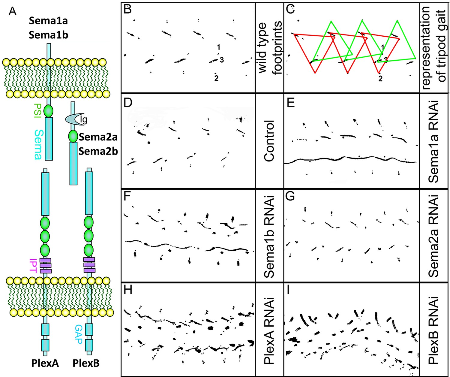
Targeted knockdown of Plexin/Semaphorin signaling in motoneurons results in walking defects.
(A) Schematic of interactions between Sema1a, Sema1b, and PlexA as well as between Sema2a, Sema2b, and PlexB. (B–I) Walking patterns monitored by footprint tracking in control and Plexin/Semaphorin RNAi knockdowns targeted to motoneurons. (B, C) Wild-type walking pattern. 1, 2 and 3 denote footprints of first, second, and third leg, respectively. Colored triangles denote the footprints of legs in the stance phase, where three legs are on the ground at any given time, representing tripod gait. The front leg and hind leg from one side move together with the middle leg on the opposite side. This alternates when the fly takes a step forward, represented by red and green triangles. Defective walking patterns are observed in (E) Sema1a, (F) Sema1b, (H) PlexA, (I) PlexB, but not in (G) Sema2a knockdowns.
-
Figure 1—source data 1
Summary of walking behavior upon Plexin/Semaphorin knockdown in motoneurons.
- https://doi.org/10.7554/eLife.11572.004
Figure 2 with 2 supplements
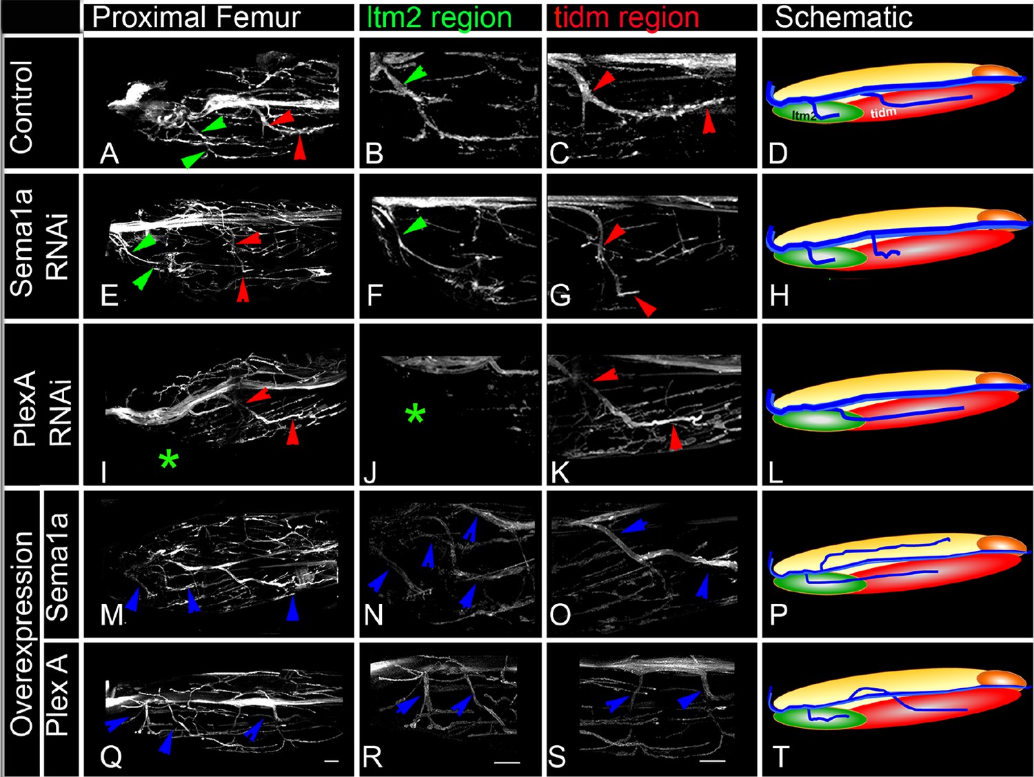
PlexA and Sema1a are required for correct axonal projections of leg motoneurons in the proximal femur.
Targeted knockdown, overexpression, and labeling mediated by motoneuron-specific OK371-Gal4 driver. (A–D) Control innervation of femur. Axon projection defects characterized by decreased innervation are observed in (E–H) Sema1a knockdown and (I–L) PlexA knockdown. Extensive defasciculation and ectopic branches exiting the main motor nerve are observed in (M–P) Sema1a overexpression and (Q–T) PlexA overexpression. (A, E, I, M, Q) show overview of proximal femur innervation. (B, F, J, N, R) show magnified views of innervation in ltm2 (long tendon muscle 2) region. (C, G, K, O, S) show magnified views of innervation of proximal tidm (tibia depressor muscle). (D, H, L, P, T) show schematic summaries of femur innervation of these two nerves. ltm2 muscle in schematic marked by green and tidm in red. The main nerve innervating ltm2 region is outlined by green arrowheads; main nerve innervating tidm outlined by red arrowheads. Green asterisk denotes absence of innervation. Multiple nerves innervating ltm2 and tidm in over-expression outlined by blue arrowheads. Scale bars = 20 microns.
Figure 2—figure supplement 1
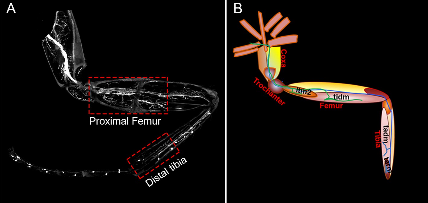
Axonal innervation of motoneurons in the leg.
https://doi.org/10.7554/eLife.11572.006
Figure 2—figure supplement 2

Plex A and Sema1a are required for correct axonal fasciculation of leg motoneurons in the proximal femur.
Defective defasciculation in Sema1a and PlexA knockdown was accompanied by increased thickness of nerve’s main axon while as extensive defasciculation observed in Sema1a and PlexA overexpression was accompanied by correspondingly reduced thickness of the nerve’s main axon.
Figure 3 with 1 supplement
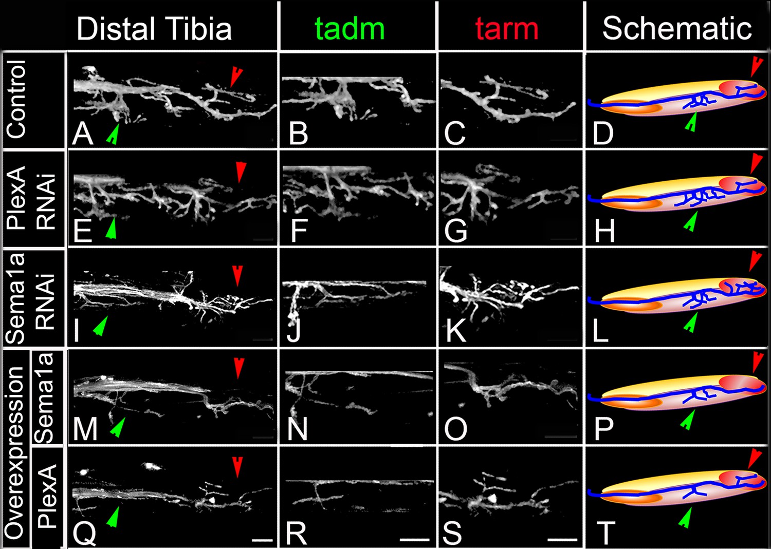
PlexA and Sema 1a are required for correct axonal projections of leg motoneurons in the distal tibia.
Targeted knockdown, overexpression, and labeling mediated by motoneuron-specific OK371-Gal4 driver. (A–D) Control innervation of distal tibia. Axon projection defects characterized by increased innervation observed in (E–H) PlexA RNAi knockdown and (I–L) Sema1a RNAi knockdown. Decreased defasciculation of axonal branches exiting the main motor nerve are observed in (M–P) Sema1a overexpression and (Q–T) PlexA overexpression. (A, E, I, M, Q) show overviews of distal tibia innervation. (B, F, J, N, R) show magnified views of innervation of tadm (tarsus depressor muscle). (C, G, K, O, S) show magnified views of innervation of tarm (tarsus reductor muscle). (D, H, L, P, T) show schematic summaries of tibia innervation. Green arrowheads point toward innervations in tadm and red arrowheads point toward tarm. Scale bars = 20 microns.
-
Figure 3—source data 1
Summary of the axon defasciculation and targeting phenotypes in proximal femur and distal tibia. File contains underlying source data for Figure 3—figure supplement 1.
- https://doi.org/10.7554/eLife.11572.009
Figure 3—figure supplement 1

Summary of the axon defasciculation and targeting phenotypes mediated by Sema1a and PlexA, in ways that differ in the proximal femur and distal tibia.
Bar graphs denote the percentage of legs showing defective motoneuron (MN) defasciculation and targeting phenotypes. Sema1a and PlexA knock down in motoneurons results in (A) decreased defasciculation and innervation of motoneuron axons in proximal femur, (B) increased defasciculation and innervation motoneuron axons in distal tibia. Sema1a and PlexA over-expression in motoneurons results in (C) increased defasciculation and innervation in proximal femur and (D) decreased defasciculation and innervation in distal tibia.
Figure 4 with 3 supplements
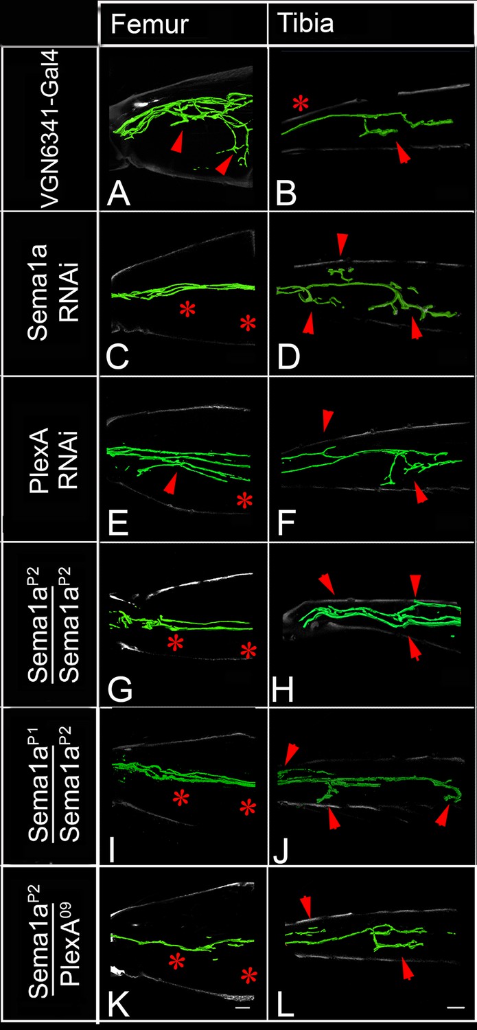
PlexA and Sema1a are required in femur motoneurons to maintain appropriate innervation of the femur and prevent inappropriate innervation of the tibia.
VGN6341-Gal4 targeted knockdown of PlexA and Sema1a in a small subset of leg motoneurons. (A, C, E, G, I, K) innervation of proximal femur. (B, D, F, H, J, L) innervation of tibia. (A, B) Control innervation by targeted motoneurons. (C, D) Sema1a knockdown in targeted motoneurons. (E, F) PlexA knockdown in targeted motoneurons. (G, H) Sema1aP2 homozygous mutant. (I, J) Sema1aP1/Sema1aP2 heteroallelic combination. (K, L) Sema1ap2/+;;PlexA09/+ trans-heterozygous mutant combination. Decreased innervation occurs in femur (C, E, G, I, K) and ectopic innervation occurs in tibia (D, F, H, J, L). Arrowheads point toward innervation and asterisk denote absence of normal innervation. Scale bars = 20 microns.
-
Figure 4—source data 1
Summary of the motoneuron axon defasciculation and targeting phenotypes in early and late-born motoneurons. File contains underlying source data for Figure 4—figure supplement 1.
- https://doi.org/10.7554/eLife.11572.012
Figure 4—figure supplement 1
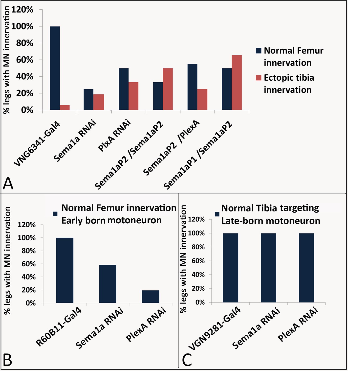
Summary of the motoneuron axon defasciculation and targeting phenotypes mediated by Sema1a and PlexA in early and late-born motoneurons.
Bar graphs denote the percentage of legs showing normal motoneuron (MN) defasciculation and targeting. (A) VGN6341-Gal4 labeled motoneurons show mis-projection phenotypes characterized by lack of innervation in ltm2, tidm muscles of the femur and ectopic innervation in ltm1, tadm muscles of the tibia in Sema1a knockdown, PlexA knockdown, Sema1a homozygous null mutant, Sema1a heteroallelic mutant combination and Sema1a/PlexA trans-heterozygous mutants. (B) R60B11-Gal4 mediated knockdown of Sema1a and PlexA results in decreased defasciculation and targeting of early-born motoneurons that innervate proximal femur. (C) VGN9281-Gal4 mediated knockdown of Sema1a and Plex A in one late-born motoneuron, does not affect the motoneuron axon targeting in distal tibia.
Figure 4—figure supplement 2
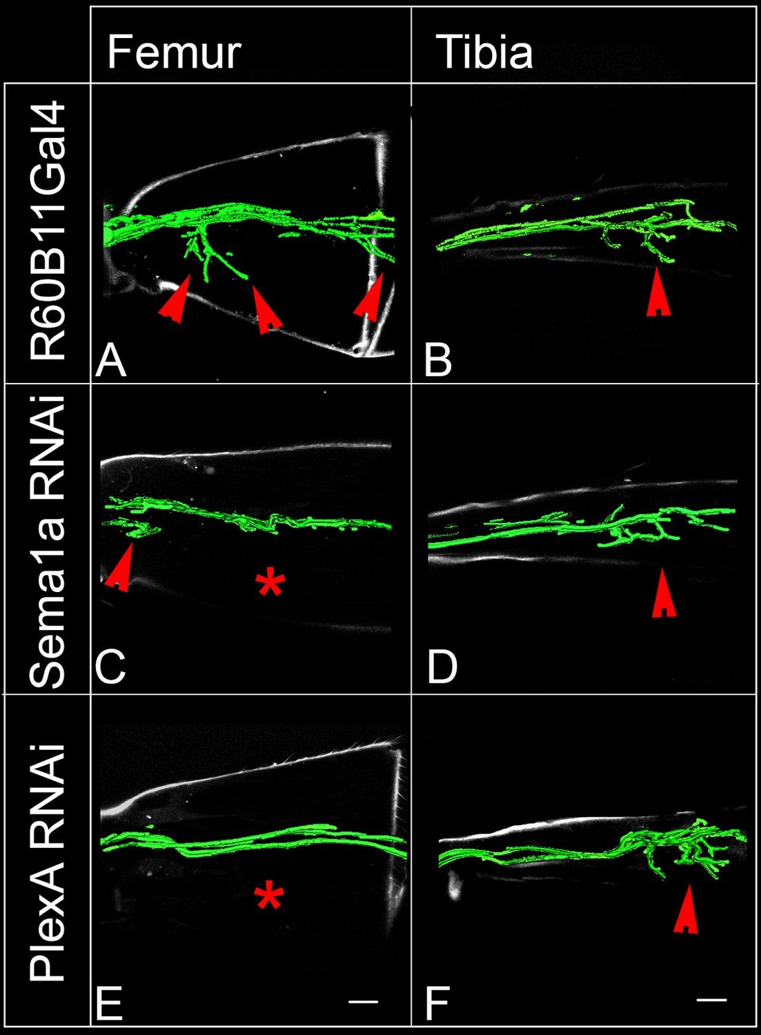
PlexA and Sema1a are required in femur motoneurons for axonal targeting and defasciculation.
R6B011-Gal4 targeted knockdown of PlexA and Sema1a in a small subset of leg motoneurons. (A, C, E) innervation of proximal femur. (B, D, F) innervation of tibia. (A, B) Control innervation by targeted motoneurons. Decreased innervation occurs in femur following (C, D) Sema1a knockdown in targeted motoneurons and (E, F) PlexA knockdown in targeted motoneurons. Arrowheads point toward innervation and asterisk denote absence of normal innervation. Scale bars = 20 microns.
Figure 4—figure supplement 3
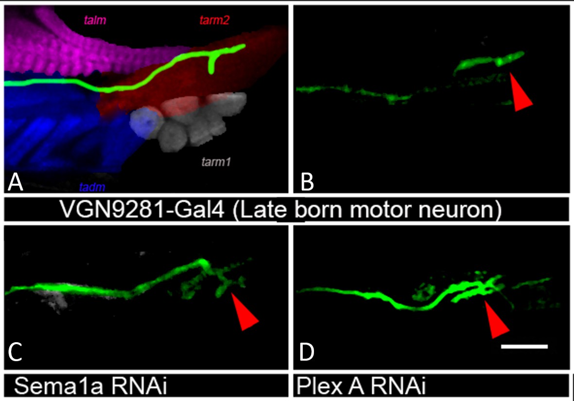
Plex A and Sema1a are not required in tibia late-born motoneurons for axonal targeting.
VGN9281-Gal4 targeted knockdown of PlexA and Sema1a in a late-born leg motoneuron. (A, B) Control innervation by targeted motoneuron. (C) Sema1a knockdown and (D) PlexA knockdown in targeted motoneuron.
Figure 5 with 1 supplement

Sema2a is highly expressed in the ganglionic midline and intermediate region.
Immunocytochemical analysis of expression of Sema1a and Sema2a in prothoracic ganglion at 25h APF (after puparium formation). (A–C) Ventral section. (D–F) Dorsal section. (A, D) Sema1a immunolabeling. (B, E) Sema2a immunolabeling. (C, F) Overlay of Sema1a and Sema2a immunolabeling. (G) Magnified view of neuropile. (H) Intensity profile of Sema1a and Sema2a immunolabeling taken along mediolateral axis of neuropile (in Z-stack; overlay of all optical sections). Yellow asterisk is placed at the ganglionic midline (ML). Sema2a level is highest at the midline and high in the intermediate regions in the neuropile and very low at the lateral edge. Scale bar = 20 microns.
Figure 5—figure supplement 1

Dorso-ventral view of Sema2a expression in the thoracic neuropil.
(A) 3D- reconstruction of Sema1a and Sema2a expression along dorso-ventral axis reveals opposing gradients of Sema1a and Sema2a along dorso-ventral (D-V) and medio-lateral (M-L) axis in prothoracic ganglion at 25h APF. (B) Intensity profile of Sema1a and Sema2a immunolabeling taken along medio-lateral axis of neuropile representing dorsal and ventral optical sections.
Figure 6 with 2 supplements
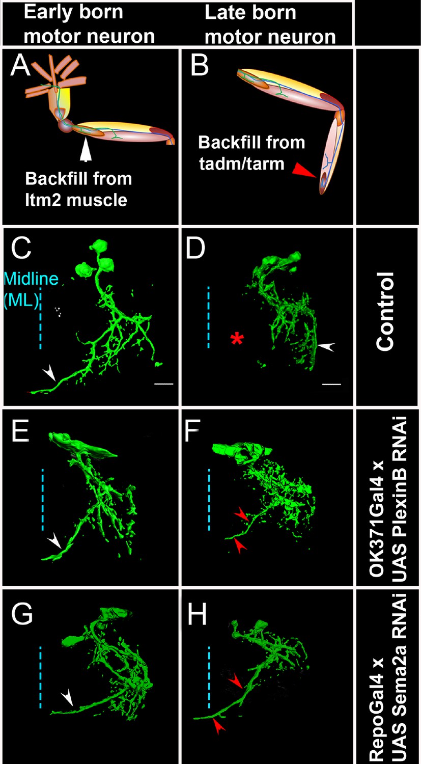
PlexB and Sema2a are required for correct dendritic targeting of leg motoneurons.
Motoneuron dendrites in prothoracic hemiganglion labeled by dye backfilling from innervated muscles. (A, C, E, G) early born motoneurons that innervate ltm2. (B, D, F, H) late born motoneurons that innervate tadm. (A, B) Schematic representation of dye backfilled leg muscles. (C, D) Dendrites of motoneurons in wild-type control. (E, F) Dendrites of motoneurons following motoneuron-specific targeted knockdown of PlexB. (G, H) Dendrites of motoneurons following glial-cell-specific targeted knockdown of Sema2a. A dendritic mis-projection phenotype occurs in late-born motoneurons (F, H) but not in early-born motoneurons (E, G). White arrowhead indicates normal dendritic projection and red arrowhead indicates mis-projection. Red asterisk marks the region where projection is normally absent in late-born neurons. Scale bars =20 microns.
Figure 6—figure supplement 1
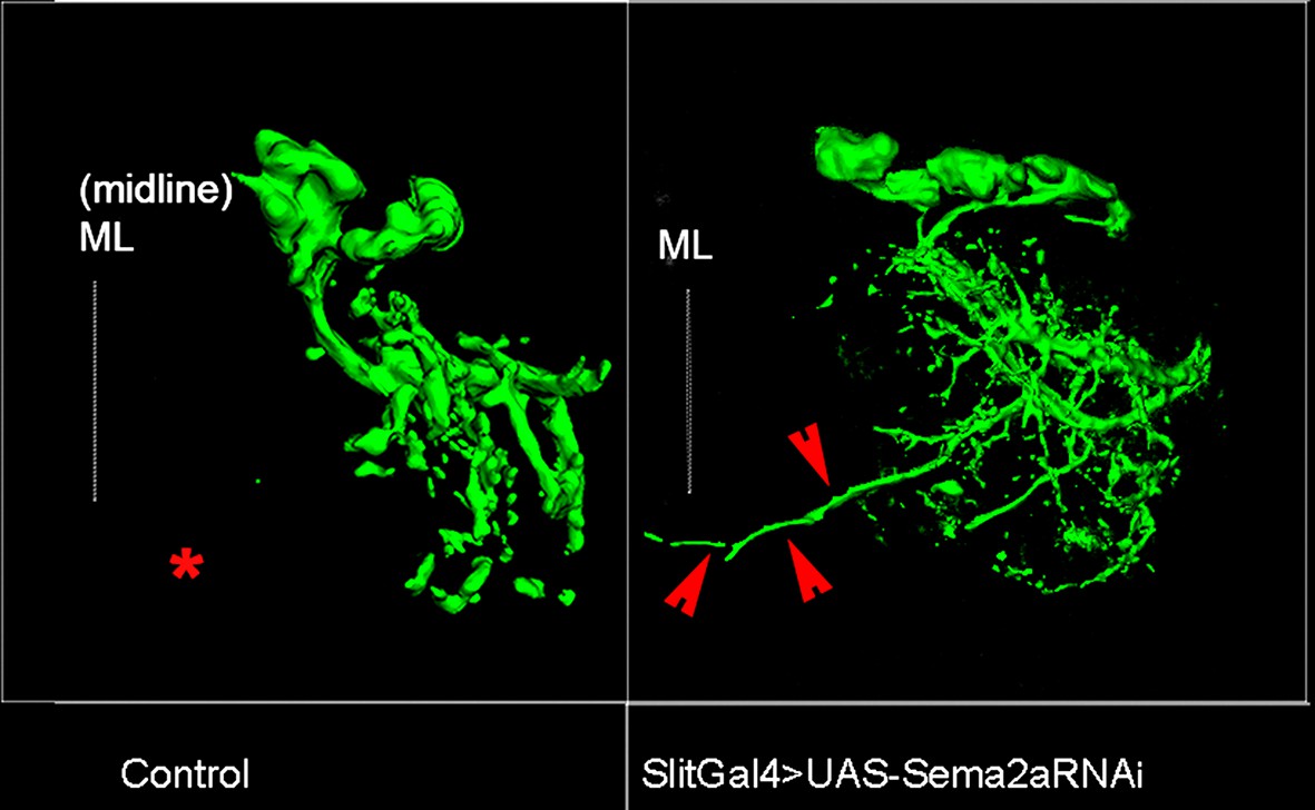
Sema2a secreted by midline cells is required for dendritic targeting of late-born motoneurons.
Dendrites of late-born motoneurons mis-project toward the midline when Sema2a is knocked down in midline cells. Asterisk marks the region near the midline where dendritic projections of late-born motoneurons are normally absent. Mis-projected branch outlined by red arrowheads.
Figure 6—figure supplement 2
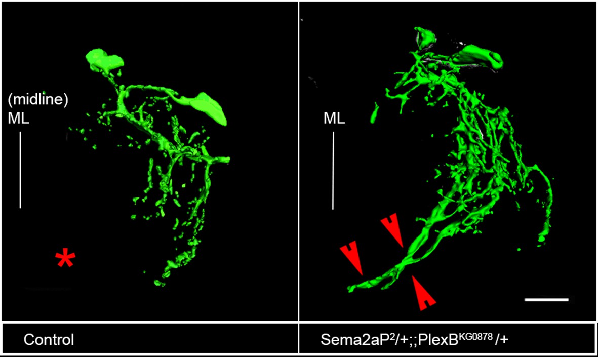
Plex B/Sema2a is required for correct dendritic targeting of late-born motoneurons.
Dendrites of late-born motoneurons mis-project toward the midline. Asterisk marks the region near the midline where dendritic projections of late-born motoneurons are normally absent. Mis-projected branch outlined by red arrowheads. Scale bar = 20 microns.
Download links
A two-part list of links to download the article, or parts of the article, in various formats.
Downloads (link to download the article as PDF)
Open citations (links to open the citations from this article in various online reference manager services)
Cite this article (links to download the citations from this article in formats compatible with various reference manager tools)
Glial and neuronal Semaphorin signaling instruct the development of a functional myotopic map for Drosophila walking
eLife 5:e11572.
https://doi.org/10.7554/eLife.11572


