A G-protein activation cascade from Arl13B to Arl3 and implications for ciliary targeting of lipidated proteins
- Max Planck Institute of Molecular Physiology, Germany
- University of Erlangen-Nuremberg, Germany
Figures
Figure 1
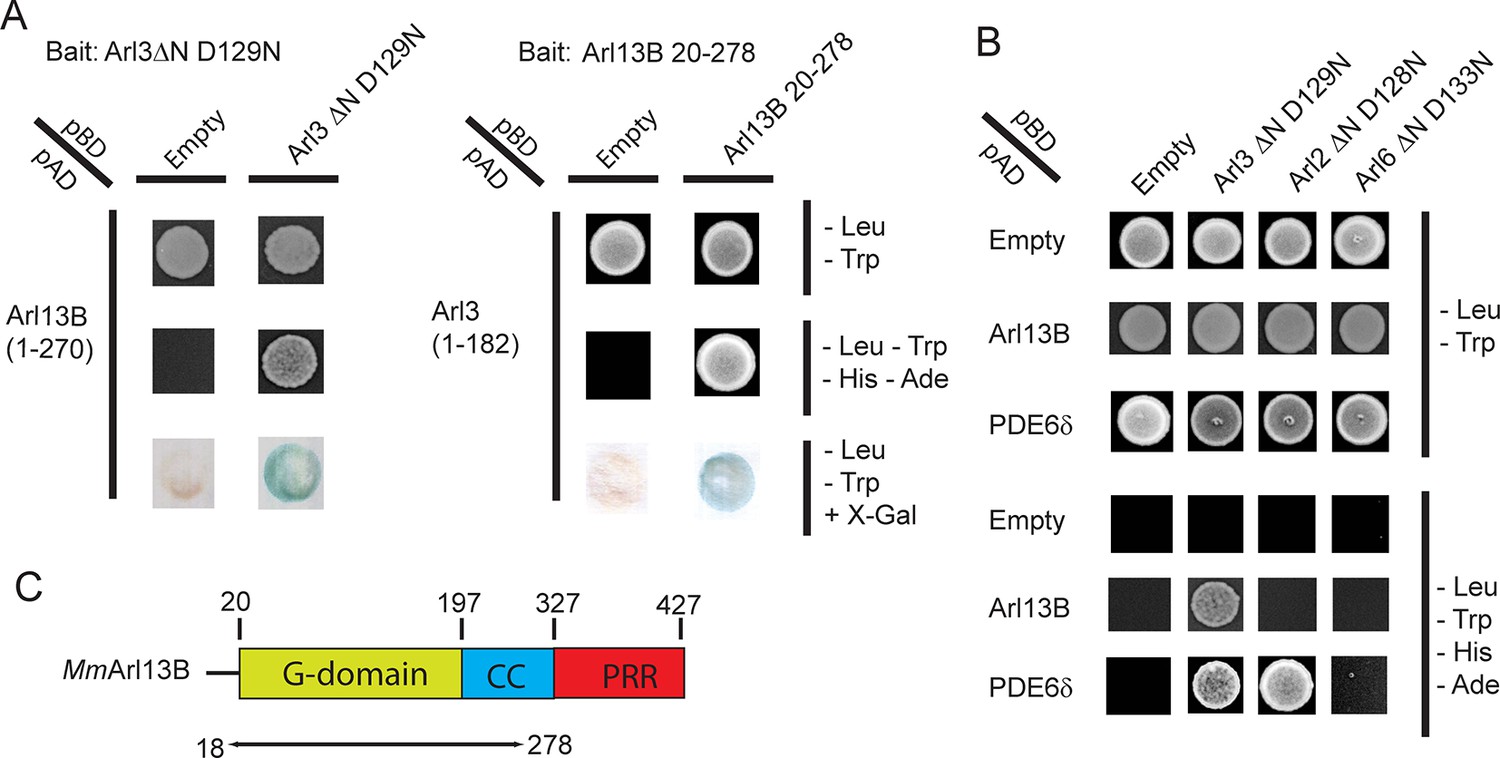
The interaction between ADP-ribosylation-factor-like (Arl) 13B (Arl13B) and Arl3 was identified in a yeast-2-hybrid (Y2H) screen.
(A) Y2H interactions between Arl3ΔN D129N -pBD and Arl13B 1-–270-pAD and between Arl13B 20- – 278-pBD and Arl3-pAD. Transformed and mated cells were grown on –Leu –Trp medium. Interaction was verified on high stringency plates (-– Leu –Trp –His –Ade) and with a β-galactosidase filter assay. (B) Interaction of Arl13B 1-–270-pAD with Arl3ΔN D129N-pBD, Arl2ΔN D128N-pBD and Arl6ΔN D133N-pBD was analyzed on low and high stringency plates. PDE6δ-pAD was used as positive control for Arl3 and Arl2. (C) Domain architecture of Arl13B, numbering derived from murine Arl13B (Mm: Mus musculus).
Figure 2
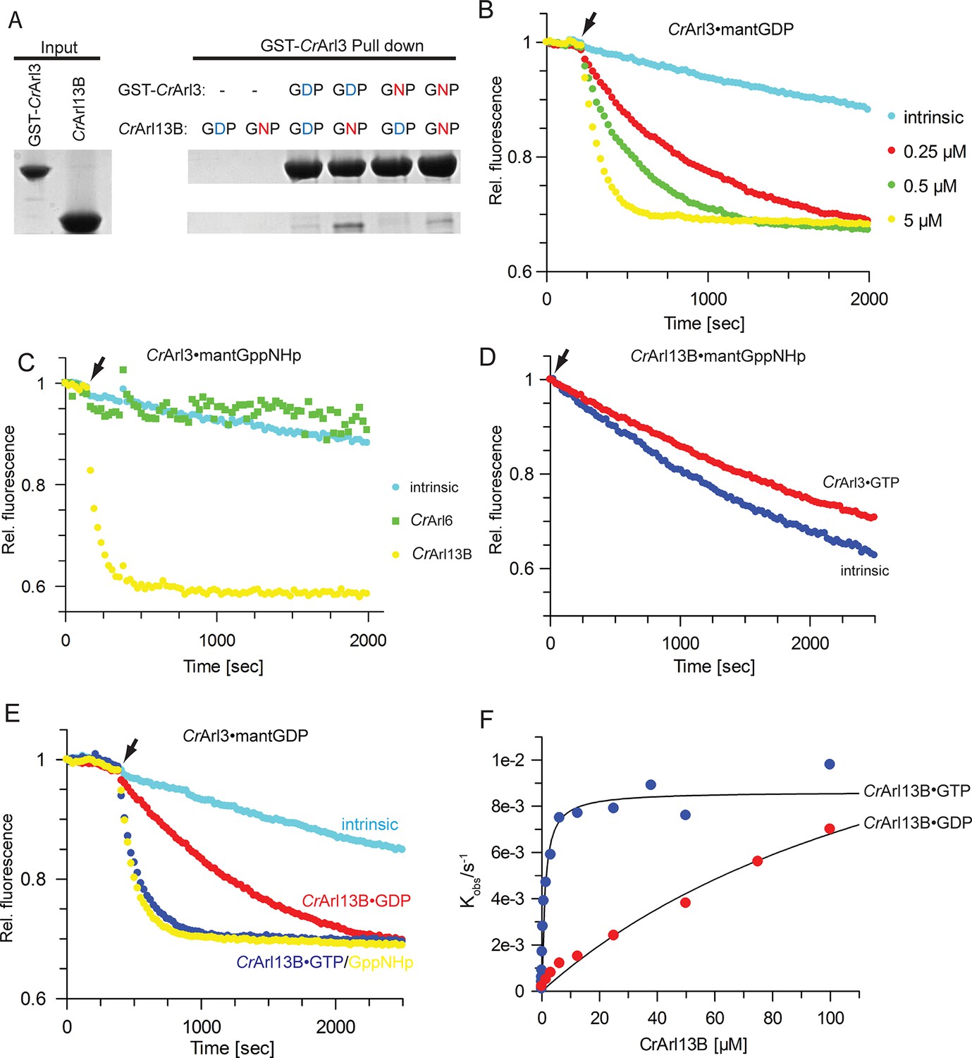
CrArl13B is the guanine nucleotide exchange factor for CrArl3.
(A) Glutathione-S-transferase (GST) pull-down assay with purified Chlamydomonas reinhardtii Arl proteins as indicated and described in detail in Material and methods. (B) Guanine nucleotide exchange factor (GEF) activity of the indicated concentrations of CrArl13B18-–278 for 500 nM CrArl3·mantGDP. Arrow designates addition of CrArl13B and excess of unlabeled nucleotide. (C) CrArl13B·GppNHp but not CrArl6·GppNHp stimulates the nucleotide release of CrArl3·mantGppNHp. (D) CrArl3·GTP does not accelerate the nucleotide dissociation of CrArl13B·mantGppNHp. (E) GEF activity of 5 µM CrArl13B18-278 loaded with GDP (red), GTP (blue), or GppNHp ((a non-hydrolyzable GTP analogue; yellow). (F) Hyperbolic dependence of the observed rate constants for mantGDP release from 500 nM CrArl3 on CrArl3B·GTP or CrArl13B·GDP concentration. Fluorescence changes in time at each concentration of CrArl13B were fitted to single exponentials, and the resulting rate constants (kobs) plotted against GEF concentration. Kobs values are summarized in Table 1.
Figure 3
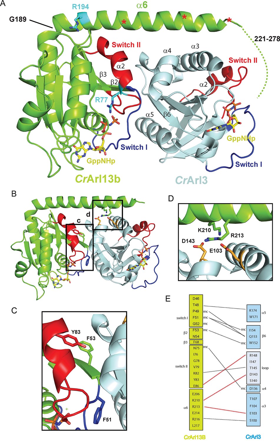
The CrArl13B-– CrArl3 complex.
(A) The CrArl13B-–CrArl3 complex structure with Arl13B (green), Arl3 (light blue), Switch I (blue), Switch II (red), GppNHp (a non-hydrolyzable (GTP) analogue; yellow). Residues analogous to Joubert syndrome mutations (R77 and R194) are depicted in cyan. Red asterisks delineate the deletion sites (V202, E212, K219) of CrArl13 used in the guanine nucleotide exchange factor (GEF) assay below (Figure 4). Other deletion sites are not resolved in the electron density. Dashed line indicates the 58 C-terminal residues not visible in the structure (B–D) Details of the interaction interface. (C) Hydrophobic residues located in Switch I and Switch II of CrArl13B are involved in the interaction with CrArl3. (D) K210 and R213 in α6Arl13B are forming salt bridges with D143Arl3 and E103Arl3(orange). Coloring as in (A). (E) Schematic representation of residues located in the interface. Hydrogen bonds between residues are depicted as black dashed line, salt bridges as red dashed line.
Figure 4
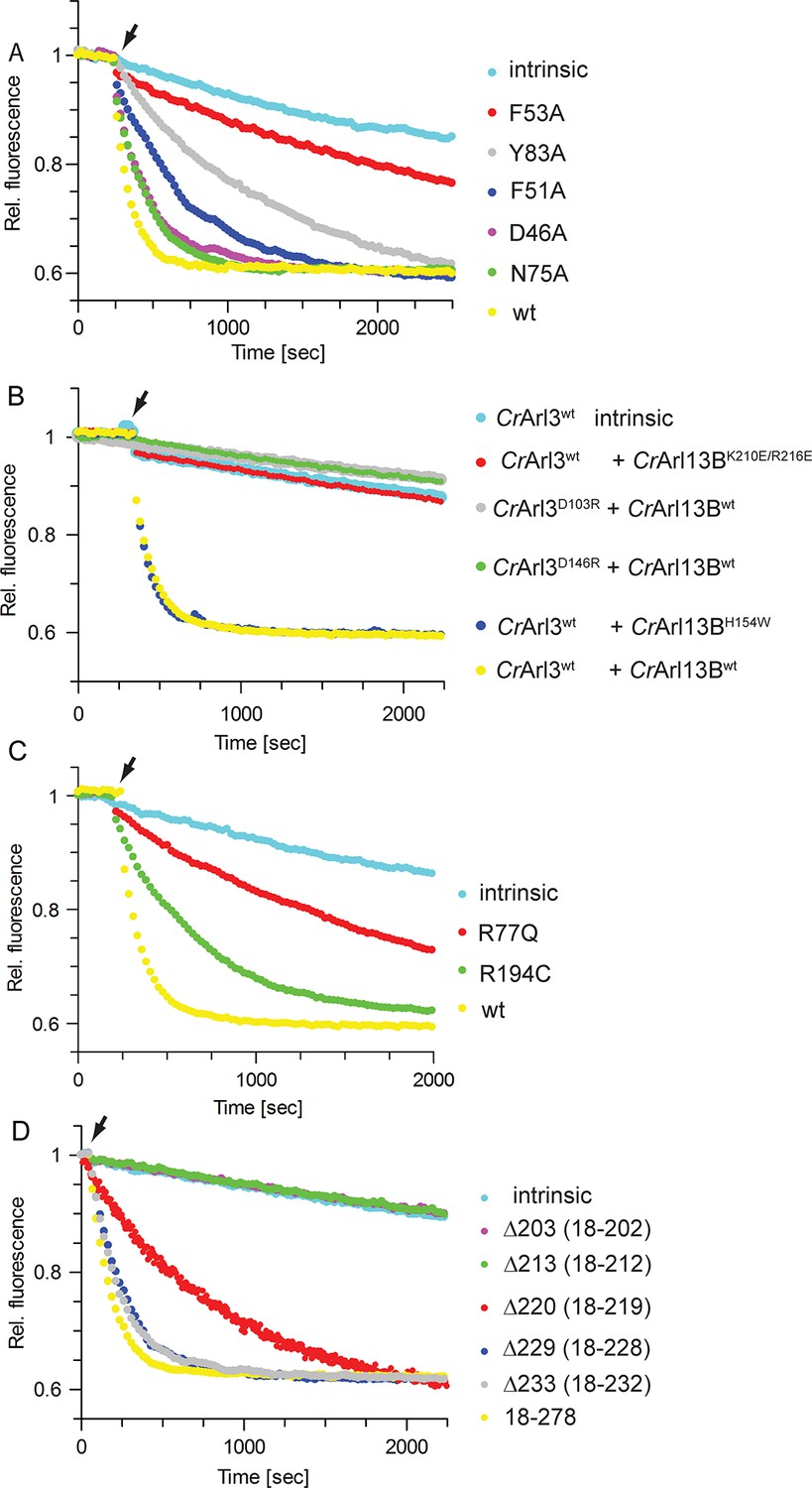
Mutations in the CrArl13B-CrArl3 interface and Joubert mutations impair guanine nucleotide exchange factor (GEF) activity.
(A) GEF activity of CrArl13B18-– 278·GppNHp (a non-hydrolyzable GTP analogue) switch I and II mutants. To CrArl3 mantGppNHp (500 nM) 5 µM of CrArl13B·GppNHp constructs and 800 µM unlabeled GppNHp were added. (B) GEF assay with CrArl13B18-278·GppNHpand CrArl3·mantGppNHp carrying charge reversal mutations located in the interface. (C) GEF activity of the analogous Joubert syndrome mutants (CrArl13BR77Q, CrArl13BR194C). Same concentrations as in (A). (D) GEF assay with CrArl13B deletion constructs. Boundaries of deletion fragments: △203: 18–202; △213: 18–213; △220: 18–219; △229: 18–228; △233: 18–232. 18–278 are the constructs used for all other GEF assays. Kobs values are summarized in Table 4.
Figure 5

Arl13B activates Arl3 in mammalian cells.
(A) Endogenous Arl3·GTP was affinity-precipitated from Human Embryonic Kidney 293 (HEK293) or murine inner medullary collecting duct 3 (IMCD3) cell lysates using GST-PDE6δ and analyzed as described in Materials and methods. HEK293 cells were transiently transfected with full length Arl13B-GFP(pGLAP5); IMCD3 cells stably expressed the same construct. (B) HEK293 cells were transiently transfected with increasing amounts of Arl13B-GFP (0, 1, 3, 6, 12 µg DNA) and constant amounts of Arl3-Flag. Arl3·GTP level determined as in (A). (C) Arl3-Flag activation in the presence of wildtype and interface mutant Arl13B-GFP was determined as in (A) and quantified in (D). (E) Arl3-Flag activation in the presence Arl13B wt and Joubert syndrome mutants R79Q and R200C. (F) Quantification of (E). Data is represented as mean ± S.E. (G) Arl3-Flag and Arl2-Flag activation in the presence of Arl13B-GFP in HEK293 cells.
Figure 6
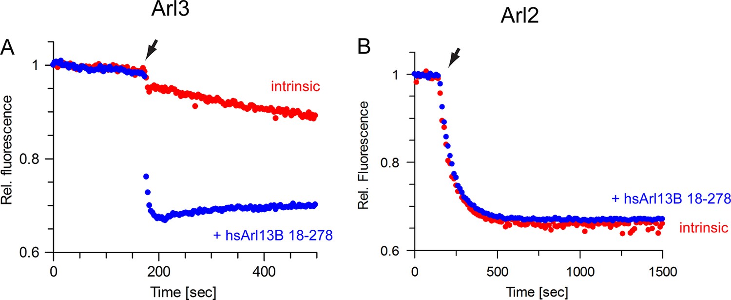
The guanine nucleotide exchange factor (GEF) activity of human Arl13B is specific for Arl3.
(A) GEF activity of human Arl13B18-–278 (purified from insect cells) for murine Arl3. To 500 nM Arl3·mantGppNHp, 5 µM hsArl13B·GTP and 800 µM GTP were added. kobs (intrinsic): 4 × 10⁻⁴ s-1, kobs(Arl13B·GTP): 0,36 s-1. (B) Human Arl13B·GTP does not accelerate nucleotide dissociation of Arl2·mantGppNHp. kobs(intrinsic):1.2 × 10⁻² s-1; kobs(Arl13B·GTP): 1.2 × 10⁻² s-1.
Figure 7
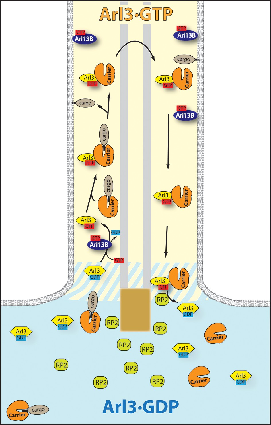
The targeting cycle of Arl3 dependent ciliary cargo.
In the cilium where Arl13B resides Arl3 gets activated. Through the exclusive localization of Arl13B (Arl3-GEF [guanine nucleotide exchange factor]) inside and retinitis pigmentosa 2 (RP2) (Arl3-GAP) outside the cilium an Arl3·GTP gradient is generated across the transition zone. The carriers PDEδ and Unc119a/b bound to ciliary lipidated cargo reach the cilium where Arl3·GTP binds to the carrier proteins and releases the cargo. RP2 -– enriched in the preciliary region – stimulates the hydrolysis of Arl3·GTP which leads to the dissociation of the carrier proteins from Arl3·GDP.
Tables
Table 1
Kobs values from data shown in Figure 2B and E.
| Concentration dependency (Figure 2B) | Kobs (s-1) ± S.E. |
|---|---|
| CrArl3 wt intrinsic | 1.2 × 10⁻⁴ ± 1 × 10⁻⁵ |
| + 0.25 µM CrArl13BGTP | 1.3 × 10⁻³± 2 × 10⁻⁵ |
| + 0.5 µM CrArl13BGTP | 2.7 × 10⁻³± 3 × 10⁻⁵ |
| + 5 µM CrArl13BGTP | 0.85 × 10⁻²± 1 × 10⁻⁴ |
| Nucleotide dependency (Figure 2E) | Kobs (s-1) ± S.E. |
| CrArl3 wt intrinsic | 1.3 × 10⁻⁴ ± 4 × 10⁻⁶ |
| + 5 µM CrArl13BGDP | 9.0 × 10⁻⁴ ± 2 × 10⁻⁵ |
| + 5 µM CrArl13BGTP | 0.6 × 10⁻² ± 8 × 10⁻⁵ |
| + 5 µM CrArl13BGNP | 0.78 × 10⁻² ± 1 × 10⁻⁴ |
-
Kobs values ± standard error (S.E.) were determined by fitting the data to single exponential functions.
Table 2
kobs values for the nucleotide dissociation of CrArl3·mGDP and CrArl3·mGppNHp in the presence of CrArl13B·GTP.
| CrArl3·mGDP vs mGppNHp | Kobs (s-1) ± S.E. |
|---|---|
| CrArl3·mGDP intrinsic | 1.3 × 10⁻⁴ ± 2 × 10⁻⁶ |
| CrArl3·mGDP + 5 µM CrArl13B·GTP | 0.84 × 10⁻² ± 7 × 10⁻⁵ |
| CrArl3·mGppNHp intrinsic | 1.3 × 10⁻⁴ ± 2 × 10⁻⁶ |
| CrArl3·mGppNHp + 5 µM CrArl13B·GTP | 1.0 × 10⁻² ± 1 × 10⁻⁴ |
-
kobs rates determined from GEF assays with 0.5 µM CrArl3 loaded with either mantGDP or mantGppNHp in the presence of 5 µM CrArl13B18-–278·GTP and 800 µM unlabeled nucleotide.
Table 3
Data collection and refinement statistics (molecular replacement).
| CrArl13B-CrArl3 (5DI3) | |
|---|---|
| Data collection | |
| Space group | P212121 |
| Cell dimensions | |
a, b, c (Å) | 57.10, 68.80, 120.00 |
α, β, γ (°) | 90.00, 90.00, 90.00 |
| Resolution (Å) | 29.84 – 2.50 (2.60-2.50) |
| Rmerge | 0.07 (0.68) |
| I / σI | 17.56 (3.26) |
| Completeness (%) | 99.9 (99.9) |
| Redundancy | 6.4 (6.8) |
| Refinement | |
| Resolution (Å) | 2.50 |
| No. reflections | 16944 (1840) |
| Rwork/Rfree | 0.199/0.236 |
| No. atoms | 2995 |
Protein | 2900 |
Ligand/ion | 2 Mg2+, 2 GMPPNP |
Water | 29 |
| B-factors | 66 |
Protein | 66.40 |
Ligand/ion | 54.30 |
Water | 55.00 |
| R.m.s. deviations | |
Bond lengths (Å) | 0.005 |
Bond angles (°) | 1.02 |
-
*Values in parentheses are for highest-resolution shell.
Table 4
Kobs values from data shown in Figure 4 A–D.
| CrArl13B Switch interface mutants | Kobs (s-1) ± S.E. |
|---|---|
| CrArl3 intrinsic | 1.4 × 10⁻⁴ ± 4 × 10⁻⁶ |
| + 5 µM CrArl13B wt GTP | 0.91 × 10⁻² ± 2 × 10⁻⁴ |
| + 5 µM CrArl13B F51A GTP | 2.0 × 10⁻³ ± 2 × 10⁻⁵ |
| + 5 µM CrArl13B F53A GTP | 4.2 × 10⁻⁴ ± 2 × 10⁻⁵ |
| + 5 µM CrArl13B Y83A GTP | 0.9 × 10⁻³ ± 1 × 10⁻⁵ |
| + 5 µM CrArl13B D46A GTP | 4.1 × 10⁻³ ± 8 × 10⁻⁵ |
| + 5 µM CrArl13B N75A GTP | 4.4 × 10⁻³ ± 3 × 10⁻⁵ |
| CrArl13B and CrArl3 Interface mutants | Kobs (s-1) ± S.E. |
| CrArl3 wt intrinsic | 1.1 × 10⁻⁴ ± 1 × 10⁻⁶ |
| CrArl3 wt + 5 µM CrArl13B K210E/R216E | 1.5 × 10⁻⁴ ± 1 × 10⁻⁵ |
| CrArl3 D103R + 5 µM CrArl13B wt | 1.4 × 10⁻⁴ ± 5 × 10⁻⁶ |
| CrArl3 D146R + 5 µM CrArl13B wt | 1.4 × 10⁻⁴ ± 6 × 10⁻⁶ |
| CrArl3 wt + 5 µM CrArl13B H154W | 0.88 × 10⁻² ± 2 × 10⁻⁴ |
| CrArl3 wt + 5 µM CrArl13B wt | 0.85 × 10⁻² ± 2 × 10⁻⁴ |
| CrArl13B Deletion constructs | Kobs (s-1) ± S.E. |
| CrArl3 wt intrinsic | 1.0 × 10⁻⁴ ± 2 × 10⁻⁵ |
| + 5 µM CrArl13B △203 | 1.0 × 10⁻⁴ ± 8 × 10⁻⁶ |
| + 5 µM CrArl13B △213 | 1.0 × 10⁻⁴ ± 1 × 10⁻⁵ |
| + 5 µM CrArl13B △220 | 1.1 × 10⁻³ ± 1 × 10⁻⁵ |
| + 5 µM CrArl13B △243 | 4.5 × 10⁻³ ± 5 × 10⁻⁵ |
| + 5 µM CrArl13B △233 | 5.0 × 10⁻³ ± 6 × 10⁻⁵ |
| + 5 µM CrArl13B 18-278 | 6.6 × 10⁻³ ± 2 × 10⁻⁴ |
| CrArl13B Joubert mutants | Kobs (s-1) ± S.E. |
| CrArl3 intrinsic | 1.4 × 10⁻⁴ ± 3 × 10⁻⁶ |
| + 5 µM CrArl13B R77Q | 5.5 × 10⁻⁴ ± 1 × 10⁻⁵ |
| + 5 µM CrArl13B R194C | 2.0 × 10⁻³ ± 2 × 10⁻⁵ |
| + 5 µM CrArl13B wt | 0.72 × 10⁻² ± 1 × 10⁻⁴ |
-
Kobs values were determined by fitting the data (Figure 4 A-D) to single exponential functions. If not stated otherwise CrArl13B 18-278 is used for the measurements.
Download links
A two-part list of links to download the article, or parts of the article, in various formats.
Downloads (link to download the article as PDF)
Open citations (links to open the citations from this article in various online reference manager services)
Cite this article (links to download the citations from this article in formats compatible with various reference manager tools)
A G-protein activation cascade from Arl13B to Arl3 and implications for ciliary targeting of lipidated proteins
eLife 4:e11859.
https://doi.org/10.7554/eLife.11859


