Direct mechanical stimulation of tip links in hair cells through DNA tethers
- Howard Hughes Medical Institute, The Rockefeller University, United States
Figures
Figure 1
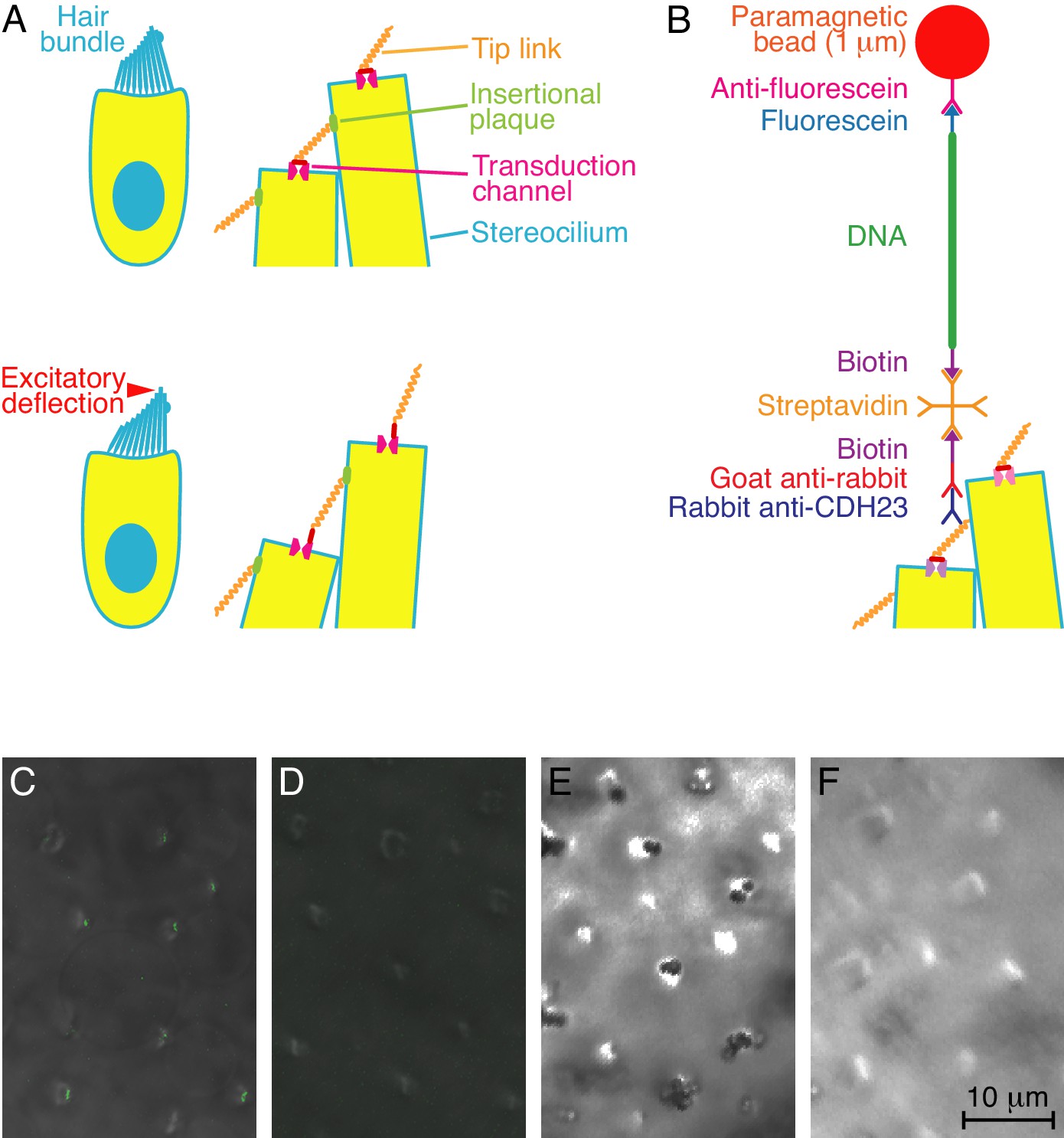
Experimental configuration and control experiments.
(A) In diagrams of a hair cell at rest (top) and during excitatory stimulation (bottom), the magnified images show how hair-bundle deflection is thought to raise the tension in tip links and thus open transduction channels. (B) A diagram, not to scale, portrays the molecular assembly that tethers a superparamagnetic bead to a tip link. (C) In overlaid fluorescence and differential-interference-contrast images of the apical surface of a bullfrog's sacculus, anti-CDH23 immunolabels the tops of hair bundles. (D) Labeling is absent under otherwise identical conditions when the primary antibody is omitted. (E) A differential-interference-contrast micrograph of a sacculus shows clusters of DNA-tethered superparamagnetic beads atop many hair bundles. (F) No beads are present in a control preparation lacking the primary antibody.
Figure 2
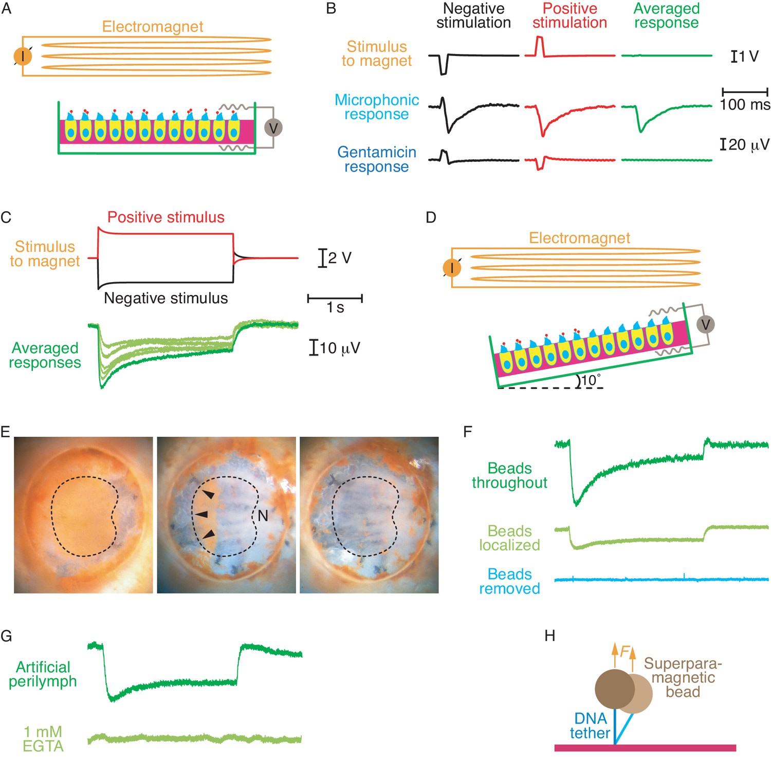
Electrical responses to magnetic stimulation.
(A) The diagram shows a sacculus mounted in a two-compartment chamber beneath an electromagnet. The otolithic membrane has been removed and superparamagnetic beads (red dots) are attached to the tip links through DNA tethers. (B) Brief magnetic stimuli of opposite polarities, which are denoted by the voltages applied to the magnet, elicit transepithelial electrical responses. Exposure of the preparation to 1 mM gentamicin blocks these physiological responses, leaving only artifacts that are eliminated by averaging the signals obtained from stimuli of opposite polarity. (C) Longer stimuli demonstrate the persistence of transduction currents without the extensive adaptation observed during conventional stimulation. The stimulus traces shown produced the largest response; successively smaller responses were generated by lowering the voltage supplied to the magnet in steps of 0.2 V. (D) To eliminate the possibility that magnetic stimulation pulls hair bundles in the excitatory direction, most of the beads can be removed and the preparation tilted by 10°. (E) Brown superparamagnetic beads initially cover the apical surface of a mounted sacculus (left). Scraping away most of the beads (center) leaves only the beads attached to hair bundles of similar orientations (arrowheads), sensitive to stimulation away from the saccular nerve (N). Further manipulation removes nearly all the beads (right). The dashed line circumscribes the region in which hair cells occur. (F) Although smaller than the control response (top), an electrical response nevertheless persists after removal of most beads (middle). With the beads localized to one region with similar hair-bundle orientations, magnetic force pulls those bundles upward and toward their short edges. Removing the remaining beads eliminates the response (bottom). The calibration bars in (C) apply to this panel as well. (G) Addition to the artificial perilymph in the top chamber of 1 mM EGTA, a Ca2+ chelator that dissociates tip links, abolished the electrical response. The calibration bars in (C) apply to this panel as well. (H) When a superparamagnetic bead is tethered by DNA to a glass surface through DNA, thermal motion causes lateral deflections that are partially suppressed by the magnetic force F.
Download links
A two-part list of links to download the article, or parts of the article, in various formats.
Downloads (link to download the article as PDF)
Open citations (links to open the citations from this article in various online reference manager services)
Cite this article (links to download the citations from this article in formats compatible with various reference manager tools)
Direct mechanical stimulation of tip links in hair cells through DNA tethers
eLife 5:e16041.
https://doi.org/10.7554/eLife.16041


