Boosting ATM activity alleviates aging and extends lifespan in a mouse model of progeria
- Shenzhen University Health Science Center, China
- Guangzhou Institutes of Biomedicine and Health, Chinese Academy of Sciences, China
- University of Hong Kong, China
- Tongji University School of Medicine, China
- Shanghai Tenth People's Hospital, Tongji University School of Medicine, China
- Tongji University, China
Figures
Figure 1 with 2 supplements
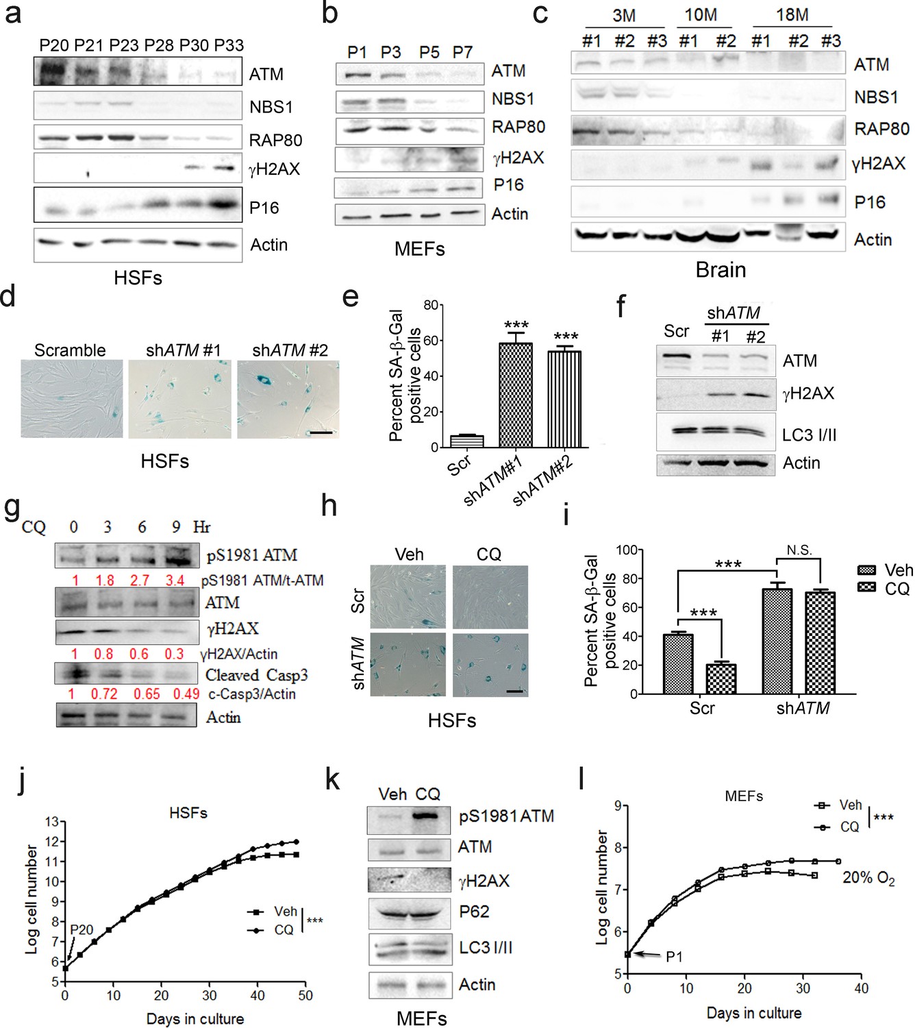
ATM activation by chloroquine alleviates senescence.
(a) Immunoblots showing protein levels of ATM, NBS1, and RAP80 in human skin fibroblasts (HSFs). A gradually increased level of p16 indicates cellular senescence, while elevated γH2AX level indicates accumulated DNA damage. (b) Immunoblots showing protein levels of ATM, NBS1, and RAP80 in mouse embryonic fibroblasts (MEFs). (c) Immunoblots showing protein levels of ATM, NBS1, and RAP80 in brain tissues isolated from 3-, 10-, and 18-month-old male mice. (d) SA-β-Gal staining in HSFs treated with sh-ATM or scramble shRNA. Scale bar, 100 µm. (e) Quantification of SA-β-Gal-positive staining of (d) from five views randomly captured for each group. Data represent means ± SEM. ***p<0.001. (f) Immunoblots showing increased γH2AX and unaffected LC3I/II in HSFs treated with sh-ATM or scramble shRNA. (g) Immunoblots showing protein levels of pS1981 ATM, γH2AX, and cleaved caspase-3 in HSFs treated with 10 μM of CQ for indicated time. (h) SA-β-Gal staining in HSFs expressing either scramble or ATM shRNA treated with 1 μM CQ or DMSO (12 hr). Scale bar, 100 µm. (i) Quantification of SA-β-Gal-positive staining of (h) from five views randomly captured for each group. Data represent means ± SEM. ***p<0.001; ‘N.S.’ indicates no significant difference. (j) HSFs at passage 20 were continuously cultured with 1 μM CQ or DMSO, and cell number was calculated at each passage. Data represent means ± SEM. ***p<0.01. (k) Immunoblots showing protein levels of γH2AX, p62, and LC3 in MEFs treated with 1 μM CQ or DMSO. Note that CQ had little effect on the expression levels of p62 and LC3. (l) MEFs at passage one were continuously cultured in 20% O2 with 1 μM CQ or DMSO, and cell number was determined at each passage. Data represent means ± SEM. ***p<0.01.
-
Figure 1—source data 1
Statistical analysis for SA-β-Gal positive staining.
- https://doi.org/10.7554/eLife.34836.006
-
Figure 1—source data 2
Statistical analysis for EdU positive staining.
- https://doi.org/10.7554/eLife.34836.007
Figure 1—figure supplement 1
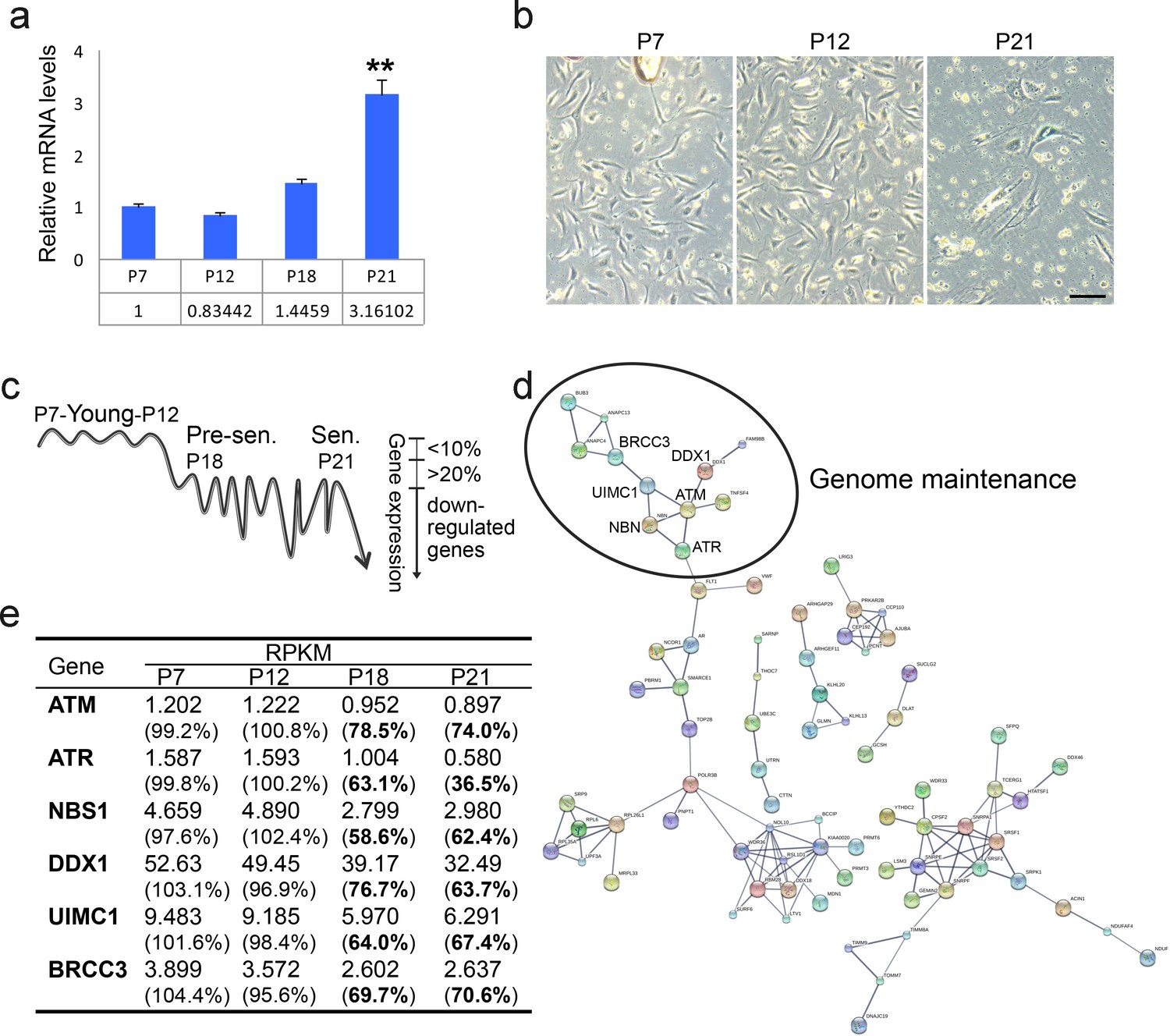
Decline of ATM-centered DNA repair machinery during senescence.
(a) Real-time PCR analysis showing progressively elevated mRNA level of p21 in continuously cultured human endothelial cells (HUVEC). **p<0.01. (b) SA-β-Gal staining of HUVEC cells at indicated passages. Scale bar, 100 µm. (c) HUVEC cells at P21, P18, P12, and P7 were subjected to transcriptome analysis. A minimum average rpkm value of 1.0 and maximum 10% fluctuation in young cells (P7 Vs P12) was set as the threshold. Genes were downregulated by more than 20% in pre-senescent, and senescent cells compared with young cells (P21/P18 Vs P12/P7) were selected. (d) Pathway analysis of genes identified in (c) by STRING v10. (e) Downregulation of ATM-related DNA repair genes during senescence.
Figure 1—figure supplement 2
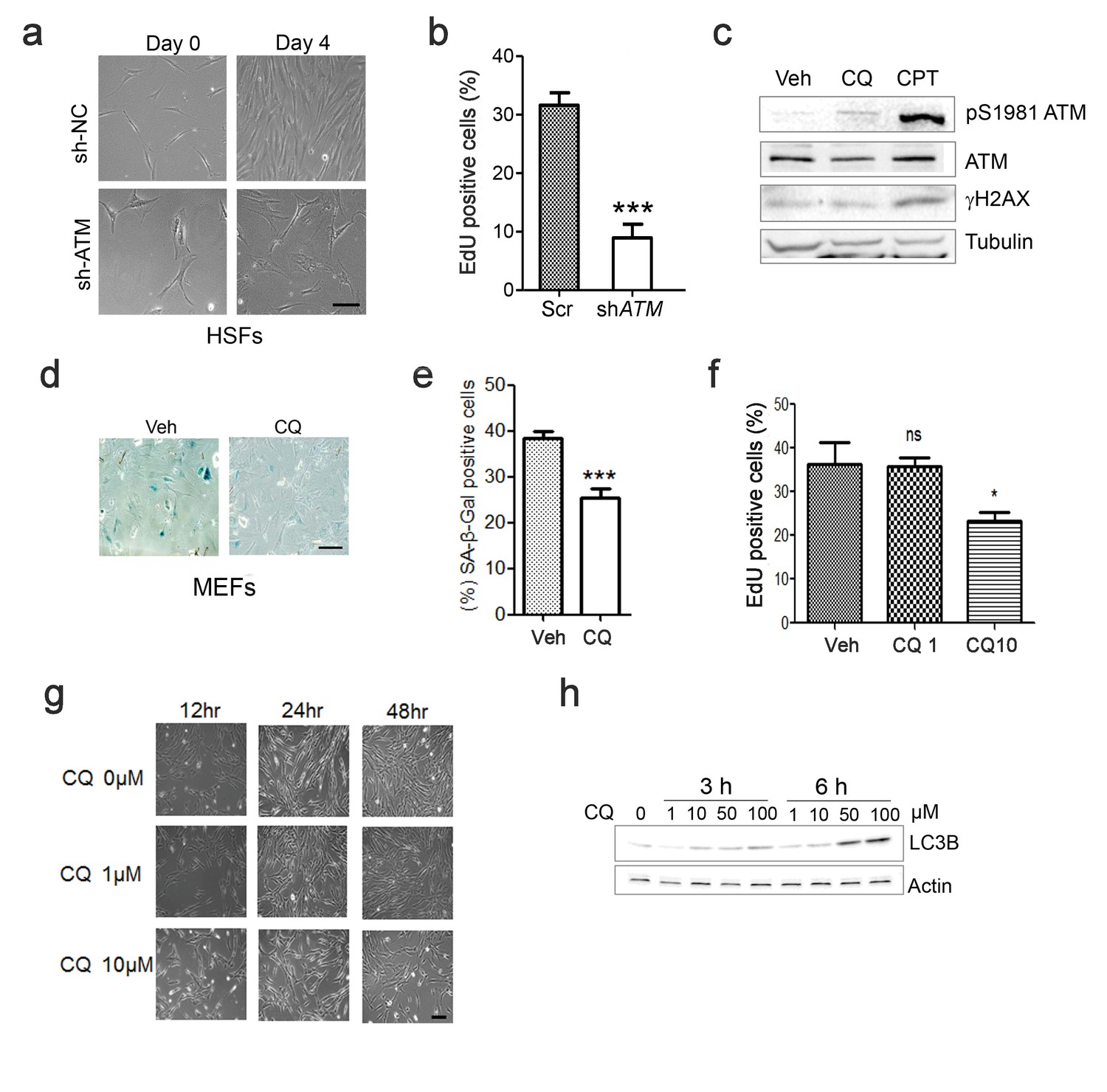
ATM regulates replicative senescence.
(a) Representative images showing cells treated with Scramble (sh-NC) or sh-ATM. (b) Percent EdU-positive cells in sh-NC or sh-ATM treated HSFs. Views were randomly captured and at least 100 cells were included in each group. Data represent means ± SEM. ***p<0.001. (c) Immunoblots showing protein levels of pS1981 ATM and γH2AX in HSFs treated with 10 μM chloroquine (CQ) or 0.4 μM CPT (4 hr). Note that CQ activated ATM (pS1981) without increasing γH2AX, while CPT activated ATM accompanied by increased γH2AX. (d) SA-β-Gal staining in primary MEFs treated with 1 μM CQ or DMSO. Scale bar, 100 µm. (e) Quantification of SA-β-Gal-positive staining of (d) from five views randomly captured for each group. Data represent means ± SEM. ***p<0.001. (f) Percent EdU-positive cells in HSFs treated with DMSO, 1 μM or 10 μM CQ. Views were randomly captured and at least 100 cells were included in each group. Data represent means ± SEM. ***p<0.001. (g) Representative images showing proliferative HSFs treated with different doses of CQ for the indicated time points. (h) Immunoblots showing LC3B levels in HSFs treated with indicated dose of CQ for indicated period of time.
Figure 2 with 2 supplements
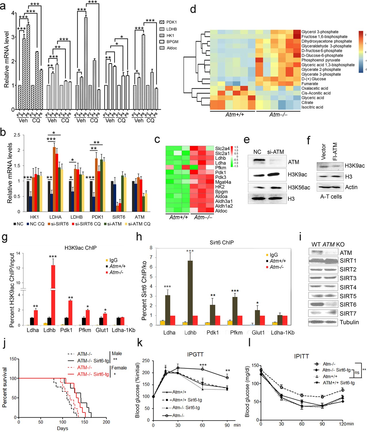
ATM-SIRT6 axis regulates age-related metabolic reprogramming.
(a) Quantitative RT-PCR analysis of mRNA levels of indicated glycolytic genes in different passages of MEFs with or without treatment of CQ. Data represent means ± SEM. *p<0.05, **p<0.01, ***p<0.001. (b) Quantitative RT-PCR analysis of mRNA levels of indicated glycolytic genes in Scramble (NC), si-SIRT6 or si-ATM HepG2 cells incubated with or without CQ (10 μM, 6 hr). Data represent means ± SEM. *p<0.05, **p<0.01, ***p<0.001. (c) Heatmap representation of RNA-Seq data (GSE109280) showing relative changes of glycolytic genes in Atm-/- MEF cells. The transcript levels are qualified in reads per kilobase of exon per million mapped sequence reads (RPKM), which is a normalized measure of exonic read density. Red and green indicate up- and downregulation, respectively. (d) Heatmap showing relative levels of metabolites in Atm+/+ and Atm-/- MEF cells of p53 null background, analyzed by LC-MS. Red and blue indicate up- and downregulation, respectively. (e) Immunoblots showing protein levels of H3K9ac and H3K56ac in ATM-deficient HepG2 cells. (f) Immunoblots showing levels of H3K9ac in A-T cells reconstituted with Flag-ATM. (g) ChIP analysis showing enrichment of H3K9ac at the promoter regions of indicated genes in Atm+/+ and Atm-/- MEFs. Data represent means ± SEM of three independent experiments. *p<0.05, **p<0.01, ***p<0.001. (h) ChIP analysis showing enrichment of Sirt6 at the promoter regions of indicated genes in Atm+/+ and Atm-/- MEFs. Data represent means ± SEM of six independent experiments. *p<0.05, **p<0.01, ***p<0.001. (i) Immunoblots showing protein levels of sirtuins in wild-type (WT) and ATM knockout (KO) HEK293 cells. (j) Kaplan-Meier survival of Atm-/- and Atm-/-;Sirt6-Tg male (n = 11 in each group) and female (n = 9 in each group) mice. **p<0.01. (k) Results of glucose tolerance tests in Atm+/+, Atm-/-, and Atm-/-;Sirt6-Tg mice. Data represent means ± SEM, n = 6. **p<0.01, ***p<0.001. (l) Results of insulin tolerance tests in Atm+/+, Atm-/-, and Atm-/-;Sirt6-Tg mice. Data represent means ± SEM, n = 6. **p<0.01. ‘ns’ indicates no significant difference.
-
Figure 2—source data 1
Differently expressed mRNA profiles of Atm-/- MEF cells.
- https://doi.org/10.7554/eLife.34836.011
-
Figure 2—source data 2
Differentially expressed Metabolites in Atm KO MEFs.
- https://doi.org/10.7554/eLife.34836.012
-
Figure 2—source data 3
Differently expressed mRNA profiles of Sirt6-/- MEF cells.
- https://doi.org/10.7554/eLife.34836.013
Figure 2—figure supplement 1
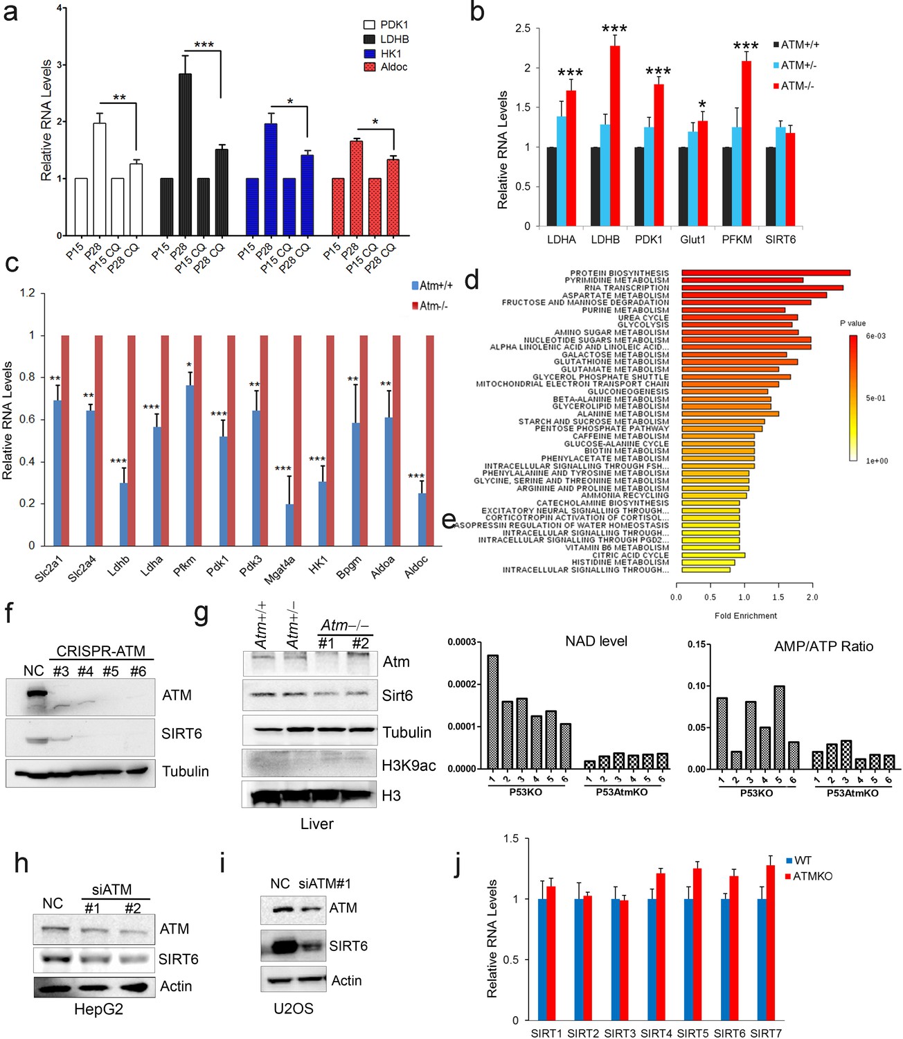
Atm deficiency promotes glycolysis.
(a) Quantitative RT-PCR analysis of mRNA levels of indicated glycolytic genes in different passages of HSFs with or without treatment of CQ. Data represent means ± SEM. *p<0.05, **p<0.01, ***p<0.001. (b) Quantitative RT-PCR analysis of mRNA levels of glycolytic genes in liver tissues from Atm+/+, Atm+/-, and Atm-/- mice. Data represent mean ± SEM. Atm-/- Vs Atm+/+, *p<0.05, ***p<0.001. (c) Quantitative RT-PCR analysis of mRNA levels of glycolytic genes to validate the RNA-seq data set of Atm+/+ and Atm-/- MEFs. Data represent means ± SEM of six independent experiments. *p<0.05, **p<0.01, ***p<0.001. (d) Metabolic pathway enrichment in Atm null MEFs compared with wild-types. (e) AMP/ATP ratio and NAD + levels in wild-type and Atm null MEFs. (f) Immunoblots showing ATM and SIRT6 protein levels in indicated clones of ATM KO HEK293 cells generated by the CRISPR/Cas9 system. (g) Immunoblots showing protein levels of Sirt6 and H3K9ac in liver tissues from 4-month-old Atm+/+, Atm+/-, and Atm-/- mice. (h–i) Immunoblots showing SIRT6 levels in si-NC and si-ATM treated HepG2 and U2OS cells. (j) Quantitative RT-PCR analysis of sirtuin mRNA levels in wild-type and ATM KO HEK293 cells. Data represent means ± SEM.
Figure 2—figure supplement 2

SIRT6 reduction underlies age-related metabolic reprogramming triggered by ATM decline.
(a) Heatmap representation of RNA-Seq data showing relative changes of glycolytic genes in Sirt6-/- MEF cells. Red and green indicate up- and downregulation, respectively. (b) Quantitative RT-PCR analysis of mRNA levels of glycolytic genes to validate the RNA-seq data set of Sirt6+/+ and Sirt6-/- MEFs. Data represent means ± SEM of six independent experiments. **p<0.01, ***p<0.001. (c) Quantitative RT-PCR analysis of mRNA levels of indicated glycolytic genes in HepG2 cells transfected with Scramble, si-ATM, si-SIRT6, or si-ATM plus Flag-SIRT6. Data represent means ± SEM. *p<0.05, **p<0.01, ***p<0.001. (d) Immunoblots showing protein levels of SIRT6 and H3K9ac in HepG2 cells treated with 10 μM CQ for indicated periods of time. (e) ChIP analysis showing enrichment of H3K9ac at promoter regions of glycolytic genes in HepG2 cells treated with 10 μM CQ for indicated periods of time. Data represent means ± SEM. *p<0.05, **p<0.01, ***p<0.001. (f) Immunoblots showing SIRT1, SIRT6, and SIRT7 protein levels in ATM knockdown (KD) or control HSFs. (g) Immunoblots showing protein levels of Atm, Sirt1, Sirt6 and γH2AX levels in Atm+/+ and Atm-/- primary MEF cells. (h) Immunoblots showing Sirt6 expression in liver tissues of Sirt6-tg mice. (i) Serum lactate levels in 3-month-old Atm+/+, Atm-/- and Atm-/-;Sirt6-tg male mice. Data represent mean ± SEM of five to six animals/group. *p<0.05.
Figure 3 with 1 supplement
ATM interacts with and phosphorylates SIRT6.
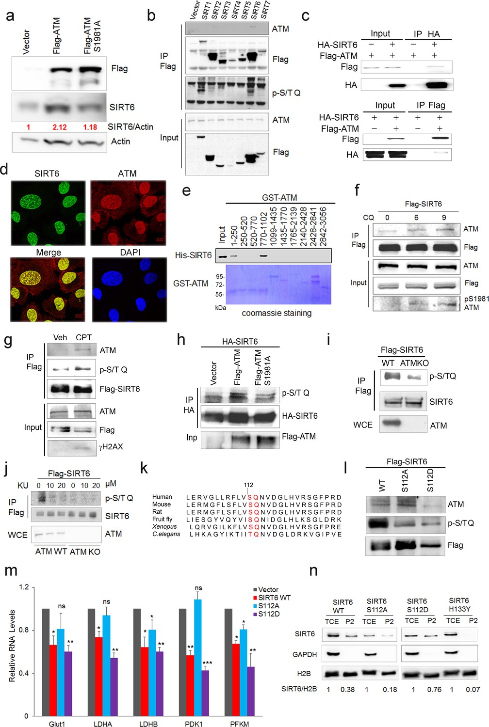
(a) Immunoblots showing protein levels of SIRT6 in HEK293 cells expressing Flag-ATM or Flag-ATM S1981A. (b) Immunoblots showing endogenous ATM and p-S/TQ motif in anti-Flag immunoprecipitates in HEK293 cells transfected with empty vector or Flag-sirtuins. (c) Immunoblots showing Flag-ATM and HA-SIRT6 in anti-HA (upper) or anti-Flag (lower) immunoprecipitates in HEK293 cells transfected with indicated constructs. (d) Representative photos of immunofluorescence staining of SIRT6 and ATM in U2OS cells, showing co-localization in the nucleus. Scale bar, 50 µm. (e) GST pull-down assay showing bacterially expressed His-SIRT6 predominantly bound to GST-ATM fragment 4 (770–1102), the N-terminal HEAT-repeat of ATM. (f) Immunoblots showing the increased binding capacity of ATM and SIRT6 under the treatment of (10 µM) CQ for the indicated time. (g) Immunoblots showing ATM and p-S/TQ in anti-Flag immunoprecipitates in HEK293 cells expressing Flag-SIRT6 treated with CPT (0.4 µM) or DMSO. (h) Immunoblots showing level of p-S/TQ SIRT6 in HEK293 cells co-transfected with HA-SIRT6 and Flag-ATM, Flag-ATM S1981A, or empty vector. (i) Immunoblots showing p-S/T Q SIRT6 in WT or ATM KO HEK293 cells. (j) Immunoblots showing p-S/T Q level of SIRT6 in ATM WT or KO HEK293 cells treated with DMSO and KU55933 (10 or 20 µM, 2 hr). (k) Alignment of protein sequence of human SIRT6 and orthologues in mouse, rat, fruit fly, Xenopus, and C. elegans. A conserved S112 Q113 motif was highlighted. (l) Immunoblots showing p-S/T Q level of Flag-SIRT6, Flag-SIRT6 S112A, or Flag-SIRT6 S112D in HEK293 cells. (m) Quantitative RT-PCR analysis of mRNA levels of indicated glycolytic genes in sh-SIRT6 HepG2 cells re-expressing SIRT6, SIRT6 S112A, or 112D mutation. Data represent means ± SEM. *p<0.05, **p<0.01, ***p<0.001. ‘ns’ indicates no significant difference. (n) Immunoblots showing SIRT6 protein level in total cell extract (TCE) and chromatin-enriched fractions (P2). Densitometry analysis was performed to determine the relative ratio of SIRT6/H2B within chromatin fractions.
Figure 3—figure supplement 1
ATM directly phosphorylates SIRT6.
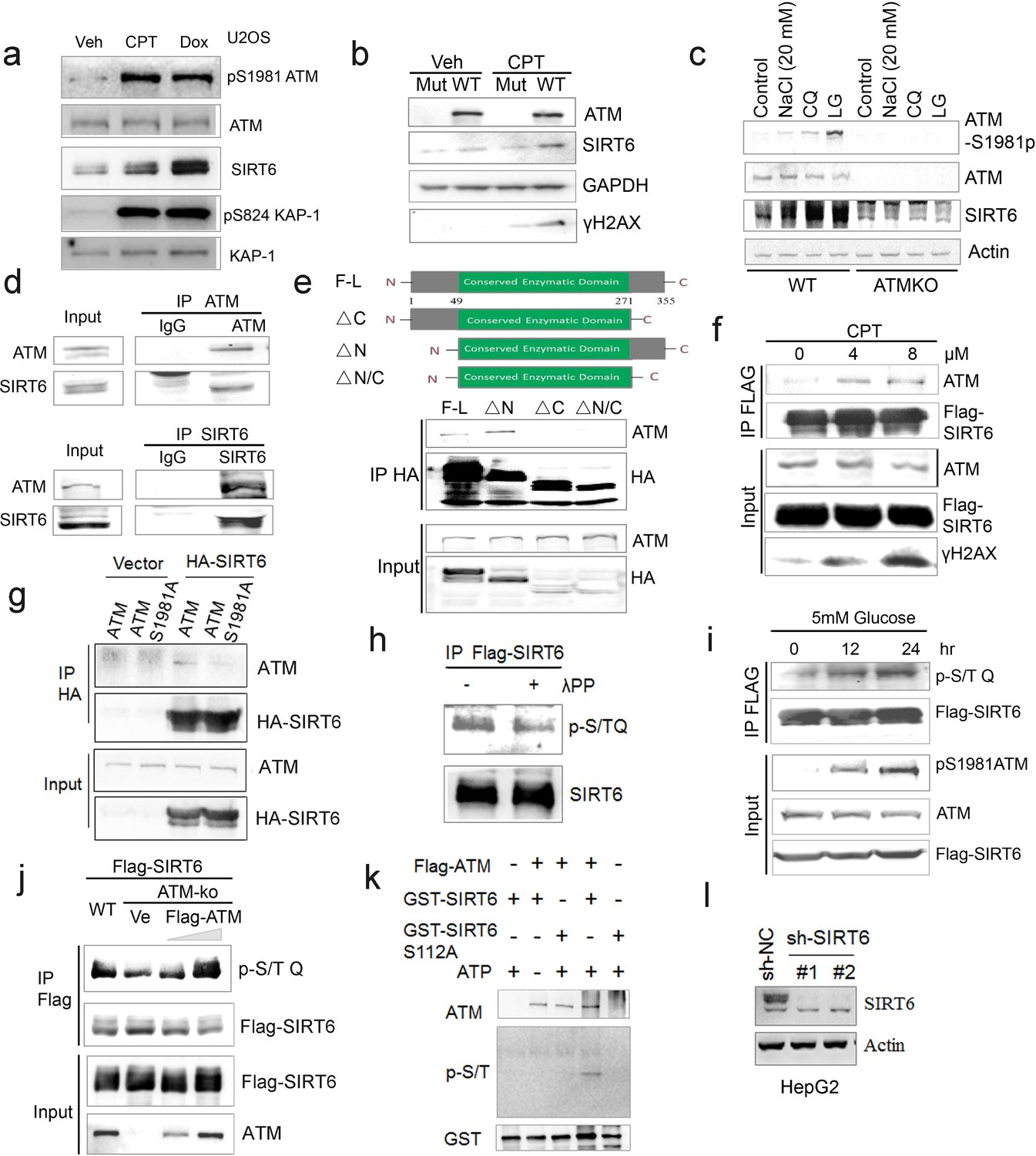
(a) Immunoblots showing a significant increase in SIRT6 protein level, pS1981ATM, and pS824KAP-1 in the presence of DNA damage reagents-CPT and Dox in U2OS cells. (b) Immunoblots showing Sirt6 protein level in immortalized wild-type and Atm null MEFs in response to 0.4 μM CPT treatment. (c) Immunoblots showing pS1981-ATM and SIRT6 levels in wild-type and ATM KO HEK293 cells treated with CQ, low glucose (LG), and hypotonic swelling (20 mM NaCl). (d) Immunoblots of indicated immunoprecipitates showing the interaction between endogenous ATM and SIRT6 in HepG2 cells. (e) Immunoblots showing interaction between ATM and truncated fragments of SIRT6 in HEK293 cells. F-L, full-length of SIRT6, ΔC, C-terminal deletion, ΔN, N-terminal deletion, ΔN-C, N-and C-terminal truncation. (f) Immunoblots showing ATM protein in anti-Flag immunoprecipitates in HEK293 cells expressing Flag-SIRT6, treated with 0 μM, 4 μM, or 8 μM of CPT for 1 hr. (g) Immunoblots showing ATM protein in anti-HA immunoprecipitates in HEK293 cells expressing Flag-ATM or Flag-ATM S1981A, as well as empty vector or HA-SIRT6. (h) Immunoblots showing p-S/T Q of Flag-SIRT6 in HepG2 cells treated with 5 mM of glucose for indicated time. (i) Immunoblots showing p-S/T Q of Flag-SIRT6 in HEK293 cells, treated with or without λPP (30 min). (j) Immunoblots showing p-S/T Q of SIRT6 in WT and ATM KO HEK293 cells, transfected with empty vector, 2 μg Flag-ATM, or 6 μg Flag-ATM. (k) Immunoblots showing p-S/T Q of GST-SIRT6 after incubation with Flag-ATM purified from CPT-treated HEK293 cells. (l) Immunoblots showing SIRT6 protein levels in HepG2 cells with lentiviral infection containing sh-NC or sh-SIRT6.
Figure 4 with 1 supplement
ATM prevents ubiquitination and degradation of SIRT6.
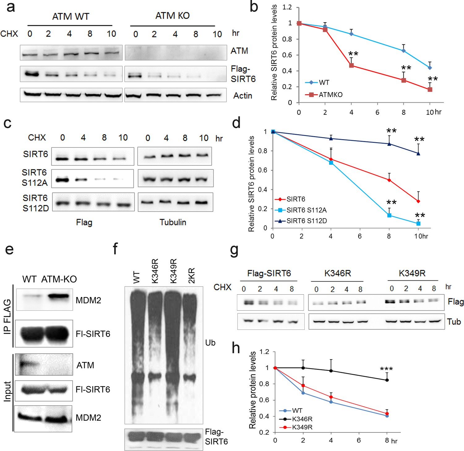
(a) Immunoblots showing protein levels of Flag-SIRT6 in WT and ATM KO HEK293 cells treated with CHX (50 µg/ml) for indicated periods of time. (b) Quantification of protein levels in (a) by ImageJ. Data represent means ± SEM of three independent experiments. **p<0.01. (c) Immunoblots showing protein levels of Flag-SIRT6, S112A, and S112D in the presence of CHX (50 μg/ml) for indicated periods of time. (d) Quantification of protein levels in (c) by ImageJ. Data represent means ± SEM of three independent experiments. ***p<0.001. (e) Immunoblots showing increased binding capacity between SIRT6 and MDM2 in ATM KO HEK293 cells. (f) Immunoblots showing ubiquitination of Flag-SIRT6, K346R, K349R, and K346/349R (2KR) in HEK293 cells. Note that 2KR and K346R abrogated the ubiquitination of Flag-SIRT6. (g, h) Upper, immunoblots showing protein levels of Flag-SIRT6, K346R, and K349R in the presence of CHX (50 μg/ml) for indicated periods of time. Lower, quantification of protein levels by ImageJ. Data represent means ± SEM of three independent experiments. ***p<0.001.
Figure 4—figure supplement 1
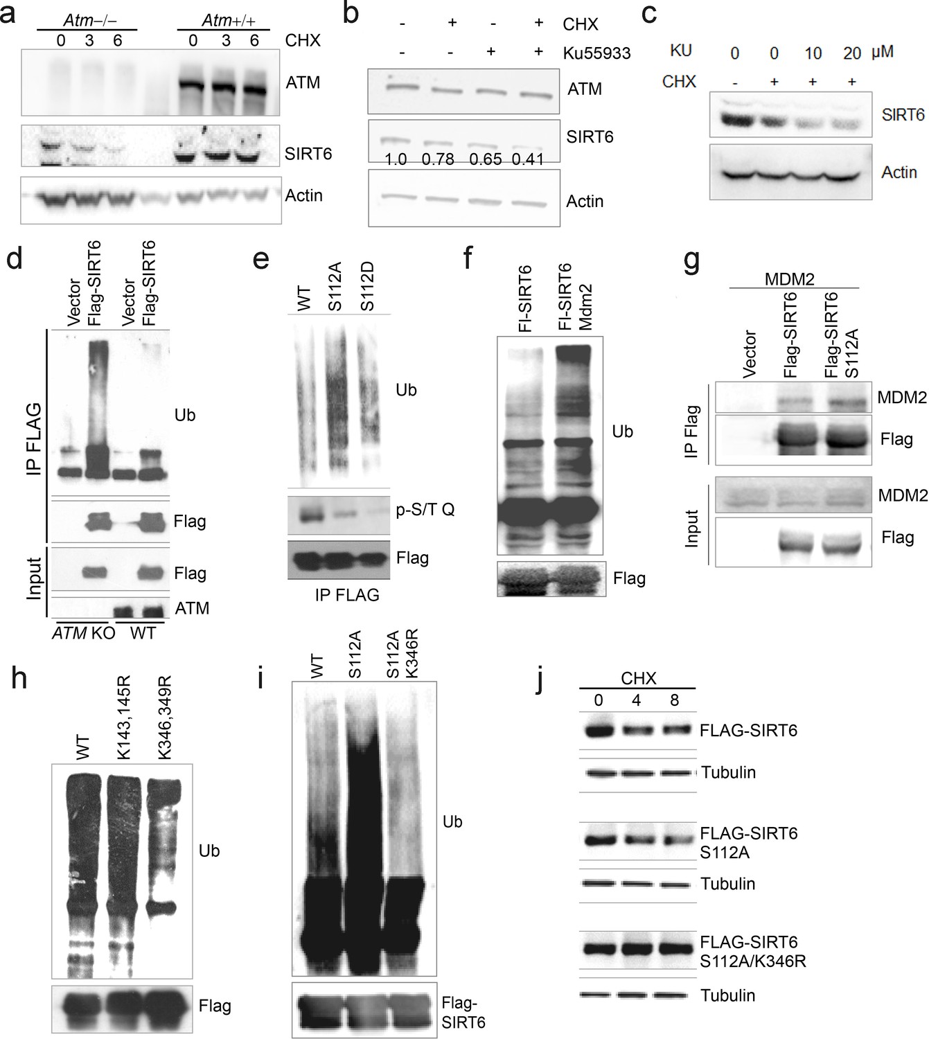
ATM-mediated phosphorylation of SIRT6 prevents its ubiquitination and degradation.
(a) Immunoblots showing Sirt6 levels in wild-type and Atm null MEFs in the presence of CHX (50 μg/ml). (b–c) Immunoblots showing SIRT6 protein level in HEK293 cells in the presence of CHX (50 μg/ml) and/or KU55933 (10 or 20 μM). Densitometry analysis was performed to determine the SIRT6/actin ratio. (d) Immunoblots showing increased ubiquitination of SIRT6 in ATM KO HEK293 cells. (e) Immunoblots showing ubiquitination of Flag-SIRT6, Flag-SIRT6 S112A, and Flag-SIRT6 S112D in HEK293 cells co-transfected Myc-Ub. (f) Immunoblots showing increased ubiquitination of Flag-SIRT6 in HEK293 cells overexpressing MDM2. (g) Immunoblots showing MDM2 in anti-Flag immunoprecipitates in HEK293 cells expressing Flag-SIRT6 or Flag-SIRT6 S112A. (h) Immunoblots showing ubiquitination of Flag-SIRT6, K143/145R, and K346/349R in HEK293 cells. Noted that K364/349R abrogated the ubiquitination of Flag-SIRT6. (i) Immunoblots showing the ubiquitination of Flag-SIRT6, Flag-SIRT6 S112A, and Flag-SIRT6 S112A/K346R. (j) Immunoblots showing protein levels of Flag-SIRT6, Flag-SIRT6 S112A, and Flag-SIRT6 S112A/K346R in the presence of CHX (50 μg/ml). Note that K346R rescued the accelerated degradation of S112A.
Figure 5 with 2 supplements
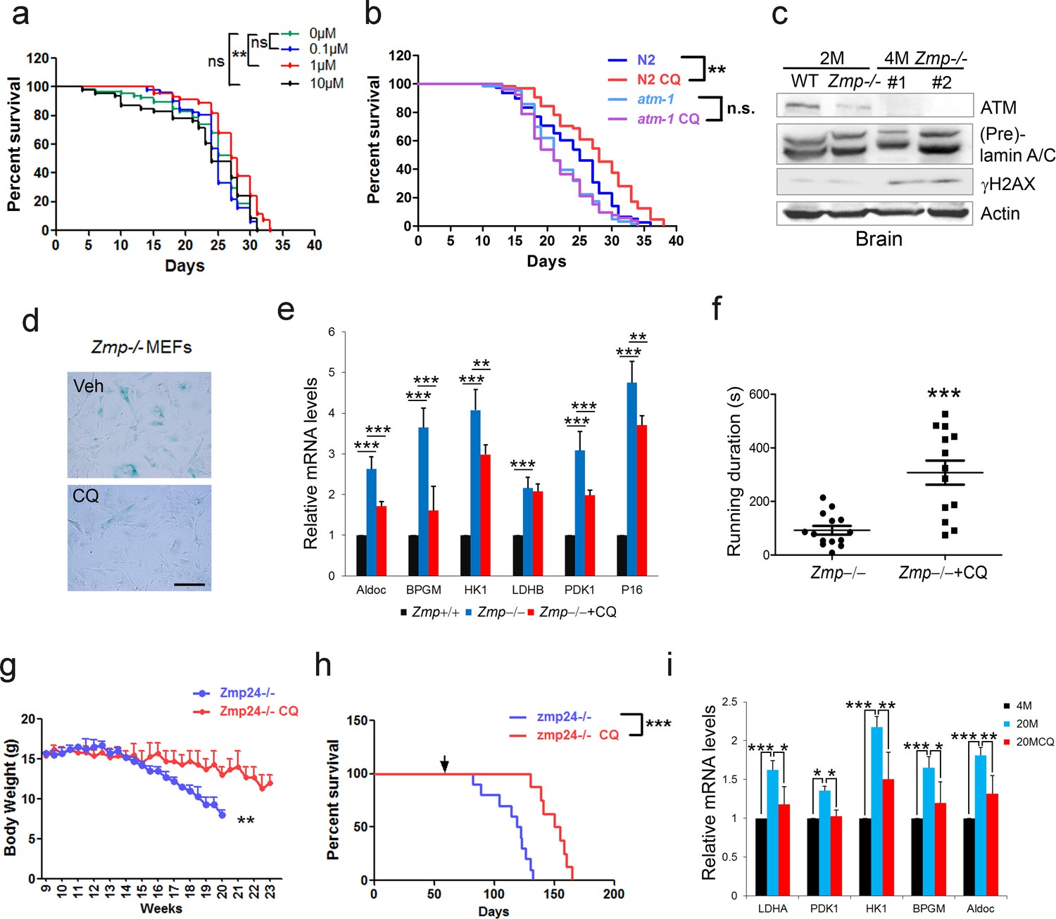
CQ extends lifespan in an ATM- dependent manner.
(a) Survival analysis of C. elegans treated with the indicated dosage of CQ. **p<0.01. NS indicates no significant difference. (b) Survival analysis of wild-type and atm-1 null C. elegans cultured in medium with or without 1 µM CQ. (c) Immunoblots showing protein levels of Atm and γH2AX in brain tissues of Zmpste24+/+ (2 months), Zmpste24-/- (2 months), and Zmpste24-/- (4 months) mice. (d) Representative images showing SA-β-Gal staining in Zmpste24-/- MEFs with or without CQ treatment. Scale bar, 100 µm. (e) Quantitative RT-PCR analysis of mRNA levels of p16Ink4a and indicated glycolytic genes in liver tissues of Zmpste24+/+, saline-treated, and CQ-treated Zmpste24-/- mice. Mice were treated for 8 weeks with two weekly intraperitoneal injections of CQ at 3.5 mg/kg. Data represent means ± SEM. *p<0.05, **p<0.01, ***p<0.001. (f) Maximum running duration in saline- and CQ-treated Zmpste24-/- mice. Data represent means ± SEM. ***p<0.001. (g) Body weight of saline- and CQ-treated male Zmpste24-/- mice. Data represent means ± SEM. **p<0.01. (h) Kaplan-Meier survival curves of saline-treated (n = 10) and CQ-treated (n = 8) Zmpste24-/- mice. ***p<0.001. (i) Quantitative RT-PCR analysis of mRNA levels of indicated glycolytic genes in the liver tissues of 4-month-old, saline-treated 12-month-old (n = 3), and CQ-treated 20-month-old (n = 3) mice. Data represent means ± SEM. *p<0.05, **p<0.01, ***p<0.001.
-
Figure 5—source data 1
Lifespan analysis of chloroquine's effect on nematodes.
- https://doi.org/10.7554/eLife.34836.021
-
Figure 5—source data 2
Lifespan analysis of chloroquine's effect on mice.
- https://doi.org/10.7554/eLife.34836.022
Figure 5—figure supplement 1
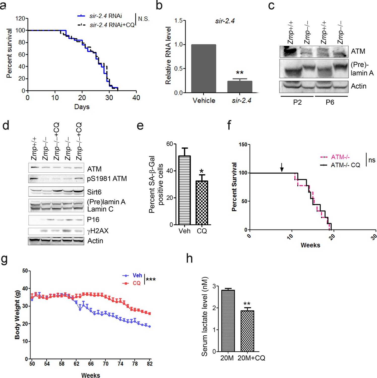
ATM activation ameliorates aging-associated features.
(a) Survival analysis of sir-2.4 downregulated C. elegans exposed to medium with or without 1 µM CQ. ‘N.S.’ indicates no significant difference. (b) Quantitative RT-PCR analysis of mRNA levels of sir-2.4 in vehicle and sir-2.4 RNAi-treated C. elegans. (c) Immunoblots showing protein levels of Atm in wild-type and Zmpste24 null MEFs. (d) Immunoblots showing levels of proteins involved in pS1981-ATM, γH2AX, Sirt6, and p16 in liver tissues of saline-treated, and CQ-treated Zmpste24-/- mice. (e) Quantification of SA-β-Gal staining in (Figure 5d) from five views randomly captured for each group. Data represent means ± SEM. *p<0.05. Scale bar, 100 µm. (f) Kaplan-Meier survival curves of saline-treated and CQ-treated Atm-/- mice (n = 9 for each group). ‘ns’ indicates no significant difference. (g) Body weight of saline- and CQ-treated male aging mice. Data represent means ± SEM. ***p<0.001. (h) Serum lactate levels in saline- and CQ-treated 20-month-old mice. Data represent means ± SEM. **p<0.01.
Figure 5—figure supplement 2

Schematic model of ATM-SIRT6 axis in regulating aging and longevity.
Left, DNA damage activates DDR cascade, and its constant activation leads to permanent cell cycle exit and senescence. Right, defective ATM-SIRT6 axis underlies premature aging in mouse models resembling HGPS and A-T, which is rescued by treatment of CQ and Sirt6 transgene, respectively. Middle, during physiological aging, DNA damage-free activation of ATM by CQ promotes longevity at organismal levels.
Author response image 1
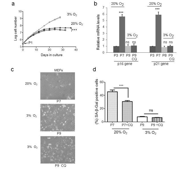
Physiological (3%) oxygen condition is inappropriate for MEFs culture to assess the replicative lifespan-extending effect of CQ.
(a) MEFs at passage 1 were cultured in 20% O2 (black) or 3% O2 (grey) with treatment of 1 μM CQ (circles) or DMSO (squares), and cell number was determined at each passage. ***P < 0.01. (b) Quantitative RT-PCR analysis of mRNA levels of p16 and p21 genes in different passages of MEFs with or without treatment of CQ in 20% O2 or 3% O2. ***P < 0.001. ‘ns’ indicates no significant difference. (c) Representative images showing morphology of MEFs under the indicated culture conditions with or without CQ treatment. (d) Quantification of SA-β-Gal-positive cells under indicated culture conditions. Data represent the means ± SEM. ***P < 0.001; ‘ns’ indicates no significant difference.
Author response image 2
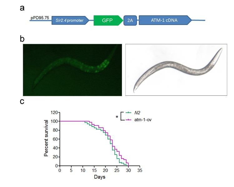
Lifespan analysis for worms overexpressing ATM-1.
(a) The strategy for constructing ATM-1 overexpressing plasmid with GFP-tag. (b) Representative images showing the detectable GFP expression in worms. (c) Survival analysis of worm line expressing GFP-ATM-1. *P < 0.05.
Tables
Key resources table
| Reagent type (species) or resource | Designation | Source or reference | Identifiers | Additional information |
|---|---|---|---|---|
| Gene (human) | ATM | National Center for Biotechnology Information | Gene ID: 472 | |
| Gene (mouse) | Atm | National Center for Biotechnology Information | Gene ID: 11920 | |
| Gene (Caenorhabditis elegans) | atm-1 | National Center for Biotechnology Information | Gene ID: 3565793 | |
| Gene (human) | SIRT6 | National Center for Biotechnology Information | Gene ID: 51548 | |
| Gene (mouse) | Sirt6 | National Center for Biotechnology Information | Gene ID: 50721 | |
| Gene (Caenorhabditis elegans) | sir-2.4 | National Center for Biotechnology Information | Gene ID: 182284 | |
| Gene (mouse) | Zmpste24 | National Center for Biotechnology Information | Gene ID: 230709 | |
| Gene (mouse) | p53 | National Center for Biotechnology Information | Gene ID: 230710 | |
| Cell line (human) | HEK293 | ATCC | Catalog number: ATCC CRL-1573; RRID:CVCL_0042 | |
| Cell line (human) | HepG2 | ATCC | Catalog number: ATCC HB-8065; RRID:CVCL_0027 | |
| Cell line (human) | U2OS | ATCC | Catalog number: ATCC HTB-96; RRID:CVCL_0042 | |
| Cell line (mouse) | Atm-/-; p53-/- MEF | from Dr. Yosef Shiloh (Tel Aviv University, Israel) | ||
| Cell line (mouse) | Sirt6-/- MEF | from Dr. Raul Mostoslavsky (Massachusetts General Hospital Cancer center, USA) | ||
| Antibody | ATM | Abcam (Cambridge, UK) | Cat# ab78; RRID:AB_306089 | Applications: WB;Immunofluorescence |
| Antibody | SIRT6 | Abcam (Cambridge, UK) | Cat# ab62739; RRID:AB_956300 | Applications: WB; Immunofluoresce;Chromatin immunoprecipitation |
| Antibody | γH2AX | Abcam (Cambridge, UK) | Cat# ab81299; RRID:AB_1640564 | Applications: WB |
| Antibody | p21 | Santa Cruz Biotechnology | Cat# sc-6246; RRID:AB_628073 | Applications: WB |
| Antibody | MDM2 | Santa Cruz Biotechnology | Cat# sc-965; RRID:AB_627920 | Applications: WB |
| Antibody | p-ATM (Ser1981) | EMD Millipore | Cat# 05–740; RRID:AB_309954 | Applications: WB |
| Antibody | H3K9ac | EMD Millipore | Cat# 07–352; RRID:AB_310544 | Applications: WB; Chromatin immunoprecipitation |
| Antibody | p-S/T Q | Cell Signaling Technology (Beverly, MA) | Cat #9607S; RRID:AB_10889739 | Applications: WB |
| Antibody | cleaved caspase-3 | Cell Signaling Technology (Beverly, MA) | Cat #9661; RRID:AB_2341188 | Applications: WB |
| Antibody | HA | Sigma-Aldrich | Cat# H3663; RRID:AB_262051 | Applications: WB |
| Antibody | Flag | Sigma-Aldrich | Cat# F1804; RRID: AB_262044 | Applications: WB |
| Antibody | LC3B | Sigma-Aldrich | Cat# L7543; RRID:AB_796155 | Applications: WB |
| Transfected construct (human) | Flag-His-ATM wt | Addgene (Cambridge, MA) | Cat #31985 | |
| Transfected construct (human) | Flag-SIRT6 | Addgene (Cambridge, MA) | Cat #13817 | |
| Transfected construct (human) | Flag-His-ATM S1981A | Addgene (Cambridge, MA) | Cat #32300 | |
| Commercial assay or kit | Senescence beta- galactosidase staining Kit | Cell Signaling Technology (Beverly, MA) | Cat #9860 | |
| Commercial assay or kit | Lactate Colorimetric Assay Kit | BioVision | Cat #K667-100 | |
| Commercial assay or kit | Click-iT EdU Alexa Fluor 488 Kit | Invitrogen | Cat #C10425 | |
| Chemical compound, drug | Cycloheximide | Sigma-Aldrich | Cat #66-81-9 | |
| Chemical compound, drug | MG-132 | Sigma-Aldrich | Cat #474787 | |
| Chemical compound, drug | Chloroquine | Sigma-Aldrich | Cat #C6628 |
Additional files
-
Transparent reporting form
- https://doi.org/10.7554/eLife.34836.023
Download links
A two-part list of links to download the article, or parts of the article, in various formats.
Downloads (link to download the article as PDF)
Open citations (links to open the citations from this article in various online reference manager services)
Cite this article (links to download the citations from this article in formats compatible with various reference manager tools)
Boosting ATM activity alleviates aging and extends lifespan in a mouse model of progeria
eLife 7:e34836.
https://doi.org/10.7554/eLife.34836


