Cryo-EM structures of the DCPIB-inhibited volume-regulated anion channel LRRC8A in lipid nanodiscs
- University of California, Berkeley, United States
- Memorial Sloan Kettering Cancer Center, United States
Figures
Figure 1 with 10 supplements
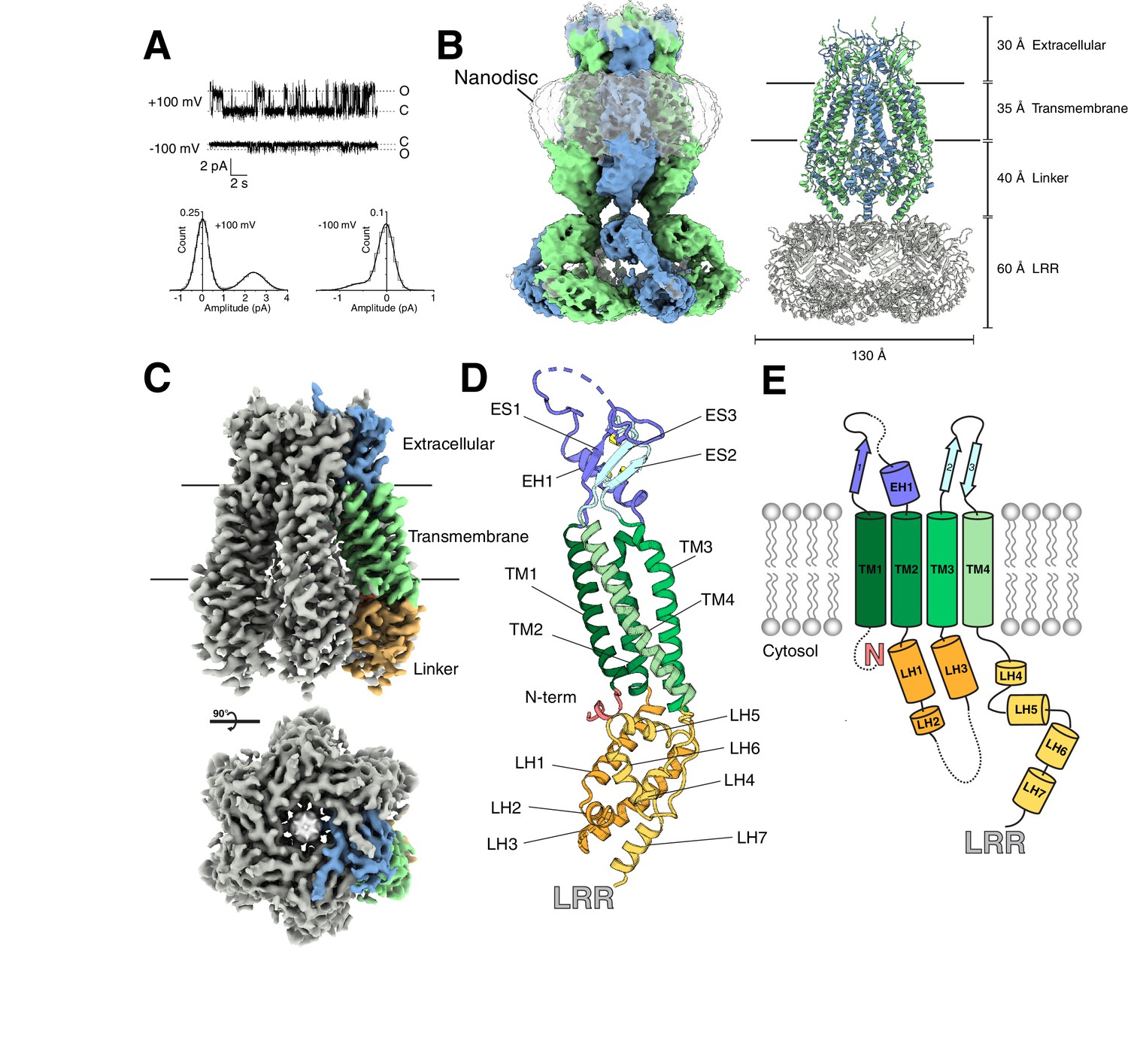
Structure of an LRRC8A-DCPIB complex in lipid nanodiscs.
(A) Representative single channel recording from an excised patch containing purified LRRC8A reconstituted into phosphatidyl choline lipids (Po = 0.3, γ = 24 pS at +100 mV; Po = 0.24, γ = 5.4 pS at −100 mV). (B, left) Cryo-EM density of LRRC8A from an unmasked refinement of the constricted state at 4.5 Å resolution and (right) corresponding atomic model viewed from the membrane plane. Individual subunits are alternatingly colored blue and green, nanodisc density is rendered transparent, and LRRs docked into the density are colored gray in the model. Dimensions of the extracellular, transmembrane, linker, and LRR regions are indicated. (C) Cryo-EM density of LRRC8A from a LRR masked refinement of the constricted class at 3.21 Å resolution. A view from the membrane (top) and extracellular space (bottom) are shown. One subunit is colored according to region and shown in (C) within the density, (D) as an isolated model with helices and N-terminus labeled and extracellular domain disulfide bonds depicted in yellow, and (E) as a cartoon.
Figure 1—figure supplement 1
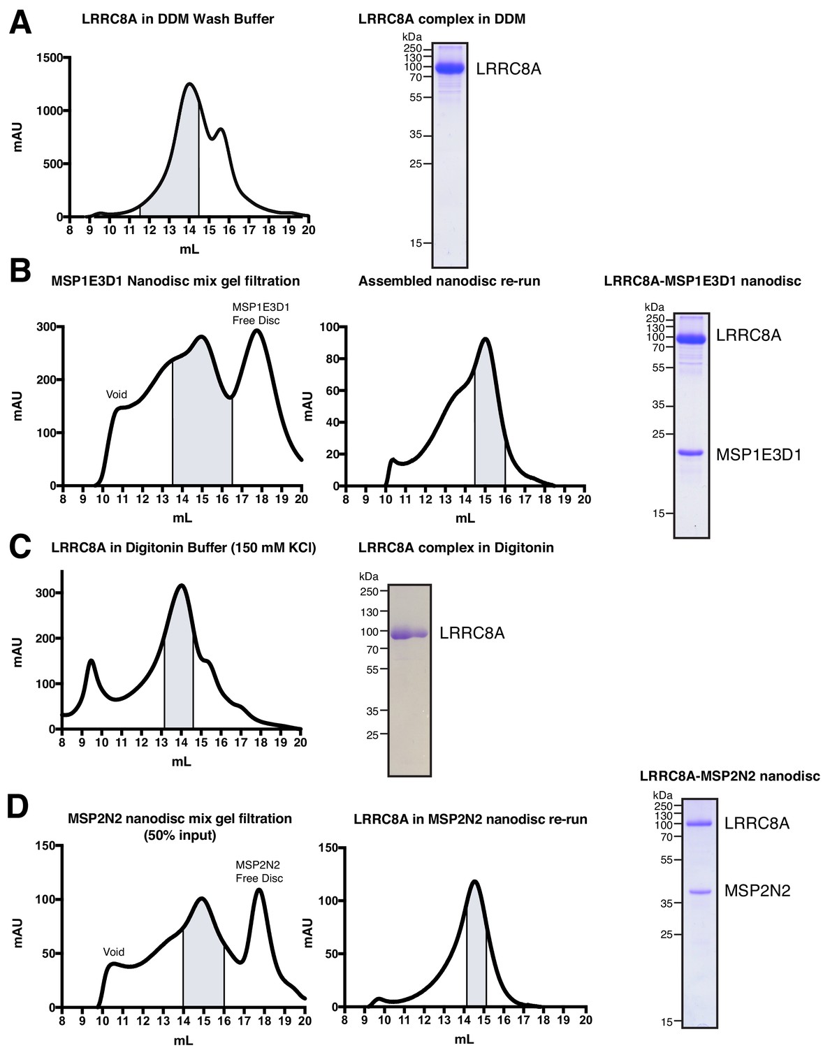
Complex purification and reconstitution.
(A) Superose six gel filtration of LRRC8A in DDM-containing wash buffer. Peak fractions corresponding to LRRC8A complex are highlighted. On the right, coomassie gel of purified LRRC8A protein in DDM. (B) First run of the MSP1E3D1 nanodisc mix on Superose six column. Peak fractions corresponding to LRRC8A in MSP1E3D1 nanodiscs is highlighted. These fractions were concentrated and re-run on the same column (middle). Indicated fractions were then pooled and concentrated for grid preparation and analysis by coomassie gel (Right). (C) Superose six gel filtration of LRRC8A in digitonin Buffer. Chosen fractions corresponding to LRRC8A complex in digitonin are highlighted. On the right, coomassie gel of purified LRRC8A in digitonin Buffer. (D) First run of the MSP2N2 nanodisc mix (50% of total input) on Superose six column. Peak fraction corresponding to LRRC8A in MSP2N2 nanodiscs is highlighted. These fractions were concentrated and re-run on the same column (middle). Indicated fractions were then pooled and concentrated for grid preparation and analysis by coomassie gel (right).
Figure 1—figure supplement 2
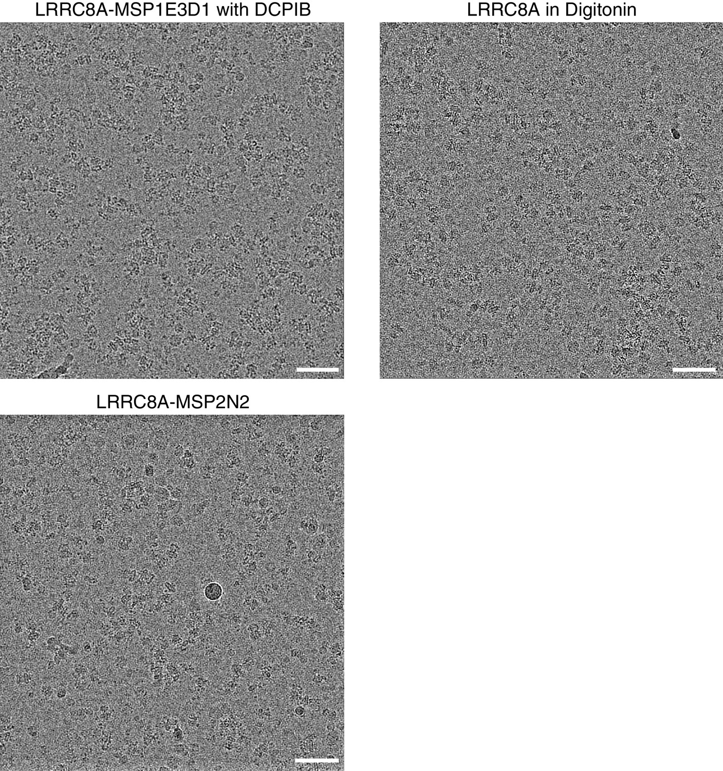
Representative micrographs from LRRC8A preparations in MSP1E3D1 nanodiscs with DCPIB, digitonin, and MSP2N2 nanodiscs.
Scale bars, 500 Å.
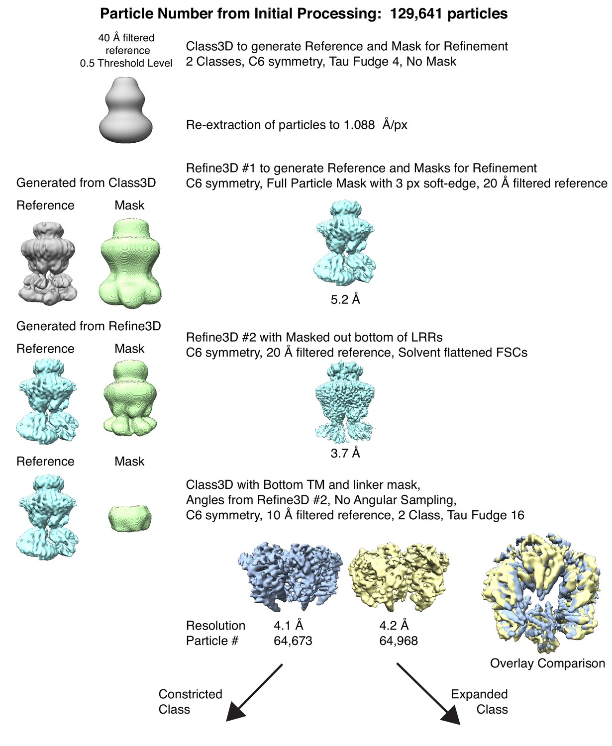
Figure 1—figure supplement 3
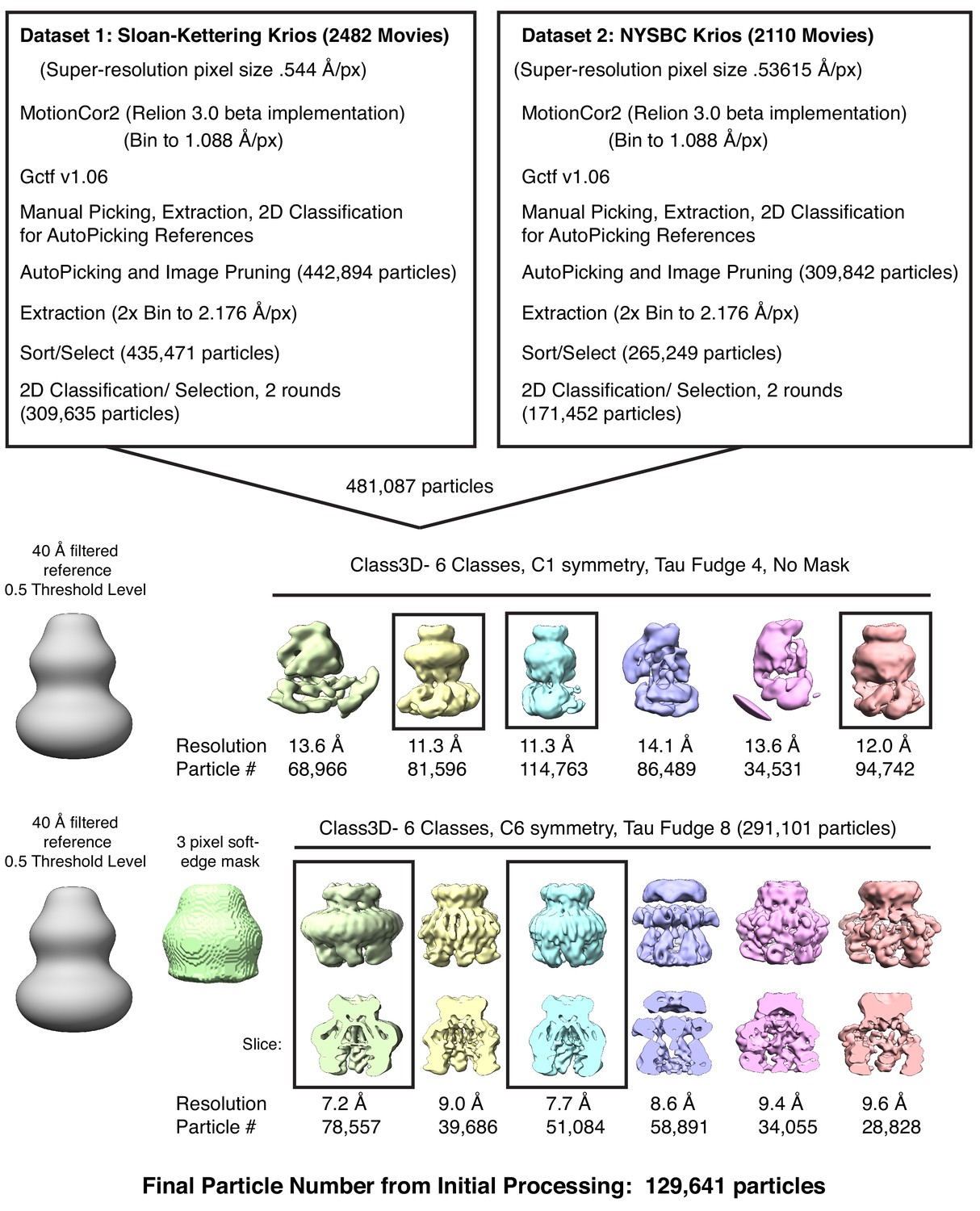
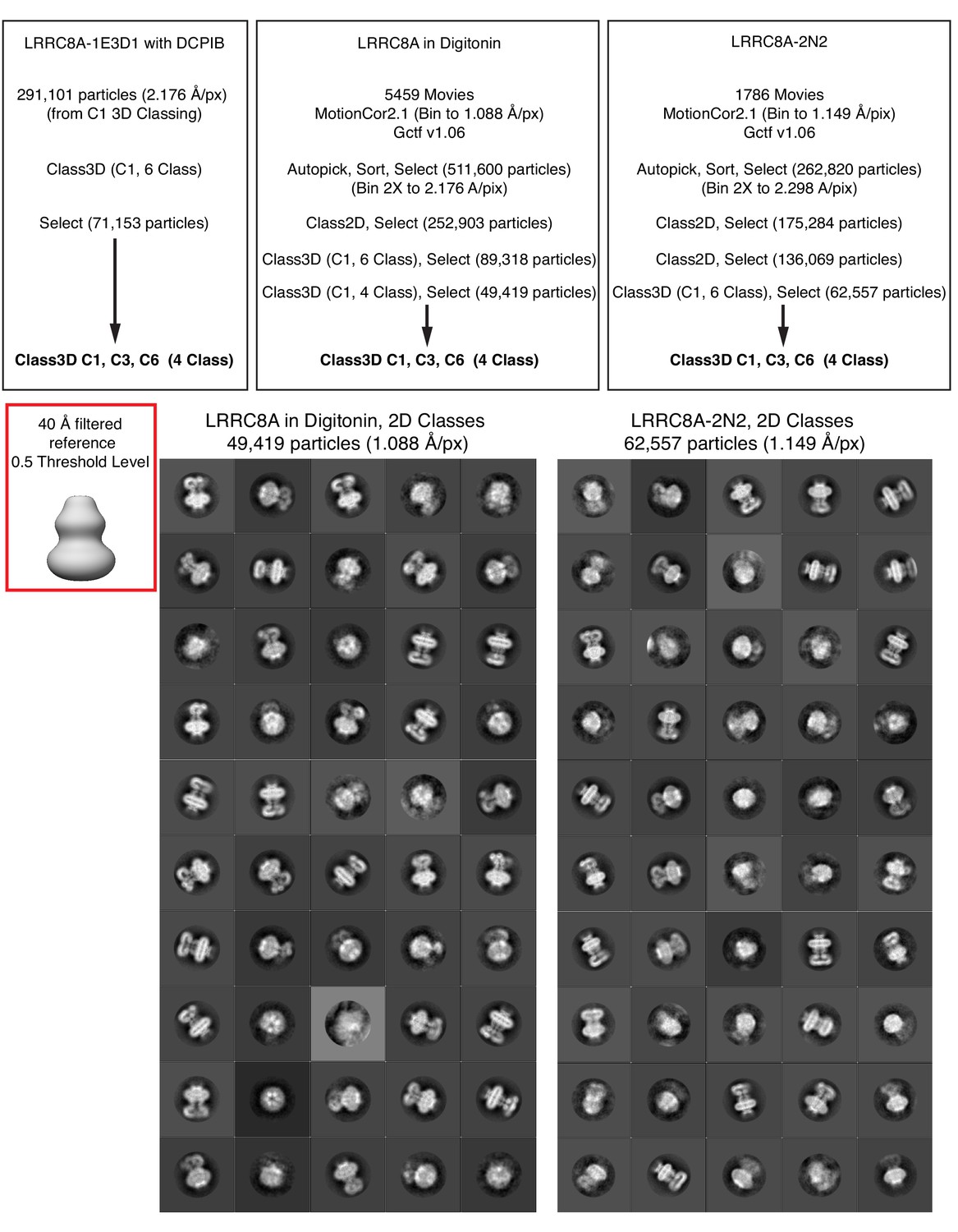
Initial processing for LRRC8A-DCPIB in MSP1E3D1 nanodisc datasets.
Particles from boxed classes were passed to the next round.
Figure 1—figure supplement 4
The refinement and classification performed to separate constricted and expanded particles for LRRC8A-DCPIB in MSP1E3D1.
https://doi.org/10.7554/eLife.42636.006
Figure 1—figure supplement 5
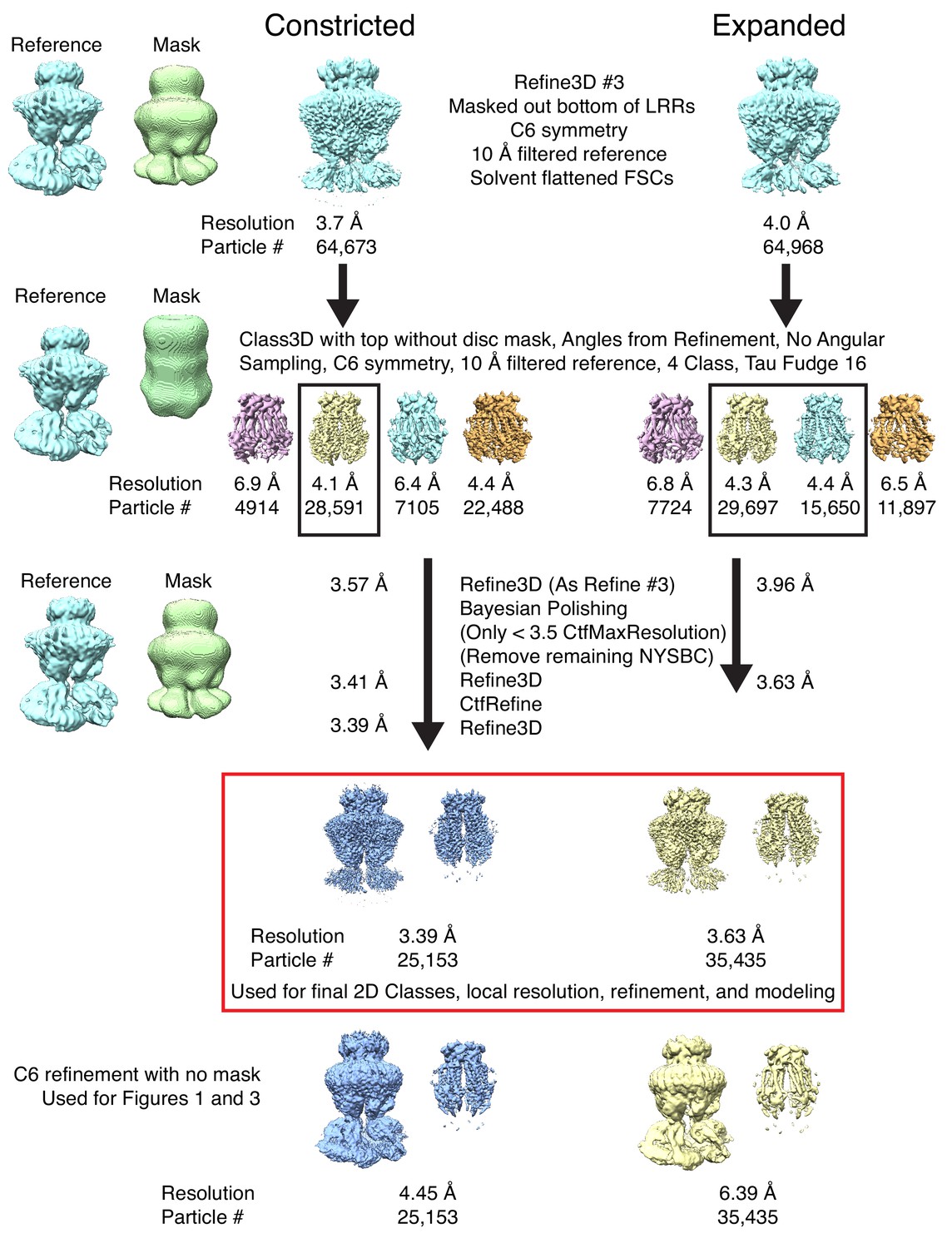
The final classification and refinement performed on constricted and expanded particles to obtain high-resolution maps for LRRC8A-DCPIB in MSP1E3D1.
Particles from boxed classes were passed to the next round. The masked refinements used for generating final maps and modeling are boxed in red. Below are the unmasked refinements used in Figures 1 and 4.
Figure 1—figure supplement 6
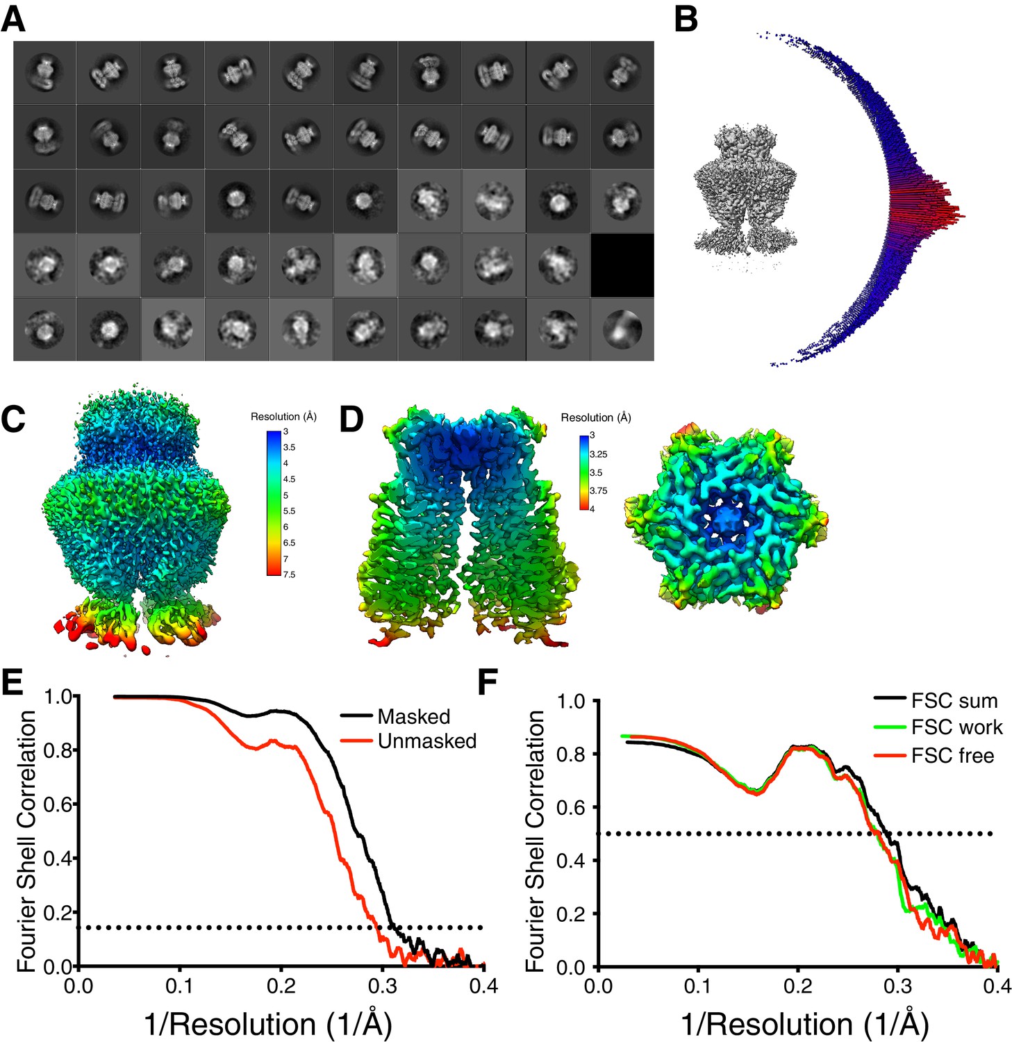
Constricted map and model validation for LRRC8A-DCPIB in MSP1E3D1.
(A) Two-dimensional classes from a fifty-class classification of the final particles. (B) Angular distribution of particle views. (C) Local resolution using Relion locally filtered map at 0.012 threshold showing resolution of the top of LRR region. (D) Local resolution using Relion locally filtered map at 0.035 threshold and dust hidden at 12 Å. Membrane view (left) and extracellular view (right). (E) Fourier Shell Correlation (FSC) of the two unfiltered half-maps from refinement used for calculating overall resolution at 0.143. (F) Model validation FSC using half-map one used for model refinement (FSC work), half-map two not used for refinement (FSC free), and full map (FSC sum).
Figure 1—figure supplement 7
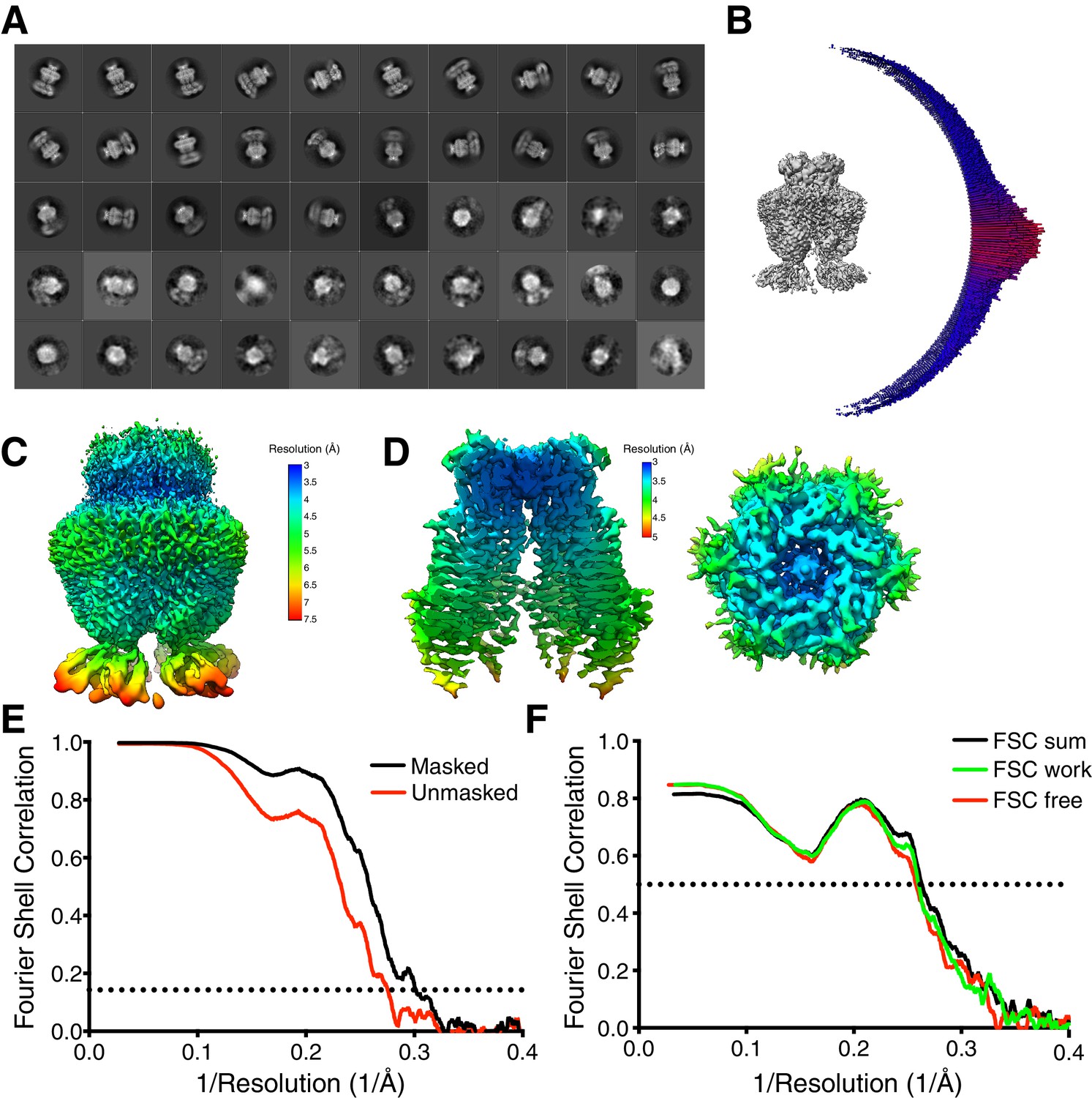
Expanded map and model validation for LRRC8A-DCPIB in MSP1E3D1.
(A) Two-dimensional classes from a fifty-class classification of the final particles. (B) Angular distribution of particle views. (C) Local resolution using Relion locally filtered map at 0.012 threshold showing resolution of the top of LRR region. (D) Local resolution using Relion locally filtered map at 0.035 threshold and dust hidden at 12 Å. Membrane view (left) and extracellular view (right). (E) Fourier Shell Correlation (FSC) of the two unfiltered half-maps from refinement used for calculating overall resolution at 0.143. (F) Model validation FSC using half-map one used for model refinement (FSC work), half-map two not used for refinement (FSC free), and full map (FSC sum).
Figure 1—figure supplement 8
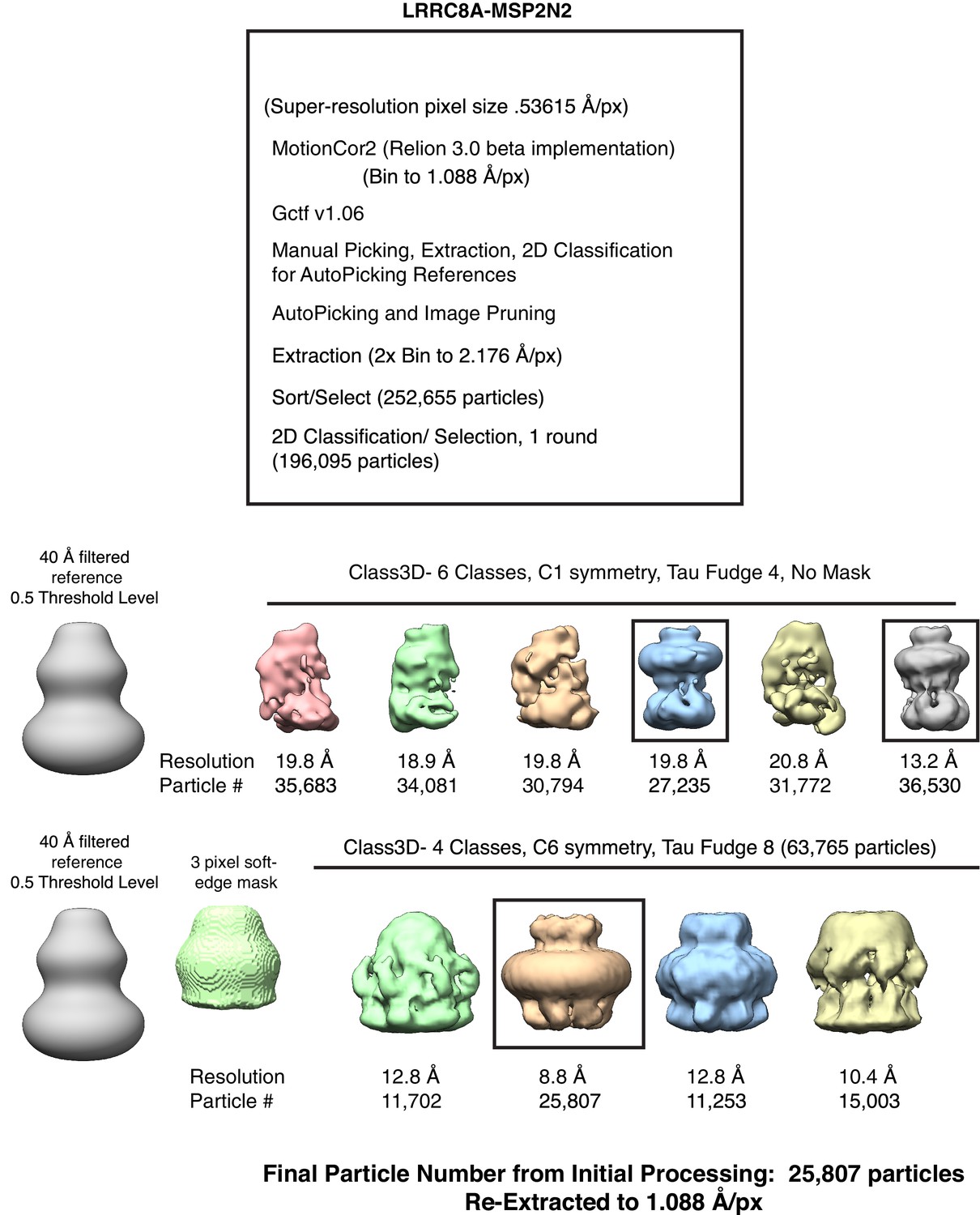
Initial processing for the LRRC8A in MSP2N2 nanodisc dataset.
Particles from boxed classes were passed to the next round.
Figure 1—figure supplement 9
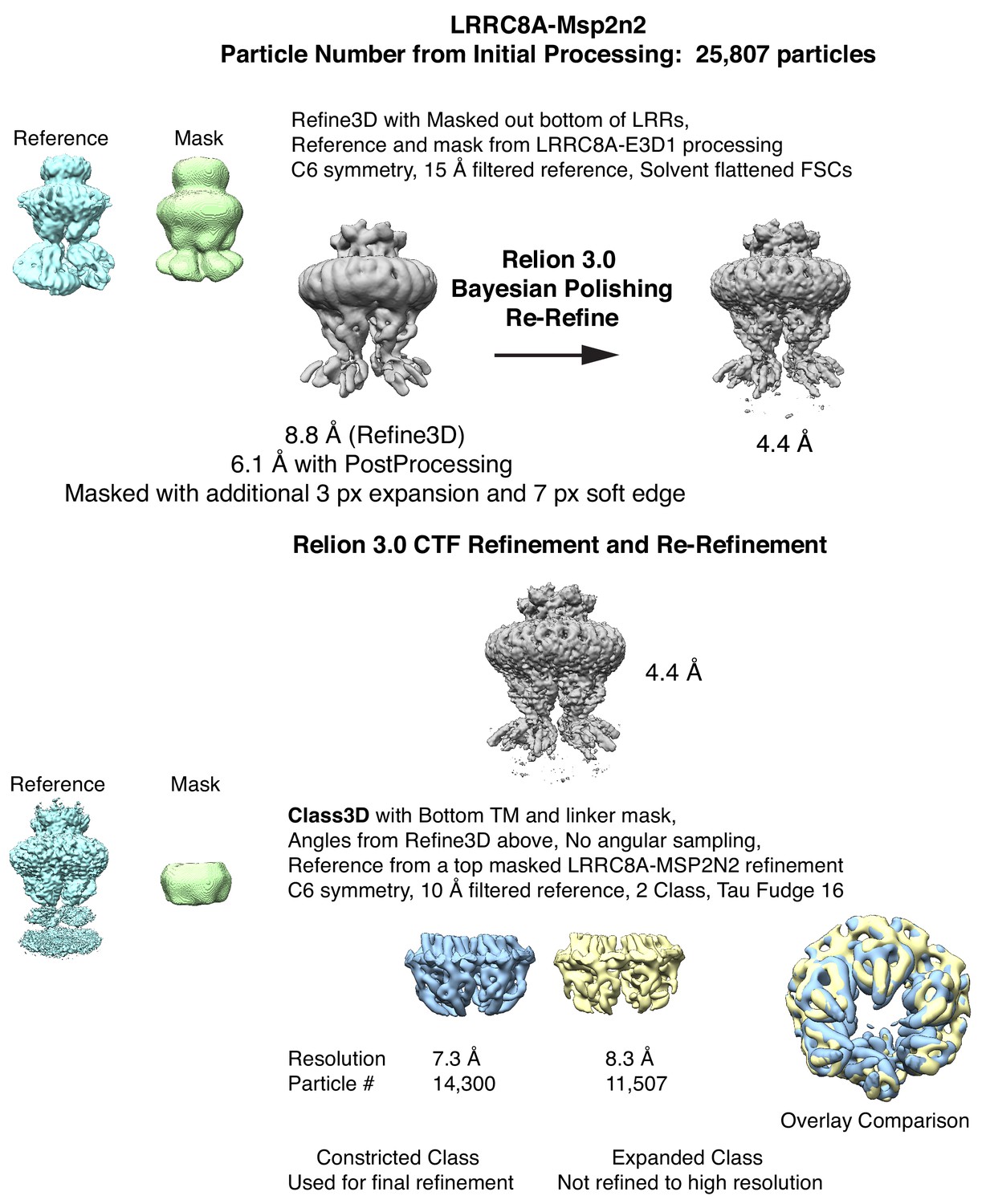
LRRC8A in MSP2N2 refinement, particle polishing, and classing into constricted and expanded linker particles.
Notably, Relion 3.0 Bayesian Polishing was crucial to obtain a high-resolution reconstruction for this dataset. The constricted particle set was refined for a final map. However, the expanded particle set did not converge to high resolution.
Figure 1—figure supplement 10
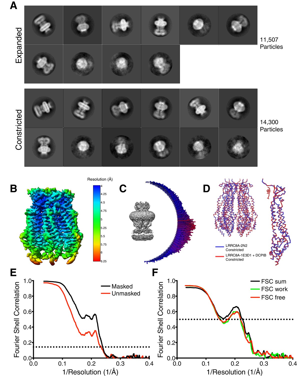
Final particle set 2D classes and the model validation for constricted map of LRRC8 in MSP2N2.
(A) 2D classes from a 12-class job for the expanded (top) and constricted (bottom) particles. For the expanded set, two sparsely populated classes were removed for this visualization. (B) Local resolution map for LRRC8A in MSP2N2 (C) Angular distribution of particle views. (D) Aligned models for the constricted states of LRRC8A-DCPIB in MSP1E3D1 (red) and LRRC8A in MSP2N2 (blue) depicted as a hexamer (left) or a single subunit (right) (E) Fourier Shell Correlation (FSC) of the two unfiltered half-maps from refinement used for calculating overall resolution at 0.143. (F) Model validation FSC using half-map one used for model refinement (FSC work), half-map two not used for refinement (FSC free), and full map (FSC sum).
Figure 2
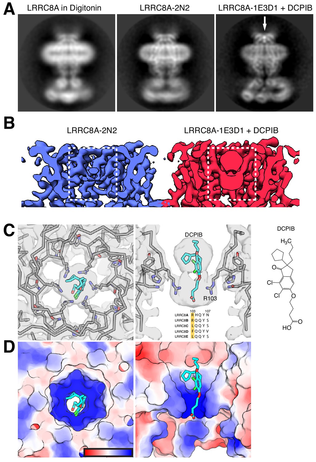
DCPIB inhibitor binding site.
(A) Representative side-view two-dimensional class averages of LRRC8A (left) solubilized in digitonin, (middle) reconstituted in MSP2N2 lipid nanodiscs, or (right) MSP1E3D1 lipid nanodiscs and complexed with DCPIB. An arrow highlights the bi-lobed feature corresponding to DCPIB in the channel selectivity filter. (B) Cryo-EM density maps of the LRRC8A selectivity filter in (left) MSP2N2 nanodiscs or (right) MSP1E3D1 nanodiscs with DCPIB. The DCPIB binding region is highlighted in white boxes for each map. (C) View of the selectivity filter with bound DCPIB from (left) the extracellular solution (top view) and (middle) the membrane plane (side view). The atomic model is shown as ribbons and sticks within the cryo-EM density with the two front and two rear subunits removed in the side view for clarity. Nitrogens are colored blue, oxygens red, chlorines green, protein carbons gray, and DCPIB carbons teal. Alignment of the residues surrounding the selectivity filter for LRRC8 paralogs is shown below the drug density with numbering for LRRC8A above. (right) The chemical structure of DCPIB. (D) Views of the DCPIB-binding site as in (B), but with the atomic surface colored by electrostatic potential from electronegative red (−5 kbTec−1) to electropositive blue (+5 kbTec−1), with the color scale drawn on the left panel.
Figure 3
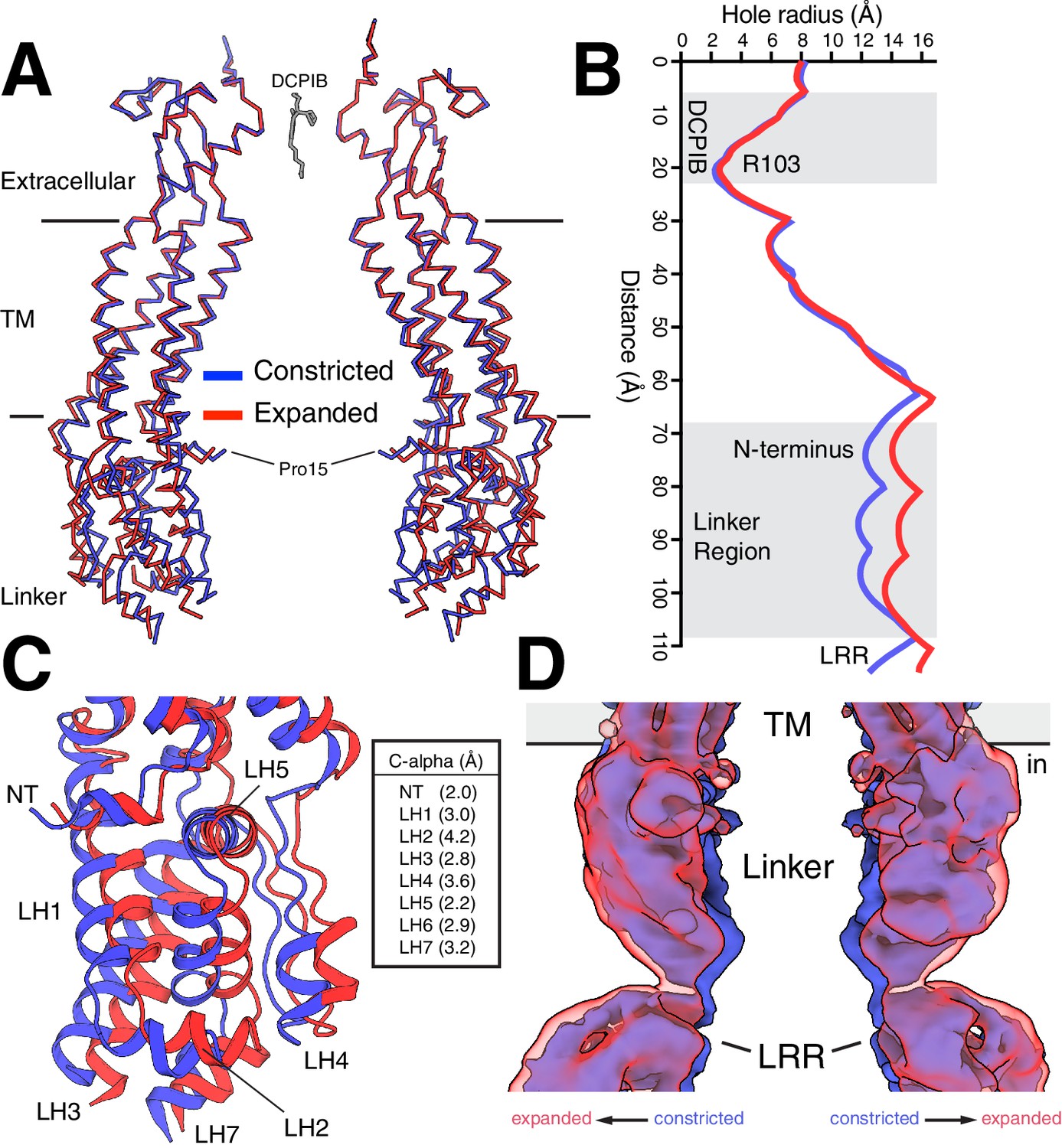
Constricted and expanded LRRC8A structures.
(A) Overlay of constricted (blue) and expanded (red) structures of LRRC8A viewed from the membrane with two opposing subunits shown for each structure in ribbon representation. DCPIB is shown in grey sticks. Proline 15, the final modeled residue at the N-terminus, is labeled. (B) Comparison of the pore radius along the conduction axis colored as in (A). (C) Close-up view of the structure overlay at the linker region with models drawn as cartoons. Helices are labeled and distances between Cα positions at the following positions are indicated: NT, R18; LH1, K162; LH2, T170; LH3, K249; LH4, I356; LH5, S379; LH6, S387; LH7, E399. (D) Overlay of unmasked cryo-EM maps from constricted and expanded particles showing correlated movement of the linker region and membrane-proximal LRR region. Density within 5 Å of two opposing chains is shown.
Figure 4 with 3 supplements
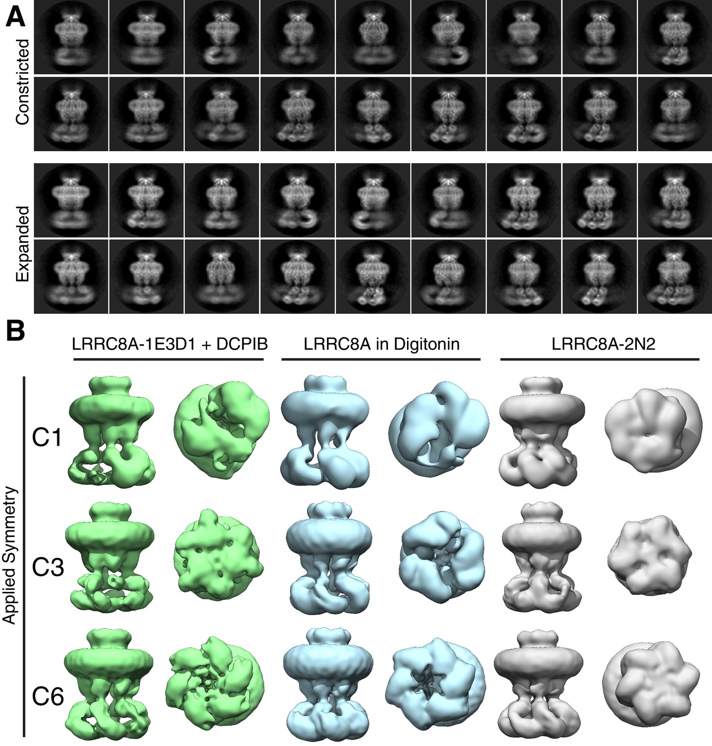
LRR position and channel symmetry differences in lipid and detergent environments.
(A) Side-views of two-dimensional class averages from the (top) constricted and (bottom) expanded particle classes illustrating variation in LRR position. Also see Videos 3 and 4. (B) Symmetry comparison of LRRC8A in (left) MSP1E3D1 + DCPIB, (middle) digitonin, or (right) MSP2N2 (displayed at a 0.015 threshold). Selected classes from three-dimensional classing jobs with the indicated symmetry are shown from the side (membrane) or bottom (cytoplasm).
Figure 4—figure supplement 1
Initial processing for symmetry testing.
(Top) The reference used for the three-dimensional classification is shown boxed in red. No masking was utilized during classification. (Bottom) Two-dimensional class averages from a fifty-class job for the final particle set are shown for digitonin and MSP2N2 data.
Figure 4—figure supplement 2
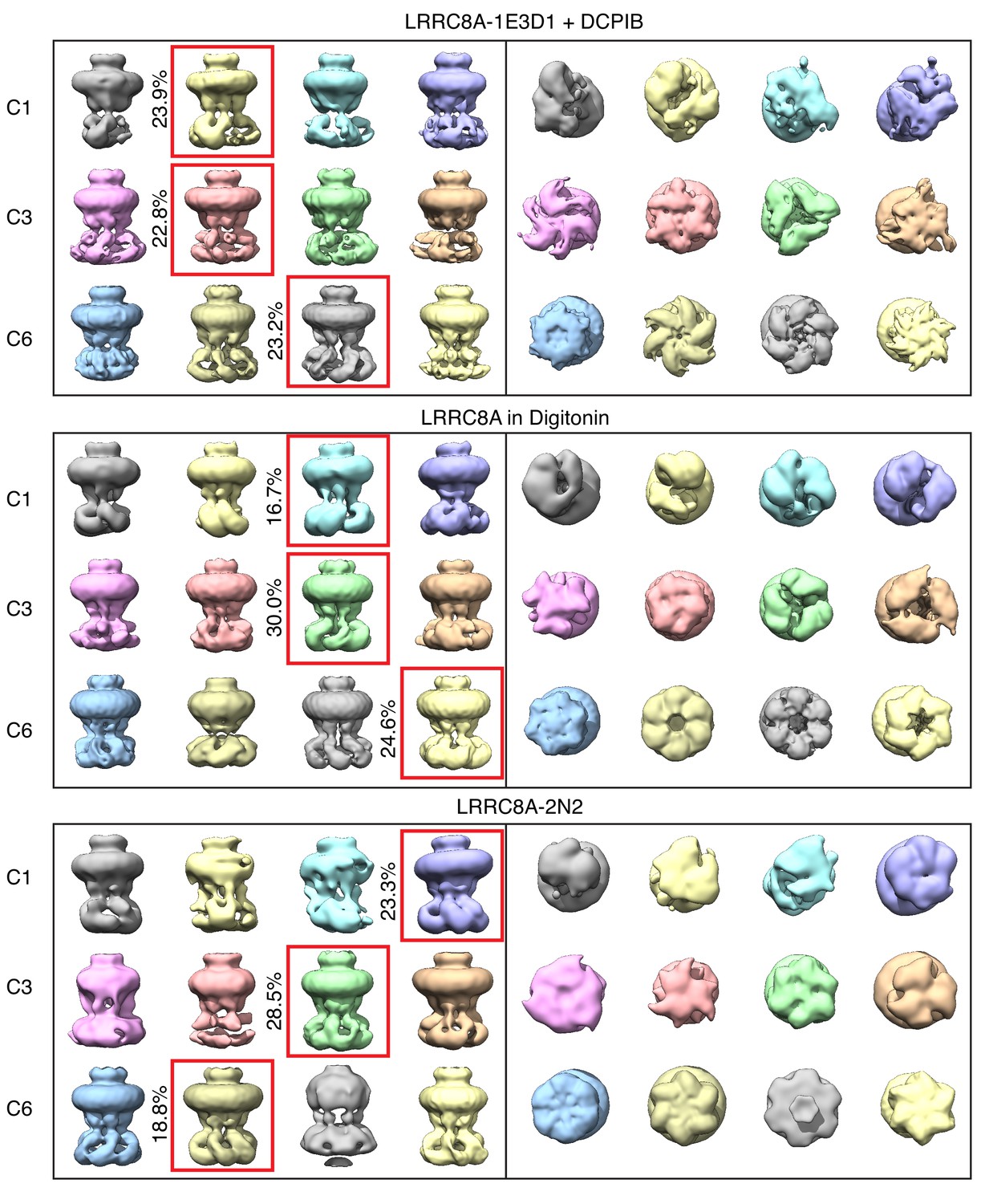
Full three-dimensional classification output for symmetry testing.
All maps are shown at 0.015 threshold. Classes selected for display in Figure 4 are boxed in red with the percentage of particles contributing to the class on the left.
Figure 4—figure supplement 3
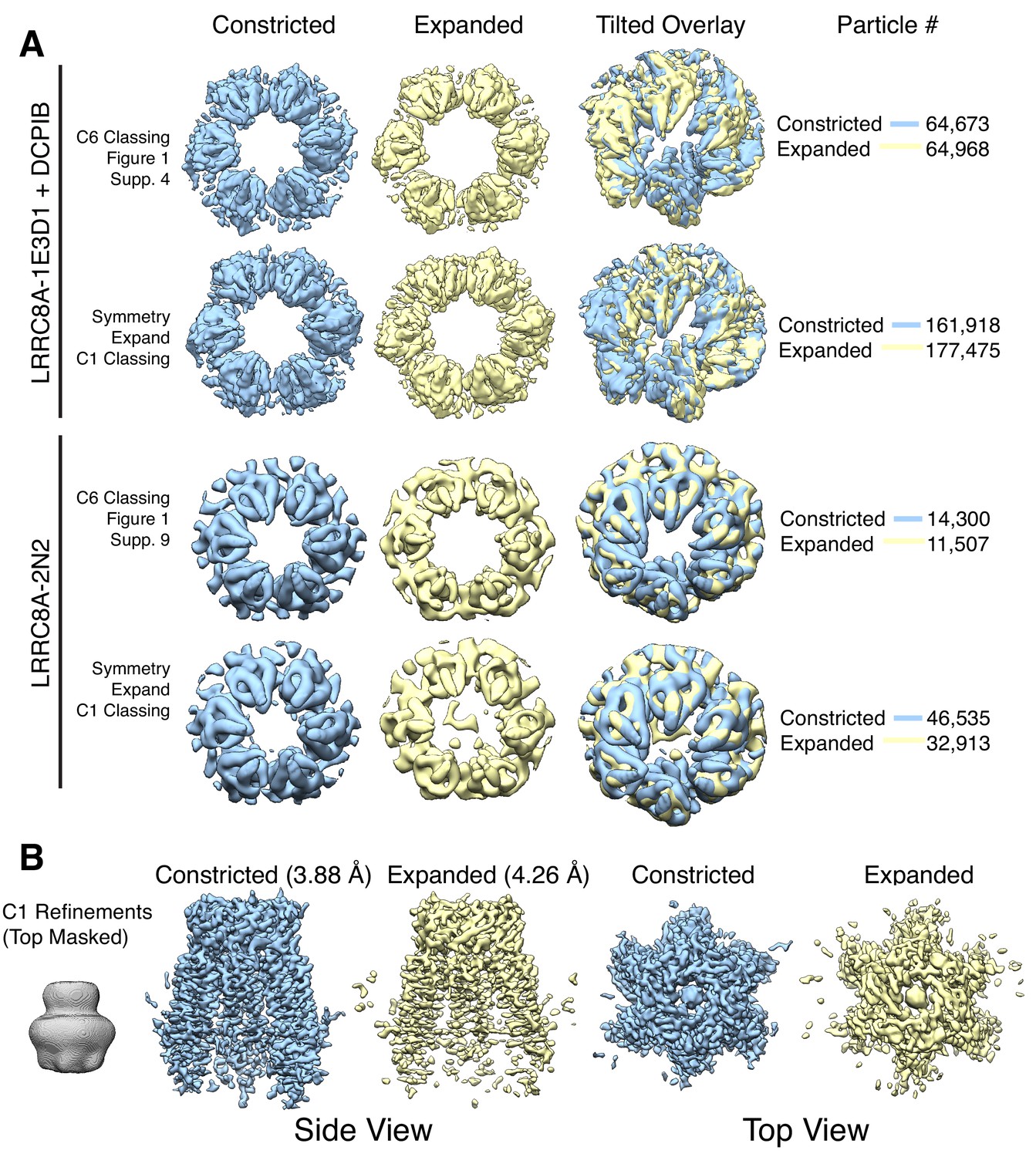
Classification of constricted and expanded states.
(A) To test if asymmetric linker states were present in the data we performed symmetry expansion followed by classing with C1 symmetry for both the LRRC8A-DCPIB in MSP1E3D1 and LRRC8A in MSP2N2 datasets. For MSP1E3D1 with DCPIB, symmetry expansion was performed using Refine3D #2 (Figure 1—figure supplement 4) with C6 symmetry to expand to 777,846 particles. Classification was performed with C1 symmetry and eight classes with the same settings as Figure 1—figure supplement 4. For apo-LRRC8A in MSP2N2, symmetry expansion was performed using the post-CtfRefine refinement (Figure 1—figure supplement 9) with C6 symmetry to expand to 154,842 particles. Classification was performed with C1 symmetry and four classes with the same settings as Figure 1—figure supplement 9. For C1 classification, comparably populated and high-resolution classes were chosen for the constricted and expanded classes displayed here. Notably, asymmetric linker sub-states were not discernible in these classes. (B) Refinements performed using C1 symmetry for the LRRC8A-DCPIB in MSP1E3D1 constricted and expanded final particle datasets.
Figure 5
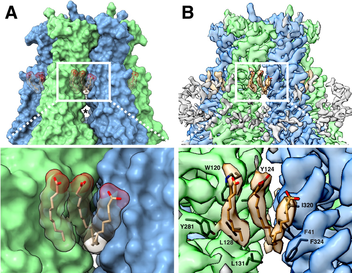
LRRC8A-lipid interactions.
(A, above) Surface representation of the constricted LRRC8A class viewed from the membrane with docked POPC lipid chains depicted in stick (tan) and space-filling (transparent) representations. The upper gap between subunits is filled by lipid density and an asterisk marks the larger lower gap. (Below) a zoomed-in view on the upper gap and bound lipid. (B, above) The corresponding cryo-EM density viewed as in (A), with lipid and the nearby hydrophobic amino-acid side chains shown in stick form. (Below), a zoomed-in view with the amino acids of the hydrophobic pocket labeled.
Figure 6
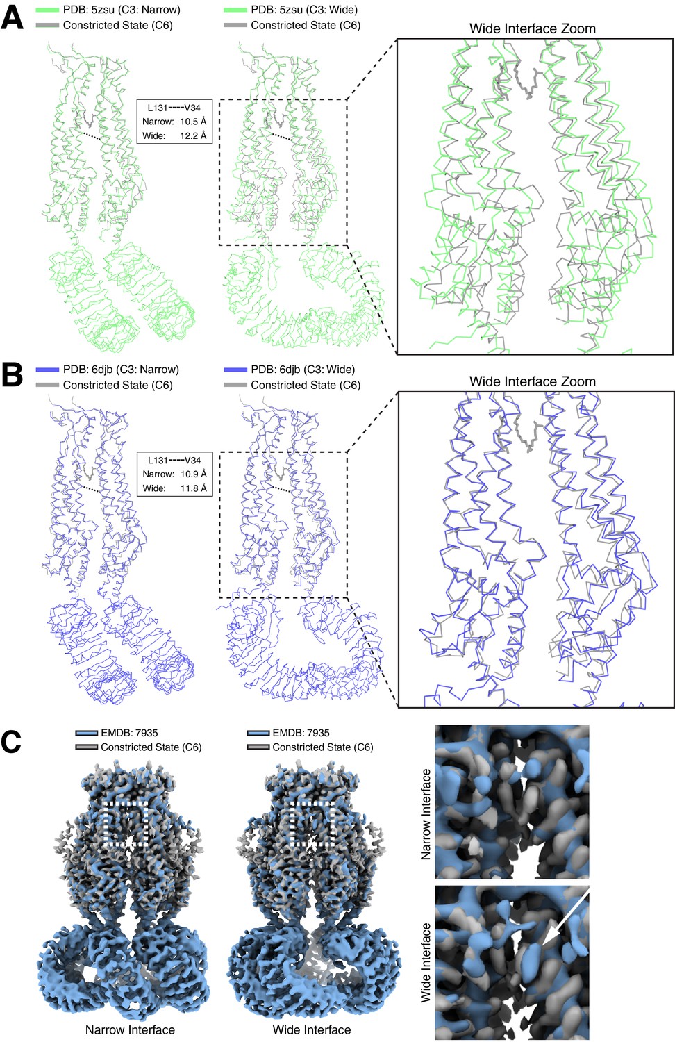
Differences in LRRC8A structures solved in lipid bilayers and detergent micelles.
Overlays of extracellular domain-aligned models of the constricted state structure determined in lipid nanodiscs (gray) and the (left) narrow and (middle) wide subunit interfaces from structures determined in digitonin: (A, PDB: 5zsu, green (Kasuya et al., 2018)), (B, PDB: 6djb, blue (Kefauver et al., 2018)). Boxed and zoomed in regions (right) illustrate the expansion in subunits of the wide interfaces extending to the LRR region. Distances between V34 and L131 Cαs in detergent-solubilized structures are indicated. (C) Aligned density between the six-fold symmetric constricted state in nanodiscs (gray) and LRRC8A in digitonin (Blue, EMDB: 7935, (Kefauver et al., 2018)). (left) View from the membrane of the narrow C3 subunit interface. (middle) View from the membrane of the wide C3 subunit interface. Boxed region and zoomed-in panels (right) show the upper gap filled with lipid density in nanodiscs or an extra density consistent with digitonin in the wide interface marked by a white arrow.
Videos
Video 1
Motion of two opposing chains (as in Figure 3A) as a cartoon-representation morph between constricted and expanded states.
Measurement in Å between the C-alpha of Pro15 is included.
Video 2
Linker region (as in Figure 3C) motion as a cartoon-representation morph between constricted and expanded states.
https://doi.org/10.7554/eLife.42636.016
Video 3
Constricted state side-views of LRRC8A-DCPIB in MSP1E3D1 nanodiscs illustrating heterogenous LRR positions.
https://doi.org/10.7554/eLife.42636.021
Video 4
Expanded state side-views of LRRC8A-DCPIB in MSP1E3D1 nanodiscs illustrating heterogenous LRR positions.
https://doi.org/10.7554/eLife.42636.022
Video 5
Overlay of the constricted state and narrow and wide interfaces of PDB: 6djb.
https://doi.org/10.7554/eLife.42636.025
Video 6
Overlay of the constricted state and narrow and wide interfaces of PDB: 5zsu.
https://doi.org/10.7554/eLife.42636.026Tables
Table 1
Cryo-EM data collection, processing, refinement, and modeling data for LRRC8A-DCPIB in MSP1E3D1 nanodiscs and LRRC8A in MSP2N2 nanodiscs.
https://doi.org/10.7554/eLife.42636.027| LRRC8A-MSP1E3D1 + DCPIB | LRRC8A-MSP2N2 | ||
|---|---|---|---|
| Data collection | Sloan-Kettering | Nysbc | Nysbc |
| Movie # | 2482 | 2110 | 1786 |
| Magnification | 22,500x | 22,500x | 22,500x |
| Voltage (kV) | 300 | 300 | 300 |
| Electron exposure (e–/Å2) | 60.8 | 55.6 | 70.3 |
| Defocus range (μm) | −1.2 ~ −2.5 | −1.2 ~ −2.5 | −1.2 ~ −2.5 |
| Super resolution pixel size (Å) | 0.544 | 0.536 | 0.536 |
| Fourier cropped pixel size (Å) | 1.088 | 1.088 | 1.088 (1.149 Figure 4) |
| Processing | Class 1 (constricted) | Class 2 (expanded) | Class 1 (constricted) |
| Symmetry imposed | C6 | C6 | C6 |
| Initial particle images (no.) | 752,736 | 752,736 | 252, 655 |
| Final particle images (no.) | 25,153 | 35,435 | 11,507 |
| Map resolution (umasked, Å)/FSC threshold | 3.39/0.143 | 3.63/0.143 | 4.28/0.143 |
| Map resolution (masked, Å)/FSC threshold | 3.21/0.143 | 3.32/0.143 | 4.18/0.143 |
| Refinement | |||
| Model resolution (Å) | 3.52/3.32 | 3.81/3.47 | 4.4/3.8 |
| FSC threshold | 0.50/0.143 | 0.50/0.143 | 0.50/0.143 |
| Map-sharpening Bfactor (Å2) | −44.538 | −134.6 | −82.8 |
| Ligands | 19 | 19 | 0 |
| Mean B factors (Å2) | |||
| Protein | 87.73 | 52.17 | 152.09 |
| Ligand | 65.05 | 27.6 | - |
| R.m.s. deviations | |||
| Bond lengths (Å) | 0.007 | 0.005 | 0.004 |
| Bond angles (°) | 0.775 | 0.823 | 0.773 |
| Validation | |||
| MolProbity score | 1.74 | 1.94 | 1.31 |
| Clashscore | 3.34 | 4.12 | 2.16 |
| Poor rotamers (%) | 3.09 | 3.17 | 0.69 |
| EMRinger score | 2.7 | 2.2 | 0.7 |
| Ramachandran plot | |||
| Favored (%) | 96.42 | 94.67 | 95.44 |
| Allowed (%) | 3.58 | 5.33 | 4.56 |
| Disallowed (%) | 0 | 0 | 0.69 |
Table 2
Cryo-EM data collection information for the digitonin datasets used in Figure 2A and Figure 4.
https://doi.org/10.7554/eLife.42636.028| Data collection | Digitonin 70 mM KCl | Digitonin 150 mM KCl | Digitonin 600 mM KCl |
|---|---|---|---|
| Movie # | 2550 | 1445 | 1464 |
| Magnification | 22,500x | 22,500x | 22,500x |
| Voltage (kV) | 300 | 300 | 300 |
| Electron exposure (e–/Å2) | 60.8 | 55.6 | 55.6 |
| Defocus range (μm) | −1.2 ~ −2.5 | −1.2 ~ −2.5 | −1.2 ~ −2.5 |
| Super resolution pixel size (Å) | 0.536 | 0.544 | 0.544 |
| Fourier cropped pixel size (Å) | 1.088 | 1.088 | 1.088 |
Key resources table
| Reagent type (species) or resource | Designation | Source or reference | Identifiers | Additional information |
|---|---|---|---|---|
| Gene (Mus musculus) | LRRC8A | Gen9 synthesis | Uniprot: Q80WG5 | Codon-optimized for Spodoptera frugiperda |
| Cell Line (Spodoptera frugiperda) | Sf9 | Expression Systems | Catalog Number: 94–001F | |
| Peptide, recombinant protein | MSP1E3D1 | Prepared as described in doi: 10.1016/ S0076-6879 (09)64011–8 | His-tag cleaved | |
| Peptide, recombinant protein | MSP2N2 | Prepared as described in doi: 10.1016/S0076-6879 (09)64011–8 | His-tag cleaved | |
| Chemical compound, drug | DDM | Anatrace | Part Number: D310S | |
| Chemical compound, drug | CHS | Anatrace | Part Number: CH210 | |
| Chemical compound, drug | Digitonin | EMD Chemicals | CAS 11024-24-1 | |
| Chemical compound, drug | 16:0-18:1 PC (POPC) lipid | Avanti Polar Lipids | SKU: 850457C | |
| Chemical compound, drug | DCPIB | Tocris | CAS Number: 82749-70-0, Catalog Number: 1540 | |
| Software, algorithm | RELION | doi: 10.7554/eLife.42166 | Relion 3.0 | |
| Software, algorithm | Gctf | doi: 10.1016/j.jsb.2015.11.003 | Gctf v1.06 | |
| Software, algorithm | UCSF Chimera | UCSF | RRID:SCR_004097 | http://plato.cgl.ucsf.edu/chimera/ |
| Software, algorithm | COOT | RRID:SCR_014222 | http://www2.mrc-lmb.cam.ac.uk/personal/pemsley/coot/ | |
| Software, algorithm | Phenix | RRID:SCR_014224 | https://www. phenix-online.org/ | |
| Software, algorithm | PyMOL | PyMOL Molecular Graphics System, Schrodinger LLC | RRID:SCR_000305 | https://www.pymol.org/ |
Additional files
-
Transparent reporting form
- https://doi.org/10.7554/eLife.42636.029
Download links
A two-part list of links to download the article, or parts of the article, in various formats.
Downloads (link to download the article as PDF)
Open citations (links to open the citations from this article in various online reference manager services)
Cite this article (links to download the citations from this article in formats compatible with various reference manager tools)
Cryo-EM structures of the DCPIB-inhibited volume-regulated anion channel LRRC8A in lipid nanodiscs
eLife 8:e42636.
https://doi.org/10.7554/eLife.42636


