Structural diversity of oligomeric β-propellers with different numbers of identical blades
- Max Planck Institute for Developmental Biology, Germany
Figures
Figure 1 with 1 supplement
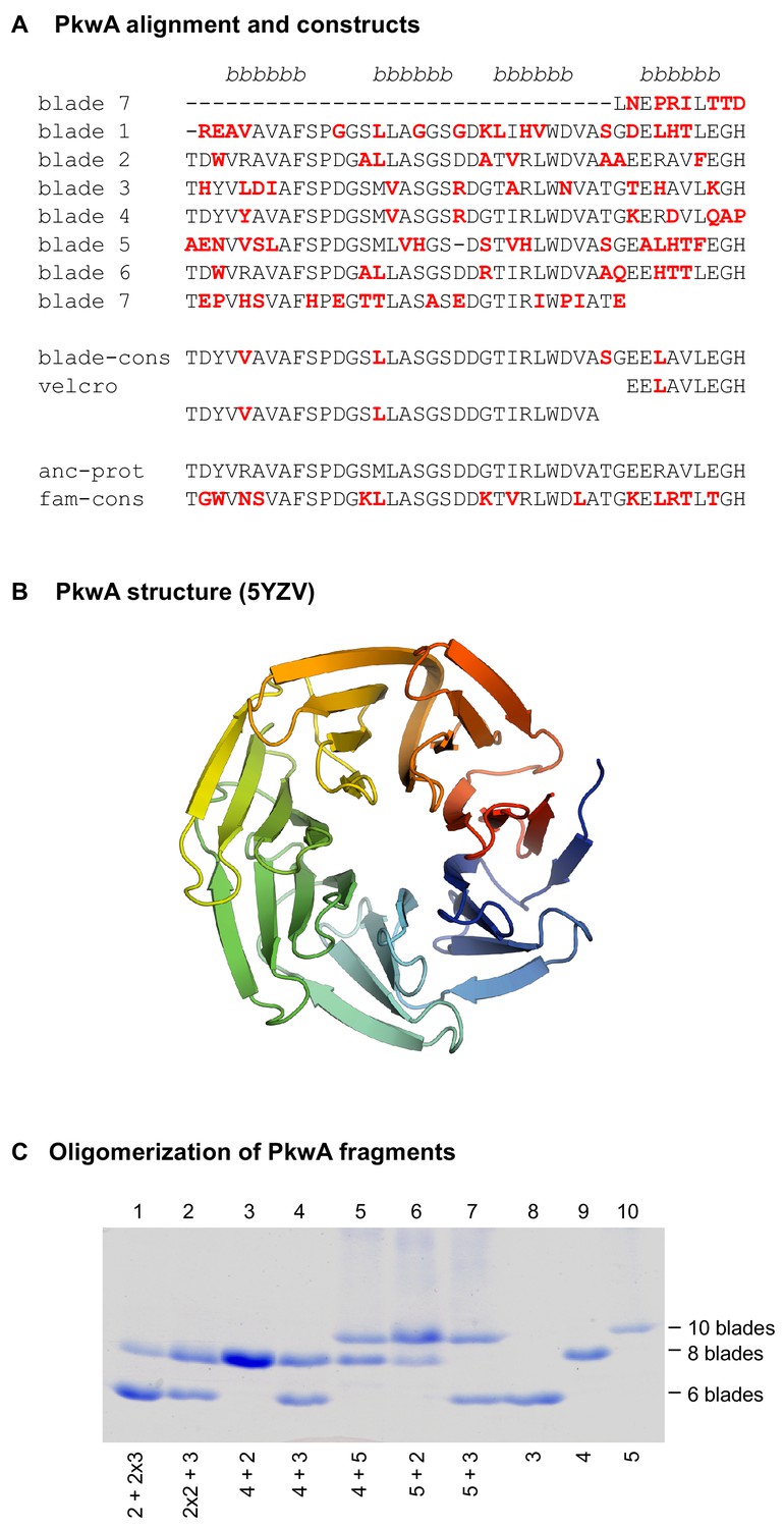
The 7-bladed propeller in the protein PkwA of Thermomonospora curvata.
(A) Multiple sequence alignment of the seven blades and sequences of the constructs used to test the formation of higher-order oligomers in vitro. Non-identical residues in the repeats are colored red. The four β−strands of the propeller blades are indicated above the alignment. (B) Crystal structure of the PkwA propeller (PDB 5YZV). The structure is colored in rainbow colors from blue at the N-terminus to red at the C-terminus. The velcro closure resulting from the last strand of the last blade being permuted to the N-terminus is clearly visible. (C) Oligomerization of PkwA consensus repeats. Differentiation of propeller sizes was achieved by native polyacrylamide gel electrophoresis. Lanes 8–10 show migration of homo-oligomeric propeller complexes assembled from 3-, 4- and 5-bladed repeats. Lanes 1–7 show mixtures of different building blocks to probe for hetero-oligomeric assembly. Proteins were mixed in equimolar ratios (lanes 3–7), unfolded and refolded together. For mixtures of 2- and 3-bladed repeats (lanes 1 and 2) 2:1 molar ratios were used. In all cases, regardless of the mixture composition, PkwA repeats re-assembled only into homo-oligomers.
Figure 1—figure supplement 1
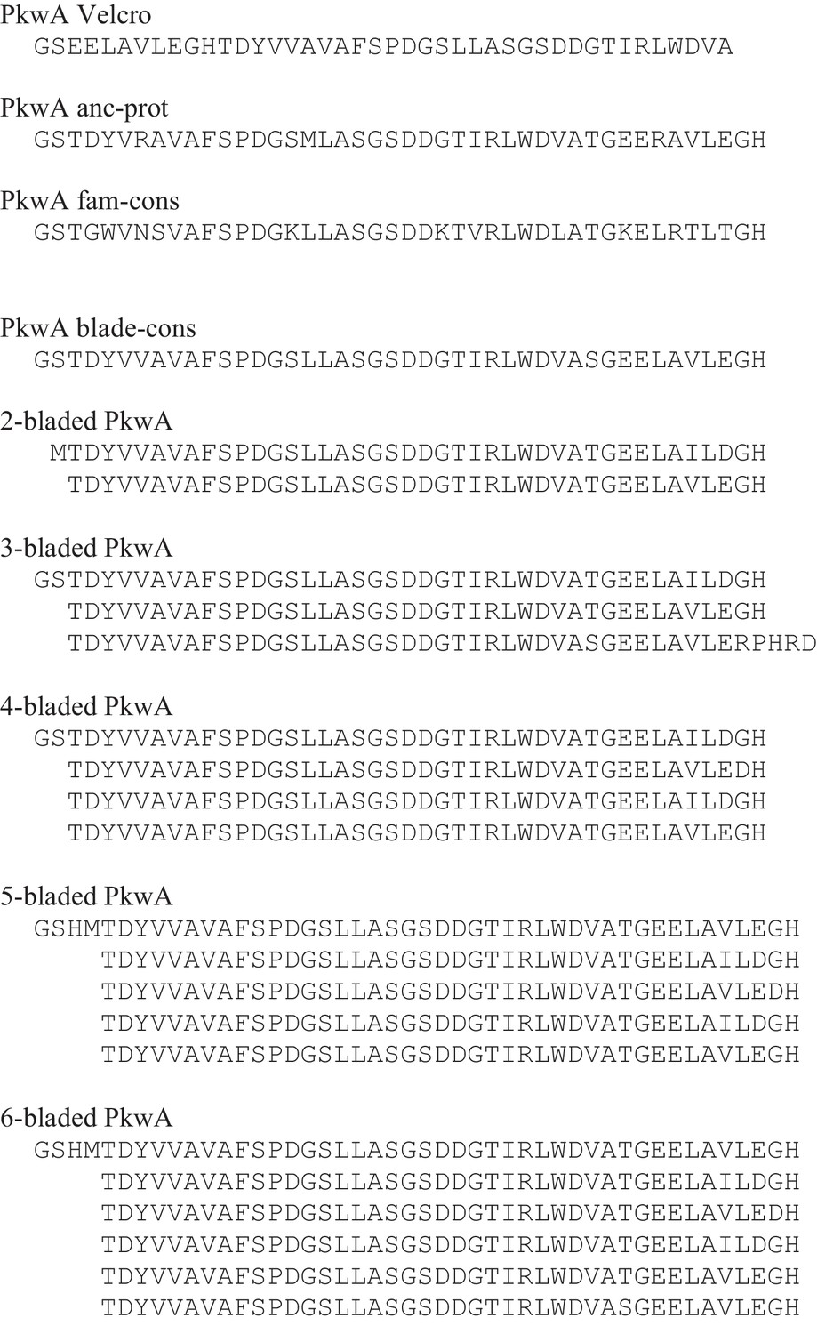
Sequences of PkwA constructs.
Due to the thrombin cleavage site motif, all PkwA proteins originally expressed as GST-fusions start with an additional GS and proteins expressed with a His6-tag start with GSHM.
Figure 2 with 2 supplements

The recently amplified WRAP propeller in Npun_R6612 of Nostoc punctiforme PCC73102.
(A) Multiple sequence alignment of the 14 blades of WRAP. Non-identical repeats are colored in red and the non-repeating β-strand 4 of the velcro blade is underlined. The four β-strands of the propeller blades are indicated above the alignment. The repeat unit chosen for in vitro studies is highlighted by a box. (B) Crystal structure of the WRAP propeller (PDB 2YMU). (C) Oligomerization of WRAP repeats. Assembly was probed by crosslinking proteins with 0.6% glutaraldehyde (GA) and subsequent analysis by SDS-PAGE and Coomassie Blue G250 staining. On the left side, non-crosslinked proteins are shown for comparison.
Figure 2—figure supplement 1
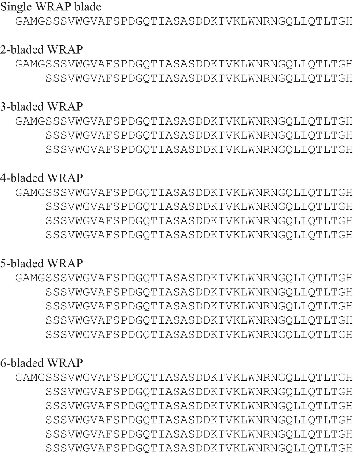
Sequences of WRAP constructs.
All constructs start with an additional GAMG after TEV-cleavage.
Figure 2—figure supplement 2
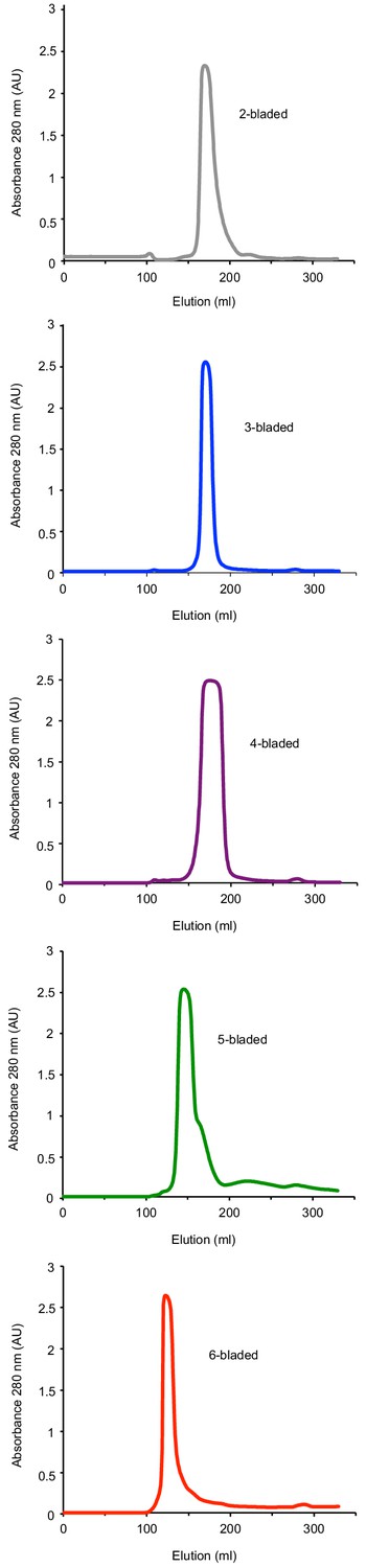
Purification of WRAP fragments.
Shown are the elution profiles of the Superdex 75 10/300 gel size-exclusion column runs.
Figure 3 with 1 supplement
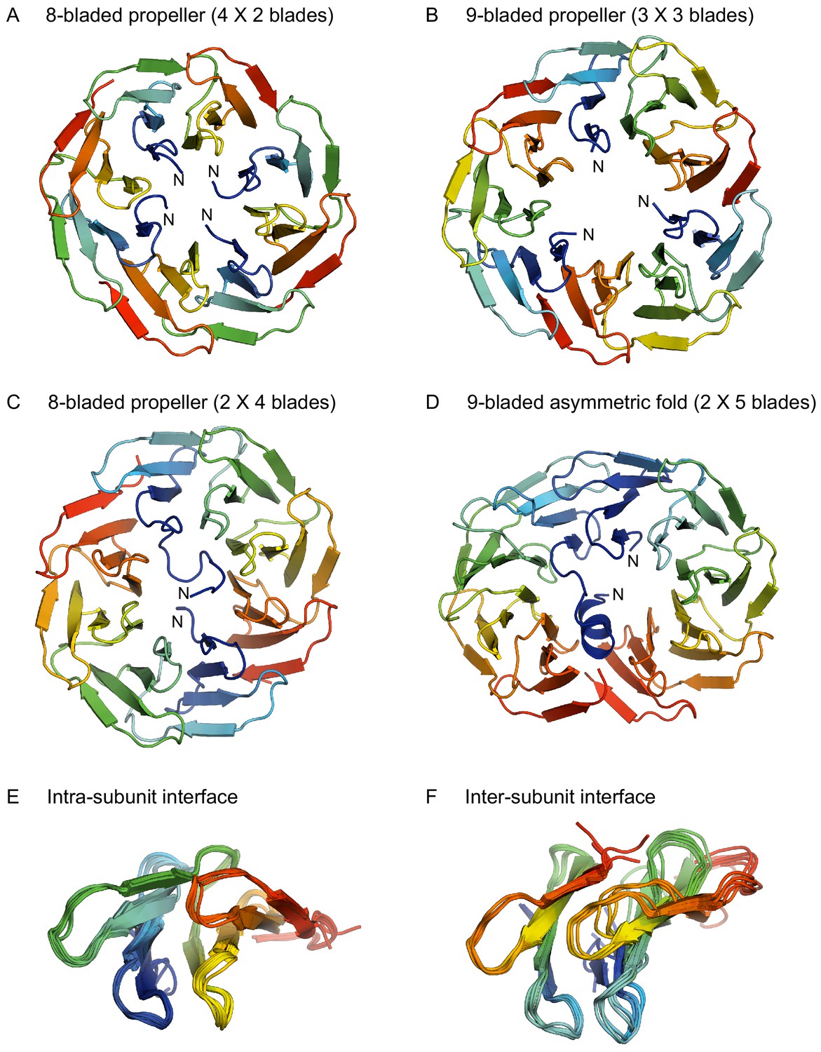
Structures of homo-oligomeric WRAP propellers.
Subunits in each propeller are colored in rainbow colors with blue at the N-terminus and red at the C-terminus. (A) 8-bladed propeller formed of four 2-bladed fragments (PDB 6R5X). (B) 9-bladed propeller formed of three 3-bladed fragments (6R5Z). (C) 8-bladed propeller formed of two 4-bladed fragments (6R5Y). (D) 9-bladed asymmetric fold formed of two 5-bladed fragments (6R60). (E) Superimposition of all intra-subunit interfaces in 2-, 3-, 4-, and 5-bladed fragments. (F) Superimposition of all inter-subunit interfaces in the two 8-bladed propellers and the 9-bladed propeller.
Figure 3—figure supplement 1
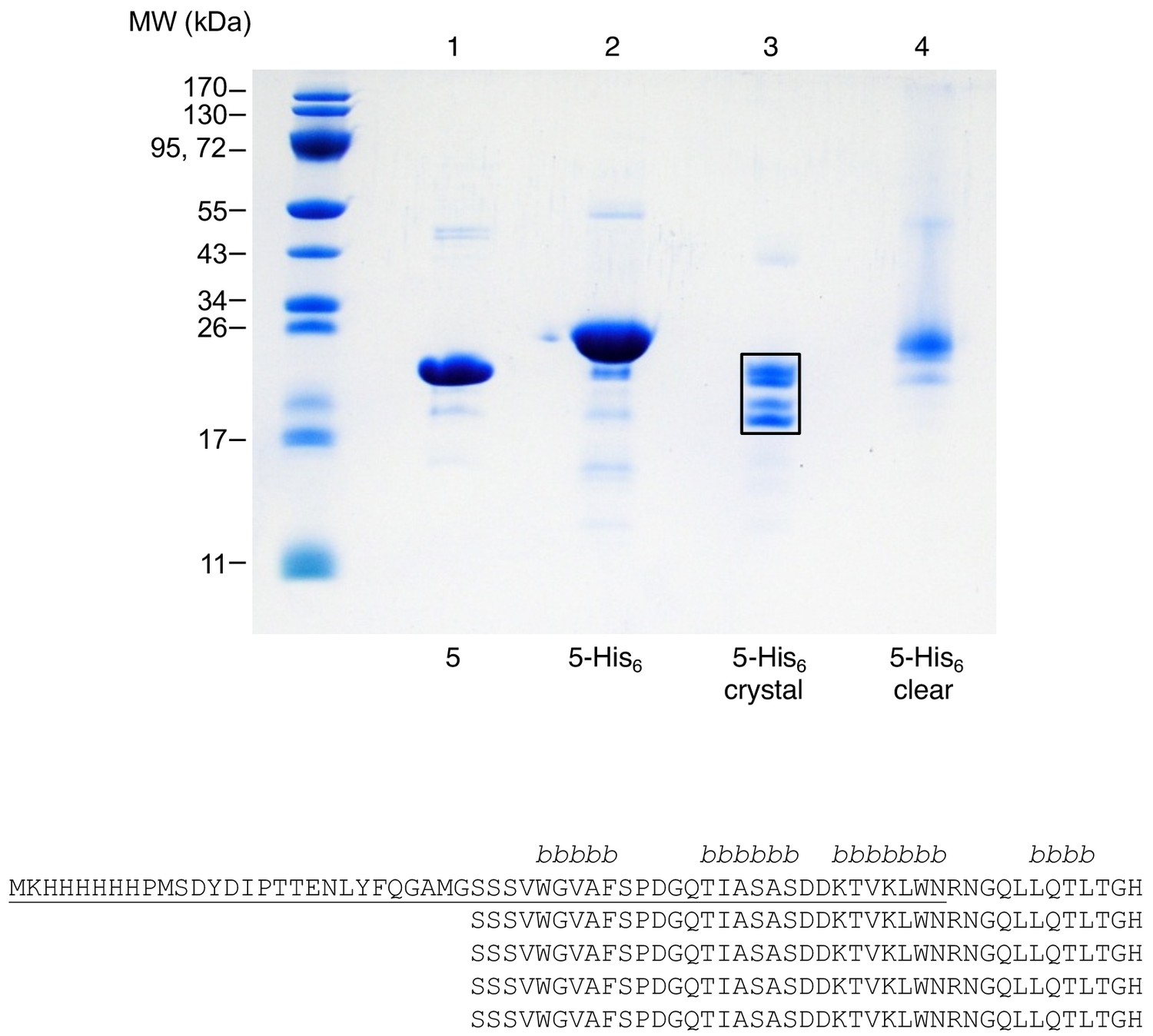
Truncation of 5-bladed WRAP repeats correlates with an asymmetric, incomplete propeller structure.
Crystals of the dimerized 5-bladed His6-tagged WRAP repeat were dissolved and analyzed by SDS-PAGE and Coomassie Blue G250 staining (lane 3). For comparison, native (lane 1) and His6-tagged (lane 2) 5-bladed repeats are shown. In lane 4, protein from a clear crystallization solution that gave no crystals is loaded. The resulting four bands of lane 3 (boxed) were analyzed by mass spectrometry and shown to contain protein species with an intact N-terminal blade (upper two bands), and protein species were the first blade is largely missing (lower two bands; underlined part in the sequence of the 5-bladed fragment). The four β−strands in an intact WRAP blade are indicated above the alignment.
Tables
Table 1
Summary of biophysical data for the different propeller constructs of PkwA (upper panel) and WRAP (lower panel).
https://doi.org/10.7554/eLife.49853.004| Propeller Blades in protomer | Molecular mass protomer calculated | Molecular mass SLS measured | Assembly state based on SLS | CD melting temperature Tm | Tryptophan fluorescence λmax |
|---|---|---|---|---|---|
| 2 | 8.8 kDa | 33.9 kDa | Tetramer | 52°C | 331 nm |
| 3 | 13.6 kDa | 27.8 kDa | Dimer | 67°C | 335 nm |
| 4 | 17.5 kDa | 32.7 kDa | Dimer | 63°C | 332 nm |
| 5 | 21.8 kDa | 42.6 kDa | Dimer | 65°C | 333 nm |
| 2 | 8.9 kDa | 28.4 kDa | Tetramer | 43°C | 345 nm |
| 3 | 13.2 kDa | 39.5 kDa | Trimer | 54°C | 341 nm |
| 4 | 17.6 kDa | 26.7 kDa | Dimer | 65°C | 341 nm |
| 5 | 22 kDa | 46.9 kDa | Dimer | 62°C | 341 nm |
| 6 | 26.3 kDa | 107 kDa | Tetramer | 63°C | 340 nm |
Table 2
Crystallization conditions and cryo protection
https://doi.org/10.7554/eLife.49853.010| Construct | Protein solution | Reservoir solution (RS) | Cryo solution |
|---|---|---|---|
| 2-blades | 8 mg/ml protein 50 mM TRIS HCl pH 8.0 150 mM sodium chloride | 200 mM sodium acetate 100 mM TRIS HCl pH 8.5 30%(w/v) PEG 4000 | n/a |
| 3-blades | 23 mg/ml protein 50 mM TRIS HCl pH 7.5 150 mM sodium chloride | 200 mM ammonium fluoride 20%(w/v) PEG 3350 | RS + 10%(v/v) PEG 400 |
| 4-blades | 23 mg/ml protein 50 mM TRIS HCl pH 7.5 150 mM sodium chloride | 10 mM zinc chloride 100 mM Hepes pH 7.0 20%(w/v) PEG 6000 | RS + 10%(v/v) PEG 400 |
| 5-blades | 4 mg/ml protein 50 mM HEPES pH 7.5 100 mM sodium chloride | 100 mM Magnesium chloride 100 mM HEPES pH 7.0 15%(w/v) PEG 4000 | RS + 15%(v/v) PEG 400 |
Table 3
Crystallographic data collection and refinement statistics
https://doi.org/10.7554/eLife.49853.011| Construct (PDB ID) | 2-blades (6R5X) | 3-blades (6R5Z) | 4-blades (6R5Y) | 5-blades (6R60) |
|---|---|---|---|---|
| Data collection | ||||
| Space group | C2221 | P21 | P21212 | C2 |
| Cell dimensions | ||||
| a, b, c (Å) | 55.24, 119.7, 84.15 | 53.55, 92.35, 61.43 | 97.72, 127.2, 72.96 | 39.75, 107.5, 179.2 |
| α, β, γ (°) | 90.00, 90.00, 90.00 | 90.00, 94.95, 90.00 | 90.00, 90.00, 90.00 | 90.00, 94.40, 90.00 |
| Resolution (Å) | 32.3–1.70 (1.80–1.70) * | 38.3–1.75 (1.85–1.75) * | 38.7–2.15 (2.28–2.15) * | 39.8–1.75 (1.85–1.75) * |
| Rmerge | 4.8 (56.7) | 6.3 (89.4) | 11.0 (76.4) | 8.8 (45.4) |
| I / σI | 17.8 (2.32) | 13.5 (1.55) | 10.1 (1.94) | 9.17 (1.92) |
| Completeness (%) | 99.3 (97.4) | 99.2 (95.9) | 99.4 (99.0) | 98.1 (95.3) |
| Redundancy | 4.31 (4.38) | 4.67 (4.44) | 3.70 (3.51) | 3.31 (3.40) |
| Refinement | ||||
| Resolution (Å) | 32.3–1.70 | 38.3–1.75 | 38.7–2.15 | 39.8–1.75 |
| No. reflections | 29426 | 56974 | 47400 | 70952 |
| Rwork/Rfree | 0.20/0.24 | 0.19/0.21 | 0.22/0.25 | 0.20/0.24 |
| No. atoms | ||||
| Protein | 2364 | 5357 | 7350 | 5639 |
| Ligands (Zn2+) | 0 | 0 | 6 | 0 |
| Water | 314 | 302 | 330 | 691 |
| B-factors | ||||
| Protein | 24.30 | 32.30 | 36.50 | 26.70 |
| Ligands (Zn2+) | - | - | 50.60 | - |
| Water | 35.30 | 36.70 | 33.70 | 35.50 |
| R.m.s. deviations | ||||
| Bond lengths (Å) | 0.012 | 0.017 | 0.011 | 0.013 |
| Bond angles (°) | 1.55 | 1.72 | 1.51 | 1.53 |
-
*Values in parentheses are for highest-resolution shell.
Download links
A two-part list of links to download the article, or parts of the article, in various formats.
Downloads (link to download the article as PDF)
Open citations (links to open the citations from this article in various online reference manager services)
Cite this article (links to download the citations from this article in formats compatible with various reference manager tools)
Structural diversity of oligomeric β-propellers with different numbers of identical blades
eLife 8:e49853.
https://doi.org/10.7554/eLife.49853


