TBX3 acts as tissue-specific component of the Wnt/β-catenin transcriptional complex

  1. Dario Zimmerli
  2. Costanza Borrelli
  3. Amaia Jauregi-Miguel
  4. Simon Söderholm
  5. Salome Brütsch
  6. Nikolaos Doumpas
  7. Jan Reichmuth
  8. Fabienne Murphy-Seiler
  9. MIchel Aguet
  10. Konrad Basler  Is a corresponding author
  11. Andreas E Moor  Is a corresponding author
  12. Claudio Cantù  Is a corresponding author
  1. Department of Molecular Life Sciences, University of Zurich, Switzerland
  2. Department of Biosystems Science and Engineering, ETH Zürich, Switzerland
  3. Wallenberg Centre for Molecular Medicine, Linköping University, Sweden
  4. Department of Biomedical and Clinical Sciences, Division of Molecular Medicine and Virology; Faculty of Medicine and Health Sciences; Linköping University, Sweden
  5. Swiss Institute for Experimental Cancer Research (ISREC), Ecole Polytechnique Fédérale de Lausanne (EPFL), School of Life Sciences, Switzerland

Abstract

BCL9 and PYGO are β-catenin cofactors that enhance the transcription of Wnt target genes. They have been proposed as therapeutic targets to diminish Wnt signaling output in intestinal malignancies. Here we find that, in colorectal cancer cells and in developing mouse forelimbs, BCL9 proteins sustain the action of β-catenin in a largely PYGO-independent manner. Our genetic analyses implied that BCL9 necessitates other interaction partners in mediating its transcriptional output. We identified the transcription factor TBX3 as a candidate tissue-specific member of the β-catenin transcriptional complex. In developing forelimbs, both TBX3 and BCL9 occupy a large number of Wnt-responsive regulatory elements, genome-wide. Moreover, mutations in Bcl9 affect the expression of TBX3 targets in vivo, and modulation of TBX3 abundance impacts on Wnt target genes transcription in a β-catenin- and TCF/LEF-dependent manner. Finally, TBX3 overexpression exacerbates the metastatic potential of Wnt-dependent human colorectal cancer cells. Our work implicates TBX3 as context-dependent component of the Wnt/β-catenin-dependent transcriptional complex.

Introduction

The Wnt pathway is an evolutionarily conserved cell signaling cascade that acts as major driving force of several developmental processes, as well as for the maintenance of the stem cell populations within adult tissues (Nusse and Clevers, 2017). Deregulation of this signaling pathway results in a spectrum of consequences, ranging from lethal developmental abnormalities to several forms of aggressive cancer (Nusse and Clevers, 2017). Most prominently, colorectal cancer (CRC) is initiated by genetic mutations that constitutively activate Wnt signaling (Kahn, 2014).

Secreted WNT ligands trigger an intracellular biochemical cascade in the receiving cells that culminates in the calibrated expression of target genes (Mosimann et al., 2009). This transcriptional response is orchestrated by nuclear β-catenin, that acts as a ‘scaffold’ to buttress a host of co-factors to cis-regulatory elements occupied by the TCF/LEF transcription factors (Valenta et al., 2012). Among the co-factors, the two paralogs BCL9 and BCL9L (referred to as BCL9/9L) and PYGO1/2 proteins reside within the Wnt/β-catenin transcriptional complex, and their concerted action is required to efficiently activate Wnt-target gene expression (Kramps et al., 2002; Parker et al., 2002; van Tienen et al., 2017Figure 1A). During vertebrate development, their requirement in the β-catenin-mediated transcription appears to be context-dependent (Cantù et al., 2018; Li et al., 2007), and they also have evolved β-catenin-independent functions (Cantù et al., 2017; Cantù et al., 2014). Curiously however, BCL9 and PYGO always seem to act as a ‘duet’ (Kennedy et al., 2010).

Figure 1 with 2 supplements see all
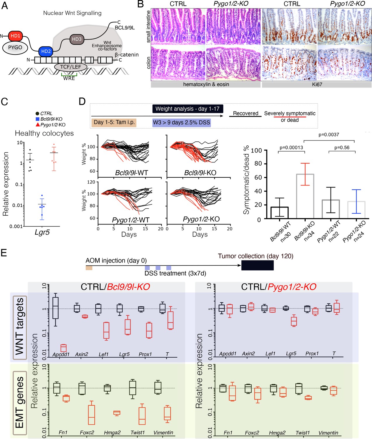
The intestinal epithelium-specific recombination of Pygo1/2 does not recapitulate the effects of deleting Bcl9/9l.

(A) Schematic representation of the Wnt/β-catenin transcriptional complex, with emphasis on the so-called ‘chain of adaptors’ components, β-catenin, BCL9/9L and PYGO; Wnt Responsive Element (WRE). The homology domains 1–3 (HD1-3) of BCL9/9L are shown. (B) Epithelial-specific Pygo1/2 deletion (via vil-Cre-ERt2; Pygo1/2-KO) does not lead to any obvious histological or functional defect, neither in the small intestine nor in the colon as seen by hematoxylin and eosin staining (left panels). The proliferative compartment, detected via Ki67 (right panels), seems also unaffected (also refer to the count in Figure 1—figure supplement 1D–E). (C) Quantitative RT-PCR detecting Lgr5 mRNA extracted from colonic epithelium of control (black), Bcl9/9l (blue) or Pygo1/2 (red) conditional mutants (KO). (D) 6–8 week-old male mice were treated with five tamoxifen (Tam) injections (i.p., 1 mg/day) for five consecutive days. 10 days later mice were treated with 2.5% dextran sodium sulfate (DSS) ad libitum in drinking water for 9 days. While 17% of control mice (N = 30) were severely affected or died due to the DSS treatment (red lines), 65% of conditional Bcl9/9l-KO (N = 34) mice performed poorly in this test. Deletion of Bcl9/9l increased significantly the death rate after DSS treatment (p-value=0.00013 in Fisher's Exact Test). No difference between Pygo1/2-KO and control mice could be measured: 27% of control mice (N = 22) and 25% of Pygo1/2-KO (N = 24) were affected upon DSS treatment (p-value=0.5626 in Fisher's Exact Test). (E) 6–8 week-old female mice were exposed to a single dose of the carcinogenic agent azoxymethane (AOM), followed by 7 days of DSS administration in the drinking water. This regimen results in the emergence of dysplastic adenomas that are collected for RNA extraction and analysis of the indicated targets via RT-PCR: Wnt target genes and genes expressed during epithelial-to-mesenchymal transition (EMT), associated with cancer metastasis.

Importantly, BCL9/9L and PYGO proteins were found to significantly contribute to the malignant traits typical of Wnt-induced CRCs (Deka et al., 2010; Gay et al., 2019; Jiang et al., 2020; Mani et al., 2009; Mieszczanek et al., 2019; Moor et al., 2015; Talla and Brembeck, 2016). These observations provided impetus to consider the BCL9/PYGO axis as relevant ‘targetable’ unit in CRC (Lyou et al., 2017; Mieszczanek et al., 2019; Talla and Brembeck, 2016; Zimmerli et al., 2017).

However, here we noticed an apparent divergence between the roles of BCL9/9L and PYGO proteins. We found that genetic abrogation of Bcl9/9l in mouse CRC cells results in broader consequences than Pygo1/2 deletion, suggesting that BCL9 function does not entirely depend on PYGO1/2. Among the putative β-catenin/BCL9 interactors we identified the developmental transcription factor TBX3. Intriguingly, we show that also during forelimb development, BCL9/9L possess a PYGO-independent role. In this in vivo context, TBX3 occupies β-catenin/BCL9 target loci genome-wide, and mutations in Bcl9/9l affect the expression of TBX3 targets. Finally, TBX3 modulates the expression of Wnt target genes in a β-catenin- and TCF/LEF-dependent manner, and increases the metastatic potential of human CRC cells when overexpressed. We conclude that TBX3 can assist the Wnt/β-catenin mediated transcription in selected developmental contexts, and that this partnership could be aberrantly reactivated in some forms of Wnt-driven CRCs.

Results and discussion

We induced intestinal epithelium-specific recombination of Pygo1/2 loxP alleles (Pygo1/2-KO), that efficiently deleted these genes in the whole epithelium, including the stem cells compartment (Figure 1—figure supplement 1A and B). Consistently with recent reports (Mieszczanek et al., 2019; Talla and Brembeck, 2016), and similarly to deletion of Bcl9/9l (Deka et al., 2010; Mani et al., 2009; Moor et al., 2015), Pygo1/2-KO displayed no overt phenotypic defects (Figure 1B; Figure 1—figure supplement 1C–E). We were surprised in noticing that the expression of Lgr5, the most important intestinal stem cell marker and Wnt target gene (Barker et al., 2007), was heavily downregulated upon loss of Bcl9/9l but unaffected in Pygo1/2-KO (Figure 1C). To address the functionality of the stem cell compartment in these two conditions, we subjected both Bcl9/9l and Pygo1/2 compound mutants (KO) to a model of intestinal regeneration by DSS treatment (Kim et al., 2012Figure 1D). While Bcl9/9l-KO mice showed a defect in regeneration after insult (Deka et al., 2010), Pygo1/2-KO proved indifferent when compared to control littermates (Figure 1D). While we cannot exclude that PYGO1/2 also contributes to the Wnt/β-catenin-dependent transcriptional regulation, our results highlight that the BCL9/9L function in the intestinal epithelium homeostasis and regeneration does not entirely depend on PYGO1/2. This was surprising, since BCL9/9L proteins were thought to act as mere ‘bridge’ proteins that tethered PYGO to the β-catenin transcriptional complex (Figure 1AFiedler et al., 2015; Mosimann et al., 2009). Both BCL9 and PYGO proteins have been implicated in colorectal carcinogenesis (Gay et al., 2019; Jiang et al., 2020; Mieszczanek et al., 2019; Talla and Brembeck, 2016). We tested if the consequence of the deletion of Bcl9/9l and Pygo1/2 genes was also different in the context of carcinogenesis. Specifically, we looked at the contribution to gene expression in chemically-induced AOM/DSS colorectal tumors (Figure 1E). As previously observed, Bcl9/9l-KO tumors exhibit a massive decrease in Wnt target gene expression, epithelial-to-mesenchymal transition (EMT) and stemness traits (Deka et al., 2010; Moor et al., 2015), which was not observed in Pygo1/2-KO tumors (Figure 1E, Figure 1—figure supplement 2A and B). This phenotypic difference is consistent with a recent study in which Bcl9/9l but not Pygo1/2 loss reduced the activation of Wnt target genes induced by APC loss-of-function (Mieszczanek et al., 2019); we interpret this as an independent validation of our observation. All these experiments open up the question of how BCL9/9L imposes its function independently of PYGO.

Surprisingly, the intestine-specific deletion of the homology domain 1 (HD1) of BCL9/9L (Figure 2A), that was previously annotated to interact only with PYGO1/2 (Cantù et al., 2014; Kramps et al., 2002), i) suppressed the metastatic phenotype of the AOM/DSS tumors while deletion of Pygo1/2 did not (Figure 2B) and ii) induced a strong downregulation of Wnt target, EMT and stemness genes (Figure 2C, Figure 2—figure supplement 1). The discrepancy between the gene expression changes induced by recombining Pygo1/2 or deleting the HD1 domain of Bcl9/9l implies that currently unknown proteins assist BCL9/9L function. We set out to identify new candidate BCL9 partners that might be responsible for the different phenotypes. To this aim, we performed a pull-down of tumor proteins expressing either a full-length or a HD1-deleted variant of BCL9, followed by mass spectrometry (Figure 2D). Among the proteins differentially pulled down by control but not by mutant BCL9 we detected TBX3 (Figure 2D and E) and selected it for further validation. The in vivo deletion of the HD1 domain (in Bcl9/9l-ΔHD1 embryos) leads to severe forelimb malformations, while Pygo1/2-KO embryonic forelimbs are unaffected (Figure 2F)(also see Schwab et al., 2007). Limb development, thus, represents another context where BCL9/9L appear to act independently of PYGO. Of note, TBX3 plays a fundamental role in the development of this structure (Frank et al., 2013).

Figure 2 with 2 supplements see all
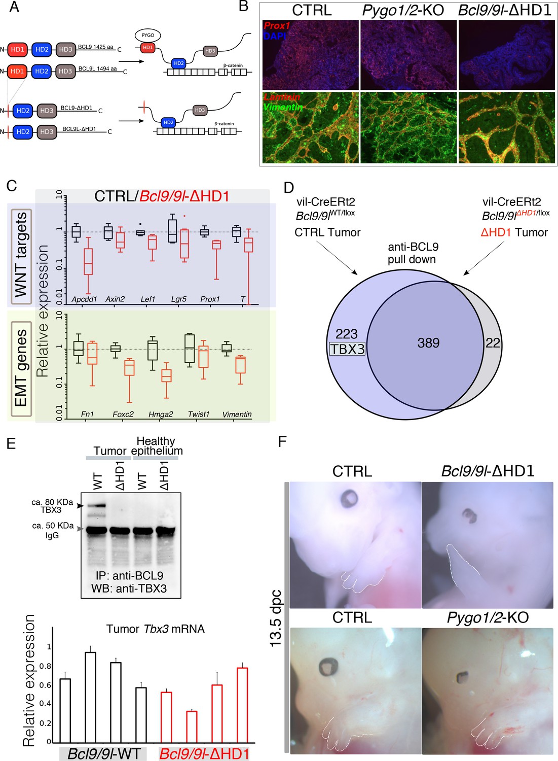
Identification of TBX3 as a putative BCL9 cofactor.

(A) The deletion of the HD1 (PYGO-interacting) domain of BCL9 and BCL9L induces a variation in the ‘chain of adaptors’ causing the loss of PYGO association with the Wnt/β-catenin transcriptional complex (Cantù et al., 2014). (B) Immunofluorescence staining of tumors collected from control or conditional Pygo1/2-KO and Bcl9/9l-KO mice. Prox1 (red) and DAPI (blue) are shown in in the top panels; Vimentin (green) and Laminin (red) in the bottom panels. (C) Quantitative RT-PCR of selected groups of targets (compare it with the same analysis of Pygo1/2-KO in Figure 1E) of RNA extracted from control or Bcl9/9l-ΔHD1 tumors. (D) Experimental outline of the tumor proteins pull-down and mass-spectrometry. TBX3 was identified among the proteins potentially interacting with BCL9 but not with BCL9-ΔHD1. (E) The IP proteins analyzed by mass spectrometry were in parallel subjected to SDS page electrophoresis and probed with an anti-TBX3 antibody (upper panel). The expression of Tbx3 in control compared to Bcl9/9l-ΔHD1 tumors (N = 4) was evaluated via qRT-PCR (bottom panel), to exclude that differential pull-down was due to lost expression in mutant tumors. (F) 13.5 dpc Bcl9/9l-ΔHD1 embryos display forelimb malformations and absence of digits (emphasized by dashed white lines) – a characteristic Tbx3-mutant phenotype (upper panels). The limb defect is absent in Pygo1/2-KO embryos (bottom panels) underscoring that BCL9/9L act, in this context, independently of PYGO1/2.

We confirmed cytological vicinity between transfected tagged versions of BCL9 and TBX3 by proximity ligation assay (PLA) (Figure 2—figure supplement 2A,B). However, overexpression-based in vitro co-immunoprecipitation experiments could not detect any stable interaction between these two proteins, suggesting absence of direct binding or a significantly lower affinity than that between BCL9 and PYGO (Figure 2—figure supplement 2C). Hence, we aimed at testing the functional association between TBX3 and BCL9 in a more relevant in vivo context. To this aim, we collected ca. 500 forelimbs from 10.5 dpc wild-type mouse embryos and subjected the crosslinked chromatin to immunoprecipitation using antibodies against BCL9 (Salazar et al., 2019) or TBX3, followed by deep-sequencing of the purified DNA (ChIP-seq, Figure 3A). By using stringent statistical parameters, and filtering with Irreproducible Discovery Rate (IDR), we extracted a list of high confidence BCL9 and TBX3 peaks (Figure 3B and C). Surprisingly, we discovered that BCL9 occupies a large fraction (ca. 2/3rd) of the TBX3-bound regions (Figure 3D). Suggestive of a role for TBX3 within the Wnt-dependent transcriptional apparatus, motif analysis of the common TBX3-BCL9 target loci identified statistical prevalence for TCF/LEF and Homeobox transcription factor consensus sequences, but not for any TBX transcription factor (Figure 3E). This suggests that TBX3 interacts with the DNA in these locations via affinity to the Wnt/β-catenin co-factors rather than via direct contact with DNA. Accordingly, TBX-specific motifs were detected within the group of TBX3 exclusive peaks (which do not display BCL9 binding, Figure 3—figure supplement 1). Notably, TBX3 and BCL9 occupancy was detected at virtually all previously described Wnt-responsive-elements (WRE) within known Wnt target genes (Figure 3F).

Figure 3 with 1 supplement see all
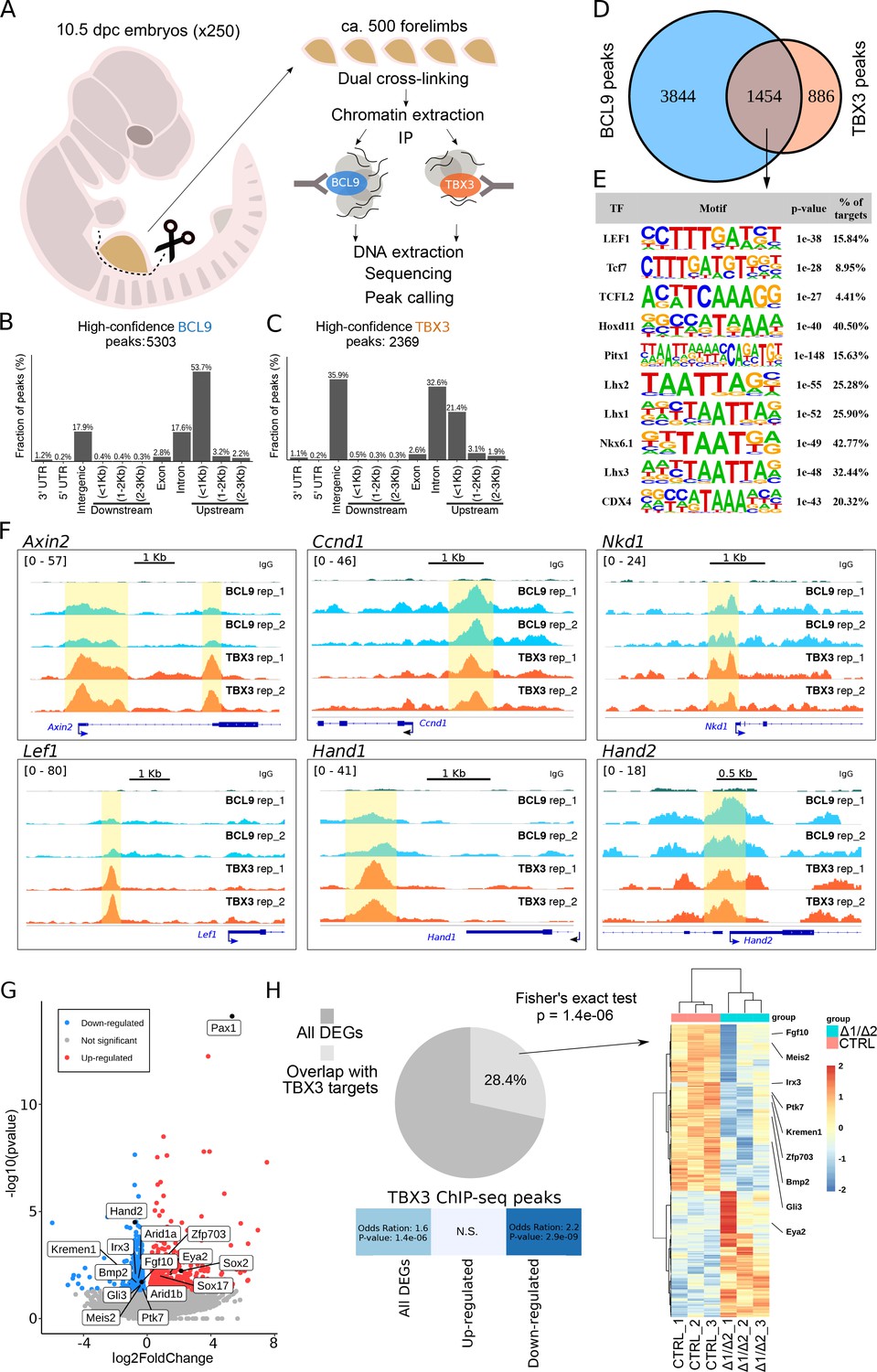
TBX3 and BCL9 occupy Wnt Responsive Elements (WRE) in vivo.

(A) Artistic representation of the ChIP-seq experimental outline. (B–C) Bar-plots showing the genomic distribution of high-confidence BCL9 peaks (B, 5303 total) and TBX3 peaks (C, 2369 total). (D) Overlap of the high-confidence peak groups between BCL9 and TBX3. (E) Selected result entries from motif analysis performed on the BCL9-TBX3 overlapping high-confidence peaks. Significant enrichment was found for TCF/LEF and Homeobox motifs. No TBX consensus sequence was detected in this analysis. (F) Select genomic tracks demonstrating occupancy of BCL9 and TBX3 within the Wnt Responsive Element (WRE) of known Wnt-target genes (Axin2, Ccnd1, Nkd1 and Lef1) and genes important in limb morphogenesis (Hand1 and Hand2). The scale of peak enrichment is indicated in the top-left corner of each group of tracks. In light blue the BCL9 (Salazar et al., 2019) and in orange the TBX3 replicates, and in green the control track (IgG). Genomic tracks are adapted for this figure upon visualization with IGV Integrative Genomic Viewer (https://igv.org/). Two independent replicates for BCL9 and TBX3 ChIP-seq experiments are shown. (G) Volcano plot displays all the differentially expressed genes (DEGs) in developing forelimbs, upon mutation of Bcl9/9l (Bcl9/9l-Δ1/Δ2 vs CTRL). DEGs were a total of 1143 (p<0.05), with 606 up-regulated and 537 down-regulated. N = 3 of individual mouse embryos for each condition were used for this analysis. (H) A significant portion (28.4%) of DEGs exhibited overlap with TBX3 ChIP-seq peaks. The overlap with TBX3 ChIP-seq peaks appeared statistically significant, in particular, when the down-regulated genes were considered. Hierarchical clustering of samples (3 CTRL versus 3 Bcl9/9l-Δ1/Δ2, right panel) based on genes overlapping between DEGs and genes annotated for TBX3 ChIP-seq peaks (normalized RNA-seq read counts, Ward’s clustering method, Euclidian distance). Annotation added for genes associated by Gene Ontology to Wnt signaling (Fgf10, Ptk7, Kremen1, Zfp703, Bmp2 and Gli3) and genes known as regulators of limb development (Meis2, Irx3 and Eya2).

To test whether the in vivo abrogation of the simultaneous interactions mediated by BCL9/9L would influence the expression of genes associated with TBX3 peaks, we set out to mutate the Bcl9/9l interaction domains, while leaving TBX3 protein unaffected. We combined different Bcl9/9l alleles in which the HD2 (β-catenin-interacting) and HD1 (PYGO/new co-factor-interacting) motifs are deleted (Cantù et al., 2018). Double heterozygous animals for the HD1 (Bcl9ΔHD1/+; Bcl9lΔHD1/+) or the HD2 (Bcl9ΔHD2/+; Bcl9lΔHD2/+) deletions are viable and fertile. The cross between them leads to a trans-heterozygous genetic configuration in which both domain deletions are present (Bcl9ΔHD1/ΔHD2; Bcl9lΔHD1/ΔHD2, referred to as Bcl9/9l-Δ1/Δ2, Figure 1A). As in these mice BCL9/9L retain both the HD1 and the HD2 domains in heterozygosity, this allelic combination is a way of testing the consequences of abrogating the tripartite complex mediated by the two interacting motifs of BCL9/9L without causing a full loss-of-function of these proteins. Bcl9/9l-Δ1/Δ2 embryos also display forelimb malformations, the cause of which cannot be due to PYGO (Figure 2F) but must be caused by the failure of recruiting the new HD1-interacting partner by BCL9/9L onto the β-catenin transcriptional complex. We collected forelimbs from control and Bcl9/9l-Δ1/Δ2 mutant embryos at 10.5, and measured gene expression via RNA-seq (Figure 3G). We found a significant enrichment (Hypergeometric test, p=1.4e-6) of TBX3 targets among the genes differentially expressed in Bcl9/9l-Δ1/Δ2 mutants (Figure 3H). The enrichment was particularly significant when considering down-regulated genes in Bcl9/9l-Δ1/Δ2 mutants, indicating that the BCL9-TBX3 partnership sustains transcriptional activation (Figure 3H). Of note, the design of our experiment directly implicates that these TBX3 transcriptional targets are also β-catenin-dependent. The overlap list includes several regulators of limb development, such as Meis2 (Capdevila et al., 1999), Irx3 (Li et al., 2014) and Eya2 (Grifone et al., 2007Figure 3H, heat map on the right). Despite being of correlative nature, this analysis supports a model in which BCL9/9L and TBX3 cooperate to the activation of target genes.

So far, we have presented genetic evidence that BCL9 proteins require additional co-factors, and that TBX3 associates with the β-catenin/BCL9 bound regions on the genome possibly influencing the expression of target genes. However, the similarity of genomic binding profiles between TBX3 and BCL9 might be due to their binding in different cells, and the decreased expression of genes with nearby enhancers bound by BCL9 and TBX3 might imply a requirement for BCL9/9L, but not necessarily for TBX3. We reasoned that our hypothesis - in which BCL9 functionally tethers TBX3 to the β-catenin transcriptional complex - raises several testable predictions that will be addressed below.

First, our model implies that TBX3 could impact on Wnt target gene expression and its activity should be dependent on the main constituents of the Wnt/β-catenin transcriptional complex. Second, if TBX3 is tethered by BCL9 to its targets, mutations in BCL9/9L should influence the ability of TBX3 to physically associate with WREs. Finally, as for BCL9, TBX3 should be capable of enhancing the metastatic potential of colorectal cancer cells.

To test our first prediction, implying a potential role of TBX3 in the transcription of Wnt target genes, we overexpressed it in HEK293T cells and monitored the activation status of Wnt signaling using the transcriptional reporter SuperTopFlash (STF). Consistent with its role as repressor, TBX3 led to a moderate but significant transcriptional downregulation that was, importantly, specific to the STF but not the control reporter plasmid (Figure 4A). Upon Wnt signaling activation achieved via GSK3 inhibition, TBX3-overexpressing cells exhibited a markedly increased reporter activity when compared to control cells, in particular at non-saturating pathway stimulating conditions (Figure 4A, left panel). Importantly, TBX3 proved transcriptionally incompetent on the STF if the cells carried mutations in TCF/LEF (Δ4TCF) or CTNNB1 (encoding for β-catenin, Δβ-CAT; Doumpas et al., 2019), strongly supporting the notion of its cell-autonomous involvement in the activation of canonical Wnt target gene transcription (Figure 4A, central and right panels, respectively). Endogenous Wnt targets showed a similar expression behaviour to that of STF upon TBX3 overexpression (Figure 4—figure supplement 1). While our experiments show that TBX3 can influence the expression of Wnt target genes, the mechanisms by which this occurs remain to be elucidated.

Figure 4 with 1 supplement see all
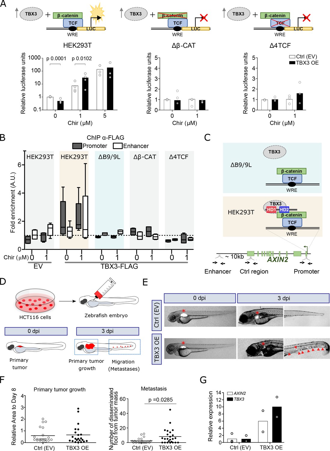
TBX3 controls the expression of Wnt target genes.

(A) β-Catenin/TCF luciferase reporter STF assay in parental (left), β-catenin knockout (Δβ-CAT, center) and TCF knockout (Δ4TCF, right) HEK293T cells. Cells were treated with the indicated concentration of Chir or DMSO, overnight. Overexpression of TBX3 (OE, black bars) compared to control (EV, empty vector, white bars) showed that TBX3 acts as a repressor on a Wnt/TCF pathway reporter, but switches to activator upon pathway induction. Only significant p-values (p<0.05) are indicated. Three independent experiments (N = 3) are shown. Note that the logarithmic scale on the y-axis of the histogram on the left is different from the linear scale of the central and middle panels. (B) ChIP followed by qPCR in HEK293T cells treated with DMSO (‘WNT-OFF’) or Chir (‘WNT-ON’). Enrichment was identified on AXIN2 promoter and the downstream enhancer. Note that the enrichment on the enhancer is only present upon pathway stimulation: we interpret this as evidence for the enhancer looping onto the promoter occurring when the Wnt-dependent transcriptional regulation is active. The data are normalized to immunoprecipitation performed in cells transfected with an empty vector (EV) and presented as the mean ± standard deviation of independent experiments. The fold enrichment of TBX3-FLAG on AXIN2 promoter and enhancer (N = 4) is lost upon mutations in BCL9/9L (ΔB9/9L, N = 2), CNTTB1 (ΔΒ-CAT, N = 2) and TCF/LEF (Δ4TCF, N = 2). (C) Schematic representation of the AXIN2 locus indicates the position of the primers used (black arrows) to test the binding of TBX3. Despite the apparent absence of direct physical interaction between TBX3 and BCL9/9L, the data support a model of TBX3 recruitment by BCL9/9L onto the β-catenin/TCF transcriptional complex. (D) Schematic diagram of the human CRC zebrafish xenografts model. Parental and TBX3- overexpressing HCT116 colorectal tumor cells were harvested and labeled with DiI dye (red). The stained cells were injected into the perivitelline space of 3 day old zebrafish embryos. Zebrafish were visualized with fluorescent microscopy at 0 day post injection (dpi) and three dpi, and primary tumor cell invasion and metastasis were counted. (E) Representative images of HCT116 tumor invasion and dissemination at 0 and 3 dpi in zebrafish xenografts, for both control and TBX3 overexpressing cells. The red asterisks indicate the position of the primary tumor. Red arrowheads point at clusters of disseminating/metastatic cells. (F) Scatter plot representing the quantification of primary tumor growth and metastasis after HCT116 xenograft. Horizontal bars represent the mean value. Only significant p-values (p<0.05) are displayed. (G) Quantitative RT-PCR confirmed continued increased expression of TBX3 while HCT116 disseminate through zebrafish tissue, and that this is accompanied by enhanced Wnt/β-catenin transcription, as seen by AXIN2 expression. Each datapoint represents the extraction of total RNA from pools of 10 zebrafish embryos. Figure Legend of Figure Supplements.

We then addressed our second hypothesis, in which BCL9/9L are required for tethering TBX3 onto WREs. We performed ChIP of TBX3 in HEK293T cells followed by quantitative PCR to detect enrichment on the WRE present in the AXIN2 promoter (Jho et al., 2002). Consistent with an effect on transcription in the absence as well as in the presence of Chir99021 (Chir) (Figure 4A), TBX3 was bound to this region both in ‘OFF’ and in ‘ON’ conditions (Figure 4B). We then exploited a HEK293T clone devoid of both BCL9 and BCL9L (ΔB9/9L; van Tienen et al., 2017), and tested if TBX3 was capable of physical association with the WRE. Of note, enrichment of TBX3 in ΔB9/9 L cells was dramatically reduced to background levels (Figure 4B). While we cannot exclude that TBX3 might act independently of BCL9/9L on several of its targets, this observation supports the notion that BCL9/9L are responsible for TBX3 recruitment on classical WREs (Figure 4C). This also suggests that the previously identified targets of both BCL9 and TBX3 (Figure 3D–F) must display simultaneous co-occupancy of these two factors. In agreement with the notion that BCL9/9L are themselves recruited by the TCF-β-catenin axis, the physical association of TBX3 with the AXIN2 promoter was also lost in Δ4TCF and Δβ-CAT cells (Figure 4B).

Finally, we evaluated the effects of TBX3 overexpression (OE) on growth and metastatic potential of HCT116 human colorectal tumor cells – a representative model of CRC driven by activating mutations in CTNNB1 (Mouradov et al., 2014) –, using a in vivo zebrafish xenograft model (Rouhi et al., 2010). Approximately 200–500 labelled control or TBX3-OE HCT116 cells were implanted in the perivitelline space of 72 hours post-fertilization (hpf) zebrafish embryos (Figure 4D). Three days after injection, TBX3-OE cells displayed a marked increase in number in the caudal hematopoietic plexus (Figure 4E–F), the main metastatic site for cells migrating from the perivitelline space (Rouhi et al., 2010). Of note, TBX3-OE HCT116 cells maintained consistently high expression of TBX3 within fish embryos throughout the experiment, and this was accompanied by increased Wnt/β-catenin-dependent transcription, as measured by AXIN2 expression (Figure 4G). While this experiment does not allow to exclude that TBX3 might also act independently of BCL9/β-catenin in this context, it shows that increased expression of TBX3 enhances proliferation and migratory capability of human CRC cells bearing constitutively active Wnt signaling, and this is associated with simultaneous enhancement of the Wnt/β-catenin-dependent transcription (Figure 4G).

Taken together, our experiments show that, in specific developmental and disease contexts, the transcription factor TBX3 can take active part in the direct regulation of Wnt target genes by functional interplay with the β-catenin/BCL9-dependent transcriptional complex. Our study suggests a new paradigm in which tissue-specific co-factors might be the key to understand the spectrum of possible transcriptional outputs observed downstream of Wnt/β-catenin signaling (Nakamura et al., 2016). Moreover, TBX3 has been linked to different cancer types (Willmer et al., 2017). Our observations suggest that TBX3, or its downstream effectors, could be considered as new relevant targets to dampen CRC progression.

Materials and methods

Treatment of mice and histological analyses

Request a detailed protocol

Homeostasis: 4–6 week-old male and female mice (Bcl9flox/flox;Bcl9lflox/flox; vil-Cre-ERt2 and Bcl9flox/flox;Bcl9lflox/flox (no Cre) littermates; Pygo1flox/flox;Pygo2flox/flox; vil-Cre-ERt2 and Bcl9flox/flox;Bcl9lflox/flox (no Cre) were treated with five tamoxifen injections (i.p., 1 mg/day, Sigma) for five consecutive days and the small intestine and colon were analyzed at different time points thereafter. Mouse experiments were performed in accordance with Swiss guidelines and approved by the Veterinarian Office of Vaud, Switzerland.

Induction of DSS Colitis: 6–8 week-old male mice were treated with five tamoxifen injections (i.p., 1 mg/day) for five consecutive days. 10 days later 2.5% DSS (MG 36–50’000, MP Biomedicals, cat. no. 160110) was administered, ad libitum, in the drinking water for 9 days.

Induction of tumors: 6–8 week-old female mice were treated with five tamoxifen injections (i.p., 1 mg/day) for five consecutive days. 10 days later they were injected i.p. with 44 mg/kg body weight DMH 2HCl (N,N’ Dimethylhydrazine dihydrochloride). After another 7 days later 2% DSS was administered, ad libitum, in the drinking water for 7 days.

Mice were monitored clinically for rectal bleeding, prolapse and general signs of morbidity, including hunched posture, apathetic behavior and failure to groom.

The relative body weight (in %) was calculated as follows: 100 X weight at a certain day/weight at the first day of DSS treatment. Epithelial damage of DSS treated mice was defined as percentage of distal colon devoid of epithelium.

To determine proliferation rates, mice were injected i.p. with 100 mg/kg BrdU (Sigma) 2 hr prior to sacrifice. Small intestines and colons (divided into three equal segments to be named proximal, middle and distal colon) were dissected, flushed with cold PBS, cut open longitudinally and fixed in 4% paraformaldehyde for 2 hr at RT and paraffin embedded. Sections (4 μm) were cut and used for hematoxylin/eosin and alcian blue staining and for immunohistochemistry. The primary antibodies used were rabbit anti-Synptophysin (DAKO; 1:100), rabbit anti-Lysozyme (1:500; DAKO), mouse anti-Ki67 (1:100; Novocastra), mouse anti-BrdU (1:500; Sigma), anti-β-Catenin (1:100; BD pharmigen), anti-active caspase 3 (1:100; Cell Signaling).

The peroxidase-conjugated secondary antibodies used were Mouse or Rabbit EnVision+ (Dako) or mouse anti-rat HRP (1:250: Biosource).

Real-time PCR genotyping

Request a detailed protocol

To determine the deletion rate, the intestinal epithelium was separated from the underlining muscle. The intestine was dissected, flushed with PBS, cut open longitudinally and incubated in 3 mM ethylenediamine tetraacetic acid (EDTA) and 0.05 mM dithiothreitol (DTT) in PBS for 1.5 hr at RT on a rotor. The tubes were shaken vigorously, the muscle removed, and the epithelium centrifuged and used for genomic DNA extraction. SYBR green real-time PCR assays were performed on each sample analyzed.

Chromatin immunoprecipitation

Request a detailed protocol

Forelimb buds were manually dissected from ca. 250 RjOrl:SWISS outbred 10.5 dpc mouse embryos. Chromatin immunoprecipitation was performed as previously described (Cantù et al., 2018). Briefly, the tissue was dissociated to a single cell suspension with collagenase (1µg/ml in PBS) for 1 hr at 37° C, washed and crosslinked in 20 ml PBS for 40 min with the addition of 1.5 mM ethylene glycol-bis(succinimidyl succinate) (Thermo Scientific, Waltham, MA, USA), for protein-protein crosslinking (Salazar et al., 2019), and 1% formaldehyde for the last 20 min of incubation, to preserve DNA-protein interactions. The reaction was blocked with glycine and the cells were subsequently lysed in 1 ml HEPES buffer (0.3% SDS, 1% Triton-X 100, 0.15 M NaCl, 1 mM EDTA, 0.5 mM EGTA, 20 mM HEPES). Chromatin was sheared using Covaris S2 (Covaris, Woburn, MA, USA) for 8 min with the following set up: duty cycle: max, intensity: max, cycles/burst: max, mode: Power Tracking. The sonicated chromatin was diluted to 0.15% SDS and incubated overnight at 4°C with 10 μg of anti-BCL9 (Abcam, ab37305) or anti-TBX3 (Santacruz, sc-17871) or IgG and 50 µl of protein A/G magnetic beads (Upstate). The beads were washed at 4°C with wash buffer 1 (0.1% SDS, 0.1% deoxycholate, 1% Triton X-100, 0.15 M NaCl, 1 mM EDTA, 0.5 mM EGTA, 20 mM HEPES), wash buffer 2 (0.1% SDS, 0.1% sodium deoxycholate, 1% Triton X-100, 0.5 M NaCl, 1 mM EDTA, 0.5 mM EGTA, 20 mM HEPES), wash buffer 3 (0.25 M LiCl, 0.5% sodium deoxycholate, 0.5% NP-40, 1 mM EDTA, 0.5 mM EGTA, 20 mM HEPES), and finally twice with Tris EDTA buffer. The chromatin was eluted with 1% SDS, 0.1 M NaHCO3, de-crosslinked by incubation at 65°C for 5 hr with 200 mM NaCl, extracted with phenol-chloroform, and ethanol precipitated. The immunoprecipitated DNA was used as input material for DNA deep sequencing. The pull downs of BCL9 and TBX3 were performed in parallel experiments. Note that the ChIP-seq of BCL9 has been already described in Salazar et al., 2019, and entirely re-analyzed here (see below).

Data analysis: Overall sequencing quality of the acquired fastq files was assessed using FastQC (version 0.11.5). Because all the samples exhibited good quality (MAPQ >30) and had no adapter contamination >0.1% trimming of reads was not deemed necessary. In addition, test alignments against several reference genomes were done using the FastQ Screen tool (version 0.13.0). Quality results were summarized using MultiQC (version 1.7). Fastq files were mapped to mouse reference genome (mm10) using the read aligner Bowtie2 (version 2.3.4.1). The mouse reference genome was downloaded from UCSC(http://hgdownload.cse.ucsc.edu/goldenpath/mm10/bigZips/). The resulting alignment files were then adjusted (conversion to binary format, removal of read aligned to mitochondrial DNA and indexing) using SamTools (version 1.9). To identify genomic regions enriched with aligned reads the peak calling tool MACS2 (version 2.2.6) was used. Calculated p-values were adjusted for false discovery rate (FDR) using Benjamini-Hochberg procedure, generating q-values. A cutoff q < 0.05 was used to assess significance. IgG sample was used as enrichment-normalization control. MACS2 generated peak files were further filtered by removal of blacklisted regions according to the ENCODE project using bedtools (version 2.26.0). Annotation and visualization were made with the R programming language (version 3.4.4) and Rstudio (version 1.1.463), using R packages ChIPpeakAnno (version 3.12.7), ChIPseeker (version 1.14.2) and ggplot2 (version 3.2.1). Genomic track visualization was done with Integrative Genomics Viewer (IGV) (version 2.4.17). Motif analysis was performed with HOMER. The data have been deposited at ArrayExpress with accession number E-MTAB-8997.

RNA-seq data analysis

Request a detailed protocol

Quality control of fastq files was done using FastQC (version 0.11.5). Trimming of reads to remove adapter remnants and low quality read (MAPQ <30) was performed with BBDuk, part of the BBTools suite (version 38.58). Test alignments against different reference genomes were done with the FastQ Screen tool (version 0.13.0).

Quality results were summarized using MultiQC (version 1.7). Sequenced reads were mapped to mouse reference genome (mm10) using the Spliced Transcript Alignment to a Reference (STAR) software (version 2.7.3a). Reference genome data (FASTA and GTF) were downloaded from GENCODE, release M24 (https://www.gencodegenes.org/mouse/). Downstream analyses, annotation and visualization were made with the R programming language (version 3.4.4) and Rstudio (version 1.1.463). Differentially expressed genes were assessed using the DESeq2 package (version 1.18.1). Significance was determined based on false discovery rate (FDR) adjusted p<0.05. Hierarchical clustered heatmap was produced with the pheatmap R package, using Ward’s Hierarchical Agglomerative Clustering Method. A complete list of bioinformatic tools and references is listed in the accompanying Supplementary file 1. The RNA-seq experiment has been deposited at ArrayExpress with accession number E-MTAB-9000.

Protein immunoprecipitation and mass spectrometry

Request a detailed protocol

Dissected mouse tumors were minced in cold PBS and treated with a hypotonic lysis buffer (20 mM tris-HCl, 75 mM NaCl, 1.5 mM MgCl2, 1 mM EGTA, 0.5% NP-40, and 5% glycerol). Protein extracts obtained were incubated with 1 μg of anti-BCL9 antibody (ab54833 or ab37305, Abcam) and protein A–conjugated Sepharose beads (GE Healthcare); they were then diluted in lysis buffer to a final volume of 1 ml. After 4 hr of incubation at 4°C on a rotating wheel, the beads were spun down and washed three times in lysis buffer. All steps were performed on ice, and all buffers were supplemented with fresh protease inhibitors (cOmplete, Roche) and 1 mM phenylmethylsulfonyl fluoride. For detecting the proteins in Western blot, the pulldown reactions were treated with Laemmli buffer, boiled at 95°C for 15 min, and subjected to SDS-PAGE separation and blotting on a polyvinylidene difluoride (PVDF) membrane. The PVDF membrane was probed with the anti–TBX3.

For liquid chromatography–MS/MS analysis, the protein samples, already dissolved in Laemmli buffer, were submitted to a filter-aided sample preparation (FASP) and digested with trypsin in 100 mM triethylammonium bicarbonate buffer overnight. Desalted samples were dried completely in a vacuum centrifuge and reconstituted with 50 μl of 3% acetonitrile and 0.1% formic acid. Each peptide solution (4 μl) was analyzed on both Q Exactive and Fusion mass spectrometers (Thermo Scientific) coupled to EASY-nLC 1000 (Thermo Scientific). Spectra acquisition and peptide count were performed as described in details in Cantù et al., 2017. The dataset is deposited at the PRIDE repository under accession number PXD018805.

Cell culture

Request a detailed protocol

The human cell lines HEK293T -parental, ΔCTNNB1 and ΔTCF/LEF (Doumpas et al., 2019), BCL9/9L double KO HEK293T (van Tienen et al., 2017), and HCT116 were cultured in DMEM (Thermo Fisher Scientific, Belmont, Massachusetts, US) supplemented with 10% fetal bovine serum (Gibco, Gaithersburg, USA) 1% L-glutamine and 1% penicillin-streptomycin at 37°C in a humidified chamber supplemented with 5% CO2. Chir99021 compound (Tocris Bioscience) was used as Wnt signaling activator. At 6 hr after transfection, the transfected cells were treated for 16 hr either with DMSO (0 µM Chir), 1 µM or 5 µM Chir. Following that, cells were collected for further experiments. All cell lines were tested and scored negative for mycoplasma infection. HCT116 were obtained from DSMZ (https://www.dsmz.de/collection/catalogue/details/culture/ACC-581) and kindly donated by Prof. Stefan Koch.

Plasmids and transfection

Request a detailed protocol

TBX3-3xflag (donated by Peter J. Hurlin), pCMV-Pygo2-HA (Cantù et al., 2013), pCDNA3.1-BCL9-GFP (kindly gifted by Mariann Bienz), pcDNA3-eGFP (kindly provided by Stefan Koch), M50 SUPER 8X TOPFLASH pTA-Luc (Addgene 12456) and M51 SUPER 8X FOPFLASH pTA-Luc (Addgene 12456) were transfected using Lipofectamine 2000 (Invitrogen, USA) following the manufacturer’s instructions. Twenty-four hours after transfection, the cells were collected for the indicated experiments.

RNA extraction and quantitative reverse transcriptase PCR (qRT-PCR)

Request a detailed protocol

Total RNAs from cells were extracted using TRIzol (Thermo Fisher Scientific, Belmont, Massachusetts, US) following the manufacturer´s instructions. After reverse transcription reaction with High Capacity cDNA Reverse Transcription Kit (Thermo Fisher Scientific), qPCR was conducted with SYBR green LightCycler 480 (Roche) on CFX96 Real-Time PCR Detection System (Bio-Rad, USA). GAPDH was used as endogenous control and the relative expression of RNAs was calculated using the 2−ΔΔCt method. The primers used in this study were designed by Primer3plus web and their sequences are listed in the Supplementary file 2.

Xenograft tumor zebrafish model

Request a detailed protocol

HCT116 cells were transfected with pCS2 (empty vector) or TBX3-expressing plasmid and labeled with 1,1′-dioctadecyl-3,3,3′,3′-tetramethylindocarbocyanine perchlorate (DiI, cat. no. D3899; Invitrogen) according to previously described method (Rouhi et al., 2010). Briefly, tumor cells were washed twice with PBS, followed by labeling with a final concentration of 5 μg/ml of DiI for 30 min at 37 °C. After rigorous washing with PBS, tumor cells were trypsinized for 5 min, counted under a phase contrast microscope, centrifuged at 300 g for 5 min and resuspended at a final concentration of 30–40 cells per µl in medium. Human cells were injected into the perivitelline space of 72 hr old fli1:EGFP transgenic zebrafish embryos. The eggs were fertilized, collected and dechorionated. After that, HCT116 cells were injected into the vitreous cavity of zebrafish embryo with non-filamentous borosilicate glass capillary needles attached to the microinjector under the stereomicroscope (Leica Microsystems). After tumor cell injection, zebrafish embryos were further selected under fluorescent microscopy to ensure that tumor cells were located only within the cavity and then incubated in aquarium water for consecutive 3 days at 36.0 °C. Primary tumor growth, invasion and metastasis in the zebrafish body were monitored at day three with a fluorescent microscope (Nikon Eclipse C1) as previously published (Rouhi et al., 2010). Briefly, each zebrafish embryo was picked up and monitored to detect tumor cell distribution. Two different sets of images from the head region and the trunk region were collected separately from each zebrafish embryo. Disseminated tumor cells in the caudal hematopoietic plexus of zebrafish embryos were counted (in a double-blind manner) and the primary tumor areas were measured using Image J software. At least 15–20 embryos were included in each experimental group.

Western blot analysis

Request a detailed protocol

Cells were lysed at 4°C for 10 min using NP-40 lysis buffer (50 mM Tris-HCl (pH 7.5), 120 mM NaCl, and 1% (v/v) Igepal), supplemented with protease inhibitor cocktail (Merck). Cells were sonicated with Q700CA Sonicator (Q Sonica) on 50 amplitude for 5 s on/off for two cycles. Protein lysates were separated by 10% SDS-polyacrylamide gel electrophoresis (SDS-PAGE) and transferred onto nitrocellulose membrane. The membrane was blocked in Odyssey TBS Blocking Buffers (LI-COR Bioscience) for 1 hr at room temperature and subsequently incubated with specific antibody against FLAG (1:1000, Merck F3165) overnight at 4°C and HA (1:1000, Merck 05–902R) and GFP (1:1000 Santa Cruz Biotechnology sc-9996) 1 hr at room temperature. Afterwards, the membrane was washed and incubated with fluorescent Goat-anti-Rabbit and goat-anti-Mouse (1:2000; LI-COR Bioscience) for 1 hr at room temperature. Protein detection was performed in LI-COR Odyssey CLx (LI-COR Bioscience).

Super-Top/Fop-Flash reporter assay

Request a detailed protocol

The β-catenin reporter plasmid (SuperTopFlash, STF) and its mutant control (Fop-flash) were constructed by Addgene (12456). Cells were plated in triplicate in a 96 well plated overnight and co-transfected with 50 ng Top flash or Fop flash expression plasmids together with other plasmids for the experiments using Lipofectamine 2000. The activities of firefly luciferase reporters were determined at 24 hr after transfection using ONE-Step Luciferase Assay System Kit (Pierce) according to the manufacturer’s instructions. The Top/Fop ratio was used as a measure of β-catenin-driven transcription. GraphPad Prism software was used for statistical analyses. Values were expressed as mean ± standard deviation. Mann Whitney U test was used to analyze the differences between two groups and p<0.05 was considered statistically significant. All experiments were performed at least three times and all samples analyzed in triplicate unless otherwise stated.

In-situ proximity ligation

View detailed protocol

Proximity ligation was performed using NaveniFlex MR assay reagents according to the supplier’s guidelines (Navinci Diagnostics AB). In brief, HEK293T cells were plated on coverslips and were transiently transfected using lipofectamine 2000 transfection reagent (ThermoFisher Scientific) with plasmids expressing the following combinations of proteins (each in a ration 1:1): BCL9-HA and TBX3-FLAG, BCL9-HA and GFP or BCL9-GFP and PYGO2-HA. An EGFP-expressing vector at 1/10th the total plasmid amount was also added in PLA assays to identify transfected cells. Paired combinations of rabbit (anti-HA) and mouse (anti-FLAG or anti-GFP) antibodies were incubated with samples for 1 hr at room temperature (1:1000 dilution each). Following addition of matched PLA probes, rolling circle amplification proceeded in presence of TEX 615 fluorophore. After washing, coverslips were mounted in Prolong Glass Antifade with NucBlue counterstain. Slides were imaged in Zeiss LSM770 confocal microscope, using 20x and 40x objective and filters set in DAPI, FITC and Texared. Specific individual protein-protein interactions can be seen as red dots. For all experimental conditions, at least three images were acquired.

Image analysis and quantification were performed using the opensource ImageJ2 software. The pipeline for signal quantification was the following: images in the green and red channels were merged and converted to a binary image (8-bit). A threshold range was set to outline the objects of interest (particles in yellow, GFP + PLA signal) and separate it from background signal. Positive dots representing the PLA signal ‘particles’ were counted using the command ‘analyze particles’. The image in the blue channel (DAPI) was used to count cell nuclei. After converting the image to binary by thresholding binary conversion, overlapping objects were separated using the plugin Watershed and counted using the command ‘analyze particles’. The minimum size of 50 micron2 was set to conservatively exclude unspecific signals.

Data availability

The ChIP-seq data have been deposited at ArrayExpress with accession number E-MTAB-8997. The RNA-seq experiment has been deposited at ArrayExpress with accession number E-MTAB-9000.

The following data sets were generated
    1. Cantù C
    2. Zimmerli D
    (2020) ArrayExpress
    ID E-MTAB-9000. RNA-seq experiment of developing mouse forelimbs, comparing Bcl9/9l mutant with control littermates.
    1. Cantù C
    2. Zimmerli D
    (2020) ArrayExpress
    ID E-MTAB-8997. ChIP-seq experiment of the beta-catenin co-factor BCL9 and the T-box transcription factor TBX3 in developing mouse forelimbs.
    1. Cantù C
    2. Moor A
    (2020) PRIDE
    ID PXD018805. Pull down of full length and mutant BCL9 (deltaHD1) in mouse colorectal tumors and colonic epithelium.
The following previously published data sets were used
    1. Cantù C
    2. Zimmerli D
    (2019) ArrayExpress
    ID E-MTAB-7652. ChIP-seq experiment of the beta-catenin co-factor Bcl9 in developing forelimbs.

References

Decision letter

  1. Roel Nusse
    Reviewing Editor; Stanford University, United States
  2. Kevin Struhl
    Senior Editor; Harvard Medical School, United States

In the interests of transparency, eLife publishes the most substantive revision requests and the accompanying author responses.

Acceptance summary:

This work analyzes the transcriptional output of the Wnt signaling pathway. Through a series a genetic and biochemical experiments, these results suggest that the TBX3 protein acts as a context-dependent component of the transcriptional complex. This is interesting as TBX3 was previously implicated as a target of the Wnt pathway. By also playing a role in the transcriptional complex activated by Wnt, TBX3 may be involved in a transcriptional circuit.

Decision letter after peer review:

Thank you for submitting your article "TBX3 acts as tissue-specific component of the Wnt/β-catenin enhanceosome" for consideration by eLife. Your article has been reviewed by three peer reviewers, one of whom is a member of our Board of Reviewing Editors, and the evaluation has been overseen by Kevin Struhl as the Senior Editor. The reviewers have opted to remain anonymous.

The reviewers have discussed the reviews with one another and the Reviewing Editor has drafted this decision to help you prepare a revised submission.

We would like to draw your attention to changes in our revision policy that we have made in response to COVID-19 (https://elifesciences.org/articles/57162). Specifically, when editors judge that a submitted work as a whole belongs in eLife but that some conclusions require a modest amount of additional new data, as they do with your paper, we are asking that the manuscript be revised to either limit claims to those supported by data in hand, or to explicitly state that the relevant conclusions require additional supporting data.

Our expectation is that the authors will eventually carry out the additional experiments and report on how they affect the relevant conclusions either in a preprint on bioRxiv or medRxiv, or if appropriate, as a Research Advance in eLife, either of which would be linked to the original paper.

Your manuscript builds upon the observation that Bcl9/Bcl9L mutants have a more severe intestinal and limb defect than Pygo1/2 double mutants. This is interesting because the Bcl9s and Pygos are often thought to act in a complex to regulate Wnt targets. A possible binding partner of Bcl9 is TBX3, a gene implicated in limb deformities that might be similar to Bcl9/9L limb conditional knockouts. Using ChIP-seq of embryonic mouse limbs, your show co-localization of TBX3 and Bcl9 at over 1400 locations (2/3s of the total TBX3 peaks). This leads to a model where TBX3 is recruited to Wnt target gene enhancers through interaction with Bcl9/9L.

Revisions for this paper:

The reviewers had a number of suggestions that would improve the manuscript. Some of these require additional experiments but the reviewers thought that these experiments would not require too much effort and time, even under the present COVID circumstances.

There is no direct evidence for co-occupancy, and the term must be changed. The binding profiles of Bcl9 and TBX3 are similar, but co-occupancy means binding of both at the same time, not just the same place. The convincing experiment here would be sequential ChIP (re-ChIP). Moreover, the authors haven't demonstrated a physical interaction between the 2 proteins, which would at least add evidence (though not proof) for co-occupancy.

Specifically, the data in Figure 4A,B do not address the model favored by you, i.e., that Bcl9/9L recruits TBX3 to enhancers. Rather it shows that genes with nearby enhancers bound by Bcl9 and TBX3 have decreased expression in Bcl9/9L mutants. This speaks to a requirement for Bcl9/9L, but doesn't address its relationship to TBX3. This can be tested by whether loss of Bcl9/9L affects TBX3 binding to enhancers. You will have the reagents to do this key experiment. In addition, it is suggested to test whether the ChIP signal at the Axin2 enhancer and promoter are dependent on β-catenin and TCFs, using the CRISPR lines used in Figure 4C. These experiments directly test their model with reagents that you have in hand. The reviewers also suggest that a proximality labeling experiment which can capture weaker interactions than co-IP could be done in Wnt-active versus inactive contexts to potentially confirm TBX3-Bcl9 interaction and identify other cofactors.

The reviewers and editors point out that the authors should eliminate the term "enhanceosome" in the title and anywhere else. "Enhanceosome" has a very specific meaning in the transcription field, namely a highly structured physical entity composed of multiple transcription factors interacting with a complex and specific arrangement of target sites.

A reviewer's comment on phenotypes in seen Pygo1/2-KO and CTRL in regard to the length of villus and depth of crypts as well as number of proliferating cells. Therefore, in the absence of more detailed morphologic and quantitative histologic information, the description of the figure should be changed from "Intestinal architecture was normal" to "Not overt phenotypic defects".

Reviewer #1:

This manuscript builds upon the interesting observation that Bcl9/Bcl9L mutants have a more severe intestinal and limb defect than Pygo1/2 double mutants. This is interesting because the Bcl9s and Pygos are often thought to act in a complex to regulate Wnt targets. Using Bcl9 as bait, the authors then identify TBX3 as an interaction partner. TBX3 has limb deformities that might be similar to Bcl9/9L limb conditional knockouts. Using ChIP-seq of embryonic mouse limbs, they show an impressive co-localization of TBX3 and Bcl9 at over 1400 locations (2/3s of the total TBX3 peaks). Finally, they attempt to support a model where TBX3 is recruited to Wnt target gene enhancers through interaction with Bcl9/9L.

The strengths of the manuscript are the convincing data demonstrating that Bcl9/9L mutants have more dramatic phenotypes than Pygo1/2. The ChIP-seq with Bcl9 and TBX3 is also very interesting. But the data functionally linking TBX3 to Bcl9 on Wnt targets is very weak and don't adequately support the molecular model that they present.

1) It's not clear how similar that limb defects in Bcl9/9L mutants are compared to the TBX3 mutant limbs described in Frank et al., 2013. I disagree that they are "strikingly similar". Without more direct experimental observation, this connection needs to be related with more careful language.

2) The data in Figure 4A,B does not address the model favored by the authors, i.e., that Bcl9/9L recruits TBX3 to enhancers. Rather it shows that genes with nearby enhancers bound by Bcl9 and TBX3 have decreased expression in Bcl9/9L mutants. This speaks to a requirement for Bcl9/9L, but doesn't address its relationship to TBX3.

3) A direct test of their model would be to see whether loss of Bcl9/9L affects TBX3 binding to enhancers. The authors have the reagents to do this key experiment.

4) The overexpression data with TBX3 is unconvincing. At the concentration used, TBX3 represses Wnt targets in the absence of signaling. It's impossible to tell whether this is a direct or indirect affect (note the TBX3 ChIP data in Figure 4D indicates TBX3 is bound to the Axin2 promoter in the absence of signaling). The very small effect of TBX3 on TopFlash or Axin2 and Nkd1 is only "significant" when this repression is taken into account. This data is not acceptable evidence that TBX3 potentiates the expression of Wnt targets.

5) The zebrafish metastasis data is interesting, but it's unclear whether the ability to spread from sight of injection is dependent on Wnt signaling. So it's impossible to interpret whether it supports that authors' model.

6) They should test whether depletion of TBX3 affects Wnt regulation of Axin2 (their ChIP data in Figure 4D suggests Axin2 might be a direct TBX3 target). They should also test whether the ChIP signal at the Axin2 enhancer and promoter are dependent on β-catenin and TCFs (with the CRISPR lines they use in Figure 4C). These experiments directly test their model with reagents that they already have in hand.

Reviewer #2:

Aberrant activation of the canonical Wnt/β-catenin pathway drives the formation of various human cancers and has emerged as a promising, yet challenging, target for cancer therapy. BCL9/9L and PYGO1/2 are transcriptional co-activator's of β-catenin and it is believed that their concerted action is required for efficient activation of Wnt/β-catenin-target gene expression and tumor phenotype. In this manuscript, by using various strains of engineered mice, Zimmerli et al. provide genetic evidence indicating that BCL9/9L function does not entirely depend on PYGO1/2. They also showed that during forelimb development, BCL9/9L have a PYGO1/2 independent function, and identified TBX3 as a candidate tissue-specific transcriptional factor that functional interact with BCL9/9L in mediating Wnt/β-catenin transcriptional output.

This is a clear, well written manuscript, with experimentally supported conclusions, which can have important basic, translational and clinical implications.

1) The histologic characterization of CTRL and Pygo1/2-KO intestine and colon should be more systematic, detailed and quantified using digital pathology (e.g. Halo Analysis platform). From the representative pictures provided in Figure 1B and Figure 1—figure supplement 1C it seems that the villi and crypts are slightly shorter and the number of Ki-67 positive cells is reduced in Pygo1/2-KO mice.

2) In Figure 2E, because the epitope recognized by the BCL9 antibody could be masked during potential BCL9/TBX3 direct interaction, this reviewer is wondering whether the IP should be done with two anti-BCL9 antibodies, and a reverse IP using anti TBX3 antibodies should be also used.

3) In Figure 3F, it would be important to know what happens with the co-occupancy of BCL9 and TBX3 within the Wnt responsive element of Lgr5.

4) In legend of Figure 4, the label (D) before (C) should be eliminated or changed according to other labels.

Reviewer #3:

Nuclear translocation of β-catenin has been established as the necessary step in Wnt pathway activation and target gene transcription. However, relatively little is understood about how this component can induce specific, context-dependent transcriptional responses. Indeed, β-catenin interacts with many more genomic loci than the number of Wnt-regulated genes, indicating that β-catenin association with cis-regulatory elements might not be sufficient for Wnt target transcription (Nakamura et al., 2016) and that other transcriptional regulators might be involved. This study identifies TBX3 as one such novel cofactor for β-catenin mediated Wnt response.

Loss of Pygopus did not phenocopy deletion of Bcl9 or the Pygopus-interacting HD1 domain of Bcl9, suggesting that additional factor(s) might be involved in Bcl9 function. IP followed by mass spec in intestine tumors identified TBX3, whose interaction with Bcl9 depends on the Bcl9 HD1 domain. TBX3 and Bcl9 ChIP-seq showed overlap among select Wnt responsive elements and TBX3 targets were enriched among genes downregulated upon deletion of the putative TBX3 binding domain of Bcl9. Two functional assays demonstrated role of TBX3 in Wnt target transcription and tumorigenesis in Wnt activated contexts: (1) TBX3 overexpression led to higher Wnt reporter induction in a manner dependent on pathway activation and β-catenin/Tcf and (2) TBX3 overexpression enhanced proliferation and migration of Wnt-dependent human colorectal tumor cells in a zebrafish xenograft assay.

This study identifies TBX3 , a transcription factor with known developmental roles, as a novel transcriptional co-regulator of Wnt targets in contexts such as colorectal cancer and limb development. It is a methodical and convincing investigation of TBX3 in multiple Wnt-activated contexts as is.

Some follow up questions include the extent of TBX3 involvement in activation of Wnt target genes in other contexts, and whether that depends on Bcl9. Does increased STF induction or tumorigenesis upon TBX3 overexpression require Bcl9, for instance? Does loss of TBX3 itself affect Wnt target activation? It would also be interesting to further probe TBX3-Bcl9 or TBX3-β-catenin interaction. An RNA-seq experiment with Bcl9 missing HD1 but intact HD2 domain, or TBX3 ChIP-seq in the context of Bcl9 HD1 deletion, would further support functional interaction of Bcl9 HD1 and TBX3 in Wnt target activation. Additionally, a proximality labeling experiment that can capture weaker interactions than co-IP could be done in Wnt-active versus inactive contexts to potentially confirm TBX3-Bcl9 interaction and identify other cofactors.

https://doi.org/10.7554/eLife.58123.sa1

Author response

Revisions for this paper:

The reviewers had a number of suggestions that would improve the manuscript. Some of these require additional experiments but the reviewers thought that these experiments would not require too much effort and time, even under the present COVID circumstances.

There is no direct evidence for co-occupancy, and the term must be changed. The binding profiles of Bcl9 and TBX3 are similar, but co-occupancy means binding of both at the same time, not just the same place. The convincing experiment here would be sequential ChIP (re-ChIP). Moreover, the authors haven't demonstrated a physical interaction between the 2 proteins, which would at least add evidence (though not proof) for co-occupancy.

We dedicated great efforts to address the concept of co-occupancy during the revision of our manuscript. We agree that ChIP-re-ChIP would have constituted convincing evidence. However, our re-ChIP attempts were inconclusive, and this was not entirely surprising due to the challenging nature of this experiment. However, we have obtained an equally informative observation by performing new ChIP experiments of TBX3 in a BCL9/9L mutant background. We exploited a HEK293T cell clone previously generated by van Tienen and colleagues (kindly provided by Mariann Bienz) in which BCL9 and BCL9L have been deleted. Using this tool, we could now show that TBX3 fails to associate with the AXIN2 promoter and enhancer in the absence of these proteins (Figure 4B of the revised manuscript). This is a very important new addition, as the most likely explanation of this observation is that BCL9 is required for TBX3 recruitment on Wnt Responsive Elements. Consistently, and in agreement with our proposition, the TBX3 ChIP signal is also lost in cell clones lacking β-catenin or the TCF/LEF transcription factors (an additional experiment we made to fulfill the reviewers’ requests).

Nevertheless, as we think this is a relevant aspect in the field of transcriptional regulation, we changed the term “co-occupy” into “occupy” throughout the description of our original ChIP-seq experiment, and made clear in the main text that this experiment does not prove co-occupancy.

Specifically, the data in Figure 4A,B do not address the model favored by you, i.e., that Bcl9/9L recruits TBX3 to enhancers. Rather it shows that genes with nearby enhancers bound by Bcl9 and TBX3 have decreased expression in Bcl9/9L mutants. This speaks to a requirement for Bcl9/9L, but doesn't address its relationship to TBX3. This can be tested by whether loss of Bcl9/9L affects TBX3 binding to enhancers. You will have the reagents to do this key experiment. In addition, it is suggested to test whether the ChIP signal at the Axin2 enhancer and promoter are dependent on β-catenin and TCFs, using the CRISPR lines used in Figure 4C. These experiments directly test their model with reagents that you have in hand.

We have now removed the emphasis previously put on the description of the RNA-seq experiment previously presented in Figure 4A-B (now in revised Figure 3G-H) and specified in the text that this experiment is of “correlative nature” and does not necessarily imply requirement of TBX3 for the expression of BCL9 targets.

On the other hand, we wish to point out that this is not a “simple” loss-of-function of Bcl9/9l. As now better explained in the main text, in the Bcl9/9l-Δ1/Δ2 embryos used for this analysis, each allele encoding for BCL9/9L retains either the HD1 or the HD2 domain. Some BCL9/9L proteins can use the HD1 for interacting (but not the HD2) and others still possess the HD2 (but not the HD1); however, no single BCL9/9L molecule could interact via the two domains simultaneously. Therefore, this allelic combination is a specific way of testing the consequences of abrogating the tripartite complex mediated by the two interacting domains of BCL9/9L. As we show that loss of Pygo1/2 does not cause malformed forelimbs, the gene expression changes observed must be caused by whatever other factor fails to be tethered by BCL9/9L onto the β-catenin transcriptional complex. We agree however, that this does not imply that this co-factor is TBX3. The limitation of this experiment, rightfully emphasized by your comments, also constituted part of the impetus that motivated us in performing the new set of ChIP experiments explained before, which now clearly show that the physical association of TBX3 with the AXIN2 promoter is dependent on BCL9/9L, β-catenin and also on TCF/LEF. These experiments, we believe, strongly support our model of TBX3 recruitment via the BCL9/β-catenin axis.

The reviewers also suggest that a proximality labeling experiment which can capture weaker interactions than co-IP could be done in Wnt-active versus inactive contexts to potentially confirm TBX3-Bcl9 interaction and identify other cofactors.

While it is true that proximity labeling assays have the potential to uncover novel dynamic and fleeting interactions, at this stage we consider that this goes beyond the scope of the present manuscript, for which we preferred to dedicate our efforts in reinforcing our understanding of the BCL9/TBX3 interaction. To this aim, we present a new set of compelling proximity ligation assays that show the cytological association between BCL9 and TBX3. Importantly, when quantified this association is comparable to that between BCL9 and PYGO (Figure 2—figure supplement 2). Moreover, please consider that the new ChIP-qPCR experiments described above, are all performed in both Wnt “ON” and “OFF” conditions, and they clearly support the simultaneous association of BCL9 and TBX3 on regulatory regions -hence proximity- in both these states. On the other hand, proximity labeling experiments are a very valuable suggestion, and performing them constitutes part of our future experimental plans.

The reviewers and editors point out that the authors should eliminate the term "enhanceosome" in the title and anywhere else. "Enhanceosome" has a very specific meaning in the transcription field, namely a highly structured physical entity composed of multiple transcription factors interacting with a complex and specific arrangement of target sites.

We agree with this concern. We have substituted the term “enhanceosome” with “Wnt/β-catenin transcriptional complex” throughout the text.

A reviewer's comment on phenotypes in seen Pygo1/2-KO and CTRL in regard to the length of villus and depth of crypts as well as number of proliferating cells. Therefore, in the absence of more detailed morphologic and quantitative histologic information, the description of the figure should be changed from "Intestinal architecture was normal" to "Not overt phenotypic defects".

We have removed the statement “Intestinal architecture was normal” and refer to the more cautious sentence “Pygo1/2 deletion does not lead to any obvious histological or functional defect” (Figure 1B legend). Importantly also, we have added a careful quantification of the number of proliferative (Ki67+) cells in each genotype, taking into account several image fields and different mice. This quantification is normalized based on crypt length, and also supports the absence of obvious defects in the intestinal epithelium upon Pygo1/2 or Bcl9/9l genetic depletion. This new analysis is shown in Figure 1—figure supplement 1D-E).

Reviewer #1:

This manuscript builds upon the interesting observation that Bcl9/Bcl9L mutants have a more severe intestinal and limb defect than Pygo1/2 double mutants. This is interesting because the Bcl9s and Pygos are often thought to act in a complex to regulate Wnt targets. Using Bcl9 as bait, the authors then identify TBX3 as an interaction partner. TBX3 has limb deformities that might be similar to Bcl9/9L limb conditional knockouts. Using ChIP-seq of embryonic mouse limbs, they show an impressive co-localization of TBX3 and Bcl9 at over 1400 locations (2/3s of the total TBX3 peaks). Finally, they attempt to support a model where TBX3 is recruited to Wnt target gene enhancers through interaction with Bcl9/9L.

The strengths of the manuscript are the convincing data demonstrating that Bcl9/9L mutants have more dramatic phenotypes than Pygo1/2. The ChIP-seq with Bcl9 and TBX3 is also very interesting. But the data functionally linking TBX3 to Bcl9 on Wnt targets is very weak and don't adequately support the molecular model that they present.

We appreciate the careful summary of our work, and the emphasis on the importance of the genetic observations as well as on the BCL9 and TBX3 ChIP-seq, which we also consider central to our finding. We also agree that the former version of our manuscript provided only weak evidence functionally linking TBX3 to BCL9 on target genes. As we will outline below, we present here a series of new ChIP-qPCR experiments strongly supporting the notion that physical occupancy of TBX3 on Wnt Responsive Elements (WREs), as well as the contribution of TBX3 to the transcription of these targets, strictly depends on the presence of β-catenin and its co-factors BCL9 and TCF/LEF.

1) It's not clear how similar that limb defects in Bcl9/9L mutants are compared to the TBX3 mutant limbs described in Frank et al., 2013. I disagree that they are "strikingly similar". Without more direct experimental observation, this connection needs to be related with more careful language.

We agree that we cannot be certain of the extent to which the defects caused by loss of Bcl9/9l recapitulate those induced by mutations in Tbx3, as this would require a fine anatomical and histological comparative analysis. As this however goes beyond our current scope, we decided to change the strength of our statement with a more cautious description. We now say: “The in vivo deletion of the HD1 domain (in Bcl9/9l-∆HD1 embryos) leads to severe forelimb malformations, while Pygo1/2-KO embryonic forelimbs are unaffected (Figure 2F)(also see Schwab et al., 2007). Limb development, thus, represents another context where BCL9/9L appear to act independently of PYGO. Of note, TBX3 plays a fundamental role in the development of this structure (Frank et al., 2013).” This change leaves unaffected the validity of our conclusion and prevents us venturing – as the reviewer suggested – into undocumented comparisons.

2) The data in Figure 4A,B does not address the model favored by the authors, i.e., that Bcl9/9L recruits TBX3 to enhancers. Rather it shows that genes with nearby enhancers bound by Bcl9 and TBX3 have decreased expression in Bcl9/9L mutants. This speaks to a requirement for Bcl9/9L, but doesn't address its relationship to TBX3.

We agree with the reviewer and have now removed the emphasis previously put in the description of this experiment. We have decided to move the presentation of this experiment “upstream” in the narrative, closer to the ChIP-seq experiment to which the comparison refers, clearly specifying the correlative nature of the data presented by saying: “Despite being of correlative nature, this analysis supports a model in which BCL9/9L and TBX3 cooperate to the activation of target genes”. The experiment is now presented in Figure 3G-H.

We still believe that this analysis provides support for a cooperation of BCL9 and an unknown factor – and the evidence points to TBX3 – for the transcription of Wnt target genes in vivo. We wish to point out that this RNA-seq analysis is executed with embryos carrying a trans-heterozygous allelic combination in which one allele has a deletion in the PYGO-binding domain (HD1) while the other lacks the β-catenin-binding domain (HD2) (Bcl9ΔHD1/ΔHD2; Bcl9lΔHD1/ΔHD2, Bcl9/9l-Δ1/Δ2). In these embryos, the resulting BCL9 proteins can form BCL9/β-catenin or BCL9/PYGO complexes, but not the full tripartite β-catenin/BCL9/PYGO. Hence, Bcl9/9l-Δ1/Δ2 embryos do not have a “simple” loss-of-function of Bcl9/9l, and in these mice BCL9/9L retain both the HD1 and the HD2 binding capacities. Therefore, using the Bcl9/9l-Δ1/Δ2 animals is a way of testing the in vivo consequences of abrogating the tripartite complex mediated by the two interacting surfaces of BCL9/9L. As we show that Pygo1/2 loss does not cause malformed forelimbs, the gene expression changes must be due to whatever other factor fails to be tethered by BCL9/9L onto the β-catenin transcriptional complex.

To clarify this, we have now added a better description of the genetic details relevant for this experiment in the manuscript.

3) A direct test of their model would be to see whether loss of Bcl9/9L affects TBX3 binding to enhancers. The authors have the reagents to do this key experiment.

We now present an entire new set of experiments in which we tested the requirement of BCL9/9L for the physical association of TBX3 on the Wnt Responsive Elements (WRE). We made use of a HEK293T cell clone devoid of BCL9/9L (kindly gifted by Mariann Bienz) and measured the enrichment of TBX3 association to the AXIN2 promoter both in a “wild-type” as well as in a BCL9/9L-knockout (DB9/9L) context. We found that the association of TBX3 with the AXIN2 promoter in both “ON” and “OFF” states was dramatically reduced down to background levels upon loss of BCL9/9L (see Figure 4B and C).

We consider this experiment as a very important addition, as it shows that BCL9/9L are likely responsible for tethering TBX3 onto WREs (a tentative model that we represent in the right panel). Importantly, it also implies that BCL9 and TBX3 co-occupy (i.e. they sit simultaneously on the same position within the same cell).

While we recognize that this experiment could have been ideally carried out in an in vivo context (such as in Bcl9/9l mutant mouse forelimbs), we wish to point out that we only have been successful in performing BCL9 and TBX3 ChIP-seq when using ca. 500 forelimbs (130/IP sample). The genetic combination of the Bcl9 and Bcl9l alleles we generated allows us to obtain a double homozygous embryo in a 1/16 ratio, which would render the collection of ca. 130 correct embryos impracticable. Additionally, the ChIP-qPCR setup in HEK293T allowed us also to test the requirement of β-catenin and TCF/LEF for TBX3 physical association in the same cellular context, by exploiting the D4TCF and Db-CAT cell clones used in this study (see Figure 4B and C, Doumpas et al., 2019). We could in fact show that also β-catenin and TCF/LEF are required for TBX3 binding on WREs (Figure 4B). While this was expected, as BCL9 requires β-catenin/TCF to sit on its targets, these experiments strongly corroborate the notion that the assembly of a Wnt/β-catenin dependent transcriptional complex is a prerequisite to allow TBX3 physical access to the WREs.

4) The overexpression data with TBX3 is unconvincing. At the concentration used, TBX3 represses Wnt targets in the absence of signaling. It's impossible to tell whether this is a direct or indirect affect (note the TBX3 ChIP data in Figure 4D indicates TBX3 is bound to the Axin2 promoter in the absence of signaling). The very small effect of TBX3 on TopFlash or Axin2 and Nkd1 is only "significant" when this repression is taken into account. This data is not acceptable evidence that TBX3 potentiates the expression of Wnt targets.

TBX3 has been previously described to act as a transcriptional repressor (Carlson et al., 2001). We consistently observe a repression effect when overexpressing TBX3, on both TOPFLASH (Figure 4A) as well as endogenous targets (Figure 4—figure supplement 1). The repression switches to an activation effect when CHIR is added to the culture (see the use of 1mM CHIR on the reporter in Figure 4A, and the considerable increase in the fold enrichment in the activation of AXIN2 and NKD1, Figure 4—figure supplement 1). Please note that the logarithmic scale on the y-axis of Figure 4A, left panel, might mask this effect. At 1mM CHIR, on average in our hands, while HEK293T cells activate the reporter 10-fold, while TBX3-expressing cells reach values of ca. 80/100, close to saturating level in this setup. We have added a brief note within the figure caption to mark the difference in scale representation.

We agree with the reviewer that the mechanism by which this happens is not clear, and that this is the only evidence we provide that TBX3 potentiates the expression of Wnt target genes. Therefore, we decided to add a statement of caution in the main text, and now suggest that: “While our experiments show that TBX3 can influence the expression of Wnt target genes, the mechanisms by which this occurs remain to be elucidated”. Overall however, we wish to point out that this does not invalidate the main conclusion of our study, consisting in the physical participation of TBX3 to the Wnt/β-catenin dependent transcriptional complex. First of all, the physical occupancy of TBX3 also in the Wnt OFF state is in line with recent studies indicating that most of the β-catenin co-factors (including BCL9) assemble before the activation of the pathway and the “arrival” of β-catenin (van Tienen et al., 2017). Moreover, the new ChIP experiments in a BCL9/9L-KO context that we present in the revised version of the manuscript strongly reinforce our model. Hence, while understanding the exact mode of action of TBX3 at these loci, as well as the quantitative effect on their transcriptional output, remain an important question that we wish to address in a more mechanistic follow-up study, we now think that the experiments presented are convincing in suggesting that the effect of TBX3 on Wnt targets is direct and it is mediated by its TCF/β-catenin/BCL9-dependent recruitment on WREs.

5) The zebrafish metastasis data is interesting, but it's unclear whether the ability to spread from sight of injection is dependent on Wnt signaling. So it's impossible to interpret whether it supports that authors' model.

We agree that this experiment does not formally prove that the TBX3-dependent enhancement of the number of metastases we observed is strictly dependent on Wnt signaling. We decided to render this explicit, by writing that: “While this experiment does not allow to exclude that TBX3 might also act independently of BCL9/β-catenin in this context, it shows that increased expression of TBX3 enhances proliferation and migratory capability of human CRC cells bearing constitutively active Wnt signalling”. On the other hand, we now also show a new analysis indicating that TBX3 overexpressing HCT116 cells maintain high expression of TBX3 throughout the course of 3 days (during which these human cells migrate through fish tissue) and this is accompanied by enhanced Wnt signalling (as measured by increased AXIN2 expression). This was made possible by measuring human-specific gene expression after total mRNA extraction from pools (2 independent experiments of N=10 fish each) of zebrafish embryos. This analysis is now shown in the new panel in Figure 4G

We interpret this as evidence that TBX3 can induce increased number of metastases in this model, and that this is associated with simultaneous enhancement of the Wnt/β-catenin-dependent transcription.

6) They should test whether depletion of TBX3 affects Wnt regulation of Axin2 (their ChIP data in Figure 4D suggests Axin2 might be a direct TBX3 target). They should also test whether the ChIP signal at the Axin2 enhancer and promoter are dependent on β-catenin and TCFs (with the CRISPR lines they use in Figure 4C). These experiments directly test their model with reagents that they already have in hand.

As described before in response to comment 3, we now present a new set of ChIP experiments clearly showing that TBX3 recruitment on the AXIN2 promoter is dependent on the presence of not only BCL9/9L, but also on β-catenin and TCF/LEF (Figure 4B and C). We are grateful for this comment, as it provided the impetus for performing this series of experiments that we now consider among the most prominent evidence in favor of our hypothesis – that the BCL9/β-catenin/TCF complex recruits TBX3 on Wnt-dependent regulatory regions.

Concerning the other point, if depletion of TBX3 affects the regulation of AXIN2: at this stage, we cannot provide any conclusive evidence. In the course of the last several months, we attempted many strategies to downregulate or knockout TBX3 from a number of cellular models, with however negative results. So far, our observations are suggestive of a strict requirement for TBX3 for the viability of the cellular models at our disposal. We wish to point out that, while this is a very interesting observation per se, and we will explore it in more depth, it is also reminiscent of the identification of TBX5 as a necessary factor required for the viability of β-catenin-dependent cells (Rosenbluh et al., 2012). Whether TBX3 and TBX5 act in concert with β-catenin in a similar fashion is an interesting research question worth of future investigation.

Reviewer #2:

Aberrant activation of the canonical Wnt/β-catenin pathway drives the formation of various human cancers and has emerged as a promising, yet challenging, target for cancer therapy. BCL9/9L and PYGO1/2 are transcriptional co-activator's of β-catenin and it is believed that their concerted action is required for efficient activation of Wnt/β-catenin-target gene expression and tumor phenotype. In this manuscript, by using various strains of engineered mice, Zimmerli et al. provide genetic evidence indicating that BCL9/9L function does not entirely depend on PYGO1/2. They also showed that during forelimb development, BCL9/9L have a PYGO1/2 independent function, and identified TBX3 as a candidate tissue-specific transcriptional factor that functional interact with BCL9/9L in mediating Wnt/β-catenin transcriptional output.

This is a clear, well written manuscript, with experimentally supported conclusions, which can have important basic, translational and clinical implications.

1) The histologic characterization of CTRL and Pygo1/2-KO intestine and colon should be more systematic, detailed and quantified using digital pathology (e.g. Halo Analysis platform). From the representative pictures provided in Figure 1B and Figure 1—figure supplement 1C it seems that the villi and crypts are slightly shorter and the number of Ki-67 positive cells is reduced in Pygo1/2-KO mice.

Our former conclusions were based on (i) the absence of any phenotypic consequence upon Pygo1/2 conditional deletion in the intestinal epithelium, and (ii) the lack of any obvious histological malformation. But we agree that a more reliable quantification of the parameters suggested is beneficial to support our conclusions. We have now measured both the number of proliferating cells in our mutants compared to control animals, and the average crypts depth in the colon and villus length in the small intestine. The Ki67+ fraction was calculated by normalizing the length of the proliferative compartment by the relative crypt villus length, and 10-15 crypts were scored for each biological replicate considering at least 3 different mice per genotype. The new data are now represented in Figure 1—figure supplement 1D-E. We are grateful for this suggestion, as these data make us more confident that, similarly to BCL9 proteins, also PYGO factors are largely dispensable for the homeostasis of the intestinal epithelium.

2) In Figure 2E, because the epitope recognized by the BCL9 antibody could be masked during potential BCL9/TBX3 direct interaction, this reviewer is wondering whether the IP should be done with two anti-BCL9 antibodies, and a reverse IP using anti TBX3 antibodies should be also used.

We have attempted several strategies to detect the interaction between BCL9 and TBX3 in a Co-IP assay, however with non-conclusive results. Our trials included the use of an anti-BCL9 or an anti-GFP antibody to pull down a BCL9-GFP fusion protein, in addition to the anti-HA antibody (against the BCL9-HA) used in this study, and the reciprocal anti-FLAG antibody when expressing a TBX3-FLAG protein. Hence it is unlikely that the epitope becomes masked by a specific antibody. Rather, it is plausible that the interaction between BCL9 and TBX3 is of dynamic nature and occurs in a rapid succession with other events. Alternatively, TBX3 and BCL9 could require other factors, such as tissue-specific additional players or the proximity to the DNA. Investigating these possibilities is among our experimental priorities for follow-up mechanistic studies. Nevertheless, we believe that throughout the study we present strong evidence in favor of their physical vicinity in a living cell. New evidence derives from the new PLA assay presented (Figure 2—figure supplement 2), showing the proximity of BCL9 and TBX3 within the nucleus of transfected cells. The second is the compelling observation – also obtained in the context of this revision – that mutations in BCL9/9L abrogate TBX3 binding on the AXIN2 promoter, as seen by ChIP of TBX3-FLAG in HEK293T carrying mutations in BCL9 and BCL9L (new Figure 4B-C). This not only suggests co-occupancy of regulatory regions – hence physical and functional proximity -, but also that BCL9 is required for recruitment of TBX3 on WREs.

3) In Figure 3F, it would be important to know what happens with the co-occupancy of BCL9 and TBX3 within the Wnt responsive element of Lgr5.

This is an interesting suggestion. We looked at the Lgr5 locus across our experimental replicates (see Author response image 1). The genomic tracks reveal two possible binding regions, also in this case presenting precisely overlapping positioning of BCL9 and TBX3 (both highlighted by transparent light blue areas). One potential binding region is in the proximity of the promoter (on the right of the image), and another close to the second exon (at the center). The peak on exon 2 is called for both TBX3 samples but for only one of the BCL9 samples, and this prevented the peak being listed among the set of common TBX3/BCL9 peaks in our conservative approach. The peak close to the promoter is not called in any of the samples, likely because the signal at this position is not high enough in comparison to the surrounding noise. Nevertheless, this observation might suggest that, even in some limb cells, Lgr5 is a TBX3>Wnt/BCL9 target gene. It is relevant to keep in mind that these are forelimb cells, while the relevance of Lgr5 as a target gene is clearly established for the intestinal epithelium. This, we think, might be an additional common feature between these two tissues.

Author response image 1

4) In legend of Figure 4, the label (D) before (C) should be eliminated or changed according to other labels.

We have corrected this mistake.

Reviewer #3:

[…]

Some follow up questions include the extent of TBX3 involvement in activation of Wnt target genes in other contexts, and whether that depends on Bcl9. Does increased STF induction or tumorigenesis upon TBX3 overexpression require Bcl9, for instance? Does loss of TBX3 itself affect Wnt target activation?

There are a number of follow up questions that our study left open, such as the important one concerning the reciprocal requirement between TBX3 and the Wnt co-factors BCL9, β-catenin and TCF/LEF raised by this and other reviewers. We now present a novel set of ChIP experiments which revealed that, in the absence of BCL9/9L, or of β-catenin and TCF/LEF, TBX3 fails in physically associating to the AXIN2 promoter (new panel in Figure 4B and C – see also response to reviewer 1). As a consequence, modulation of TBX3 expression does not affect STF transcription in the absence of the main Wnt-dependent transcriptional transducers β-catenin and TCF/LEF (Figure 4A). This is an important addition to our study, as it clearly shows the requirement of the Wnt transcriptional complex for the binding of TBX3 on Wnt Responsive Elements (WREs), and its role in transcriptional regulation at these loci. It remains to be determined what the fraction in, on a genome-wide scale, of the TBX3 target loci for which the axis BCL9/β-catenin/TCF is necessary for direct binding. We consider this an interesting new question for future follow up studies. Ideally, this experiment should be carried out in an in vivo context, such as in Bcl9/9l mutant mouse forelimbs. At this stage however, we have been successful in performing BCL9 and TBX3 ChIP-seq when using ca. 500 forelimbs (130/sample). The genetic combination of the Bcl9 and Bcl9l alleles we generated allows us to obtain a double homozygous embryo in a 1/16 ratio among littermates; this renders the collection of ca. 130 Bcl9 and Bcl9l double mutant embryos impracticable at the moment.

Concerning the important question if a loss of TBX3 affects the regulation of target genes: while we consider this a very relevant aspect of our investigation, at this stage we cannot provide any evidence describing the consequence of TBX3 loss. In the course of the last months, we attempted many strategies to downregulate or knockout TBX3 from a number of cellular models, with however negative results. So far, our observations are suggestive of a requirement of TBX3 for viability of the cellular models in our hands. While this is an interesting observation per se, it is currently preventing us to fully grasp the consequences of TBX3 loss-of-function in the context of Wnt signaling. Also, our observation is reminiscent of the identification of TBX5 as a necessary factor required for viability of β-catenin-dependent cell (Rosenbluh et al., 2012). Whether TBX3 and TBX5 act in concert with β-catenin in a similar fashion is a powerful research question worth future investigation. We have started to explore this possibility more in depth, and our plans include the use of mouse models and the induction of tumors in vivo, benefiting of conditional Tbx3 alleles (Frank et al., 2013). But the time necessary for the execution of these experiments (and the mouse crosses) imposes us to consider them for a subsequent report.

It would also be interesting to further probe TBX3-Bcl9 or Tbx3-β-catenin interaction.

As described in response to a comment from other reviewers, we have attempted to detect the interaction between BCL9 and TBX3 in a Co-IP assay in many ways, but our results are not conclusive. For example, we used anti-BCL9 or anti-GFP antibodies to pull down endogenous BCL9 or a BCL9-GFP fusion protein, in addition to the anti-HA described in the manuscript. We have also assessed the reciprocal IP, using an anti-FLAG against a TBX3-FLAG protein and probing the presence of BCL9 in the IP reaction. In several of these experiments, TBX3 appeared “sticky”, but its affinity for BCL9 was comparable to that for GFP alone, and the use of higher salt concentration abrogated both these interactions. We speculate that, in vivo, the interaction between BCL9 and TBX3 might be dynamic, perhaps occurring in rapid succession with other events during transcriptional regulation. Alternatively, the interaction itself could require the presence of other co-factors (present in limbs but not in other cells) or be even conditional to the proximity of DNA. Investigating these possibilities is among our experimental priorities in follow up studies. On the other hand, we already present strong evidence in favor of their physical vicinity and functional interplay. The first evidence derives from the quantification of new PLA assays (Figure 2—figure supplement 2 – see also the response to reviewer 1) showing that the proximity of transfected BCL9 and TBX3 occurs within the nucleus at levels comparable to that between BCL9 and PYGO2. The second, even more compelling, and also obtained in the context of this revision, is the new finding that mutations in BCL9/9L abrogate TBX3 binding on AXIN2 promoter, as seen by ChIP-qPCR assays (Figure 4B, C). This, in our opinion, now constitutes one of the main evidence presented in our study, as it suggests physical proximity on the same molecule of DNA (i.e. co-occupancy), but also that BCL9 is necessary for the recruitment of TBX3 on WREs.

An RNA-seq experiment with Bcl9 missing HD1 but intact HD2 domain, or TBX3 ChIP-seq in the context of Bcl9 HD1 deletion, would further support functional interaction of Bcl9 HD1 and TBX3 in Wnt target activation.

During the execution of the current study we have chosen to focus on the trans-heterozygous allelic combination in which embryos carry deletions in the PYGO-binding domain (HD1) on one allele and deletion of the β-catenin-binding domain (HD2) on the other (Bcl9ΔHD1/ΔHD2; Bcl9lΔHD1/ΔHD2, Bcl9/9l-Δ1/Δ2). In these embryos, the resulting BCL9 proteins can form BCL9/β-catenin or BCL9/PYGO complexes, but not the full tripartite β-catenin/BCL9/PYGO. Hence, Bcl9/9l-Δ1/Δ2 embryos do not carry a “simple” loss-of-function of Bcl9/9l, because in these mice BCL9/9L retain both the HD1 and the HD2 domains. As BCL9/9L produced by one allele can use the HD1 for interacting (but not the HD2) and the other still possesses the HD2 (but not the HD1), this allelic combination is a specific way of testing the consequences of abrogating the tripartite complex mediated by the two interacting surfaces of BCL9/9L. Therefore, as we show that Pygo1/2 loss does not cause malformed forelimbs, the gene expression chances must be due to whatever other factor fails to be tethered by BCL9/9L onto the β-catenin transcriptional complex. We add a clearer description of the genetic details relevant for this experiment in more depth in the manuscript. For this reason, we think that the allelic combination that we used is a more informative experiment for the current purpose of this study.

However, we agree that the HD1 deletion – leaving the HD2 intact – would also be a way of abrogating the interaction with the new interactors (e.g. TBX3). This is exemplified by the observation that the homozygous deletion of HD1 in Bcl9/9l also causes forelimbs malformations (Figure 2F). An RNA-seq analysis of forelimbs with only the HD1 mutated would be of certain interest, as it would lead to a likely broader set of consequences many of which might not depend on the action of BCL9/9L in the context of Wnt signalling. We have previously shown that BCL9/9L, especially via the HD1, entertain Wnt/β-catenin independent roles (Cantù et al., 2014; Cantù et al., 2017). However, we could not consider this experiment a priority for the present study. This is also due to the low probability of the breeding outcomes (each double mutant embryo occurs with a probability of 1/16), that would have made it very difficult to perform both types of breedings in parallel, or the second one (to obtain mutation only in HD1) in the limited time for this revision.

Additionally, a proximality labeling experiment that can capture weaker interactions than co-IP could be done in Wnt-active versus inactive contexts to potentially confirm TBX3-Bcl9 interaction and identify other cofactors.

While it is true that proximity labeling assays have the potential to uncover novel dynamic and fleeting interactions, at this stage we considered this as a scope that goes beyond that of the present manuscript. During this revision, we dedicated our efforts in reinforcing our understanding of the BCL9/TBX3 interaction rather than discovering new potential candidates. To this aim, we present a new set of compelling proximity ligation assays that show a cytological association between BCL9 and TBX3 that, when quantified, is comparable to that between BCL9 and PYGO (Figure 2—figure supplement 2). Moreover, please consider that the new ChIP-qPCR experiments described above, are all performed in both Wnt “ON” and “OFF” conditions, and they clearly support the simultaneous association of BCL9 and TBX3 on regulatory regions in both these states.

However, we consider the use of proximality labeling assays as a very valuable suggestion. This would imply the generation of APEX2 or BirA fusion proteins for BCL9 and TBX3, and we will consider this as an important part of our future experimental plans.

https://doi.org/10.7554/eLife.58123.sa2

Article and author information

Author details

  1. Dario Zimmerli

    Department of Molecular Life Sciences, University of Zurich, Zürich, Switzerland
    Present address
    Division of Molecular Pathology, The Netherlands Cancer Institute, Amsterdam, Netherlands
    Contribution
    Conceptualization, Data curation, Formal analysis, Validation, Investigation, Methodology, Writing - review and editing
    Contributed equally with
    Costanza Borrelli and Amaia Jauregi-Miguel
    Competing interests
    No competing interests declared
  2. Costanza Borrelli

    Department of Biosystems Science and Engineering, ETH Zürich, Basel, Switzerland
    Contribution
    Data curation, Formal analysis, Validation, Investigation, Methodology, Writing - review and editing
    Contributed equally with
    Dario Zimmerli and Amaia Jauregi-Miguel
    Competing interests
    No competing interests declared
  3. Amaia Jauregi-Miguel

    1. Wallenberg Centre for Molecular Medicine, Linköping University, Linköping, Sweden
    2. Department of Biomedical and Clinical Sciences, Division of Molecular Medicine and Virology; Faculty of Medicine and Health Sciences; Linköping University, Linköping, Sweden
    Contribution
    Data curation, Formal analysis, Validation, Investigation, Methodology, Writing - review and editing
    Contributed equally with
    Dario Zimmerli and Costanza Borrelli
    Competing interests
    No competing interests declared
    ORCID icon "This ORCID iD identifies the author of this article:" 0000-0003-0938-7734
  4. Simon Söderholm

    1. Wallenberg Centre for Molecular Medicine, Linköping University, Linköping, Sweden
    2. Department of Biomedical and Clinical Sciences, Division of Molecular Medicine and Virology; Faculty of Medicine and Health Sciences; Linköping University, Linköping, Sweden
    Contribution
    Resources, Data curation, Software, Formal analysis, Investigation, Writing - review and editing
    Competing interests
    No competing interests declared
    ORCID icon "This ORCID iD identifies the author of this article:" 0000-0001-5350-7102
  5. Salome Brütsch

    Department of Molecular Life Sciences, University of Zurich, Zürich, Switzerland
    Contribution
    Investigation, Methodology, Writing - review and editing
    Competing interests
    No competing interests declared
  6. Nikolaos Doumpas

    Department of Molecular Life Sciences, University of Zurich, Zürich, Switzerland
    Contribution
    Investigation, Methodology, Writing - review and editing
    Competing interests
    No competing interests declared
  7. Jan Reichmuth

    Department of Molecular Life Sciences, University of Zurich, Zürich, Switzerland
    Contribution
    Investigation, Methodology, Writing - review and editing
    Competing interests
    No competing interests declared
  8. Fabienne Murphy-Seiler

    Swiss Institute for Experimental Cancer Research (ISREC), Ecole Polytechnique Fédérale de Lausanne (EPFL), School of Life Sciences, Lausanne, Switzerland
    Contribution
    Data curation, Formal analysis, Investigation, Methodology, Writing - review and editing
    Competing interests
    No competing interests declared
  9. MIchel Aguet

    Swiss Institute for Experimental Cancer Research (ISREC), Ecole Polytechnique Fédérale de Lausanne (EPFL), School of Life Sciences, Lausanne, Switzerland
    Contribution
    Data curation, Supervision, Investigation, Methodology, Writing - review and editing
    Competing interests
    No competing interests declared
  10. Konrad Basler

    Department of Molecular Life Sciences, University of Zurich, Zürich, Switzerland
    Contribution
    Conceptualization, Resources, Data curation, Supervision, Funding acquisition, Validation, Visualization, Project administration, Writing - review and editing
    For correspondence
    konrad.basler@imls.uzh.ch
    Competing interests
    No competing interests declared
  11. Andreas E Moor

    Department of Biosystems Science and Engineering, ETH Zürich, Basel, Switzerland
    Contribution
    Conceptualization, Resources, Data curation, Formal analysis, Supervision, Funding acquisition, Validation, Investigation, Visualization, Methodology, Writing - original draft, Project administration, Writing - review and editing
    For correspondence
    andreas.moor@bsse.ethz.ch
    Competing interests
    No competing interests declared
    ORCID icon "This ORCID iD identifies the author of this article:" 0000-0001-8715-8449
  12. Claudio Cantù

    1. Wallenberg Centre for Molecular Medicine, Linköping University, Linköping, Sweden
    2. Department of Biomedical and Clinical Sciences, Division of Molecular Medicine and Virology; Faculty of Medicine and Health Sciences; Linköping University, Linköping, Sweden
    Contribution
    Conceptualization, Resources, Data curation, Formal analysis, Supervision, Funding acquisition, Validation, Investigation, Visualization, Methodology, Writing - original draft, Project administration, Writing - review and editing
    For correspondence
    claudio.cantu@liu.se
    Competing interests
    No competing interests declared
    ORCID icon "This ORCID iD identifies the author of this article:" 0000-0003-1547-5415

Funding

Knut och Alice Wallenbergs Stiftelse

  • Claudio Cantù

Cancerfonden (CAN 2018/542)

  • Claudio Cantù

Schweizerischer Nationalfonds zur Förderung der Wissenschaftlichen Forschung (PCEPP3_181249)

  • Andreas E Moor

Schweizerischer Nationalfonds zur Förderung der Wissenschaftlichen Forschung

  • Konrad Basler

The funders had no role in study design, data collection and interpretation, or the decision to submit the work for publication.

Acknowledgements

The authors thank Peter J Hurlin for kindly donating the TBX3 expressing plasmid, Mariann Bienz for donating the BCL9-GFP plasmid, George Hausmann, Stefan Koch, Lavanya Moparthi for sharing reagents and critical input, and Lasse Jensen for helping with zebrafish injection. We are also grateful to Mariann Bienz and Melissa Gammons for donating – and shipping very rapidly – the BCL9/9L-KO HEK293T cell clone. CC is a Wallenberg Molecular Medicine fellow and receives generous financial support from the Knut and Alice Wallenberg Foundation. This work was supported by Cancerfonden to CC (CAN 2018/542), by the Swiss National Science Foundation and the Canton of Zurich to KB and by the Swiss National Science Foundation (grant PCEPP3_181249) to AEM.

Ethics

Animal experimentation: Tumor and colitis induction experiments in the mouse were performed in Lausanne, in accordance with Swiss guidelines and approved by the Veterinarian Office of Vaud, Switzerland. Embryological studies were performed in Linköping, Sweden, under the ethical animal work license obtained by C.C. at Jordbruksverket (Dnr 2456-2019).

Senior Editor

  1. Kevin Struhl, Harvard Medical School, United States

Reviewing Editor

  1. Roel Nusse, Stanford University, United States

Publication history

  1. Received: April 21, 2020
  2. Accepted: August 6, 2020
  3. Version of Record published: August 18, 2020 (version 1)

Copyright

© 2020, Zimmerli et al.

This article is distributed under the terms of the Creative Commons Attribution License, which permits unrestricted use and redistribution provided that the original author and source are credited.

Metrics

  • 2,436
    Page views
  • 274
    Downloads
  • 21
    Citations

Article citation count generated by polling the highest count across the following sources: Crossref, PubMed Central, Scopus.

Download links

A two-part list of links to download the article, or parts of the article, in various formats.

Downloads (link to download the article as PDF)

Open citations (links to open the citations from this article in various online reference manager services)

Cite this article (links to download the citations from this article in formats compatible with various reference manager tools)

  1. Dario Zimmerli
  2. Costanza Borrelli
  3. Amaia Jauregi-Miguel
  4. Simon Söderholm
  5. Salome Brütsch
  6. Nikolaos Doumpas
  7. Jan Reichmuth
  8. Fabienne Murphy-Seiler
  9. MIchel Aguet
  10. Konrad Basler
  11. Andreas E Moor
  12. Claudio Cantù
(2020)
TBX3 acts as tissue-specific component of the Wnt/β-catenin transcriptional complex
eLife 9:e58123.
https://doi.org/10.7554/eLife.58123

Further reading

    1. Developmental Biology
    2. Stem Cells and Regenerative Medicine
    Brian Silver, Kevin Gerrish, Erik Tokar
    Research Article

    Cell-free DNA (cfDNA) present in the bloodstream or other bodily fluids holds potential as a non-invasive diagnostic for early disease detection. However, it remains unclear what cfDNA markers might be produced in response to specific tissue-level events. Organoid systems present a tractable and efficient method for screening cfDNA markers. However, research investigating the release of cfDNA from organoids is limited. Here, we present a scalable method for high-throughput screening of cfDNA from cardiac organoids. We demonstrate that cfDNA is recoverable from cardiac organoids, and that cfDNA release is highest early in differentiation. Intriguingly, we observed that the fraction of cell-free mitochondrial DNA appeared to decrease as the organoids developed, suggesting a possible signature of cardiac organoid maturation, or other cardiac growth-related tissue-level events. We also observe alterations in the prevalence of specific genomic regions in cardiac organoid-derived cfDNA at different timepoints during growth. In addition, we identify cfDNA markers that were increased upon addition of cardiotoxic drugs, prior to the onset of tissue demise. Together, these results indicate that cardiac organoids may be a useful system towards the identification of candidate predictive cfDNA markers of cardiac tissue development and demise.

    1. Developmental Biology
    2. Neuroscience
    Sweta Parab, Olivia A Card ... Ryota L Matsuoka
    Research Article Updated

    Fenestrated and blood-brain barrier (BBB)-forming endothelial cells constitute major brain capillaries, and this vascular heterogeneity is crucial for region-specific neural function and brain homeostasis. How these capillary types emerge in a brain region-specific manner and subsequently establish intra-brain vascular heterogeneity remains unclear. Here, we performed a comparative analysis of vascularization across the zebrafish choroid plexuses (CPs), circumventricular organs (CVOs), and retinal choroid, and show common angiogenic mechanisms critical for fenestrated brain capillary formation. We found that zebrafish deficient for Gpr124, Reck, or Wnt7aa exhibit severely impaired BBB angiogenesis without any apparent defect in fenestrated capillary formation in the CPs, CVOs, and retinal choroid. Conversely, genetic loss of various Vegf combinations caused significant disruptions in Wnt7/Gpr124/Reck signaling-independent vascularization of these organs. The phenotypic variation and specificity revealed heterogeneous endothelial requirements for Vegfs-dependent angiogenesis during CP and CVO vascularization, identifying unexpected interplay of Vegfc/d and Vegfa in this process. Mechanistically, expression analysis and paracrine activity-deficient vegfc mutant characterization suggest that endothelial cells and non-neuronal specialized cell types present in the CPs and CVOs are major sources of Vegfs responsible for regionally restricted angiogenic interplay. Thus, brain region-specific presentations and interplay of Vegfc/d and Vegfa control emergence of fenestrated capillaries, providing insight into the mechanisms driving intra-brain vascular heterogeneity and fenestrated vessel formation in other organs.