POMK regulates dystroglycan function via LARGE1-mediated elongation of matriglycan
- Howard Hughes Medical Institute, Senator Paul D. Wellstone Muscular Dystrophy Specialized Research Center, Department of Molecular Physiology and Biophysics and Department of Neurology, Roy J. and Lucille A. Carver College of Medicine, The University of Iowa, United States
- Dubowitz Neuromuscular Centre, UCL Great Ormond Street Institute of Child Health & Great Ormond Street Hospital, United Kingdom
- Medical Nuclear Magnetic Resonance Facility, University of Iowa Roy J. and Lucille A. Carver College of Medicine, United States
- The State Key Laboratory of Protein and Plant Gene Research, School of Life Sciences, Academy for Advanced Interdisciplinary Studies, Peking-Tsinghua Center for Life Sciences, Peking University, China
- Department of Pharmacology, Department of Cellular and Molecular Medicine, Department of Chemistry and Biochemistry, University of California, San Diego, United States
- National Institute for Health Research Great Ormond Street Hospital Biomedical Research Centre, UCL Great Ormond Street Institute of Child Health, United Kingdom
Figures
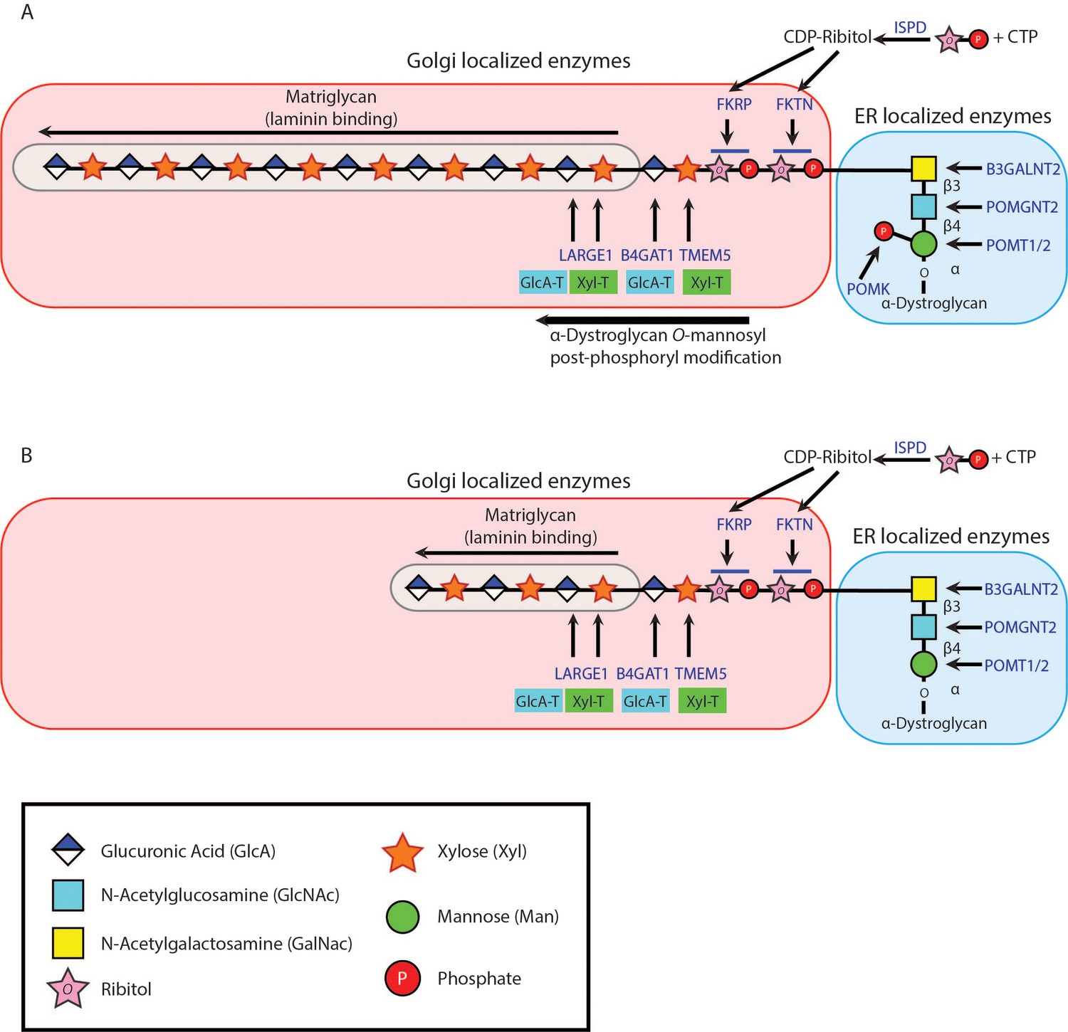
Figure 1
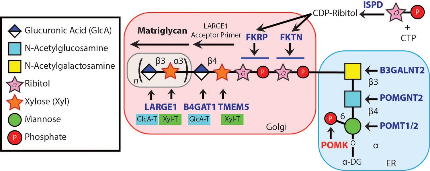
Synthesis of the α-DG Laminin-Binding Modification and Enzymes Involved.
Synthesis of the laminin-binding modification begins with the addition of the core M3 trisaccharide (GalNAc-β3-GlcNAc-β4-Man) on α-DG by the sequential actions of Protein O-Mannosyltransferase 1 and 2 (POMT1/2), Protein O-linked Mannose N-Acetyl-glucosaminyltransferase 2 (POMGNT2), and β1,3-N-Acetylgalactosaminyltransferase 2 (B3GALNT2), in the ER. POMK phosphorylates the C6 hydroxyl of mannose after synthesis of core M3. The phosphorylated core M3 is further elongated in the Golgi by Fukutin (FKTN), Fukutin related protein (FKRP), Transmembrane Protein 5 (TMEM5), β1,4-Glucuronyltransferase 1 (B4GAT1), and Like-acetyl-glucosaminyltranserase 1 (LARGE1). Isoprenoid synthase domain-containing (ISPD) produces cytidine diphosphate (CDP)-ribitol in the cytosol, and this serves as a sugar donor for the reactions catalyzed by FKTN and FKRP. LARGE1 synthesizes matriglycan, which directly interacts with the LG domains of matrix ligands.
Figure 2 with 2 supplements
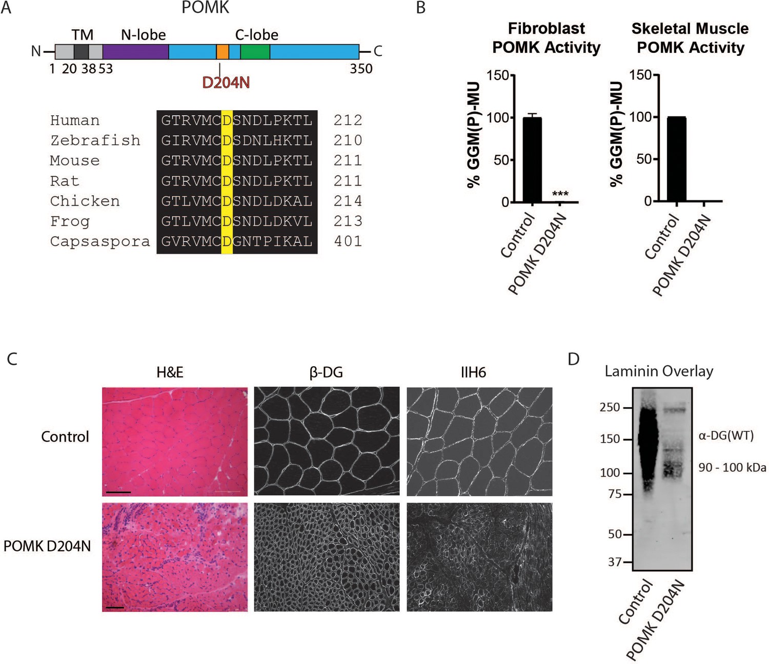
Characterization of a Patient with a Loss-of-Function Mutation in POMK.
(A) (above) Human POMK consists of a transmembrane domain (TM) and a kinase domain (N-lobe and C-lobe). The kinase domain contains the catalytic loop (orange) and activation segment (green). (below) Alignment of protein sequences flanking the D204N mutation. The mutation alters a highly conserved aspartate that is the catalytic base of the phosphorylation reaction catalyzed by the kinase. (B) POMK activity in control and patient NH13-284 (POMK D204N) fibroblasts (left) and skeletal muscle (right). n = 3 experiments were performed in fibroblasts. Triple asterisks: statistical significance with Student’s unpaired t-test (p-value<0.0001). Due to limited skeletal muscle, n = 1 experiment was performed. (C) Histology and immunofluorescence of control and POMK D204N skeletal muscle using IIH6 (anti-matriglycan) and a β-DG antibody. (Scale bars: Control- 200 µM, POMK D204N- 75 µM). (D) Laminin overlay of control and POMK D204N skeletal muscle.
Figure 2—figure supplement 1

Structural Modeling of POMK D204N Mutation.
This figure shows structural modeling of wild-type POMK and the POMK D204N mutation using human POMK protein sequence numbering, based on the crystal structure of zebrafish POMK. The green spheres indicate manganese ions. The phosphorus, oxygen, nitrogen, and carbon atoms are colored in orange, red, blue, and white, respectively. The D204 and N204 carbon atoms are colored dark. The gamma phosphate of ATP is not shown.
Figure 2—figure supplement 2
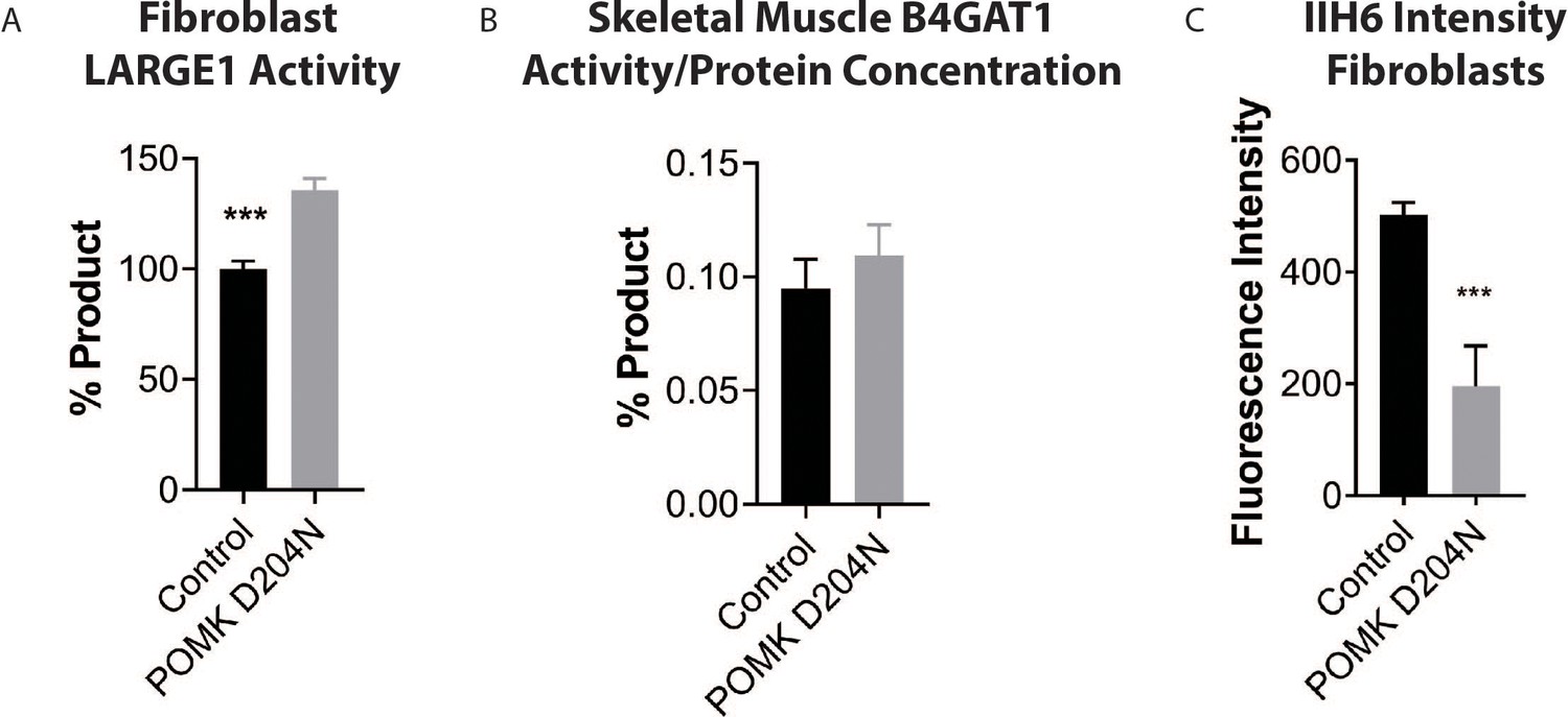
Supplemental Analysis of POMK D204N Fibroblasts and Muscle.
(A) LARGE1 activity in control human fibroblasts and fibroblasts from patient NH13-284 (POMK D204N). Triple asterisks indicate statistical significance using Student’s unpaired t-test (three replicates, p-value=0.0007). (B) B4GAT1 activity (normalized to protein concentration) from control skeletal muscle and POMK D204N muscle. (C) Mean fluorescence intensity of control human fibroblasts and POMK D204N fibroblasts. Flow cytometry analyses were performed using an antibody against matriglycan (IIH6). Triple asterisks indicate statistical significance using Student’s unpaired t-test (three replicates. p-value<0.0001).
Figure 3 with 4 supplements
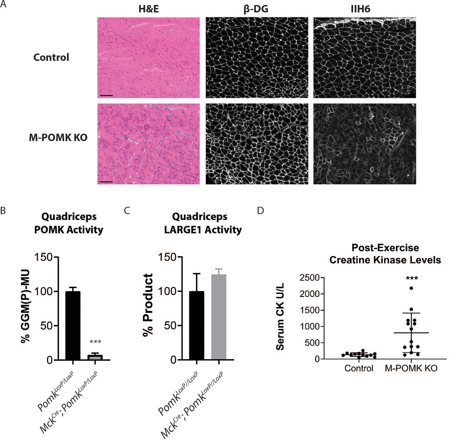
Mice with a Muscle-Specific Loss of Pomk Develop Hallmarks of a Mild Muscular Dystrophy.
(A) H&E and immunofluorescence analyses using IIH6 (anti-matriglycan) and an anti-β-DG antibody of quadriceps muscles of 4–6 week-old PomkLoxP/LoxP (Control) and MckCre; Pax7Cre; PomkLoxP/LoxP (M-POMK KO) mice. Scale bars: 100 µM. (B) POMK and (C) LARGE1 activity in extracts of MckCre; PomkLoxP/LoxP and PomkLoxP/LoxP quadriceps skeletal muscles. Triple asterisks indicate statistical significance using Student’s unpaired t-test (p-value<0.0001, three replicates). (D) Creatine kinase levels of 8-week-old M-POMK KO and Control mice. p-values were calculated with Student’s unpaired t-test. Triple asterisks: statistical significance with p-value<0.05 (p-value=0.0008), n = 12 Control and 14 M-POMK KO mice.
Figure 3—figure supplement 1
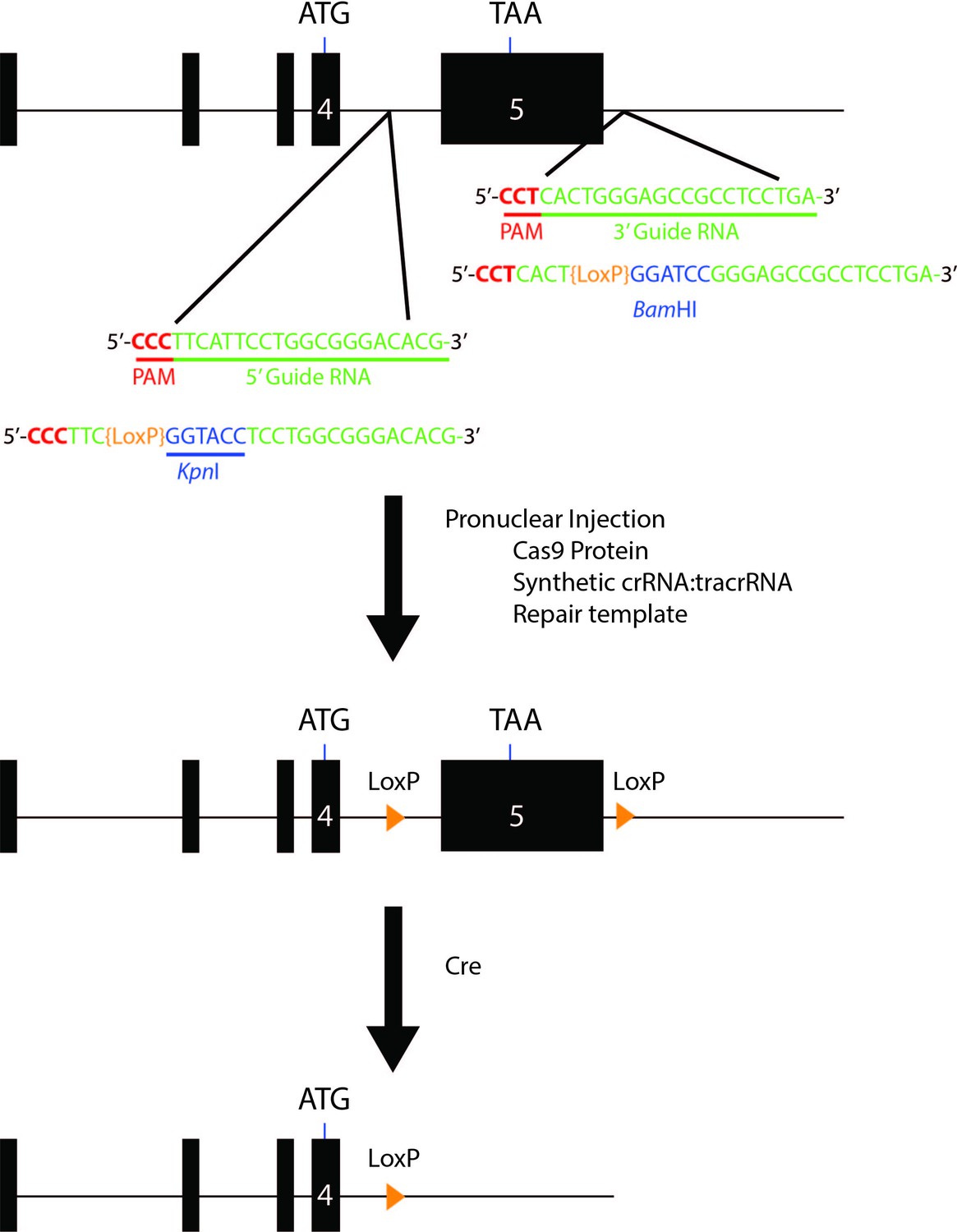
Schematic for Generation of Floxed Alleles of Pomk.
Map of 5’ and 3’ LoxP sites (orange). LoxP sites flanking exon 5 of Pomk (large black box), which encodes the majority of the kinase domain of POMK, were inserted using CRISPR/Cas9. Cre-mediated recombination of the floxed allele of Pomk is predicted to lead to a loss of exon 5.
Figure 3—figure supplement 2
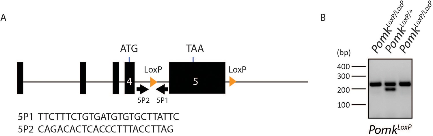
Results of PomkLoxP/LoxP Genotyping.
(A) Genotyping strategy for floxed Pomk Allele. PCR Primers were designed to flank the 5’ LoxP site. (B) The wild-type allele of Pomk is 197 bp, while the floxed allele is 235 base pairs.
Figure 3—figure supplement 3
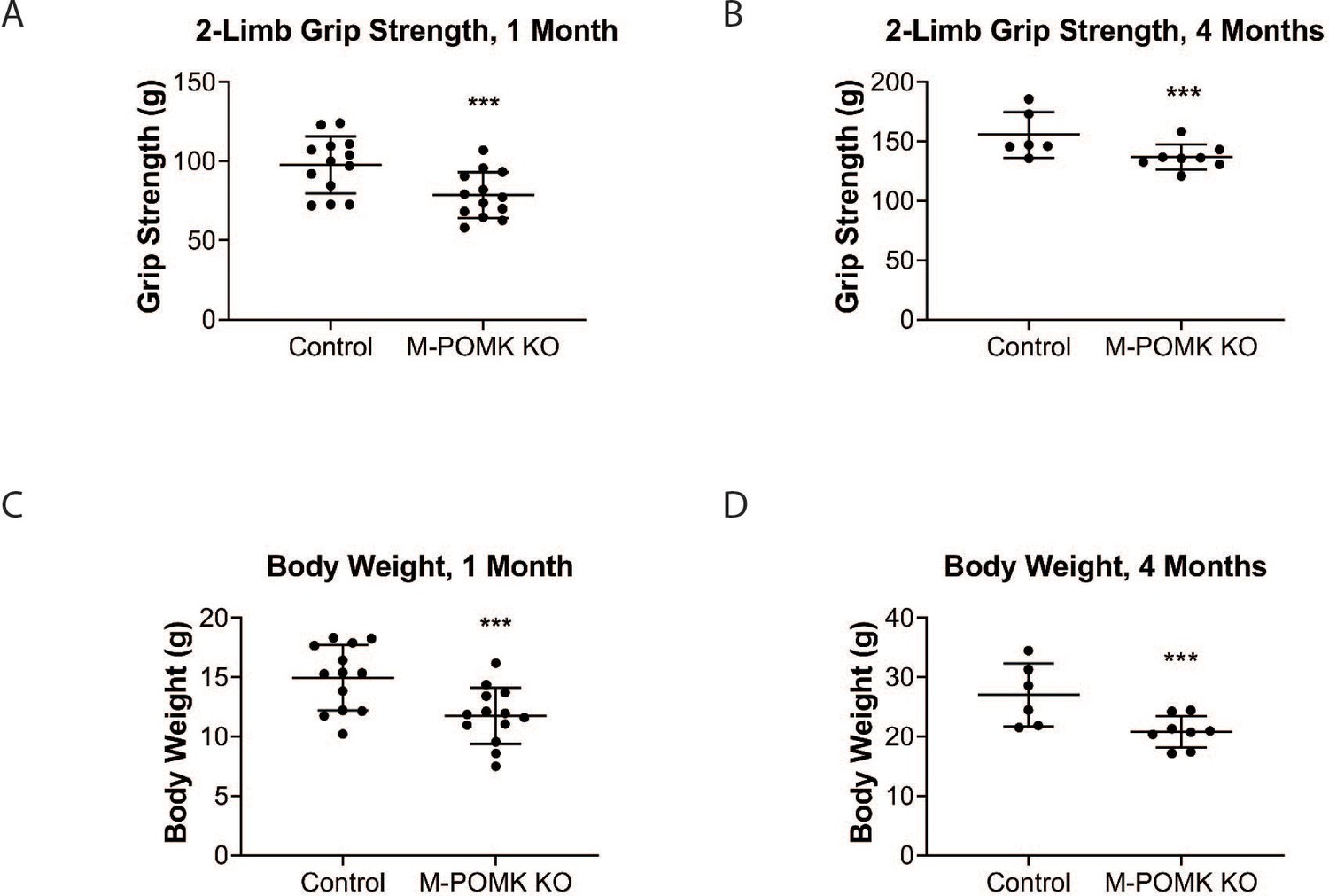
Muscle-Specific Pomk Knockout Mice Have Reduced Grip Strength and Body Weight.
(A, B) 2-limb grip strength of 1-month-old (A) and 4-month-old (B) PomkLoxP/LoxP (Control) and MckCre; Pax7Cre; PomkLoxP/LoxP (M-POMK KO) mice. Triple asterisks indicate statistical significance using Student’s unpaired t-test, p-value=0.0069 (A) p-value=0.038 (B). (C, D) Body weights of 1-month-old (C) and 4-month-old (D) Control and M-POMK KO mice. Triple asterisks indicate statistical significance with p-value<0.05 using Student’s unpaired t-test, p-value=0.0038 (C) p-value=0.0134 (D).
Figure 3—figure supplement 4

Supplemental Biochemical Analysis of Pomk-null Skeletal Muscle.
(A, B) POMK (A) and LARGE1 (B) activity of PomkLoxP/LoxP (Control) and MckCre; Pax7Cre; PomkLoxP/LoxP (M-POMK KO) quadriceps muscle extracts (three replicates). Asterisks indicate statistical significance with p-value<0.05 (p-value=0.0144) using Student’s unpaired t-test. (C) B4GAT1 activity in MckCre; PomkLoxP/LoxP and control quadriceps muscle extracts (three replicates).
Figure 4
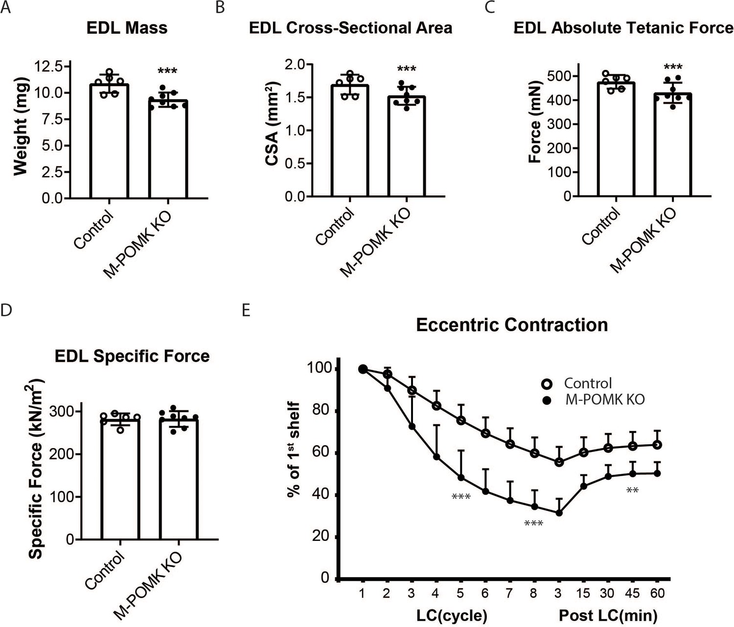
MckCre; Pax7Cre; PomkLoxP/LoxP Extensor Digitorum Longus (EDL) Muscle Demonstrates Eccentric Contraction-Induced Force Loss.
(A) Mass (milligrams) of PomkLoxP/LoxP (Control) and MckCre; Pax7Cre; PomkLoxP/LoxP (M-POMK KO) EDL muscles tested for force production. ***Statistical significance with Student’s unpaired t-test with p-value<0.05 (p=0.0031). (B) Cross-sectional area (CSA) of EDL muscles. ***Statistical significance using Student’s unpaired t-test with p-value<0.05 (p=0.0463). (C) Maximum Absolute Tetanic Force production by Control and M-POMK KO EDL muscles. ***Statistical significance using Student’s unpaired t-test with a p-value<0.05 (p=0.0395). (D) Specific Force production in Control and M-POMK KO EDL muscles (p=0.921). (E) Force deficit and force recovery in Control (n=3) and M-POMK KO (n=4) mice after eccentric contractions. EDL muscles from 18- to 20-week-old male mice were tested and are represented by open (Control) or closed (M-POMK KO) circles. ***Statistical significance using Student’s unpaired t-test (p-value<0.0001) compared to Control EDL at given LC cycle. **Statistical significance using Student’s unpaired t-test (p-value=0.0027) compared to Control EDL at given LC cycle. Error bars represent SD.
Figure 5 with 2 supplements
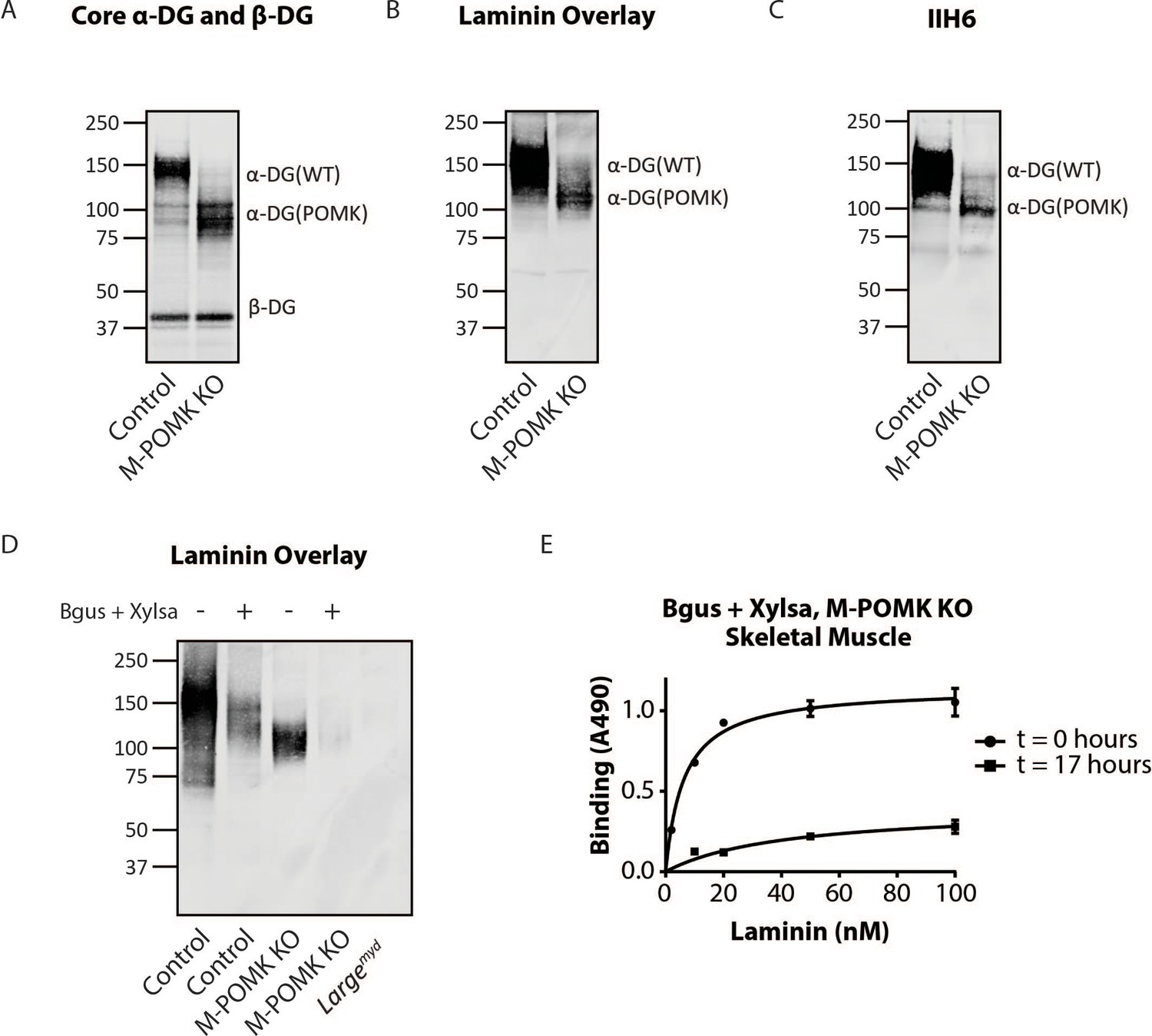
Mice with a Muscle-Specific Loss of Pomk Express Matriglycan.
(A) Biochemical analysis of Control and M-POMK KO skeletal muscle. Glycoproteins were enriched from quadriceps skeletal muscles of mice using wheat-germ agglutinin (WGA)-agarose. Immunoblotting was performed with antibody AF6868, which recognizes core α-DG and β-DG (three replicates). (B) Laminin overlay of quadriceps muscles of Control and M-POMK KO mice (three replicates). (C) IIH6 immunoblotting of Control and M-POMK KO quadriceps muscle. (D, E) Laminin overlay (D) and solid-phase analysis (E) of skeletal muscles of M-POMK KO mice treated in combination with two exoglycosidases, α-xylosidase (Xylsa) and β-glucuronidase (Bgus) for 17 hr (three replicates).
Figure 5—figure supplement 1

Solid-Phase Binding Analyses of Pomk-null Skeletal Muscle.
(A) Solid-phase binding analysis (relative Bmax for laminin-111) of PomkLoxP/LoxP (Control), MckCre; Pax7Cre; PomkLoxP/LoxP (M-POMK KO), MckCre; PomkLoxP/LoxP, and Largemyd skeletal muscle (three replicates). Error bars: standard deviation. (B) Solid-phase binding analysis of MckCre; PomkLoxP/LoxP skeletal muscle treated in combination with α-xylosidase (Xylsa) and β-glucuronidase (Bgus) for 0 or 20 hr. Results from three independent experiments are shown. Error bars: standard deviation.
Figure 5—figure supplement 2
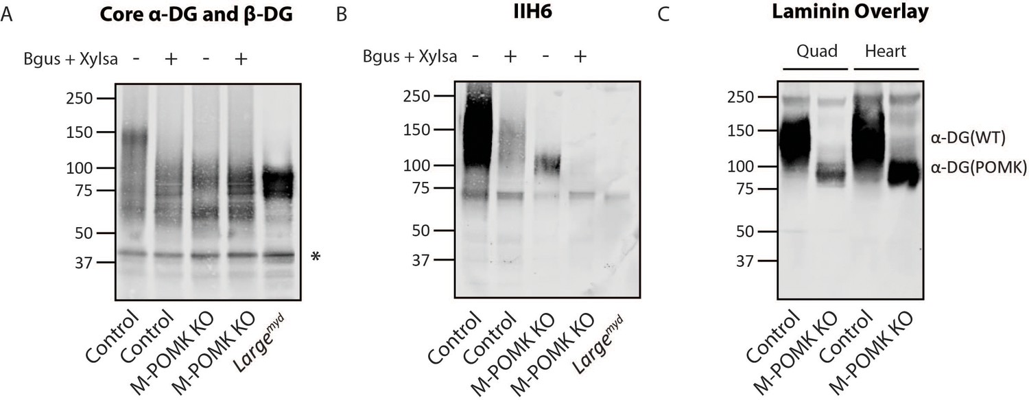
Pomk-null Muscle Expresses Matriglycan.
(A, B) Glycoproteins were enriched from skeletal muscles of Control, M-POMK KO, and Largemyd mice and treated in combination with α-xylosidase (Xylsa) and β-glucuronidase (Bgus). Immunoblotting was performed with (A) AF6868 (Core α-DG and β-DG) and (B) IIH6 (matriglycan). Results from three independent experiments are shown. Asterisk: β-DG. (C) A laminin overlay was performed of Control and M-POMK KO skeletal muscle and heart. Glycoproteins from heart were enriched as above (three replicates).
Figure 6 with 2 supplements
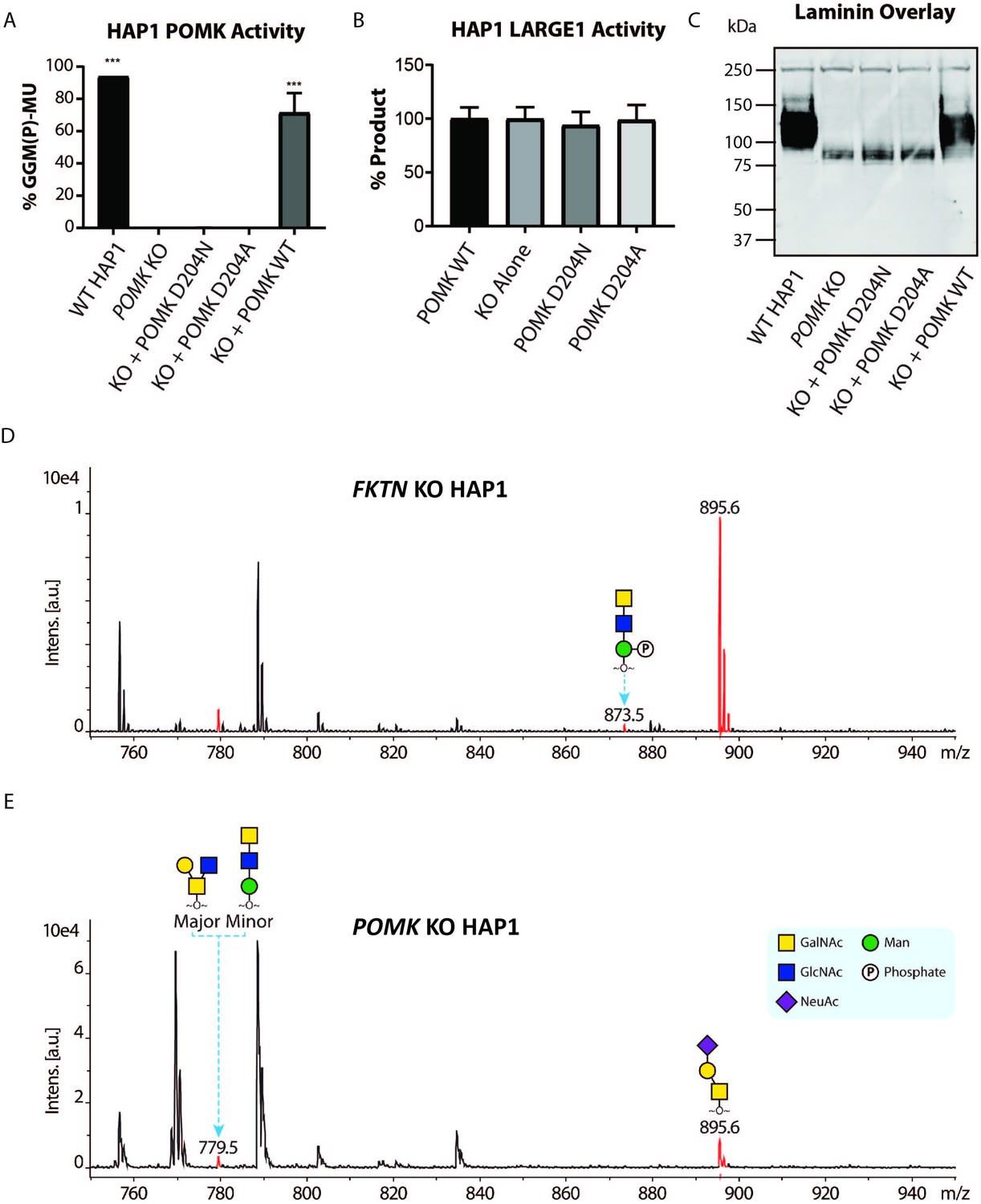
POMK D204N lacks Catalytic Activity.
(A) POMK or (B) LARGE1 activity in POMK KO HAP1 cells transduced with adenoviruses encoding POMK D204N, POMK D204A, or POMK WT. Triple asterisks: statistical significance (p-value<0.0001) compared to POMK KO alone using one-way ANOVA with Dunnett’s test for multiple comparisons (three replicates, 95% Confidence intervals for POMK KO vs. WT HAP1: −106.7 to −81.0, POMK KO vs. POMK KO + POMK WT: −84.25 to −58.54). (C) Laminin overlay of POMK KO HAP1 cells expressing the indicated POMK mutants. (D, E) Mass Spectrometry (MS)-based O-glycomic analyses of DG mucin-like domain (DG390TevHis) expressed in Fukutin (FKTN) (D) or POMK (E) KO HAP1 cells. O-glycans were released from the protein backbone and permethylated prior to matrix-assisted laser desorption/ionization time-of-flight (MALDI-TOF) analyses. MS peaks at m/z 779.5 (779.6) correspond to a mixture of core 2 and core M3 O-glycan, and at 873.5, phosphorylated core M3 O-glycan (red). MALDI-TOF is unable to determine anomeric or epimeric configurations of annotated O-glycans.
Figure 6—figure supplement 1
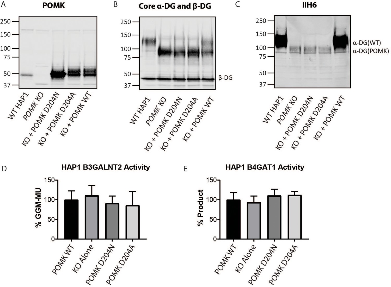
Supplemental Biochemical Analysis of POMK D204N and POMK KO HAP1 Cells.
(A, B, C) POMK KO HAP1 cells were transduced with the indicated adenoviruses and immunoblotting was performed for: (A) POMK, (B) Core α-DG and β-DG, and (C) matriglycan (IIH6), (three replicates). (D) B3GALNT2 and (E) B4GAT1 activity of POMK KO HAP1 cells expressing POMK mutants. Activity of each mutant relative to WT POMK is depicted. (Error bars: standard deviation). Results from three independent experiments are shown.
Figure 6—figure supplement 2
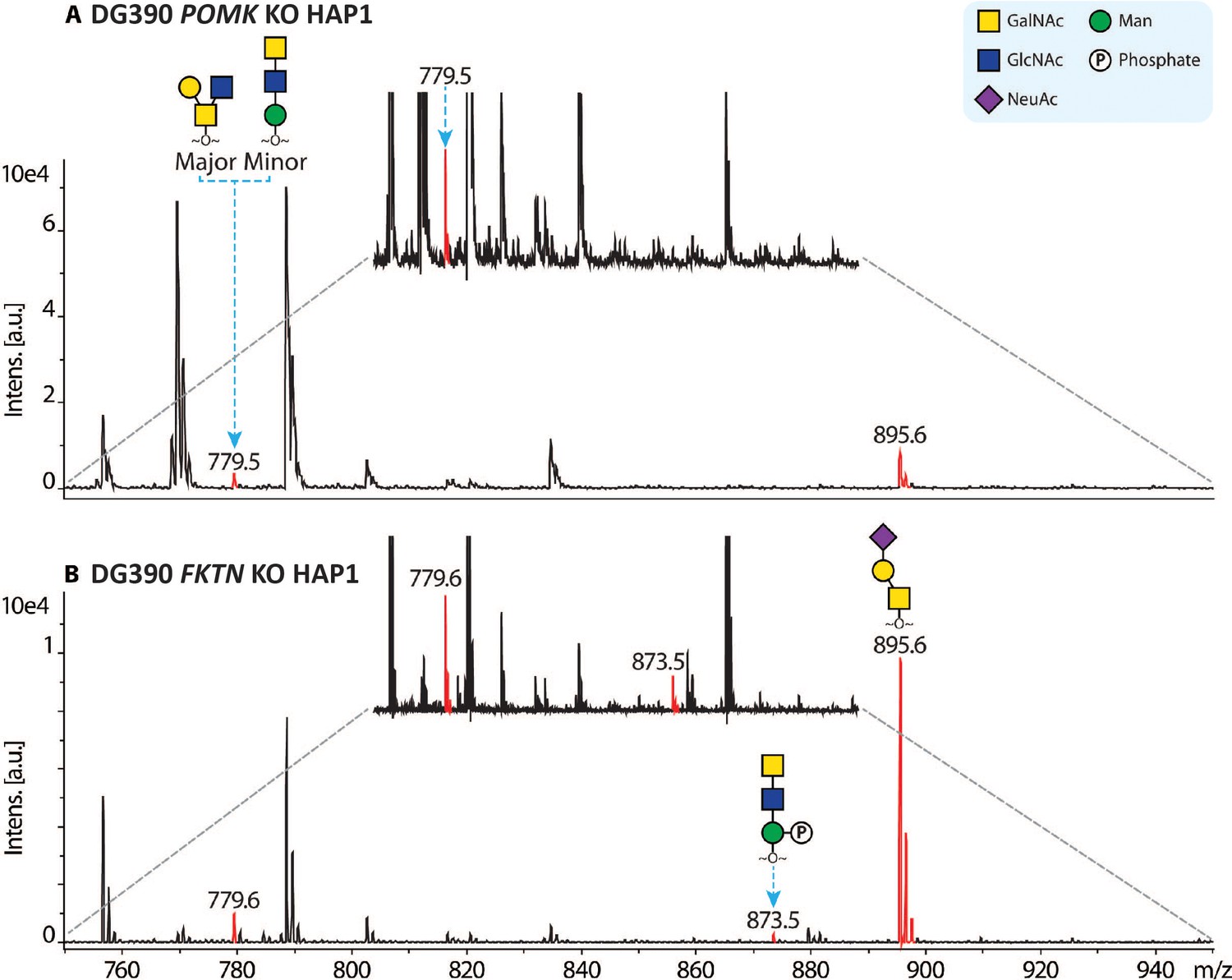
Mass spectra of O-glycans carried by a DG mucin-like domain model (DG390) expressed in POMK KO (A) or Fukutin (FKTN) KO (B) HAP1 cells.
The glycans were reductively released from the protein backbone and permethylated prior to matrix-assisted laser desorption/ionization time-of-flight (MALDI-TOF) analyses. Mass spectrometry (MS) peaks corresponding to sodiated permethylated O-glycans were colored red and annotated with glycan structures. The annotation was based on previous knowledge of human O-glycan structure and biosynthesis. MS peaks at m/z 779.5 correspond to a mixture of core 2 and core M3 O-glycan, and at 873.5, phosphorylated core M3 O-glycan. In addition, mucin-type core 1 O-glycan was also observed (m/z 895.6). Non-annotated peaks are contaminants from matrix and/or samples. The spectra were further zoomed (the spectra between the gray-dashed lines) to facilitate the relative intensity comparison between core M3 and phosphorylated core M3 O-glycans in the two samples. Under the current experimental set-up, our MALDI-TOF data are not sufficient to determine the stereochemistries of monosaccharides in the observed O-glycans. Raw MS data has been included as a supplement for more information (Source data 1 and Source data 2).
Figure 7 with 6 supplements
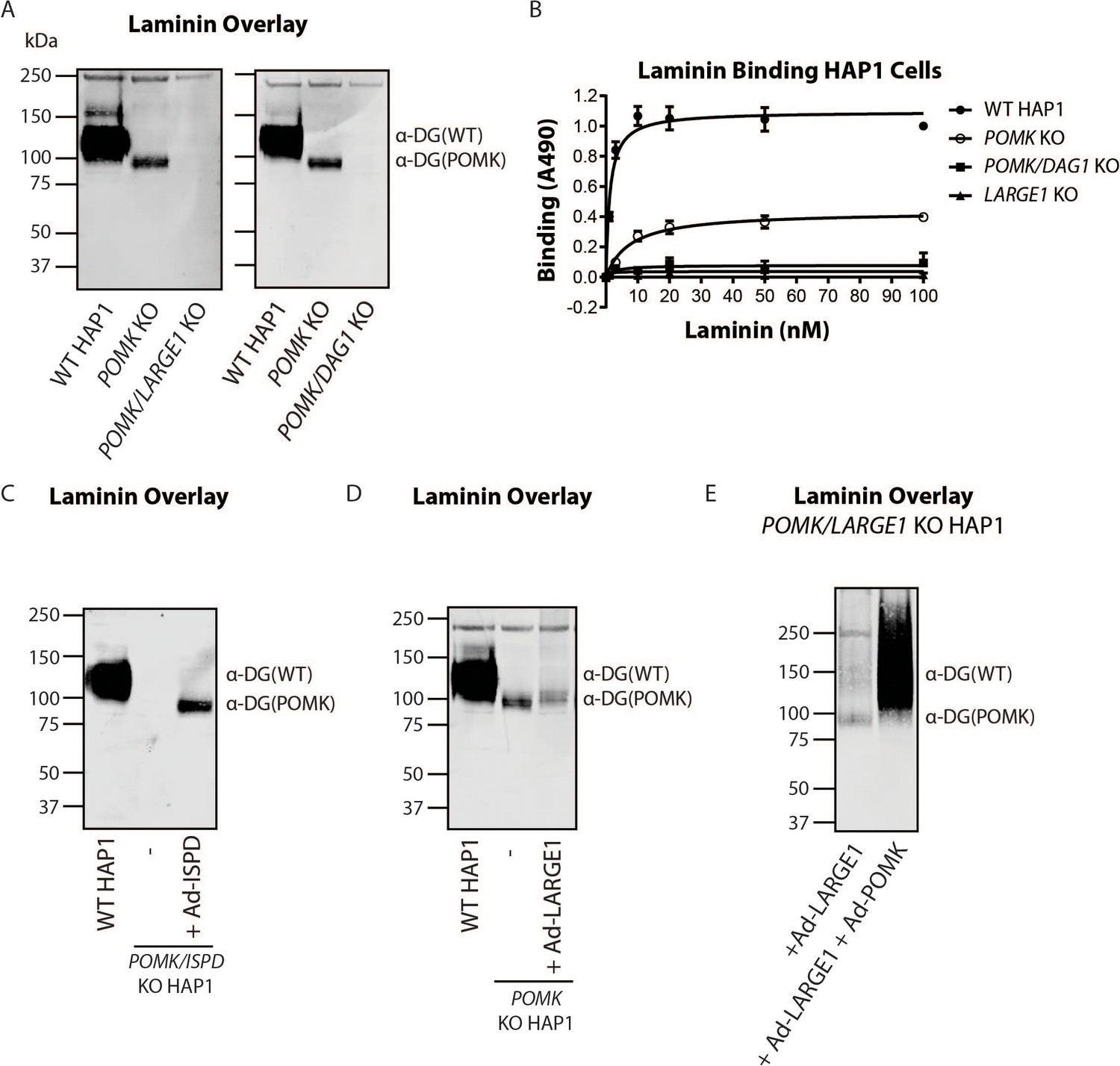
LARGE1 requires POMK to Elongate Matriglycan.
(A) WT, POMK KO, and POMK/LARGE1 KO HAP1 cells (left) or POMK/DAG1 KO HAP1 cells (right) (three replicates). (B) Solid-phase analysis of WT, POMK KO, POMK/DAG1 KO, and LARGE1 KO HAP1 cells (three replicates). (C, D, E) Laminin overlays of the following KO HAP1 cells (three replicates): POMK/ISPD expressing Ad-ISPD (C); POMK expressing Ad-LARGE1 (D); POMK/LARGE1 expressing Ad-LARGE1 with or without Ad-POMK (E).
Figure 7—figure supplement 1
Supplemental Biochemical Analysis of POMK/LARGE1 KO and POMK/DAG1 KO HAP1 Cells.
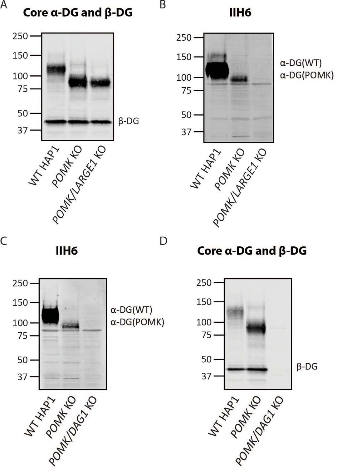
(A, B) Immunoblotting of WT, POMK KO and POMK/LARGE1 KO HAP1 cells with antibodies AF6868 (A) (Core α-DG and β-DG) or IIH6 (B). Glycoproteins were enriched using WGA-agarose as described in the Methods. (C, D) Immunoblotting of WT, POMK KO, and POMK/DAG1 KO HAP1 cells with antibodies IIH6 (C) or AF6868 (D) (Core α-DG and β-DG). Representative results from three independent experiments are shown.
Figure 7—figure supplement 2

Requirement for Ribitol-Phosphate in the Synthesis of the Non-Extended Form of Matriglycan.
(A, B, C) POMK KO HAP1 cells were transduced with an adenovirus encoding isoprenoid synthase domain-containing (Ad-ISPD). Immunoblotting was performed using antibodies AF6868 (A) or IIH6 (C). (B) A laminin overlay was also performed. Representative results from three independent experiments are shown. (D, E) HAP1 cells lacking expression of ISPD and POMK (POMK/ISPD KO) were transduced with Ad-ISPD. Immunoblotting was performed with an anti-Myc antibody (D) or antibody AF6868 (E) (Core α-DG and β-DG). Representative results from three independent experiments are shown.
Figure 7—figure supplement 3
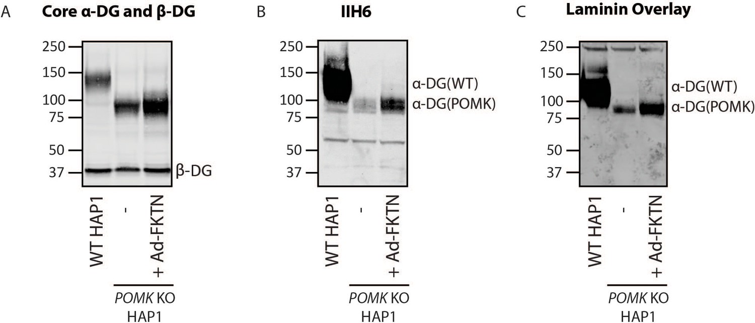
Fukutin Overexpression Enhances Synthesis of the Non-Extended Matriglycan.
(A, B, C) POMK KO HAP1 cells transduced with an adenovirus encoding Fukutin (FKTN), Ad-FKTN. Immunoblotting was performed using antibodies AF6868 (A) (Core α-DG and β-DG) or IIH6 (B) (three replicates). (C) A laminin overlay was also performed (three replicates).
Figure 7—figure supplement 4
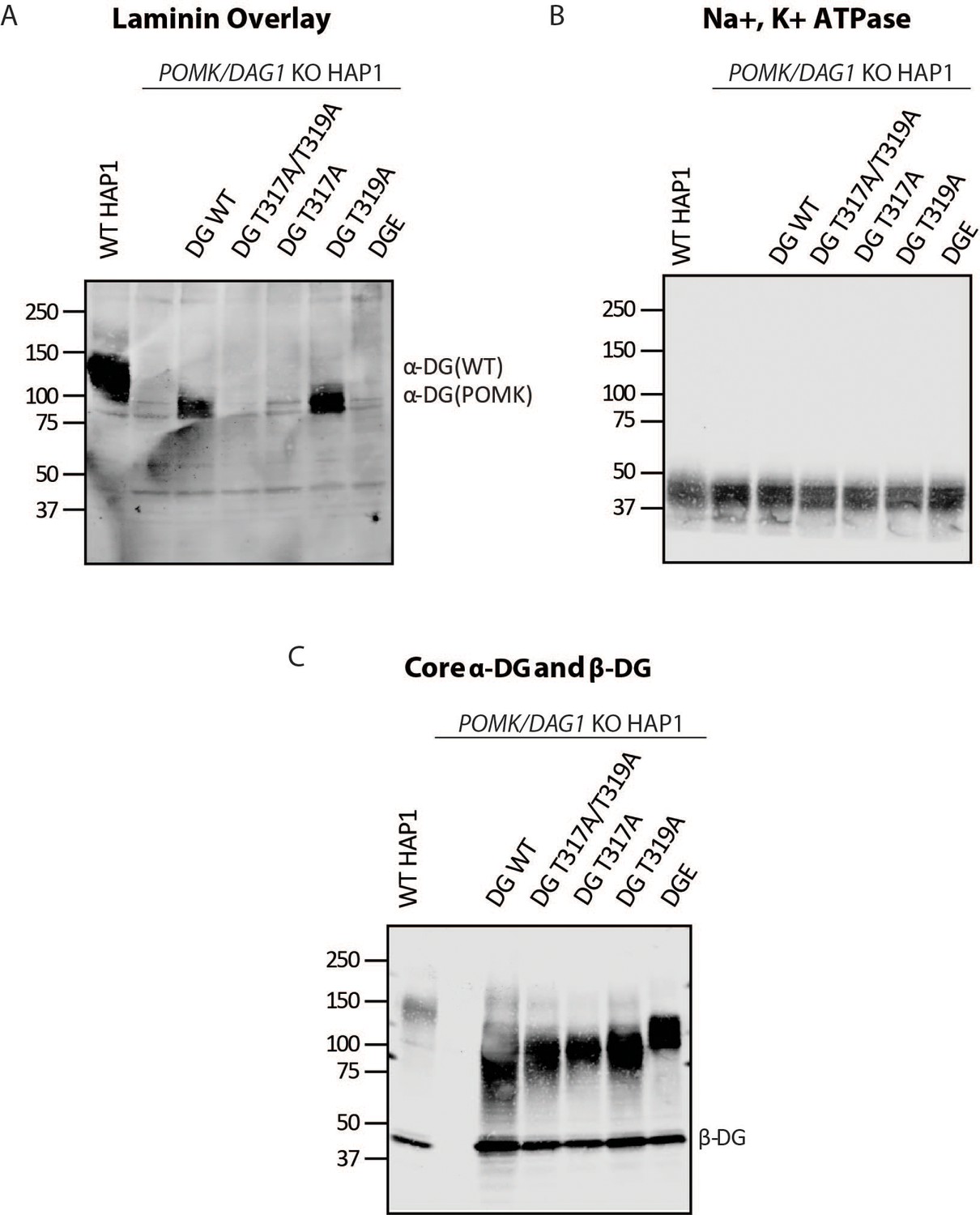
T317 is Required for Synthesis of the Non-Extended Matriglycan.
(A, B, C) Biochemical analysis of POMK/DAG1 KO HAP1 cells expressing the indicated adenoviruses (three replicates). DGE is for viral expression of α-DG that lacks the Dystroglycan N-terminal domain (DGN). (A) A laminin overlay was performed. Immunoblotting was performed with an Na+/K+ ATPase antibody (B) and antibody AF6868 (C) (Core α-DG and β-DG).
Figure 7—figure supplement 5
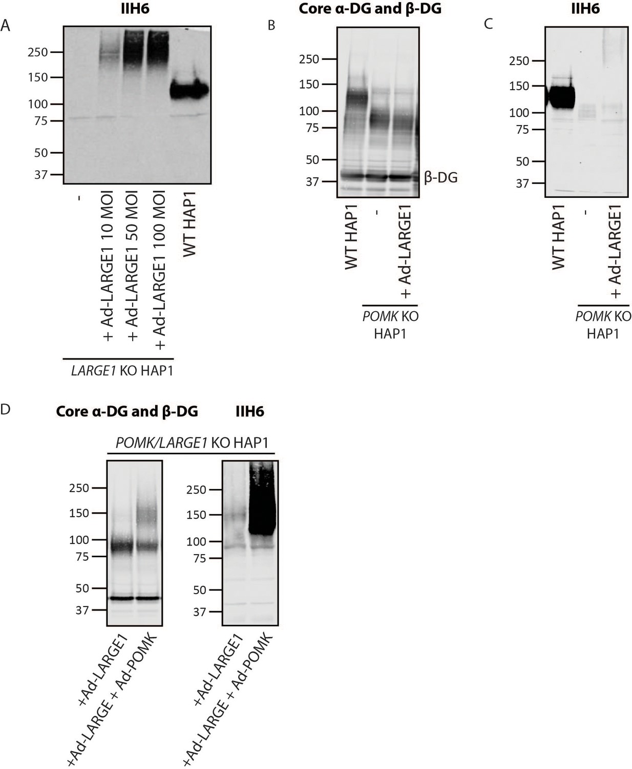
POMK Enables LARGE1-mediated Elongation of Matriglycan.
(A, B, C) Immunoblots of the following HAP1 cells: (A) LARGE1 KO, overexpressing Ad-LARGE1; (B, C) POMK KO, overexpressing Ad-LARGE1; (D) POMK/LARGE1 KO, overexpressing Ad-LARGE1 with or without Ad-POMK. Immunoblotting was performed with antibodies AF6868 (Core α-DG and β-DG) or IIH6 (three replicates).
Figure 7—figure supplement 6
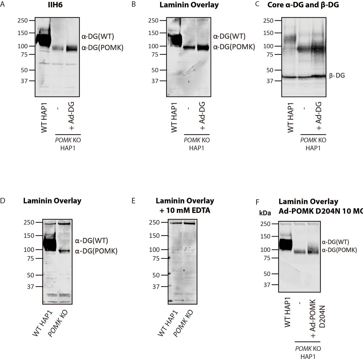
Supplemental Characterization of POMK-null Matriglycan Synthesis.
(A, B, C) POMK KO HAP1 cells were transduced with an adenovirus encoding DG WT (Ad-DG) and immunoblotting was performed with antibodies IIH6 (A) and AF6868 (C) (three replicates). A laminin overlay was also performed (B) (three replicates). (D, E) Laminin overlays of WT and POMK KO HAP1 cells were performed without (D) or with (E) EDTA (three replicates). (F) A laminin overlay of WT HAP1, POMK KO HAP1, or POMK KO HAP1 cells transduced with 10 MOI Ad-POMK D204N was performed (three replicates).
Figure 8 with 3 supplements
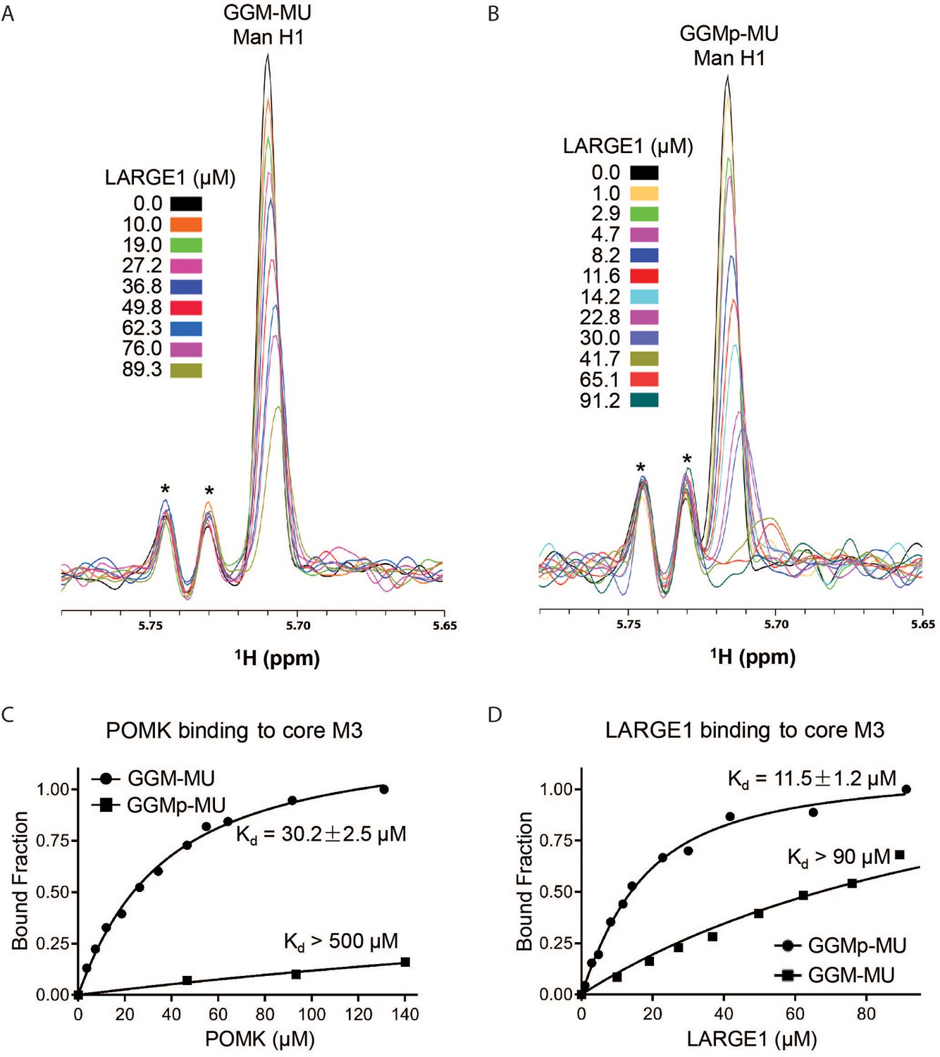
NMR Analyses of POMK and LARGE1 Binding to GGM-MU and GGMp-MU.
(A, B) 1D 1H NMR spectra of the anomeric region of GGM-MU (A) and GGMp-MU (B) were acquired for the glycan concentration of 10.0 µM in the presence of various concentrations of LARGE1 as indicated. The peak Man H1 is derived from the mannose anomeric H1 proton. Stars indicate impurity peaks derived from buffer. (C, D) Fitting of the NMR binding data of POMK (C) and LARGE1 (D) to core M3 glycans of GGM-MU and GGMp-MU, respectively. The bound fraction was obtained from the NMR titration data by measuring the difference in the peak intensity of the anomeric proton Man H1 in the absence (free form) and presence (bound form) of POMK or LARGE1, then divided by the peak intensity of the free form.
Figure 8—figure supplement 1

NMR Spectra of POMK Binding to GGMp-MU and Structure of GGMp-MU.
(A) 1D 1H NMR spectra of the glycan sample (10.0 µM GGMp-MU) were acquired in the presence of various concentrations of zebrafish POMK as indicated. The 13C and 1H resonances of GGMp-MU have been assigned before (Yoshida-Moriguchi et al., 2013). The peak AH1 is derived from the residue A (Man) anomeric H1 proton. (B) Chemical structure of GGMp-MU.
Figure 8—figure supplement 2
Model of Full-Length and Non-extended Matriglycan Synthesis.
(A) Mature matriglycan is a long polysaccharide that is synthesized by LARGE1. (B) In the absence of the core M3 phosphate added by POMK, LARGE1 generates a shorter, non-extended form of matriglycan.
Figure 8—figure supplement 3
Biochemical and Histologic Analysis of MckCre; PomkLoxP/LoxP Quadriceps Muscle.
(A, B, C) Representative biochemical analysis of glycoproteins enriched from quadriceps skeletal muscles of PomkLoxP/LoxP, MckCre; PomkLoxP/LoxP, and Largemyd mice using WGA-agarose (three replicates). For immunoblotting, antibodies AF6868 (A) and IIH6 (C) were used, and a laminin overlay was also performed (B). (D) Immunofluorescence and H&E analyses of PomkLoxP/LoxP and MckCre; PomkLoxP/LoxP quadriceps muscle sections from 8 month old mice. Sections were stained with antibodies against β-DG (middle) and matriglycan (IIH6) (right). Histologic abnormalities in the sections were evaluated by means of hematoxylin and eosin (H&E) staining (left). Scale bars- 50 µM (three replicates).
Tables
Appendix 1—key resources table
| Reagent type (species) or resource | Designation | Source or reference | Identifiers | Additional information |
|---|---|---|---|---|
| Genetic reagent Mus musculus | PomkLoxP/LoxP ICR | This paper | Campbell Lab | Materials and Methods ‘Generation of PomkLoxP/LoxP Mice’ |
| Genetic reagent Mus musculus | Pax7Cre C57BL/6J | The Jackson Laboratory, Bar Harbor ME. | JAX:010530, RRID:IMSRJAX:010530 | Pax7tm1(cre)Mrc |
| Genetic reagent Mus musculus | MckCre C57BL/6J | The Jackson Laboratory, Bar Harbor ME. | JAX:006475, RRID:IMSR_JAX:006475 | B6.FVB(129S4)-Tg(Ckmm-cre)5Khn/J |
| Genetic reagent Mus musculus | Largemyd C57BL/6J | The Jackson Laboratory, Bar Harbor ME. | JAX:000300, RRID:IMSR_JAX:000300 | MYD/Le-Os +/+ Largemyd/J myd |
| Antibody | anti-DG (Sheep polyclonal) | R and D Systems | Cat# AF6868, RRID:AB_10891298 | WB (1:500) |
| Antibody | anti-α-DG (IIH6C4) (Mouse monoclonal) | DSHB Campbell Lab | Cat# IIH6 C4, RRID:AB_2617216 | WB (1:10-1:100) |
| Antibody | anti-myc clone 4A6 (Mouse monoclonal) | Millipore | Cat# 05–724, RRID:AB_309938 | WB (1:2000) |
| Antibody | anti-α-DG (IIH6C4) (Mouse monoclonal) | Millipore Campbell Lab | Cat# 05–593, RRID:AB_309828 | IF (1:1000-1:2000) |
| Antibody | anti-Laminin (Rabbit polyclonal) | Sigma-Aldrich | Cat# L9393, RRID:AB_477163 | WB (1:1000), Solid-Phase Assay (1:5000) |
| Antibody | anti-β-DG (Rabbit polyclonal) | Campbell Lab PMID:1741056 DOI:10.1038/355696a0 | AP83 | IF (1:50) |
| Antibody | anti-β-DG mouse IgM (Mouse monoclonal) | Leica Biosystems | Cat# NCL-b-DG, RRID:AB_442043 | IF (1:50 to 1:200) |
| Antibody | anti-Na+,K+ ATPase (Mouse monoclonal) | BD Biosciences | Cat# 610993 RRID:AB_398306 | WB (1:1000) |
| Antibody | anti-sheep IgG (Donkey polyclonal) | Rockland | Cat# 613-731-168, RRID:AB_220181 | WB (1:2000) |
| Antibody | anti-mouse IgG (H + L) (Donkey polyclonal) | LI-COR Biosciences | Cat# 926–32212, RRID:AB_621847 | WB (1:15,000), IF (1:800) |
| Antibody | anti-rabbit IgG (H + L) (Donkey polyclonal) | LI-COR Biosciences | Cat# 926–32213, RRID:AB_621848 | WB (1:15,000), IF (1:800) |
| Antibody | anti-mouse IgM (Goat polyclonal) | LI-COR Biosciences | Cat# 926–32280, RRID:AB_2814919 | WB (1:2500) |
| Antibody | anti-mouse IgG1 (Goat polyclonal) | LI-COR Biosciences | Cat# 926–32350, RRID:AB_2782997 | WB (1:2000, 1:10,000) |
| Antibody | anti-rabbit IgG (H+L) (Goat polyclonal) | Thermo Fisher Scientific | Cat# A-11034, RRID:AB_2576217 | IF (1:1000 to 1:2000) |
| Antibody | anti-mouse IgM (Goat polyclonal) | Thermo Fisher Scientific | Cat# A-21042, RRID:AB_2535711 | IF (1:1000 to 1:2000) |
| Antibody | anti-human FLJ23356 (Mouse monoclonal) | Novus | Cat# H00084197-M03, RRID:AB_2188284 | WB (1:500) |
| Commercial assay or kit | Creatine Kinase (CK) Liqui-UV Test | Fisher Scientific/Stanbio | Cat# 22-022-630 | |
| Cell line (Homo-sapiens) | Parental cell line C631 | Horizon Discovery | Cat# C631 | Mycoplasma testing passed |
| Cell line (Homo-sapiens) | POMK/DAG1 KO | Horizon Discovery | HZGHC001338c004, RRID:CVCL_TF19 | Authenticated by Sanger sequencing. Mycoplasma testing passed. |
| Cell line (Homo-sapiens) | POMK/LARGE1 KO | Horizon Discovery | HZGHC007364c011 | Authenticated by Sanger sequencing. Mycoplasma testing passed. |
| Cell line (Homo-sapiens) | POMK KO | Horizon Discovery | HZGHC001338c004, RRID:CVCL_TF19 | Authenticated by Sanger sequencing. Mycoplasma testing passed. |
| Cell line (Homo-sapiens) | POMK/ISPD KO | Horizon Discovery | HZGHC001338c001, RRID:CVCL_TF18 | Authenticated by Sanger sequencing. Mycoplasma testing passed. |
| Cell line (Homo-sapiens) | FKTN KO | Horizon Discovery | HZGHC000721c010, RRID:CVCL_SN68 | Authenticated by Sanger sequencing. Mycoplasma testing passed. |
| Cell line (Homo-sapiens) | LARGE1 KO | Horizon Discovery | HZGHC000122c007, RRID:CVCL_SV31 | Authenticated by Sanger sequencing. Mycoplasma testing passed. |
| Cell line (Homo-sapiens) | Primary dermal fibroblasts, human | ATCC | PCS-201–012 | |
| Cell line (Homo-sapiens) | Human fibroblasts (POMK D204N) | This paper | NH13-284 | Dubowitz Neuromuscular Center, Campbell Lab |
| Peptide, recombinant protein | β-Glucuronidase | PMID:16303119 DOI:10.1016/j.carres.2005.10.005 | ||
| Peptide, recombinant protein | α-Xylosidase | PMID:10801892 DOI:10.1074/jbc.M910392199 | ||
| Biological sample (Homo-sapiens) | Control human skeletal muscle | This paper | Dubowitz Neuromuscular Center, Campbell Lab | |
| Biological sample (Homo-sapiens) | Human skeletal muscle | This paper | (NH13-284, POMK D204N) | Dubowitz Neuromuscular Center, Campbell Lab |
| Chemical compound, drug | Purified Danio rerio POMK | PMID:27879205 DOI:10.7554/eLife.22238 | ||
| Chemical compound, drug | Purified mammalian dTMLARGE1 | PMID:22223806 DOI:10.1126/science.1214115 | ||
| Chemical compound, drug | GGM-MU and GGMp-MU | PMID:23929950 DOI:10.1126/science.1239951 | ||
| Chemical compound, drug | UDP-Xylose | CarboSource | https://www.ccrc.uga.edu/~carbosource/CSS_substrates.html | |
| Chemical compound, drug | 4-Methylumbelliferyl-β-D-xylopyranoside | Sigma/Millipore | Cat# M7008 | |
| Chemical compound, drug | UDP-Glucuronic acid | Sigma/Millipore | Cat# U6751 | |
| Chemical compound, drug | Uridine 5′-diphospho-N-acetylgalactosamine disodium salt | Sigma/Millipore | Cat# U5252 | |
| Chemical compound, drug | Uridine 5′-diphospho-N-acetylglucosamine sodium salt | Sigma/Millipore | Cat# U4375 | |
| Chemical compound, drug | 4-methylumbelliferyl α-(GlcNAc-β(1-4)Man) GM-MU | Sussex Research | https://www.sussex-research.com/ | |
| Chemical compound, drug | Xylose-α1,3-GlcA-β-MU | Sussex Research | https://www.sussex-research.com/ | |
| Chemical compound, drug | Pepstatin A | Sigma/Millipore | Cat# 516481 | |
| Chemical compound, drug | Calpain Inhibitor I (25 mg) | Sigma/Millipore | Cat# A6185 | |
| Chemical compound, drug | Aprotinin from bovine lung | Sigma/Millipore | Cat# A1153 | |
| Chemical compound, drug | Leupeptin (25 mg) | Sigma/Millipore | Cat# 108975 | |
| Chemical compound, drug | PMSF | Sigma/Millipore | Cat# P7626-25G | |
| Chemical compound, drug | Immobilon-FL PVDF | Sigma/Millipore | Cat# IPFL00010 | |
| Chemical compound, drug | Calpeptin | Fisher Scientific | Cat# 03-340-05125M | |
| Chemical compound, drug | Bis-acrylamide solution-30% (37:1) | Fisher Scientific/Hoefer | Cat# HBGR337500X | |
| Chemical compound, drug | Benzamidine Hydrochloride Hydrate | MP Biochemicals | Cat# 195068 | |
| Chemical compound, drug | WGA agarose bound | Vector Labs | Cat# AL-1023, RRID:AB_2336862 | |
| Chemical compound, drug | Precision Plus Protein All Blue Standards-500ul | Bio-Rad | Cat# 161–0373 | |
| Software, algorithm | SigmaPlot | SigmaPlot | RRID:SCR_003210 | |
| Software, algorithm | Excel | Microsoft | RRID:SCR_016137 | |
| Software, algorithm | GraphPad Prism | https://www.graphpad.com/scientific-software/prism/ | RRID:SCR_002798 | Version 8.3 |
| Software, algorithm | FlowJo | https://www.flowjo.com/solutions/flowjo/downloads | RRID:SCR_008520 | Version 7.6.5 |
| Software, algorithm | Li-Cor Image Studio Software | https://www.licor.com/bio/image-studio-lite/download | RRID:SCR_015795 | |
| Other | Streptavidin, Alexa Fluor 594 conjugate | Thermo Fisher Scientific | Cat# S11227 | IF (1:1000 to 1:2000) |
| Other | Adenovirus: DGC (DG, delta H30-A316) | PMID:21987822 DOI:10.1073/pnas.1114836108 | ||
| Other | Adenovirus: DG T317A | PMID:21987822 DOI:10.1073/pnas.1114836108 | ||
| Other | Adenovirus: DG T319A | PMID:21987822 DOI:10.1073/pnas.1114836108 | ||
| Other | Adenovirus: DG T317A/319A | PMID:21987822 DOI:10.1073/pnas.1114836108 | ||
| Other | Adenovirus: DG Wild-Type (WT) | PMID:21987822 DOI:10.1073/pnas.1114836108 | ||
| Other | Adenovirus: POMK WT | PMID:27879205 DOI:10.7554/eLife.22238 | ||
| Other | Adenovirus: POMK D204A | PMID:27879205 DOI:10.7554/eLife.22238 | ||
| Other | Adenovirus: POMK D204N | This paper | Campbell Lab | Materials and methods ‘Adenovirus Production’ |
| Other | Adenovirus: DG390TEVHis | This paper | Campbell Lab | Materials and methods ‘Adenovirus Production’ |
| Other | Adenovirus: Fukutin | PMID:22522420 DOI:10.1038/ng.2252 | ||
| Other | Adenovirus: Isoprenoid Synthase Domain-Containing (ISPD) | PMID:22522420 DOI:10.1038/ng.2252 | ||
| Other | Adenovirus: LARGE1 | PMID:22522420 DOI:10.1038/ng.2252 | ||
| Other | NMR spectrometer | Bruker | Avance II 800 MHz | |
| Other | Rodent Treadmill | Columbus Instruments | Exer 3/6 Treadmill | |
| Other | Western Blot Imager | Li-Cor | Odyssey CLx | |
| Other | Mouse treadmill | Omnitech Electronics | Accupacer Treadmill | |
| Other | Isolated Mouse Muscle System | Aurora Scientific | 1200A | |
| Other | Mouse Grip Strength Meter | Columbus Instruments | 1027 Mouse | |
| Other | Prominence HPLC | Shimadzu | LC-20 system | |
| Other | Tabletop ultracentrifuge | Beckman Coulter | Optima max, 130K | |
| Other | Ultracentrifuge | Beckman Coulter | Optima-L-100 XP | |
| Other | Centrifuge | Beckman Coulter | Avanti J-E HPC | |
| Other | HPLC LC18 column | Supelco | 58368 |
Additional files
-
Source data 1
Raw MALDI-TOF data of DG390 expressed in FKTN KO HAP1 cells (Figure 6—figure supplement 2B).
Data were exported to the TXT format by FlexAnalysis 3.3 (Bruker Daltonics). The mass spectra in the article were zoomed into the range of m/z 750–950 to better present MS signals corresponding to core M3 glycan structures.
- https://cdn.elifesciences.org/articles/61388/elife-61388-data1-v4.txt.zip
-
Source data 2
Raw MALDI-TOF data of DG390 expressed in POMK KO HAP1 cells (Figure 6—figure supplement 2A).
Data were exported to the TXT format by FlexAnalysis 3.3 (Bruker Daltonics). The mass spectra in the article were zoomed into the range of m/z 750–950 to better present MS signals corresponding to core M3 glycan structures.
- https://cdn.elifesciences.org/articles/61388/elife-61388-data2-v4.txt.zip
-
Transparent reporting form
- https://cdn.elifesciences.org/articles/61388/elife-61388-transrepform-v4.docx
Download links
A two-part list of links to download the article, or parts of the article, in various formats.
Downloads (link to download the article as PDF)
Open citations (links to open the citations from this article in various online reference manager services)
Cite this article (links to download the citations from this article in formats compatible with various reference manager tools)
POMK regulates dystroglycan function via LARGE1-mediated elongation of matriglycan
eLife 9:e61388.
https://doi.org/10.7554/eLife.61388


