Differentiation of mouse fetal lung alveolar progenitors in serum-free organotypic cultures
Abstract
Lung epithelial progenitors differentiate into alveolar type 1 (AT1) and type 2 (AT2) cells. These cells form the air-blood interface and secrete surfactant, respectively, and are essential for lung maturation and function. Current protocols to derive and culture alveolar cells do not faithfully recapitulate the architecture of the distal lung, which influences cell fate patterns in vivo. Here, we report serum-free conditions that allow for growth and differentiation of mouse distal lung epithelial progenitors. We find that Collagen I promotes the differentiation of flattened, polarized AT1 cells. Using these organoids, we performed a chemical screen to investigate WNT signaling in epithelial differentiation. We identify an association between Casein Kinase activity and maintenance of an AT2 expression signature; Casein Kinase inhibition leads to an increase in AT1/progenitor cell ratio. These organoids provide a simplified model of alveolar differentiation and constitute a scalable screening platform to identify and analyze cell differentiation mechanisms.
Introduction
Lung alveolar progenitor cells differentiate at saccular stages (E17 in mouse, 26 weeks in human) into flattened alveolar type 1 (AT1) cells, constituting the surface for gas exchange, and secretory type 2 (AT2) cells, which prevent alveolar collapse by secreting pulmonary surfactant (Morrisey and Hogan, 2010; Chao et al., 2015). Fetal distal lung progenitors are thought to be bipotent and they co-express markers of both AT1 and AT2 cells, including Podoplanin/PDPN and Advanced Glycosylation End product-specific Receptor/RAGE as well as Prosurfactant Protein C/SFTPC, respectively (Desai et al., 2014; Treutlein et al., 2014). Differentiation of progenitors into AT1 and AT2 cells is regulated by multiple signals including WNT (Wingless and Int-1) and FGF (Fibroblast Growth Factor) from the mesenchyme (Volckaert and De Langhe, 2015; Li et al., 2018), mechanical forces (Li et al., 2018), epigenetic modifications (Wang et al., 2016), and the extracellular matrix (ECM) (Kim et al., 2018).
In particular, WNT signaling was shown to promote the expansion of Axin2+ AT2 cells during alveologenesis and prevent their conversion to the AT1 lineage (Frank et al., 2016). Similarly, WNT activity marks lung progenitors in homeostasis and regeneration, and WNT inhibition leads to AT1 differentiation (Nabhan et al., 2018; Zacharias et al., 2018). During mouse fetal development, β-catenin is required for the maintenance of distal lung progenitors (Ostrin et al., 2018); however, a mechanistic understanding of how WNT signaling controls AT1/AT2 cell differentiation or fate selection is currently lacking (Aros et al., 2021). A more detailed picture of how WNT/β-catenin signaling influences early alveolar differentiation could lead to better derivation protocols for alveolar cells and reveal new ways to stimulate differentiation in the fetal lung.
3D cultures of lung- or iPSC-derived alveolar progenitor cells have been used to model respiratory cell fate specification and signaling/cell interactions (reviewed by Gkatzis et al., 2018). However, these models fail to recapitulate the defined epithelial architecture of the mouse lung at canalicular and saccular stages, with HOPX+ cells residing in the stalk and SOX9+/SFTPC+ cells in the tip region (Frank et al., 2019). Until recently (Vazquez‐Armendariz et al., 2020), most models have displayed a spheroid or unpatterned morphology, making it difficult to assess localized differentiation from progenitors to alveolar cells. In addition, despite the central role of AT1 cells in lung development (Zepp et al., 2021), AT1 cell isolation and generation in vitro have been longstanding obstacles, and the efficiency of AT1 cell generation in vitro, or the properties of the cells obtained in these cultures, have not been analyzed in detail, limiting the relevance of these models for cell differentiation studies. These shortcomings highlight the need for an alveolar organoid model with morphological and cellular features as observed in vivo, which would accelerate the identification of differentiation-promoting factors.
To help address these questions, we developed a serum-free, rapid, and scalable distal lung progenitor organotypic culture system that recapitulates both AT1 cell differentiation and the endogenous tissue architecture of the alveolizing lung, which will facilitate mechanistic investigations of alveolar development.
Results and discussion
Mouse fetal alveolar epithelial progenitors are maintained in 3D serum-free cultures
To generate alveolar organoids by self-organization of endogenous progenitors, we manually dissociated E14.5 mouse lungs and collected distal epithelial fragments. At E14.5, lung pseudoglandular development is almost complete in mouse and NKX2.1+ alveolar progenitors become specified (Frank et al., 2019), while the distal SOX9+ epithelial domain begins to expand compared with the proximal airway (Alanis et al., 2014).
We embedded the epithelial tips into diluted Matrigel/Collagen I domes and established cultures in defined medium supplemented with growth factors (Figure 1A). Three main soluble growth factors have been implicated in distal lung growth and differentiation: FGF7, FGF10, and BMP4 (Bone Morphogenetic Protein 4) (Bellusci et al., 1997; Weaver et al., 1999; Weaver et al., 2000; Chao et al., 2016; Li et al., 2018; Danopoulos et al., 2019). We tested combinations of these factors and observed that treatment with FGF7 and FGF10 led to epithelial growth and lumenization over three days without inducing hyperproliferation of residual mesenchymal cells (Figure 1—figure supplement 1A, B). We further found that cryopreserved epithelial tips could be expanded in culture and formed organoids in FGF-supplemented medium (52% efficiency compared with 75% using freshly dissociated tissue, Figure 1B), thereby allowing for more experimental flexibility.

Lung distal progenitor marker expression is progressively lost in 3D explant cultures.
(A) Schematic of the organoid establishment procedure. (B) Organoid forming efficiency from fresh (black bar) and cryopreserved (gray bar) epithelial tips. (C) Representative organoids over time (brightfield imaging). Isolated tips lumenize by day 2 and subsequently form digit-like branches. (D) Distal epithelial progenitor marker SOX9 expression becomes progressively restricted by day 6 of culture, correlating with a reduced number of proliferating KI67+ cells. (E) Distal epithelial progenitor markers are upregulated by day 2 and downregulated by day 6. Expression values are normalized to transcript levels in isolated E13.5 distal epithelial tips. Each box depicts one biological replicate, showing the mean value among two technical replicates; 27 tip cultures were used for each time point. Scale bars: 100 µm (A, C), 50 µm (D). (B) Mean values are displayed; error bars represent S.D.
-
Figure 1—source data 1
Organoid forming efficiency counts, fresh vs. frozen tissue. Normalized expression values of progenitor markers.
- https://cdn.elifesciences.org/articles/65811/elife-65811-fig1-data1-v1.xlsx
To promote progenitor differentiation in longer-term cultures, we developed a six-day culturing protocol with a one-time change of medium on day 3, when the concentration of supplemented growth factors was halved (Figure 1A). This reduction slowed down organoid expansion and promoted an increase in the number and density of epithelial branches. Morphologically, on day 1, the epithelial tips reorganized to form a closed system (cyst) and began a process of lumen expansion. On day 2, a single large lumen was observed, along with newly formed epithelial branches. By day 6, many epithelial branches had extended into the surrounding matrix (Figure 1C). Although at this stage mesenchymal cells can be observed, organoid morphogenesis did not appear to depend on their prevalence. A role for interstitial cells in local matrix remodeling and/or epithelial cell proliferation or migration, however, cannot be excluded.
To determine the location and dynamics of progenitor cells in the organoid cultures, we assessed the expression of SOX9, a transcription factor restricted to the distal progenitor population in mouse and human (Rock et al., 2009; Alanis et al., 2014; Nikolić et al., 2017; Frank et al., 2019), and which is required for lung epithelial differentiation (Rockich et al., 2013). SOX9+ cells were prevalent in isolated structures on day 0, and SOX9 expression was maintained throughout organoid lumenization until day 2. During this process, the majority of cells in the organoids were proliferating. By day 6, the tips of newly formed epithelial branches contained fewer SOX9+ cells and also fewer proliferating cells as assessed by KI67 immunostaining (Figure 1D). These data indicate that the distal progenitor state is initially maintained in the 3D cultures.
Next, we assessed progenitor marker mRNA levels in organoids by comparing multiple culture stages with distal epithelial tips collected at E13.5, one embryonic day prior to sample collection for organoid culture. The distal epithelial markers Id2, Bmp4 and Etv5 displayed higher expression levels on day 0, possibly reflecting the enrichment in distal characteristics or the endogenous activation of a distal developmental program between E13 and E14 (Frank et al., 2019). A marker of the proximal epithelium, Sox2, showed variable expression on day 0, suggesting that a variable number of proximal epithelial cells was isolated in the different experiments. Sox2 was downregulated by day 2, suggesting that FGF10-supplemented media selectively support distal epithelial progenitors, as described previously (Bellusci et al., 1997; Danopoulos et al., 2019). Although SOX2 expression could be identified in the organoid core throughout the six-day culture period, suggesting that proximal airway cells can be maintained in culture (Figure 1—figure supplement 1C, D), this region did not appear to expand compared to the distal SOX9 expression domain (Figure 1—figure supplement 1C), which may explain the progressive reduction in Sox2 mRNA levels. Conversely, other markers of distal lung progenitors, such as Id2, Bmp4, and to a lesser extent Sox9, displayed upregulation by day 2, which may indicate expansion or conversion into distal progenitor cell identity. By day 6, all analyzed distal progenitor markers, as well as Sox2, were downregulated compared with E13.5 epithelial tips, suggesting that the distal progenitor cell identity is progressively lost during culture (Figure 1D and E). Since on day 6 epithelial branches appear to be patterned in the proximo-distal axis, are morphologically elongated, and have lost most proliferative capacity, similar to distal lung epithelium at late gestation (Frank et al., 2019), we hypothesized that distal epithelial progenitors undergo differentiation in culture.
Collagen I promotes AT1 cell differentiation in culture
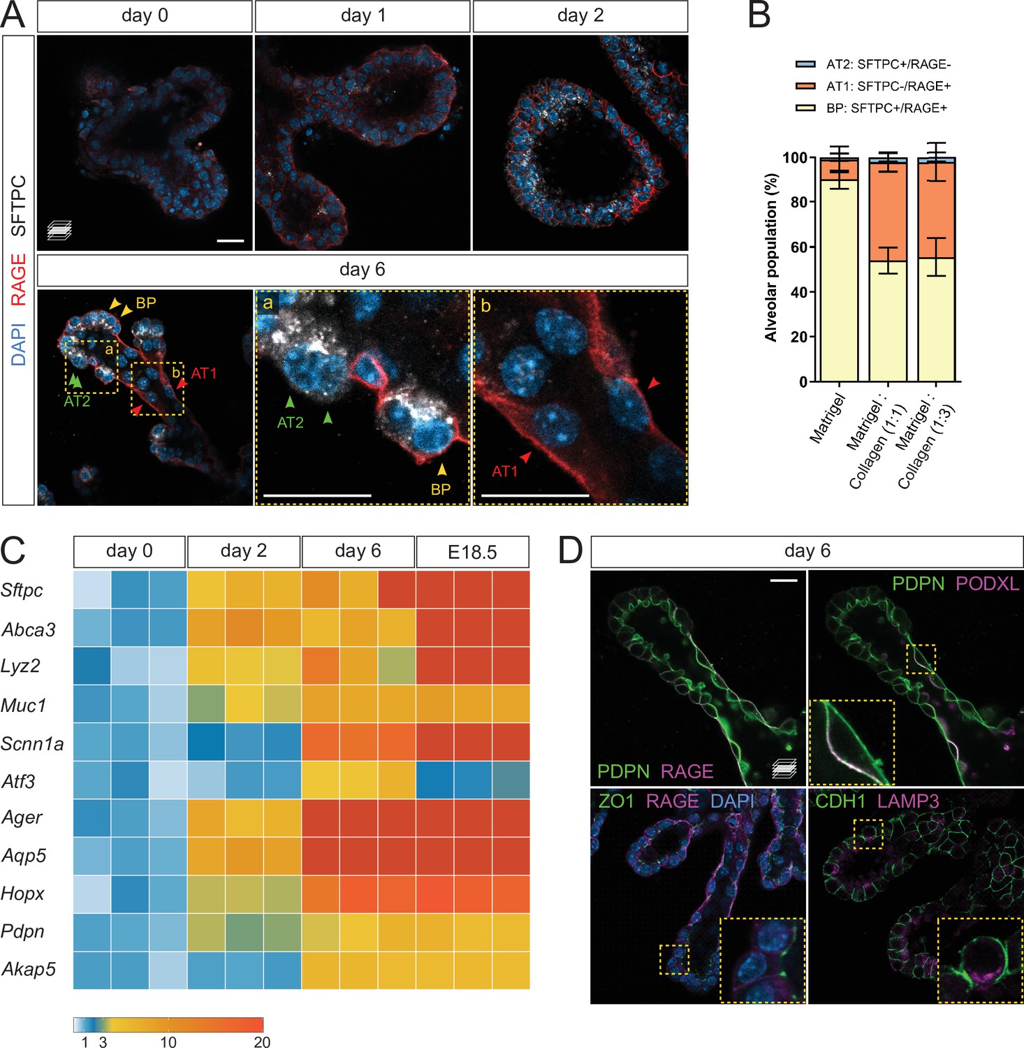
To assess epithelial differentiation, we performed whole-mount organoid immunostaining for SFTPC and RAGE, marking AT2 and AT1 cells, respectively. Low staining intensity was observed on days 0 and 1, indicating that within the first days of culture, the organoids are mainly composed of undifferentiated progenitors. On day 2, SFTPC/RAGE were co-expressed in a majority of the cells, similar to what is observed with alveolar bipotent progenitors (Desai et al., 2014; Treutlein et al., 2014). By day 6, two cell populations emerged: SFTPC-/RAGE+ cells in the stalk region of the elongated branches, and SFTPC+/RAGE- cells in the tip region or at new sites of branching. The tip domain also comprised cells co-expressing SFTPC and RAGE, suggesting the maintenance of a bipotent progenitor (BP) population (Treutlein et al., 2014; Figure 2A). Quantification of the differentiated and progenitor cells on day 6 revealed approximately equal numbers of AT1 and BP cells in epithelial branches while AT2 cells were less prevalent (Figure 2B).

Lung distal progenitor cells differentiate in 3D explant cultures.
(A) Whole-mount immunostaining for the alveolar cell markers SFTPC and RAGE at consecutive culture days. By day 2, SFTPC and RAGE are expressed by most cells. Day 6 epithelial branches comprise both bipotent progenitor (BP) cells (SFTPC+/RAGE+) and differentiated alveolar cells (AT1, SFTPC-/RAGE+ and AT2, SFTPC+/RAGE-) in an organotypic pattern. (B) Collagen I promotes AT1 cell differentiation, compared with Matrigel alone. Cell identities were scored on day 6. (C) Markers of differentiated AT1 and AT2 cells are transcriptionally upregulated by day 6 of culture, at levels comparable with E18.5 distal lung tissue. Expression values are normalized to culture day 0. Each box depicts one biological replicate, showing the mean value among two technical replicates; 27 tip cultures were used for each time point. (D) AT1 and AT2 cells in organoids are morphologically distinct and display apico-basal polarity. AT1 cells are elongated and often populate stalk regions. Upper row: AT1 cells display basolateral localization of the AT1 cell marker RAGE and apical localization of PODXL. Lower row: epithelial branches in organoids display adhesions typical of intact epithelia, such as tight junctions (ZO-1). The AT2 cell marker LAMP3 is localized to the apical secretory compartment of surfactant-producing cells. Scale bars: 20 µm. (B) Mean values are displayed; error bars represent S.D.
-
Figure 2—source data 1
Counts of alveolar cell types in organoid culture, Matrigel vs. Collagen. Normalized expression values of alveolar markers.
- https://cdn.elifesciences.org/articles/65811/elife-65811-fig2-data1-v1.xlsx
In the absence of Collagen I, the organoids displayed a reduced number of AT1 cells in favor of BP cells, indicating that a collagenous matrix (at 1:1 or 1:3 Matrigel/Collagen I ratio) promotes the differentiation of AT1 cells in vitro (Figure 2B), consistent with the reported role of the ECM in alveolar epithelial differentiation (Kim et al., 2018). We next asked to what extent the organoids represent mature distal lung tissue, and compared differentiation marker expression with distal lung tissue at E18.5, a time when differentiated alveolar epithelial cells can be unequivocally identified. The expression level of differentiated cell markers, including Sftpc, Ager, Aqp5, and Hopx, progressively increased until day 6, at which point they reached levels comparable with those observed in E18.5 distal lung tissue (Figure 2C). These data indicate that distal progenitors differentiate within six days of organoid culture, offering a simple model of early alveolar formation.
We further asked whether the AT1 and AT2 cells identified in the organoid cultures displayed morphological hallmarks of a differentiated respiratory epithelium. By immunostaining, we found that SFTPC-/RAGE+ cells displayed exclusive basolateral localization of RAGE (Figure 2A and D) and apical localization of Podocalyxin (PODXL) (Figure 2D), similar to observations in intact lung tissue at E19 (Yang et al., 2016). SFTPC-/RAGE+ cells were also marked by uniform plasma membrane expression of Podoplanin/T1alpha (PDPN) (Figure 2D). Immunostaining for Zonula Occludens-1 (ZO-1), a marker of tight junctions, labeled intercellular adhesions at the luminal/apical surface (Figure 2D) as previously reported (Yang et al., 2016), suggesting that the epithelial branches form an intact cellular monolayer and display barrier function. In addition, we found that Lysosome-Associated Membrane glycoProtein 3 (LAMP3), a lysosomal marker localized to lamellar bodies in differentiated AT2 cells (Chang et al., 2013; Desai et al., 2014), was localized in apical secretory organelles in SFTPC+ cells, indicating that these cells can differentiate into AT2 cells (Figure 2D). Altogether, these data show that cultured alveolar progenitors differentiate morphologically and molecularly into AT1 and AT2 cells, suggesting that fetal mouse alveolar organoids maintain the distal airway architecture found at late gestation stages (Frank et al., 2019).
Casein Kinase modulates epithelial differentiation in lung organoids
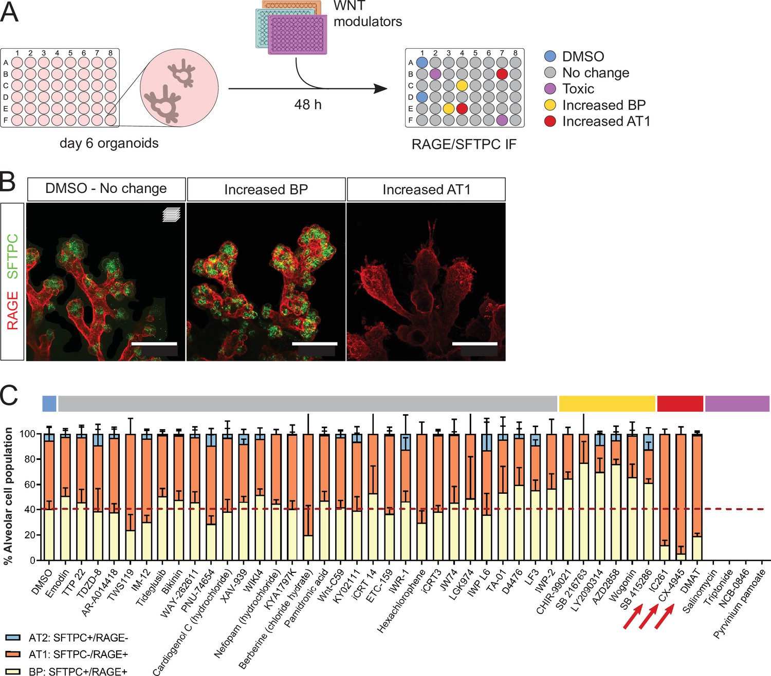
We used fetal alveolar organoids to dissect the mechanisms by which WNT signaling controls the differentiation of AT1 and AT2 cells. To this end, we performed a chemical screen of WNT modulators from days 6 to 8 in the absence of supplemented growth factors (Figure 3A) and determined the relative alveolar cell composition by immunostaining for SFTPC and RAGE (Figure 3B). In control conditions, the proportion of SFTPC+/RAGE+ progenitors decreased from 54% (day 6, Figure 2B) to 40% (day 8, Figure 3C), with a concomitant increase in differentiated cell numbers (more pronounced for AT1 cells), suggesting that progenitors are actively differentiating in culture at the stages chosen for screening.

A WNT pathway chemical screen identifies modulators of alveolar cell differentiation.
(A) Screening workflow schematic. Organoids at day 6 were treated for 48 h with WNT-modulating compounds. (10 µM). Phenotypes were scored by cell counting after immunostaining for SFTPC and RAGE and F-actin co-staining (Phalloidin). (B) Phenotypic classes observed in the chemical screen comprise an increased proportion of SFTPC+/RAGE+ cells (BP, middle panel) and increased proportion of SFTPC-/RAGE+ cells (AT1, right panel). (C) Casein Kinase inhibitors IC261, CX-4945, and DMAT (arrows) led to a higher proportion of AT1 cells at the expense of progenitors, while a majority of the compounds tested did not alter the proportion of differentiated alveolar cells in culture. Four biological replicates were analyzed, 12 tip cultures per compound. Scale bars: 50 µm. (C) Mean values are displayed; error bars represent S.D.
-
Figure 3—source data 1
Counts of alveolar cell types in the WNT modulators screen. Percentages of alveolar cell types in the WNT modulators screen.
- https://cdn.elifesciences.org/articles/65811/elife-65811-fig3-data1-v1.xlsx
Unexpectedly, the majority of tested compounds (gray highlight) did not lead to significant changes in the proportion of AT1, AT2, or progenitor cells compared with DMSO controls (Figure 3C). Belonging to this group are first-in-class WNT pathway inhibitors such as IWR-1, IWP-2, and XAV-939 (Chen et al., 2009; Huang et al., 2009). Active WNT signaling is a feature of alveolar progenitors in homeostasis and regeneration, and blocking the WNT pathway by Ctnnb1 knockout in AT2 cells was shown to induce AT1 differentiation at postnatal stages (Frank et al., 2016; Nabhan et al., 2018). In addition, an expansion of the AT1 cell population was observed in vitro after treatment of hiPSC-derived AT2 cells with XAV-939 (Kanagaki et al., 2021). Differentiation of cultured adult human alveolar progenitors (TM4SF1+) also showed WNT responsiveness, with the AT1 fate induced after XAV-939 treatment. In contrast with these data, the results of our chemical screen suggest that WNT inhibition via Axin stabilization is not sufficient to alter fate selection or cell differentiation of mouse fetal distal lung progenitors in our organoid cultures. This discrepancy with previous findings could be explained in part by differences in the phenotypic outcome of pharmacological versus genetic inhibition of WNT signaling, or by distinct regulation of the WNT pathway between pre- and postnatal stages. Limitations in our screening approach and the cellular composition of the organoid cultures could have influenced the results, as follows. First, a single concentration of 10 µM was used for all tested compounds, possibly leading to false negatives. Second, the heterogeneous cell composition of the organoids, in particular a variable number of mesenchymal cells, may have contributed to some of the variability in the data. Indeed, mesenchymal cells are fundamental players throughout lung development and engage in signaling crosstalk with epithelial cells, notably through FGF and WNT signaling (McCulley et al., 2015; Volckaert and De Langhe, 2015). Because of this variability in the relative cell composition of the organoids, four biological replicates were used for the screening experiments. Six compounds led to an increase in SFTPC+/RAGE+ BP cells (yellow highlight), including several Glycogen Synthase Kinase 3 (GSK3) inhibitors such as CHIR-99021, and a natural flavonoid, Wogonin. In line with published data (Ostrin et al., 2018; de Carvalho et al., 2019), we found that sustained WNT activity was associated with progenitor maintenance. Finally, three compounds induced a greater percentage of AT1 cells (SFTPC-/RAGE+) at the expense of bipotent progenitors (Figure 3C). Mechanistically, these compounds inhibit Casein Kinase (CK) 1 (IC261) and CK2 (CX-4945, DMAT), central regulators of WNT signaling and cell cycle progression (St-Denis and Litchfield, 2009; Cruciat, 2014; Venerando et al., 2014).
Casein Kinase inhibition leads to downregulation of AT2 marker gene expression and WNT-dependent transcription
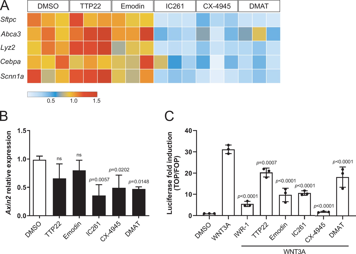
Casein Kinase (CK) acts in stem cell maintenance by phosphorylating transcriptional regulators, WNT signaling components, and cell cycle factors (St-Denis and Litchfield, 2009; Cruciat, 2014). We found that CK inhibition by IC261, CX-4945, and DMAT led to the downregulation of markers of the AT2 fate, including Sftpc and Abca3, compared with control organoids (Figure 4A), suggesting that upon CK inhibition, progenitor cells lose AT2 fate potential and convert to AT1 cells. Similarly, WNT signaling activation was reduced in organoids treated with the same CK inhibitors for 6 h as assessed by measuring mRNA levels of Axin2 (Figure 4B), a β-catenin-dependent WNT target gene. Pharmacological CK inhibition could also significantly reduce the transcriptional response to WNT/β-catenin signaling in HEK293T cells, as shown by SuperTOPFlash luciferase reporter assays (Figure 4C). Collectively, these data indicate that progenitor cells convert to AT1 cells by combined loss of β-catenin-dependent WNT activity and AT2 marker expression, and suggest that Casein Kinases antagonize cell differentiation by modulating WNT signaling transduction and transcriptional regulation. However, since Casein Kinases mediate a plethora of housekeeping cellular functions, determining which specific CK targets are involved in alveolar differentiation requires further investigation. Recently, CK2 has emerged as a prominent target of SARS-CoV-2 infection in the lung, and its overactivation was associated with increased viral transmission by cytoskeletal remodeling and formation of cell protrusions mediating virus budding (Bouhaddou et al., 2020). Combined with our data, these recent findings suggest that SARS-CoV-2 infection could affect lung cell fate establishment and regeneration of the alveolar compartment directly by targeting CK2 activity.

Compounds promoting AT1 cell differentiation lead to concomitant loss of AT2 marker gene expression and WNT-dependent transcription.
(A) IC261, CX-4945, and DMAT treatments (48 h) lead to downregulation of AT2 cell markers. TTP22 and Emodin (also CK2 inhibitors) do not lead to the same phenotype, possibly due to differences in potency/target affinity. Expression levels relative to DMSO control. Three biological replicates (consisting of 2 technical replicates each) were analyzed per compound. (B) Casein Kinase inhibition by IC261, CX-4945, and DMAT (6 h) leads to reduced Axin2 relative expression. Treatment with TTP22 or Emodin does not significantly affect Axin2 levels. More than three biological replicates were analyzed. (C) CK inhibition reduced WNT/β-catenin-dependent transcription in HEK293T epithelial cells (SuperTOPFlash-based luciferase assay). (B) Mean values are displayed; error bars represent S.D.; p-values from one-way ANOVA, Tukey’s multiple comparisons test. (C) Mean values displayed; error bars represent S.D.; p-values from one-way ANOVA, Tukey’s multiple comparisons test; displayed p-values refer to comparisons with WNT3A-treated condition.
-
Figure 4—source data 1
Normalized expression values of AT2 cell markers, CK inhibitors vs. DMSO control. Normalized Axin2 expression values, CK inhibitors vs. DMSO control and related statistics. Raw luciferase data. Normalized luciferase values and related statistics.
- https://cdn.elifesciences.org/articles/65811/elife-65811-fig4-data1-v1.xlsx
In summary, our fetal mouse lung alveolar organoids constitute an ideal system to assess cell differentiation at the single-cell level and will allow the investigation of this process by live imaging and chemical screening. This new model should also facilitate the identification of modulators of alveolar cell differentiation, and help discover treatments to promote lung development and maturation in premature newborns.
Materials and methods
Reagent type (species) or resource | Designation | Source or reference | Identifiers | Additional information |
---|---|---|---|---|
Strain, strain background (Mus musculus) | C57BL/6J mice | https://www.jax.org/strain/000664 | RRID:IMSR_JAX:000664 | |
Cell line (Homo sapiens) | HEK-293T | ATCC | ATCC:CRL-3216; RRID:CVCL_0063 | |
Other | Rat-tail Collagen I | Corning | Corning:354236 | |
Other | Growth factor reduced Matrigel | Corning | Corning:356231 | |
Other | DMEM/F12 | Sigma | Sigma:D6434 | |
Other | L-Glutamine | Sigma | Sigma:G7513 | |
Other | Penicillin/Streptomycin | Sigma | Sigma:P4333 | |
Other | DNAse I | Roche | Roche:10104159001 | |
Other | 10% BSA | Sigma | Sigma:A1595 | |
Other | MEM Non-Essential Amino Acids solution | Gibco | Thermo Fisher Scientific:11140050 | |
Other | Insulin-Transferrin-Selenium-Ethanolamine (ITS-X) | Gibco | Thermo Fisher Scientific:51500056 | |
Other | Primocin | Invivogen | Invivogen:ant-pm-1 | |
Other | 10x DMEM | Sigma | Sigma:D2429 | |
Chemical compound, drug | 1 N NaOH | Sigma | Sigma:S2770 | |
Peptide, recombinant protein | Human recombinant FGF10 | R&D Systems | R&D Systems:345-FG | |
Peptide, recombinant protein | Human recombinant FGF7 | Peprotech | Peprotech:100–19 | |
Other | CryoStor CS10 | Stem Cell Technologies | Stem Cell Technologies:07959 | |
Other | Normal donkey serum | Jackson ImmunoResearch | Jackson Immuno Research:017-000-121; RRID:AB_2337258 | |
Antibody | Anti-SOX9 (rabbit polyclonal) | Millipore | Millipore:AB5535; RRID:AB_2239761 | (1:250) |
Antibody | Anti-KI67 (rat monoclonal) | Invitrogen | Thermo Fisher Scientific:14-5698-82; RRID:AB_10854564 | (1:250) |
Antibody | Anti-SOX2 (goat polyclonal) | R&D Systems | R&D Systems:AF2018; RRID:AB_355110 | (1:250) |
Antibody | Anti-KRT5 (rabbit polyclonal) | Abcam | Abcam:ab53121; RRID:AB_869889 | (1:250) |
Antibody | Anti-FOXJ1 (mouse monoclonal) | Invitrogen | Thermo Fisher Scientific:14-9965-82; RRID:AB_1548835 | (1:250) |
Antibody | Anti-ProSP-C (rabbit polyclonal) | Millipore | Millipore:AB3786; RRID:AB_91588 | (1:500) |
Antibody | Anti-RAGE (rat monoclonal) | R&D Systems | R&D Systems:MAB1179; RRID:AB_2289349 | (1:250) |
Antibody | Anti-PDPN (sheep polyclonal) | R&D Systems | R&D Systems:AF3670; RRID:AB_2162070 | (1:250) |
Antibody | Anti-PDPN (syrian hamster monoclonal) | DSHB | DSHB:8.1.1; RRID:AB_531893 | (1:20) |
Antibody | Anti-PODXL (goat polyclonal) | R&D System | R&D Systems:AF1556; RRID:AB_354858 | (1:250) |
Antibody | Anti-ZO1/TJP1 (mouse monoclonal) | Invitrogen | Thermo Fisher Scientific:33–9100; RRID:AB_2533147 | (1:250) |
Antibody | Anti-CDH1 (goat polyclonal) | R&D Systems | R&D Systems:AF748; RRID:AB_355568 | (1:250) |
Antibody | Anti-LAMP3 (rat monoclonal) | Dendritics | IMGENEX:DDX0192; RRID:AB_1148779 | (1:250) |
other | Phalloidin, Alexa Fluor 488 conjugate | InvitrogenThermo Fisher Scientific:A12379 | Thermo Fisher Scientific:A12379 | (1:500) |
other | DAPI | Sigma | Sigma:D9542 | (1 µg/ml) |
Antibody | Alexa 488-, 568-, 647- or Cy3-conjugated secondaries | Invitrogen | (1:500) | |
Antibody | Alexa 488-, 568-, 647- or Cy3-conjugated secondaries | Jackson ImmunoResearch | (1:500) | |
Other | DMEM + Glutamax | Gibco | Thermo Fisher Scientific:31966021 | |
Other | FBS superior | Sigma | Sigma:S0615 | |
Other | Lipofectamine 3000 Transfection Reagent | Invitrogen | Thermo Fisher Scientific:L3000001 | |
Transfected construct (Homo sapiens) | M50 Super 8x TOPFlash (plasmid) | Addgene | Addgene:12456; http://n2t.net/addgene:12456; RRID:Addgene_12456 | |
Transfected construct (Homo sapiens) | M51 Super 8x FOPFlash (TOPFlash mutant) (plasmid) | Addgene | Addgene:12457; http://n2t.net/addgene:12457; RRID:Addgene_12457 | |
Transfected construct (Homo sapiens) | pRL-TK (plasmid) | Promega | Promega:E2241 | |
Peptide, recombinant protein | Recombinant human WNT3A | Proteintech | Proteintech:HZ-1296 | (500 ng/ml) |
Chemical compound, drug | Dimethyl sulfoxide (DMSO) | Sigma | Sigma:D2650 | |
Chemical compound, drug | Emodin | Tocris | Tocris:3811 | (10 µM) |
Chemical compound, drug | TTP22 | Tocris | Tocris:4432 | (10 µM) |
Chemical compound, drug | IC261 | Sigma | Sigma:I0658 | (10 µM) |
Chemical compound, drug | CX-4945 | Enzo Life Sciences | Enzo Life Sciences: ENZ-CHM151 | (10 µM) |
Chemical compound, drug | DMAT | Sigma | Sigma:SML2044 | (10 µM) |
Chemical compound, drug | IWR-1 | Sigma | Sigma:I0161 | (10 µM) |
Commercial assay or kit | Dual-Luciferase Reporter Assay System | Promega | Promega:E1910 | |
Commercial assay or kit | NucleoSpin RNA kit | Macherey-Nagel | Macherey- Nagel:740955.50 | |
Commercial assay or kit | Superscript III Reverse Transcriptase system | Invitrogen | Thermo Fisher Scientific:18080093 | |
Commercial assay or kit | DyNAmo ColorFlash SYBR green qPCR kit | Thermo Scientific | Thermo Fisher Scientific:F416XL | |
Sequence- based reagent | qPCR | This paper | Supplementary file 1 | |
Software, algorithm | Fiji/ImageJ 1.53 c | Schindelin et al., 2012; doi:10.1038/nmeth.2019 | RRID:SCR_002285 | |
Software, algorithm | GraphPad Prism 8 | GraphPad | RRID:SCR_002798 |
Media preparation
Request a detailed protocolFrozen aliquots of rat-tail Collagen I (500 µl, Corning 354236) and growth factor reduced (GFR) Matrigel (550 µl, Corning 356231) were thawed on ice during lung epithelial tip isolation. One aliquot of Collagen I and Matrigel were sufficient for two dissociations (two 48-well plates).
Dissection Medium (DM) composition: DMEM/F12 (Sigma D6434), 1 mM L-Glutamine (Sigma G7513), 1x Penicillin/Streptomycin (Sigma P4333), 100 µg/ml DNAse I (Roche 10104159001).
Complete Medium (CM) composition: DMEM/F12, 1 mM L-Glutamine, 0.25% BSA (Sigma A1595), 1x MEM Non Essential Amino Acids (Gibco 11140050), 0.1x Insulin-Transferrin-Selenium-Ethanolamine (Gibco 51500056), 100 µg/ml Primocin (Invivogen ant-pm-1).
Media were prepared fresh just before tissue isolation.
Lung epithelial tip isolation
Request a detailed protocolE14.5 C57BL/6J mouse embryos (staged according to Theiler, 1989) were harvested and washed in ice-cold PBS. Embryos were quickly decapitated and lungs extracted and washed in fresh ice-cold PBS. Lobes were isolated by cutting them away from the proximal bronchi using microscissors (Fine Science Tools 15003–08).
Using a P1000 pipette and tips coated with fresh 2% BSA (Sigma A1595), lobes from three to five lungs were transferred to freshly prepared ice-cold DM in a 60 mm petri dish. Lobes were pipetted up and down using a coated-tip P1000 pipette until fragmented. Next, if necessary, a coated-tip P200 pipette was used to triturate the tissue more finely. The degree of dissociation was monitored using a Zeiss Stemi 305 stereomicroscope. The petri dish was placed over ice-cold black metal blocks, allowing for simultaneous temperature control and contrast for visual inspection.
Fragments including one to threeepithelial tips were collected using a coated-tip P20 pipette and transferred in fresh DM on ice, to dilute out dissociated mesenchymal cells. Epithelial tips were re-collected, counted and transferred into a 1.5 ml tube on ice (about 100–120 per isolation). Tips were allowed to sink to the bottom of the tube on ice for 1–2 min and then washed twice in 100 µl DM and twice in 100 µl CM.
Organoid embedding
Request a detailed protocolDuring epithelial tips washes, thawed Collagen I (500 µl) was neutralized by adding 57 µl 10x DMEM (Sigma D2429) and approximately 16–17 µl 1 M NaOH (Sigma S2770). Once mixed to homogeneity, neutralized collagen was allowed to polymerize on ice for 10 min. Next, neutralized collagen was mixed to an isovolume of GFR Matrigel and kept on ice.
Washed epithelial tips were resuspended in 500 µl ice-cold CM, to which 500 µl of Matrigel/Collagen solution was added. Epithelial tips were carefully and thoroughly resuspended on ice and plated using cooled P20 tips onto a 48-well tissue culture plate (20 µl/well) over a 39°C heat block. Domes were allowed to solidify over the heat block for 30 min and plates were moved to a standard tissue culture incubator at 37°C for 45 min-1 h before CM with growth factors (CM+) was added as follows.
Organoid culture
Request a detailed protocolA total of 50 ml CM was warmed up to 37°C and human recombinant FGF proteins were added (50 ng/ml FGF10, R&D 345-FG and 25 ng/ml FGF7, Peprotech 100–19). 300 µl CM with growth factors (CM+) was added to each culture well.
At day 3, organoid medium was changed to diluted CM+ (1:1 with CM, halved growth factor concentration).
At day 6, organoid medium was changed to CM without supplemented growth factors.
For long-term maintenance of organoid cultures, organoid medium was changed every two days starting at day 6. Organoids could be maintained in media devoid of growth factors for up to 30 days.
Organoids with overgrown mesenchymal cells (attached to the plastic) were excluded from further analysis.
RT-qPCR
Request a detailed protocolTotal RNA was isolated from pooled organoids (9–15 domes) or tissue using the NucleoSpin RNA kit (Macherey-Nagel 740955.50). cDNA was synthesized from total RNA using the Superscript III Reverse Transcriptase system (Thermo Scientific 18080093). qPCR was performed using the DyNAmo ColorFlash SYBR green qPCR kit (Thermo Scientific F416XL) on a CFX Connect Real-Time System (Bio-Rad). qPCR reactions were performed in technical duplicates; data from at least three biological replicates were collected. Gene expression values were normalized to the mouse Actb gene. Heatmaps were generated using R/Bioconductor and ggplot2 packages. Mean values among technical replicates were color-coded and each biological replicate was represented.
Organoid whole-mount immunostaining
Request a detailed protocolMatrix domes were quickly washed in PBS and fixed in 4% PFA for 15 min at room temperature (RT). Domes were detached by the well bottom with a flat microspatula and transferred to tubes or staining baskets. After several PBS washes, domes were permeabilized for 1 h at RT in sterile-filtered permeabilization solution: 0.5% Triton-X100, 0.5% Tween20, 3% Donkey Serum (Jackson ImmunoResearch 017-000-121), 1% BSA. Samples were incubated with primary antibodies in permeabilization solution diluted 1:1 in PBS for two days at 4°C, with mild agitation. Domes were then washed in PBST (PBS + 0.05% Tween20) for 6 h at RT or overnight at 4°C with mild agitation. Next, domes were incubated with secondary antibodies, Alexa488-Phalloidin (Invitrogen A12379, both at 1:500) and DAPI (1 µg/ml) in diluted permeabilization solution (1:1 in PBS) for 3 h at RT, followed by 3–4 h wash in PBST. Domes were mounted in mounting medium (Dako S3023) on glass slides using a thin vacuum grease ring as coverslip spacer. After overnight incubation at 4°C, imaging was performed on a Leica SP8 or Zeiss LSM 800 Observer confocal microscope, ×25 and ×40 magnification.
Antibodies
The following primary antibodies were used: rabbit anti-SOX9 (Millipore AB5535), rabbit anti-ProSP-C (Millipore AB3786), rat anti-RAGE (R&D MAB1179), goat anti-PDPN (R&D AF3670), syrian hamster anti-PDPN (DSHB 8.1.1-SN), goat anti-PODXL (R&D AF1556), mouse anti ZO-1/TJP1 (Invitrogen 33–9100), goat anti-CDH1 (R&D AF748), rat anti-LAMP3 (Dendritics DDX0192), rat anti-KI67 (Invitrogen 14-5698-82), goat anti-SOX2 (R&D AF2018), mouse anti-FOXJ1 (Invitrogen 14-9965-82), rabbit anti-KRT5 (Abcam ab53121). All primary antibodies were used at 1:250 dilution, except anti-ProSP-C (1:500). All secondary antibodies (Invitrogen, Jackson ImmunoResearch) were used at 1:500 dilution.
Cryostorage
Request a detailed protocolFreshly isolated E14.5 epithelial tips were mixed with 500 µl of CryoStor CS10 (Stem Cell Technologies 07959) and stored at –80°C for up to 6 months.
Chemical screen
Request a detailed protocolOrganoids were plated into 48-well plates and incubated from day 6 for 48 h in the presence of compounds. Chemicals were dissolved in CM devoid of additional growth factors at a final concentration of 10 µM (from a DMSO stock). Screened compounds belong to the Stem Cell Signaling Compound Library (MCE HY-L017). Treatment with 0.1% DMSO (Sigma D2650) was used as internal control in each organoid plate. On day 8, organoids were washed and fixed in 4% PFA and three or four technical replicates were pooled for immunostaining. Confocal scans were followed by cell counting as detailed below. Four biological replicates (12 tip cultures in total) were analyzed per compound.
Cell counts
Request a detailed protocolFor each confocal stack, three optical sections were selected corresponding to 25, 50, and 75% of the confocal Z stack range. For each of these planes, SFTPC+/RAGE+, SFTPC-/RAGE+, and SFTPC+/RAGE- cells were manually counted using the Cell Counter plugin in Fiji/ImageJ (Schindelin et al., 2012) and quantified as a percentage of DAPI+ cells. Percentages were averaged among the three planes, and mean alveolar cell percentages of at least nine organoids among three technical replicates derived from at least three biological replicates (different pregnant dams) were then plotted and error was calculated as standard deviation. Day 6 tip cultures contained on average 54% SFTPC+/RAGE+ cells, 43.9% SFTPC-/RAGE+ cells, 2.1% SFTPC+/RAGE- cells (Figure 2—source data 1). Tip cultures treated from day 6 to day 8 with 0.1% DMSO contained on average 40.5% SFTPC+/RAGE+ cells, 54.1% SFTPC-/RAGE+ cells, 5.4% SFTPC+/RAGE- cells (Figure 3—source data 1).
Cell lines
Request a detailed protocolHuman Embryonic Kidney cells (HEK293T, ATCC CRL-3216) were certified by STR profiling by ATCC and tested negative for mycoplasma contamination. Cells were maintained in DMEM (Gibco 31966021), 10% FBS (Sigma S0615), 1% Penicillin/Streptomycin and passaged using TrypLE Express (Gibco 12604021).
Luciferase assay
Request a detailed protocolSuperTOPFlash reporter assays were performed as in Veeman et al., 2003. In brief, 15,000 HEK293T cells/well were seeded into gelatin-coated 96-well plates and allowed to grow overnight. Cells were transfected with a combination of 100 ng SuperTOPFlash or 100 ng SuperFOPFlash and 10 ng pRL-TK (Promega E2241) plasmids. M50 Super 8x TOPFlash and M51 Super 8x FOPFlash (TOPFlash mutant) were gifts from Randall Moon (Addgene plasmid # 12456; http://n2t.net/addgene:12456; RRID:Addgene_12456 and Addgene plasmid # 12457; http://n2t.net/addgene:12457; RRID:Addgene_12457). Transfections were carried out using Lipofectamine 3000 Reagent (Invitrogen L3000001) according to manufacturer’s instructions. Transfected cells were serum-starved for 6 h and treated with 500 ng/ml human recombinant WNT3A (Proteintech HZ-1296) and 10 µM chemical compounds or 0.1% DMSO for 12 h. Cells were washed with PBS on ice to remove phenol red and lysed. Firefly and Renilla luciferase assays were carried out using Dual-Luciferase Reporter Assay System (Promega E1910) in technical triplicates. Raw Firefly luminescence values were normalized to corresponding Renilla values. Technical triplicates were averaged, normalized over FOPFlash values and normalized over DMSO-treated control (no WNT3A stimulation). Experiments were carried out in biological triplicates.
Data availability
All data generated or analyzed during this study are included in the manuscript and supporting files. Source data files have been provided for Figures 1, 2, 3, 4 and Figure 1-figure supplement 1.
References
-
Fibroblast Growth Factor 10 (FGF10) and branching morphogenesis in the embryonic mouse lungDevelopment 124:4867–4878.
-
Alveologenesis: key cellular players and fibroblast growth factor 10 signalingMolecular and Cellular Pediatrics 3:17.https://doi.org/10.1186/s40348-016-0045-7
-
Small molecule-mediated disruption of Wnt-dependent signaling in tissue regeneration and cancerNature Chemical Biology 5:100–107.https://doi.org/10.1038/nchembio.137
-
Casein kinase 1 and Wnt/β-catenin signalingCurrent Opinion in Cell Biology 31:46–55.https://doi.org/10.1016/j.ceb.2014.08.003
-
Discordant roles for FGF ligands in lung branching morphogenesis between human and mouseThe Journal of Pathology 247:254–265.https://doi.org/10.1002/path.5188
-
Use of three-dimensional organoids and lung-on-a-chip methods to study lung development, regeneration and diseaseThe European Respiratory Journal 52:00876.https://doi.org/10.1183/13993003.00876-2018
-
The pulmonary mesenchyme directs lung developmentCurrent Opinion in Genetics & Development 32:98–105.https://doi.org/10.1016/j.gde.2015.01.011
-
Fiji: An open-source platform for biological-image analysisNature Methods 9:676–682.https://doi.org/10.1038/nmeth.2019
-
From birth to death: The role of protein kinase CK2 in the regulation of cell proliferation and survivalCellular and Molecular Life Sciences 66:1817–1829.https://doi.org/10.1007/s00018-009-9150-2
-
Casein kinase: The triple meaning of a misnomerThe Biochemical Journal 460:141–156.https://doi.org/10.1042/BJ20140178
-
Wnt and FGF mediated epithelial-mesenchymal crosstalk during lung developmentDevelopmental Dynamics 244:342–366.https://doi.org/10.1002/dvdy.24234
Decision letter
-
Melanie KönigshoffReviewing Editor; University of Pittsburgh, United States
-
Edward E MorriseySenior Editor; University of Pennsylvania, United States
-
Aimee K RyanReviewer; McGill University, Canada
-
Amy FirthReviewer; University of Southern California, United States
In the interests of transparency, eLife publishes the most substantive revision requests and the accompanying author responses.
Acceptance summary:
This manuscript describes a newly developed method to culture mouse lung distal progenitor cells in a 3D serum-free condition. By modulating the concentration of supplied growth factors, distal lung progenitor cells could branch and then gradually differentiate into AT1 and AT2 cells, a process very similar to alveolar development in mice. The authors also found that collagen I could significantly promote progenitor cells to differentiate into AT1 cells. Based on this culture system, the authors performed a Wnt signaling-related-drug screening and identified that casein kinases are essential for the specification of alveolar epithelial cells. The manuscript is well-written and discusses the method and its potential limitations nicely. This is an important study and will be of interest to the readership of eLife.
Decision letter after peer review:
Thank you for submitting your article "Differentiation of mouse fetal lung alveolar progenitors in serum-free organotypic cultures" for consideration by eLife. Your article has been reviewed by three peer reviewers, and the evaluation has been overseen by a Reviewing Editor and Edward Morrisey as the Senior Editor. The following individuals involved in review of your submission have agreed to reveal their identity: Aimee K Ryan (Reviewer #2); Amy Firth (Reviewer #3).
The reviewers have discussed their reviews with one another, and the Reviewing Editor has drafted this to help you prepare a revised submission.
The reviewers have raised several important questions about the model system, which should be addressed and integrated into the revised discussion. Please address specifically the questions raised with respect to the potential role of mesenchymal/other non-epithelial cells in the organoid system, the effect of Wnt inhibitors on cell fate specification, and provide additional data to further strengthen the Wnt readouts upon screening and the CK/Wnt link.
Essential revisions:
(1) Additional experimental data on Wnt readouts and Wnt/CK link
(2) Discussion on the potential role of mesenchymal/other non-epithelial cells in the organoid system
(3) Discussion on the effect of Wnt inhibitors on cell fate specification
(4) Data/discussion on potential Sox2 differentiation
Reviewer #1:
In this manuscript, the authors presented a newly developed method that the mouse lung distal progenitor cells can be cultured in a 3D serum-free condition. By modulating the concentration of supplied growth factors, these distal progenitor cells could branch and then gradually differentiate into AT1 and AT2 cells, a process is very similar to the alveolar development process in mice. The authors also found that collagen I could significantly promoted progenitor cells to differentiate into AT1 cells. Based on this culture system, the authors performed a Wnt signaling-related-drug screening and identified that casein kinases are essential for the specification of alveolar epithelial cells. Modeling alveolar development in vitro is very important to facilitate future studies on the cellular and molecular mechanisms of alveolar development.
1. In this culture system, the authors found that distal progenitor cells could expand and become alveolar lineage epithelial cells (bipotent cells, AT1 and AT2). Considering that distal progenitor cells at E14.5 also could also differentiate into proximal airway lineage cells, the authors should provide information on whether these cells could differentiate into Sox2+ proximal progenitor cells and other proximal cell types.
2. Based on their dissection method, the distal progenitor cells-derived organoids may contain mesenchymal cells or other non-epithelial cell types, which may significantly influence the proliferation and differentiation of distal progenitor epithelial cells. The authors should at least consider this possibility and discuss whether these drug-induced phenotypes are caused by modulation of mesenchymal cells or other non-epithelial cell types.
3. The electron microscopy analysis can better demonstrate the cell identity of bipotent cells, AT1 and AT2 cells in the organoids.
4. Using this culture system to perform a Wnt signaling-related drug screening experiment, the authors found that Wnt inhibitors do not affect the cell fate specification, which does not agree with previous results. The authors should provide a discussion. Further details of this drug screening experiment should be provided in the manuscript.
5. The link between Casein Kinase (CK) inhibition and Axin2 mRNA expression is not strong enough. The authors should provide more evidence to support their observed relationship between CK and Wnt signaling. Furthermore, the authors should discuss and propose the mechanisms of CK in regulating alveolar epithelial cell fate specification.
Reviewer #2:
The authors have developed an ex vivo organoid culture system using distal epithelial fragments from the mouse lung that permits investigation of studied cell type differentiation and that can be manipulated to evaluate the role of signaling pathways on the process of alveolization. The organoid cultures were established using manually dissected and dissociated distal epithelial fragments from E14.5 mouse lungs. At this point of development, pseudoglandular development is almost complete and alveolar progenitors have been specified. In this manuscript the authors have defined culture conditions under which the tissue fragments can develop in three-dimensional culture to form an epithelial structure with a lumen. Within 6 days of culture the organoids formed from the distal lung epithelial fragments are capable of forming epithelial branches and undergo cell type differentiation similar to what is observed in vivo. The observed epithelial growth and lumenization occurs in the absence of hyperproliferation of mesenchymal cells. Of interest is the observation that tissue that has been cryopreserved can also be expanded to form organoids, albeit with a reduced efficiency.
During the six day culture period, the organoids assume lung-like structures and display epithelial buds and branches. Sufficient details are provided to permit others to establish the lung organoids in their own labs. Morphological analysis of these structures demonstrates that the distal fragments are able to reorganize to form a closed lumen and expand the number of epithelial branches within an individual organoid. Throughout the culture period, the differentiation status of cells within the organoids were assayed using immunofluorescence to identify SOX9 +ve progenitor cells and differentiated alveolar Type 1 and Type 2 cells and quantitative RT-PCR to measure changes in expression levels of distal epithelial markers. Expression of SOX9 and ID2 were analyzed to identify the distal tip progenitor population. SOX9 +ve progenitors were initially observed at the tips on day 0, became widespread by day 1 and localized to discrete patches on day 2 before becoming once again restricted to the distal tips of the epithelial branches on day 6. The increased number of cells expressing SOX9 is supported by the increase in Sox9 mRNA expression observed at day 2. It is not clear if the changes in expression and localization of SOX9 positive cells reflect the reorganization of cells when the distal tips are place in organoid culture, a general transient increase in cells assuming a progenitor cell phenotype or if they reflect differences in the proportion of progenitor cells in the tissue used for the organoid culture versus the E13.5 lung distal tips.
Gene expression analysis of the differentiation of alveolar cell types AT1 and AT2 in the distal epithelium organoids and show similar gene expression levels as compared to E18.5 embryonic lungs. The cells in the organoid epithelium express AT1 and AT2-specific marker proteins and are polarized with apical localization of tight and adherens junction proteins. The presence of collagen clearly promotes the differentiation of bipotential progenitors to differentiate into AT1 cells. There also appears to be an increase in AT2 positive cells but these are a much smaller proportion of the total cell population.
The effect of altering WNT signaling on cell type differentiation is determined using the organoid cultures in a chemical screen. Chemicals are categorized based on their ability to shift the proportion of bipotential progenitors versus differentiated AT1 cells or their toxicity. The effects on cell type differentiation were confirmed by quantitative RT-PCR. The principal findings from this study is that sustaining WNT activity promoted maintenance of the progenitor population. In contrast, compounds that inhibit Casein Kinase 1 or 2, central regulators of WNT signaling and cell cycle progression pushed differentiation of the progenitor population in the direction of AT1 cells. This was associated with an inhibition of expression of genes associated with AT2 cell fate and decreased WNT signaling, as assessed by decreased mRNA expression of the Β-catenin target gene Axin2.
In summary, this manuscript describes the development of an organoid culture method for distal lung epithelium and demonstrated its applicability for understanding the role of ECM and WNT signaling in differentiated cell types. The branching process was not examined in detail but this culture system should permit further analysis of the molecular and morphological events that underlie this process and to compare to what has been determined in vivo and perhaps identify treatments that have important implications for lung development in premature babies.
Specific points to address:
1. Expression of SOX9 and ID2 were analyzed to identify the distal tip progenitor population. SOX9 +ve progenitors were initially observed at the tips on day 0, became widespread by day 1 and localized to discrete patches on day 2 before becoming once again restricted to the distal tips of the epithelial branches on day 6. The increased number of cells expressing SOX9 is supported by the increase in Sox9 expression observed by QRT-PCR at day 2. It would be helpful to further expand on this to discuss the potential implications for the transient widespread expression of SOX9. Is this an artifact of culture? Is this associated with proliferation and growth of the organoid? Or is it that the cells placed in culture have a higher percentage of SOX9 +ve cells and only those at what becomes the equivalent of the distal tip retain SOX9 expression? Did ID2 show the same expansion of expression domains before becoming restricted as suggest to the increase in Id2 expression? Does the relative decrease in mRNA expression at day 6 reflect a more limited number of SOX9 +ve cells as compared to the percentage of SOX9 +ve cells in the distal tips cells in the E13.5 population?
2. In addition to the % of bipotential, AT1 and AT2 cells in Figure 2B, the absolute numbers for the cells that were counted in each population should be included either in the text or in the figure legend. There also seems to be an increase in the percentage of AT2 cells but it is not clear if this is a significant increase.
3. The authors state that the proportion of bipotential progenitors and AT1 and AT2 cells do not change in a 30-day culture. Addition of a figure showing this would be great.
Reviewer #3:
Gkatzis and colleagues have developed a method for the expansion and cell fate specification of fetal lung alveolar progenitor cells from the developing mouse lung. Their refined methods focus on a serum free approach and they find that culture in collagen I promotes the differentiation to alveolar type 1 (AT1) cells with a typical flattened morphology. They also found, through a chemical screen, that Casein Kinase was a critical component of the media to maintain alveolar type 2 (AT2) cells and when it was inhibited, the AT2 cells differentiated to AT1 cells. The authors have successfully described a simple model system that can be used to characterize mechanisms regulating cellular stemness and cell fate decisions during fetal lung development. The data also provide and increased understanding of WNT signaling pathways in the support of distal airway specification. The paper provides a new model that has potential for screening of compounds regulating alveolar development and maturation.
The conclusions of the paper are supported by the data; however, it would benefit from some functional validation, increased characterization and further evaluation and mechanistic follow through on the chemical screen.
Strengths of the manuscript
The manuscript presents solid data evaluating the growth conditions essential to stimulate fetal organoid budding and branching elongation that maps nicely to E18.5 of fetal mouse development. They show good images of AT1 and AT2 cell differentiation and evidence of more primitive bipotent progenitors expressing both RAGE and SFTPC. To confirm maturity, nice apical and basolateral localization of alveolar cell markers including podoplanin, LAMP3, RAGE and ZO1 in differentiated AT1 cells. The authors demonstrate the utility of the model by performing a chemical screen of WNT modulators identifying compounds that increase the bipotent progenitors and compounds that increase AT1 cell specification.
Major points to consider:
It is unclear whether by day 6 of ex vivo growth the organoids stop growing and have the branches stopped elongating. If they are still growing, would it not be expected to still observe all the progenitor markers at the tips of the branches? It would be interesting to know what is restricting further growth of the branches and whetehr the progenitor cells are exhausted.
Only a couple of the markers used for characterization are discussed. While the majority of these genes follow a pattern similar to E18.5, it is notable that ATF3 is considerably higher in day 6 organoids than in E18.5 cells. As ATF3 is a transcription factor that can regulate many downstream pathways, such as epithelial mesenchymal transition and is itself regulated by oxidative stress – is its upregulation a result of artificial stress induced by the organoid system and is its higher expression impacting extended function of these cells?
There is a lack of data on the functional properties of the cells at Day 6. If these are to be considered mature, it would interesting to know whether they are capable of surfactant secretion, are lamellar bodies present.
It is interesting that the authors chose to evaluate the wnt modulators on day 6 of their experiment and not before if they were expecting them to impact mature cell formation, which they preciously describe as being at Day 6 with no information on day 8 cultures provided. Some additional rationale as to the choice of timepoint would be helpful to the flow of the paper.
While the strengths of the manuscript suggest the value of the model in understanding alveolar development, there are a number of points that should be considered in strengthening the impact and robustness of the manuscript.
In addition to the suggestions above the authors should consider the following additional data:
The characterization of the WNT signaling is restricted to a small amount of RNA analysis. Can the cultures be pushed toward mature AT2 cells? Can the cells grown in the presence of the modulators that increase the number of BP cells then re differentiate when the modulator is removed? Are the BP cells more proliferative? This would be interesting to study in the context of modulating an airways ability of proliferate and repair.
How important are the mesenchymal cells in this differentiation? At the beginning of the manuscript the authors state that the tips have mesenchymal and epithelial cells – what happened to the mesenchymal cells and how are they distributed in the day 6 organoids?
It would be nice to see some colocalization of the markers in panel 1E in addition to the SOX9 staining shown in Panel D.
As for Figure 1 it would be nice to see more extensive co-staining of the cells such as HTII-280 and SFTPC colocalization of AT2 cells and AQP5, RAGE, HOPX on the AT1 cells. EM images showing the presence of lamellar bodies and westerns for the presence of Pro surfactant B and C and SPB and SPC would be a nice addition as proof of functional differentiation.
In figure 4A it would be nice to see a wider panel of markers for AT1, AT2 and WNT signaling.
The characterization of the WNT signaling is a little disappointing and restricted to a small amount of RNA analysis. It would be nice to validate the data by in situ hybridization looking at co-localization between Axin2 and AT2/1/BP cells.
[Editors' note: further revisions were suggested prior to acceptance, as described below.]
Thank you for submitting your article "Differentiation of mouse fetal lung alveolar progenitors in serum-free organotypic cultures" for consideration by eLife. Your article has been reviewed by three peer reviewers, and the evaluation has been overseen by a Reviewing Editor and Edward Morrisey as the Senior Editor. The following individuals involved in review of your submission have agreed to reveal their identity: Aimee K Ryan (Reviewer #2); Amy Firth (Reviewer #3).
The reviewers have discussed their reviews with one another, and the Reviewing Editor has drafted this to help you prepare a revised submission.
The manuscript has improved significantly and the editor and reviewers alike believe this study is novel and impactful. Reviewer #3 points out some of the limitations of the study, which we would like to ask you to explicitly address in the discussion part of the manuscript, for example in form of a paragraph focused on the limitations. These include potential influence of residual supportive mesenchymal cells as well as potential limitations of the wet screen including missing validation studies, limited concentrations used and potential heterogeneity of cells in the organoid assay.
No additional experiments are needed.
Reviewer #1:
The authors have addressed my critiques to satisfaction. This is an important study and will be of interest to the readership of eLife.
Reviewer #2:
I am satisfied with the authors' responses to my previous comments and have nothing additional to add.
Reviewer #3:
The authors have addressed several of my concerns and have overall improved the quality of the manuscript. I do still have some concerns relating to my initial comments that still dampen my enthusiasm for the impact of the data presented.
Overall, however, the model does offer a platform for potentially evaluating signaling mechanisms controlling alveolar fate decisions which would be of value to the community.
In response to my concern over the influence of the mesenchyme in alveolar specification and the potential influence of residual supportive mesenchymal cells, the authors acknowledge that organoid morphogenesis did not appear to depend on their prevalence, however there is no data to support this speculation. The amount of mesenchyme present could be vastly different per organoid and have significant impact on the data outcomes. Relating to this, the lack of impact of many of the wnt signaling regulators on the differentiation is interesting based on current data in the field. Unfortunately, the impact of the wnt screening is still hard to interpret as there is a lack of validation of the compounds on multiple biological and experimental replicates.
There is also no proof that the inhibitors, at the concentrations used for the screening, were optimal or actively inhibiting wnt signaling in this particular assay (a western to show the level of inhibition would be supportive). The rationale for the different CK2 inhibitors not having the same impact, is described as being due to their potential differences in potency/target affinity – why not perform a dose response curve?
Finally, the screen is presented as the % of alveolar cells, this population is likely vastly different in each organoid at the start of the assay and thus may have a significant impact on the outcomes. This should at least be described in some of the limitations of the assay and may lead to some regulators being overlooked.
https://doi.org/10.7554/eLife.65811.sa1Author response
Essential revisions:
(1) Additional experimental data on Wnt readouts and Wnt/CK link
To strengthen our finding of Casein Kinase inhibitors downregulating the transcriptional response to WNT, we performed luciferase assays in HEK293T cells (Figure 4C) using the SuperTOPFlash reporter system (Veeman et al., 2003).
(2) Discussion on the potential role of mesenchymal/other non-epithelial cells in the organoid system.
To discuss the role of interstitial cells, we added a paragraph (page 3) stating that a possible contribution of mesenchymal cells to the growth and morphogenesis of organoids could not be excluded. Although mesenchymal cells with fibroblast morphology can be observed in our organoid cultures, in our chemical screen we did not observe any obvious correlation between the prevalence of fibroblasts and the manifestation of phenotypes.
(3) Discussion on the effect of Wnt inhibitors on cell fate specification
We have now included a paragraph discussing published data on the role of WNT signaling in alveolar cell fate specification (page 5). We refer to work showing that Axin2-positive AT2 cells differentiate into AT1 cells upon blockade of WNT transduction postnatally (Frank et al., 2016; Nabhan et al., 2018). We also mention similar work using hiPSC-derived AT2 cells and cultured human alveolar progenitors (Kanagaki et al., 2020 and Zacharias et al., 2018, respectively). To our knowledge, published data refer to postnatal alveolar progenitors, while the mechanisms underlying fetal progenitor differentiation remain to a large extent uncharacterized. Since Axin2positive AT2 progenitors have been described only in the context of postnatal alveologenesis, the fetal mechanisms of alveolar differentiation may differ at least in part. In addition to differences between the biological systems used in previous publications and our work, potential methodological limitations could also be part of the reasons for the observed discrepancy. We provide a discussion of these issues in the revised manuscript.
(4) Data/discussion on potential Sox2 differentiation
To assess whether cells in the organoids can express markers of proximal differentiation, we performed immunostaining for SOX2 and other proximal epithelial cell markers (FOXJ1, KRT5) at several stages of organoid development (Figure 1-Figure suppl. 1C, D). At all stages analyzed, we observed SOX2 expression specifically in the central portion of the organoids (core). In contrast to the distal SOX9+ region, the proximal Sox2+ domain did not appear to expand during culture. Although variable, FOXJ1 and KRT5 expression could be observed in sparse cells in the organoid core on day 6. This result suggests that, to some extent, proximal airway cells can be maintained or differentiate in our organoid model. We do not exclude the possibility that proximal airway cell composition could be enriched under different culture conditions. However, proximal cell marker expression was invariably observed to be localized in the central portion of the organoids, suggesting that distal epithelial branches provide a specific model for alveolar cell differentiation.
Reviewer #1:
[…] 1. In this culture system, the authors found that distal progenitor cells could expand and become alveolar lineage epithelial cells (bipotent cells, AT1 and AT2). Considering that distal progenitor cells at E14.5 also could also differentiate into proximal airway lineage cells, the authors should provide information on whether these cells could differentiate into Sox2+ proximal progenitor cells and other proximal cell types.
We thank the Reviewer for this question. The treatment of explanted lung tissue with FGF7 and FGF10 promotes distal epithelial expansion (Bellusci et al., 1997), as observed in our model. To characterize proximal airway cell composition in our organoids, we performed immunostaining for SOX2, FOXJ1 and KRT5. This analysis revealed a proximal domain of SOX2 expression in the organoid core, persisting at all culture stages analyzed (i.e., at least until day 6). In addition, FOXJ1 and KRT5, markers of ciliated and basal cells, respectively, were also variably expressed in sparse cells in the central portion of the organoids on day 6. Since proximal epithelial cells can be maintained and perhaps to some degree differentiate in our organoid model, we mention this point in the revised manuscript (page 3).
2. Based on their dissection method, the distal progenitor cells-derived organoids may contain mesenchymal cells or other non-epithelial cell types, which may significantly influence the proliferation and differentiation of distal progenitor epithelial cells. The authors should at least consider this possibility and discuss whether these drug-induced phenotypes are caused by modulation of mesenchymal cells or other non-epithelial cell types.
We thank the Reviewer for bringing up this important point. We now mention in the revised manuscript that mesenchymal cells are associated with the organoid epithelial branches and that a role for these cells in the morphogenesis of our model cannot be excluded. However, since we did not observe any clear correlation between the prevalence of mesenchymal cells and the phenotypes in our screen, we suggest that mesenchymal cells do not contribute significantly to the observed phenotypes. In addition, the reduction in WNT-dependent transcription observed in organoids (Figure 4B) could be recapitulated in cultured epithelial cells (HEK293T) expressing a WNT-reporter construct (Figure 4C), consistent with the model that the drugs are acting at least on epithelial cells (Figure 1-Figure suppl. 1B).
3. The electron microscopy analysis can better demonstrate the cell identity of bipotent cells, AT1 and AT2 cells in the organoids.
We thank the Reviewer for this suggestion. Although electron microscopy data would certainly help identify progenitors and differentiated cell types in the organoids, we have not yet established the required protocol.
4. Using this culture system to perform a Wnt signaling-related drug screening experiment, the authors found that Wnt inhibitors do not affect the cell fate specification, which does not agree with previous results. The authors should provide a discussion. Further details of this drug screening experiment should be provided in the manuscript.
We thank the Reviewer for this comment and have now addressed this issue by providing a discussion on WNT modulation of alveolar cell fate specification (page 5). Although WNT activity has been shown to control cell fate establishment in the context of postnatal development, we point out that the role of WNT signaling in alveolar differentiation at fetal stages is incompletely understood and may differ with what happens at postnatal stages. In addition, published work has probed the role of WNT signaling in vivo by genetic deletion or stabilization of Ctnnb1, which could result in distinct phenotypes compared with those caused by pharmacological inhibition of the pathway.
5. The link between Casein Kinase (CK) inhibition and Axin2 mRNA expression is not strong enough. The authors should provide more evidence to support their observed relationship between CK and Wnt signaling. Furthermore, the authors should discuss and propose the mechanisms of CK in regulating alveolar epithelial cell fate specification.
We thank the Reviewer for these comments and suggestions. To support our model that CK inhibition can lead to inhibition of β-catenin-dependent transcription, we performed SuperTOPflash-based luciferase assays and determined the effect of CK inhibitor compounds in HEK293T cells (Figure 4C). These experiments showed that IC261, CX-4945 and DMAT induced a significant decrease in WNT-dependent transcription.
Regarding the mechanisms of CK in regulating alveolar epithelial cell fate specification, we now mention in the revised manuscript: “[…] our results suggest that Casein Kinases antagonize cell differentiation by modulating WNT signaling transduction and transcriptional regulation […]” (page 6).
Reviewer #2:
[…] Specific points to address:
1. Expression of SOX9 and ID2 were analyzed to identify the distal tip progenitor population. SOX9 +ve progenitors were initially observed at the tips on day 0, became widespread by day 1 and localized to discrete patches on day 2 before becoming once again restricted to the distal tips of the epithelial branches on day 6. The increased number of cells expressing SOX9 is supported by the increase in Sox9 expression observed by QRT-PCR at day 2. It would be helpful to further expand on this to discuss the potential implications for the transient widespread expression of SOX9. Is this an artifact of culture? Is this associated with proliferation and growth of the organoid? Or is it that the cells placed in culture have a higher percentage of SOX9 +ve cells and only those at what becomes the equivalent of the distal tip retain SOX9 expression?
We thank the Reviewer for these questions. We have now analyzed in more detail the variation in SOX9 expression between days 0, 1, 2 and 6 of organoid culture and included these data in Figure 1D. To investigate the possible correlation with the proliferation of cultured progenitors, we have also now included confocal images showing co-expression of SOX9 and KI67. The transient widespread increase in SOX9 expression during the first days of culture closely follows cell proliferation dynamics in the organoids. By day 6, the reduction in SOX9 expression is accompanied by a reduced number of proliferating cells in the distal portion of the organoids, providing independent evidence that cell cycle exit and cell differentiation are likely occurring by this stage.
Did ID2 show the same expansion of expression domains before becoming restricted as suggest to the increase in Id2 expression? Does the relative decrease in mRNA expression at day 6 reflect a more limited number of SOX9 +ve cells as compared to the percentage of SOX9 +ve cells in the distal tips cells in the E13.5 population?
Unfortunately, ID2 expression could not be clearly analyzed in all samples due to technical issues with the mouse monoclonal antibody used, and thus we have now removed the ID2 data from the manuscript.
The revised manuscript now includes clearer information regarding the prevalence of SOX9+ cells at day 6 (new data included in Figure 1D). Indeed, as pointed out by the Reviewer, a reduction in the number of SOX9+ cells is apparent on day 6 compared to day 0 (E14.5), which correlates with the observed decrease in mRNA levels (Figure 1E).
2. In addition to the % of bipotential, AT1 and AT2 cells in Figure 2B, the absolute numbers for the cells that were counted in each population should be included either in the text or in the figure legend. There also seems to be an increase in the percentage of AT2 cells but it is not clear if this is a significant increase.
We thank the Reviewer for this comment and have now included absolute cell counts as supporting data (Figure 2-source data.xlsx and Figure 3-source data.xlsx). Unfortunately, the number of AT2 cells in our organoids is too low, making it difficult to determine the significance of potential increases in AT2 cell percentages.
3. The authors state that the proportion of bipotential progenitors and AT1 and AT2 cells do not change in a 30-day culture. Addition of a figure showing this would be great.
We thank the Reviewer for the suggestion. We believe that additional work would be needed to solidify our claim. Since the strength of our model lies in the rapid maturation and cell differentiation observed, we think that more information on the prolonged maintenance of organoids will not benefit the conclusions of this manuscript. For this reason, we have removed our original claim about long-term organoid cultures.
Reviewer #3:
[…] Major points to consider:
It is unclear whether by day 6 of ex vivo growth the organoids stop growing and have the branches stopped elongating. If they are still growing, would it not be expected to still observe all the progenitor markers at the tips of the branches? It would be interesting to know what is restricting further growth of the branches and whether the progenitor cells are exhausted.
We thank the Reviewer for these questions. To assess cell proliferation in the organoids, we performed KI67 immunostaining at several stages and included the data in Figure 1D. This analysis confirmed that whereas most cells are proliferating during the first two days of organoid culture, by day 6 the number of KI67+ cells has decreased substantially. This observation suggests that organoids are only slowly growing at this stage, although distal branches further elongate until day 8. In terms of progenitor marker expression at the tips of the branches, we observed SOX9 expression confined to a small subset of distal cells at day 6 (Figure 1D).
Only a couple of the markers used for characterization are discussed. While the majority of these genes follow a pattern similar to E18.5, it is notable that ATF3 is considerably higher in day 6 organoids than in E18.5 cells. As ATF3 is a transcription factor that can regulate many downstream pathways, such as epithelial mesenchymal transition and is itself regulated by oxidative stress – is its upregulation a result of artificial stress induced by the organoid system and is its higher expression impacting extended function of these cells?
We thank the Reviewer for these questions. We included Atf3 in our analysis as a potential readout of the cellular stress response. It is possible that higher Atf3 levels simply reflect transient oxidative or nutritional stress preceding medium change on day 6. Additional work will be required to localize Atf3 expression in the organoids and determine whether the cellular stress response plays a role in cell differentiation.
There is a lack of data on the functional properties of the cells at Day 6. If these are to be considered mature, it would interesting to know whether they are capable of surfactant secretion, are lamellar bodies present.
We thank the Reviewer for these questions. We fully agree that a more detailed characterization of the cellular features of this system would be very interesting; however, a thorough analysis is preferable and will take some time to complete.
It is interesting that the authors chose to evaluate the wnt modulators on day 6 of their experiment and not before if they were expecting them to impact mature cell formation, which they preciously describe as being at Day 6 with no information on day 8 cultures provided. Some additional rationale as to the choice of timepoint would be helpful to the flow of the paper.
We thank the Reviewer for this comment. On day 6, approximately equal numbers of RAGE+/SFTPC- cells and double positive cells are found in distal epithelial branches. This cellular composition evolved during the next two days and in the absence of growth factors into a significant increase in AT1 cell number at the expense of progenitors (see Author response image 1). Day 6 was therefore selected as an ideal time point for our chemical screen, since differentiation is ongoing at this stage. We have now included this important information in the revised manuscript (page 5).
While the strengths of the manuscript suggest the value of the model in understanding alveolar development, there are a number of points that should be considered in strengthening the impact and robustness of the manuscript.
In addition to the suggestions above the authors should consider the following additional data:
The characterization of the WNT signaling is restricted to a small amount of RNA analysis. Can the cultures be pushed toward mature AT2 cells? Can the cells grown in the presence of the modulators that increase the number of BP cells then re differentiate when the modulator is removed? Are the BP cells more proliferative? This would be interesting to study in the context of modulating an airways ability of proliferate and repair.
We thank the Reviewer for these very interesting questions which will definitely be worth addressing in the future.
How important are the mesenchymal cells in this differentiation? At the beginning of the manuscript the authors state that the tips have mesenchymal and epithelial cells – what happened to the mesenchymal cells and how are they distributed in the day 6 organoids?
We thank the Reviewer for these questions. By day 6 mesenchymal cells can be observed between epithelial branches (see Author response image 2, arrowheads point to mesenchymal cells); however, we did not observe a correlation between the number of mesenchymal cells and organoid development/morphology. We have now included an explanation on the possible role of mesenchymal cell in our system in the revised manuscript (page 3).
It would be nice to see some colocalization of the markers in panel 1E in addition to the SOX9 staining shown in Panel D.
We thank the Reviewer for this suggestion. We could not find working antibodies for most of the targets reported in panel 1E. However, we have now included a confocal series detailing the different expression domains of SOX9 and SOX2 in organoids at different stages (Figure 1-Figure suppl. 1C), which suggest that the organoids are patterned in the proximo-distal axis.
As for Figure 1 it would be nice to see more extensive co-staining of the cells such as HTII-280 and SFTPC colocalization of AT2 cells and AQP5, RAGE, HOPX on the AT1 cells. EM images showing the presence of lamellar bodies and westerns for the presence of Pro surfactant B and C and SPB and SPC would be a nice addition as proof of functional differentiation.
We thank the Reviewer for these suggestions. As noted above, further analysis of cell differentiation and maturation in the organoids will be most valuable.
In figure 4A it would be nice to see a wider panel of markers for AT1, AT2 and WNT signaling.
The characterization of the WNT signaling is a little disappointing and restricted to a small amount of RNA analysis. It would be nice to validate the data by in situ hybridization looking at co-localization between Axin2 and AT2/1/BP cells.
We thank the Reviewer for these comments. Although we have not yet developed robust in situ hybridization techniques in our organoid model, we have validated the effect of CK inhibitors on WNT signaling by using a SuperTOPFlash luciferase reporter of WNT/β-catenin transcriptional activity (Figure 4C), which we hope will in part address the Reviewer’s concerns.
[Editors' note: further revisions were suggested prior to acceptance, as described below.]
The manuscript has improved significantly and the editor and reviewers alike believe this study is novel and impactful. Reviewer #3 points out some of the limitations of the study, which we would like to ask you to explicitly address in the discussion part of the manuscript, for example in form of a paragraph focused on the limitations. These include potential influence of residual supportive mesenchymal cells as well as potential limitations of the wet screen including missing validation studies, limited concentrations used and potential heterogeneity of cells in the organoid assay.
No additional experiments are needed.
We thank the Editors and Reviewers for their supportive comments and helpful suggestions. We have now revised the manuscript to include a paragraph discussing the limitations of our screening approach, which should facilitate the interpretation of the presented data.
Reviewer #3:
The authors have addressed several of my concerns and have overall improved the quality of the manuscript. I do still have some concerns relating to my initial comments that still dampen my enthusiasm for the impact of the data presented.
Overall, however, the model does offer a platform for potentially evaluating signaling mechanisms controlling alveolar fate decisions which would be of value to the community.
In response to my concern over the influence of the mesenchyme in alveolar specification and the potential influence of residual supportive mesenchymal cells, the authors acknowledge that organoid morphogenesis did not appear to depend on their prevalence, however there is no data to support this speculation. The amount of mesenchyme present could be vastly different per organoid and have significant impact on the data outcomes. Relating to this, the lack of impact of many of the wnt signaling regulators on the differentiation is interesting based on current data in the field. Unfortunately, the impact of the wnt screening is still hard to interpret as there is a lack of validation of the compounds on multiple biological and experimental replicates.
There is also no proof that the inhibitors, at the concentrations used for the screening, were optimal or actively inhibiting wnt signaling in this particular assay (a western to show the level of inhibition would be supportive). The rationale for the different CK2 inhibitors not having the same impact, is described as being due to their potential differences in potency/target affinity – why not perform a dose response curve?
Finally, the screen is presented as the % of alveolar cells, this population is likely vastly different in each organoid at the start of the assay and thus may have a significant impact on the outcomes. This should at least be described in some of the limitations of the assay and may lead to some regulators being overlooked.
We thank the Reviewer for their helpful suggestions. In the revised manuscript, we have included a paragraph outlining the limitations of our screen. We agree that the variable mesenchymal cell content in the organoids and the single concentration used for the compounds could have influenced the results. And while the screened compounds were not pre-validated, the screen was performed in four biological replicates (different pregnant dams), which allowed us to identify compounds that significantly shifted the differentiation of alveolar progenitors. This important information is included in the legend of Figure 3 and we have now also included it in the Methods section, under Chemical screen. We further show that while the relative composition of alveolar cells at the start of the assay (day 6) is variable, it is not vastly different among different replicates (Figure 2B).
https://doi.org/10.7554/eLife.65811.sa2Article and author information
Author details
Funding
Max-Planck-Gesellschaft
- Konstantinos Gkatzis
- Paolo Panza
- Sofia Peruzzo
- Didier YR Stainier true
Cardio-Pulmonary Institute (Flexible outbreak project grant)
- Paolo Panza
- Didier YR Stainier true
Deutsche Forschungsgemeinschaft (SFB 834)
- Didier YR Stainier true
Leducq Foundation
- Didier YR Stainier true
H2020 European Research Council (ZMOD 694455)
- Didier YR Stainier true
The funders had no role in study design, data collection and interpretation, or the decision to submit the work for publication.
Acknowledgements
We thank Emma Rawlins, Saverio Bellusci and Chi-Chung Wu for discussions, and Saverio Bellusci, Chi-Chung Wu, Felix Gunawan and Simon Perathoner for suggestions and comments on the manuscript. We thank Hyun-Taek Kim, Alessandra Gentile and Till Lautenschläger for technical suggestions and help. Paolo Panza and Didier Stainier are recipients of a CPI flexible outbreak project grant. Research in the Stainier lab is supported in part by the Max Planck Society, the DFG (Sonderforschungsbereich) (SFB 834), the Leducq Foundation, and the European Research Council (AdG project: ZMOD 694455).
Senior Editor
- Edward E Morrisey, University of Pennsylvania, United States
Reviewing Editor
- Melanie Königshoff, University of Pittsburgh, United States
Reviewers
- Aimee K Ryan, McGill University, Canada
- Amy Firth, University of Southern California, United States
Version history
- Received: December 15, 2020
- Accepted: September 16, 2021
- Version of Record published: September 29, 2021 (version 1)
Copyright
© 2021, Gkatzis et al.
This article is distributed under the terms of the Creative Commons Attribution License, which permits unrestricted use and redistribution provided that the original author and source are credited.
Metrics
-
- 2,142
- Page views
-
- 216
- Downloads
-
- 3
- Citations
Article citation count generated by polling the highest count across the following sources: Crossref, PubMed Central, Scopus.
Download links
Downloads (link to download the article as PDF)
Open citations (links to open the citations from this article in various online reference manager services)
Cite this article (links to download the citations from this article in formats compatible with various reference manager tools)
Further reading
-
- Chromosomes and Gene Expression
- Developmental Biology
Imaging experiments reveal the complex and dynamic nature of the transcriptional hubs associated with Notch signaling.
-
- Cell Biology
- Developmental Biology
Cylicins are testis-specific proteins, which are exclusively expressed during spermiogenesis. In mice and humans, two Cylicins, the gonosomal X-linked Cylicin 1 (Cylc1/CYLC1) and the autosomal Cylicin 2 (Cylc2/CYLC2) genes, have been identified. Cylicins are cytoskeletal proteins with an overall positive charge due to lysine-rich repeats. While Cylicins have been localized in the acrosomal region of round spermatids, they resemble a major component of the calyx within the perinuclear theca at the posterior part of mature sperm nuclei. However, the role of Cylicins during spermiogenesis has not yet been investigated. Here, we applied CRISPR/Cas9-mediated gene editing in zygotes to establish Cylc1- and Cylc2-deficient mouse lines as a model to study the function of these proteins. Cylc1 deficiency resulted in male subfertility, whereas Cylc2-/-, Cylc1-/yCylc2+/-, and Cylc1-/yCylc2-/- males were infertile. Phenotypical characterization revealed that loss of Cylicins prevents proper calyx assembly during spermiogenesis. This results in decreased epididymal sperm counts, impaired shedding of excess cytoplasm, and severe structural malformations, ultimately resulting in impaired sperm motility. Furthermore, exome sequencing identified an infertile man with a hemizygous variant in CYLC1 and a heterozygous variant in CYLC2, displaying morphological abnormalities of the sperm including the absence of the acrosome. Thus, our study highlights the relevance and importance of Cylicins for spermiogenic remodeling and male fertility in human and mouse, and provides the basis for further studies on unraveling the complex molecular interactions between perinuclear theca proteins required during spermiogenesis.