Generation of a CRF1-Cre transgenic rat and the role of central amygdala CRF1 cells in nociception and anxiety-like behavior
- Department of Physiology, Louisiana State University Health Sciences Center, United States
- Department of Pharmacology, University of North Carolina, United States
- Institute of Molecular Medicine, University of Texas Health Sciences Center, United States
- Bowles Center for Alcohol Studies, University of North Carolina, United States
- Department of Integrative Biology and Pharmacology, McGovern Medical School at UT Health, United States
- Neuroscience Center of Excellence, Louisiana State University Health Sciences Center, United States
- Alcohol & Drug Abuse Center of Excellence, Louisiana State University Health Sciences Center, United States
- Southeast Louisiana VA Healthcare System (SLVHCS), United States
Figures
Figure 1
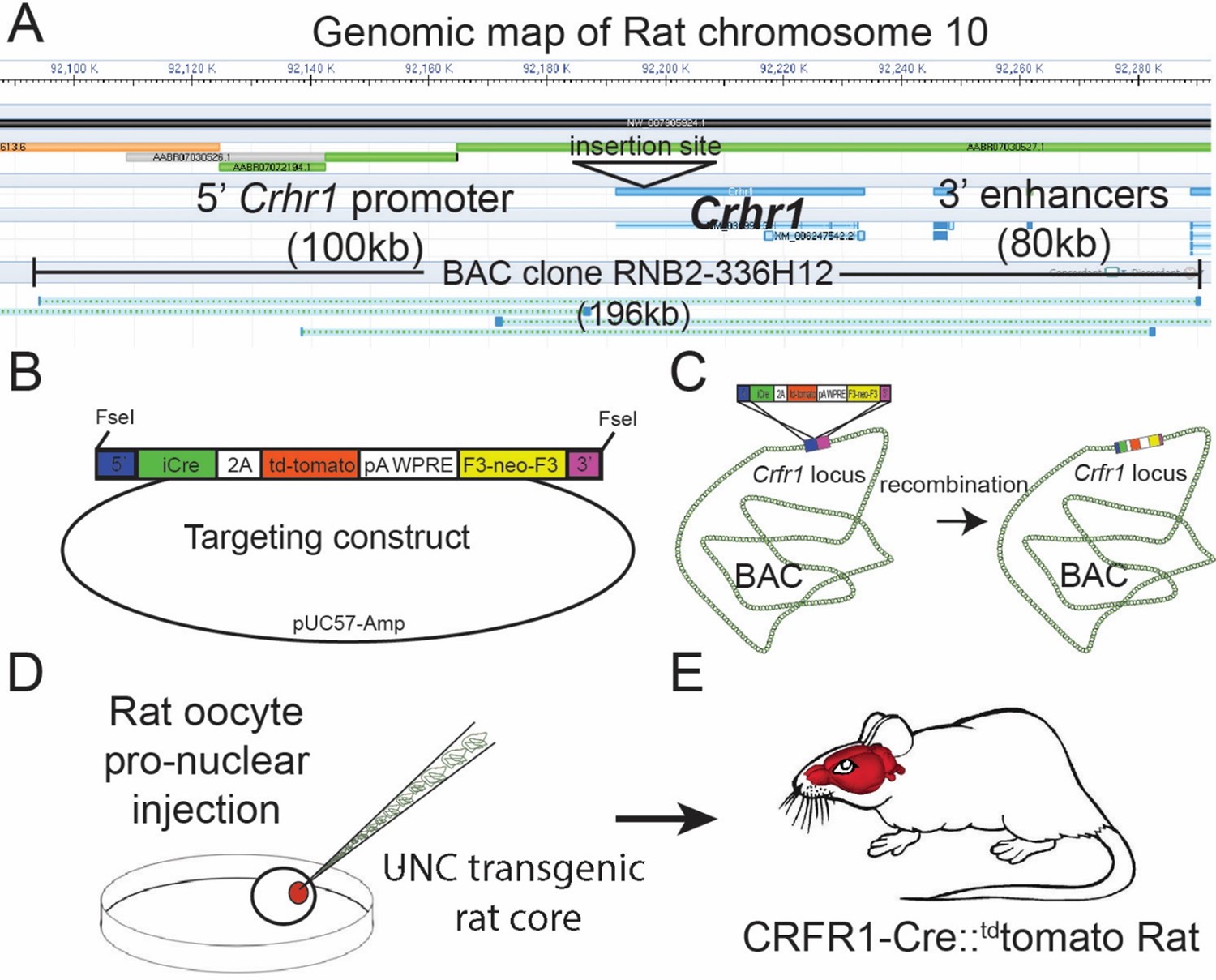
Design of the Crhr1-Cre2aTom BAC transgene.
(A)Crhr1 is located on chromosome 10 in the rat. A BAC clone containing 196 kb of DNA surrounding the Crhr1 coding region includes 100 kb of upstream and 80 kb of downstream DNA, where the majority of promoter and enhancer sequences that control Crhr1 expression are located, was obtained (Riken, RNB2-336H12). There are no other sequences within this 196 kb DNA clone have been annotated as coding sequences for genes other than Crhr1. (B) A transgene containing 5’ (blue) and 3’ (magenta) targeting sequences, a bicistronic iCre 2 A fused tdTomato (red) sequence, 3’ polyA/WPRE stabilizing sequence, and a F3 flanked neomycin resistance sequence (yellow) was constructed then transformed into E. coli containing the RNB2-336H12 BAC construct. (C) Using recombineering techniques we isolated BAC clones in which targeted insertion of the transgene at the translation start site of Crhr1 (ATG) was confirmed by PCR/sequencing. A single Bacterial clone containing the transgene inserted BAC was sent to the UNC transgenic facility where BAC DNA was purified and injected into single cell, fertilized rat oocytes. Two independent rat lines were recovered in which the entire BAC sequence (confirmed by PCR) was inserted into genomic DNA, of which one line displayed transgenic expression in a pattern representative of known Crhr1 expression patterns.
Figure 2
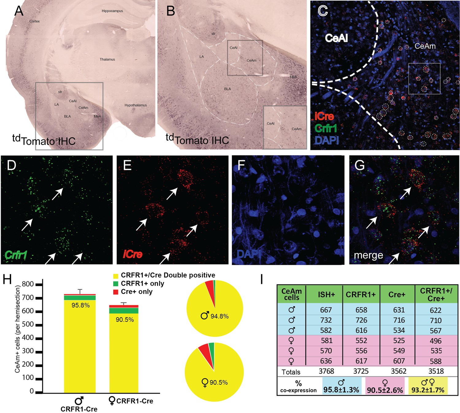
Validation of transgenic expression of iCre/tdTomato in CRF1+ expressing neurons located in the medial central nucleus of the amygdala (CeAm).
(A) A low-magnification image of a section containing the amygdala from a CRF1-Cre rat, immunohistochemically labeled for tdTomato. Expression of the CRF1-Cre transgene is broadly very similar to previous reports of CRF1 expression in both rat and mouse. (B) Within the boxed region of panel A, higher magnification reveals CRF1+ cells in the lateral amygdala (LA), basolateral amygdala (BLA), medial portion central amygdala (CeAm), and medial amygdala (MeA). The lack of significant labeling in the CeAl is consistent with reports using both in situ hybridization and transgenic reporters to detect CRF1 expression. (C) Micrograph of the CeA from the region boxed in panel B allows visualization of mRNA encoding iCre (red) and Crhr1 (green) along with nuclei stained with DAPI (blue). (D–G) Higher magnification images of the boxed region in (C) allows visualization of mRNA for Crhr1 (D, green), and iCre (E, red), with nuclei visualized by DAPI staining (F). (G) Merged images reveals that many CeAm neurons that are positive for Crhr1 mRNA are also positive for iCre mRNA (arrows point to double positive neurons). Quantification of coincidence of in situ hybridization for both Crhr1 and iCre mRNAs demonstrates that >90% of Crhr1-positive cells are also positive for iCre in the CeAm (n = 3). (H) Graphical representation of quantification of coincident labeling, or (I) a table of the precise counts from each of three male and three female CRF1-Cre transgenic animals. We observed greater than 90% of neurons positive for both Crhr1 and iCre in the CeAm.
Figure 3
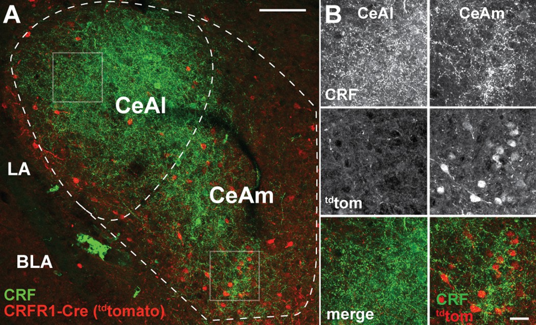
CRF1-driven expression of Cre/tdTomato in the CeA.
(A) Visualization of CRF using immunofluorescent labeling (green) in a rat carrying the CRF1-Cre2aTom transgene (red) reveals minimal cellular expression of CRF1 in the lateral central nucleus of the amygdala (CeAl) where CRF is highly abundant. This discrepancy in CRF localization compared to CRF1 expression is consistent with previous reports of CRF1 expression in both rat and mouse. In contrast to the CeAl, the medial central nucleus of the amygdala (CeAm) contains many CRF1+ neurons (reported by the CRF1-Cre2Atom transgene), in contact with puncta positive for CRF peptide. (B) High-resolution images from the boxed regions of CeAl and CeAm in panel A. CRF staining is dense in both the CeAl and CeAm (top panels); however, cellular expression of the CRF1-Cre2aTom transgene is low in the CeAl, while many neurons in the CeAm are positive for CRF1 expression (middle panels). Merged images (lower panels) display the coincident staining of CRF1+neurons with CRF puncta in the CeAm, suggesting that stress driven CRF release directly signals to CRF1+ neurons in the CeAm to modulate neural excitability to influence the output of CeAm neurons. LA – lateral amygdala, BLA – basolateral amygdala.
Figure 4
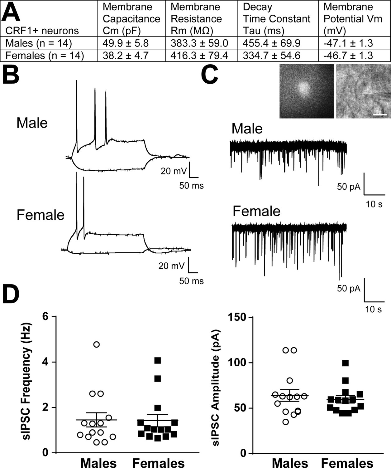
Basal membrane properties and inhibitory synaptic transmission in CeAm CRF1-Cre-tdTomato neurons.
(A) Basal membrane properties (Membrane Capacitance, Cm; Membrane Resistance, Rm; Decay Constant, Tau; Membrane Potential, Vm) from male and female CRF1+ CeAm neurons. (B) Representative current-evoked spiking properties from male (top) and female (bottom) CRF1+ CeAm neurons. (C) Basal spontaneous inhibitory postsynaptic currents (sIPSCs) from male (top) and female (bottom) CRF1+ CeAm neurons (right). Inset: representative fluorescent (left) and infrared differential interference contract (IR-DIC, right) image of a CRF1+ CeAm neuron targeted for recording. Scale bar = 20 μm. (D) Average sIPSC frequency (left) and sIPSC amplitude (right) from male and female CRF1+ CeAm neurons. Raw data are available in Source Data File 1.
Figure 5
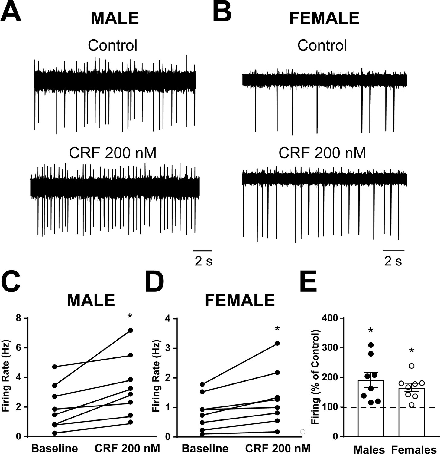
Spontaneous firing activity and CRF sensitivity of CeAm CRF1-Cre-tdTomato neurons.
(A) Representative cell-attached recording of spontaneous firing activity in a CRF1+ CeAm neuron from a male CRF1-Cre-tdTomato rat before and during CRF (200 nM) application. (B) Representative cell-attached recording of spontaneous firing activity in a CRF1+ CeAm neuron from a female CRF1-Cre-tdTomato rat before and during CRF (200 nM) application. (C) Summary of changes in spontaneous firing activity with CRF application in CRF1+ CeAm neurons from male CRF1-Cre-tdTomato rats.*p < 0.05 by paired t-test. (D) Summary of changes in spontaneous firing activity with CRF application in CRF1+ CeAm neurons from female CRF1-Cre-tdTomato rats.*p < 0.05 by paired t-test. (E) Normalized change in firing activity in CRF1+ CeAm neurons from male and female CRF1-Cre-tdTomato rats.*p < 0.05 by one-sample t-test. Raw data are available in Source Data File 1.
Figure 6
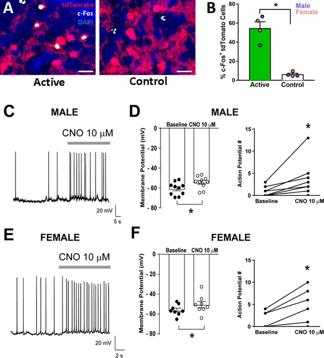
Validation of DREADD expression and function in CeA CRF1-Cre-tdTomato neurons.
(A) Representative images of CRF1-Cre-tdTomato cells (red) and c-Fos immunostaining (white) in CeAm of rats that were given intra-CeA microinjections of AAV8-hSyn-DIO-HA-hM3D(Gq)-IRES-mCitrine (active virus) or AAV5-hSyn-DIO-EGFP (control virus). Scale bar: 50 µm. (B) CNO treatment 90 min before sacrifice increased the percentage of c-Fos+ tdTomato cells in CeAm of rats that were given active virus compared to rats that were given control virus microinjections. *p < 0.05. (C) Representative whole-cell current clamp recording of membrane potential and firing activity in a CRF1+ CeAm neuron from a male CRF1-Cre-tdTomato rat before and during CNO (10 μM) application. (D) Summary of the change in membrane potential (left) and action potentials (right) in male CRF1+ CeAm neurons after CNO application. *p < 0.05 by paired t-test. (E) Representative whole-cell current clamp recording of membrane potential and firing activity in a CRF1+ CeAm neuron from a female CRF1-Cre-tdTomato rat before and during CNO (10 μM) application. (D) Summary of the change in membrane potential (left) and action potentials (right) in female CRF1+ CeAm neurons after CNO application. *p < 0.05 by paired t-test. Raw data are available in Source Data File 1.
Figure 7

Effects of chemogenetic stimulation of CeA CRF1-Cre-tdTomato neurons on nociception and anxiety-like behaviors.
(A) Representative image of AAV8-hSyn-DIO-HA-hM3D(Gq)-IRES-mCitrine expression (green) in the CeA. Scale bar: 500 µm. BLA: basolateral amygdala, Opt: optic tract. (B) Timeline of experimental procedures. (C) CNO treatment decreased paw withdrawal thresholds in the Von Frey test of mechanical nociception in rats that were given intra-CeA hM3D(Gq) virus microinjections. There were no effects of treatment on paw withdrawal thresholds in the EGFP control group. (D) CNO treatment had no effects on paw withdrawal latencies in either the hM3D(Gq) or EGFP groups in the Hargreaves test of thermal nociception. (E) CNO treatment decreased the percent time spent in open arms in the EPM test in the hM3D(Gq) group, but had no effect in the EGFP group. (F) CNO treatment decreased the percent time spent in the center of the arena in the OF test in the hM3D(Gq) group, but had no effect in the EGFP group. (G) CNO treatment had no effect on percent time spent in the light box in the LD test. *p < 0.05. Raw data are available in Source Data File 2. iCre and Crhr1 mRNA are highly co-expressed in BLA.
Figure 8
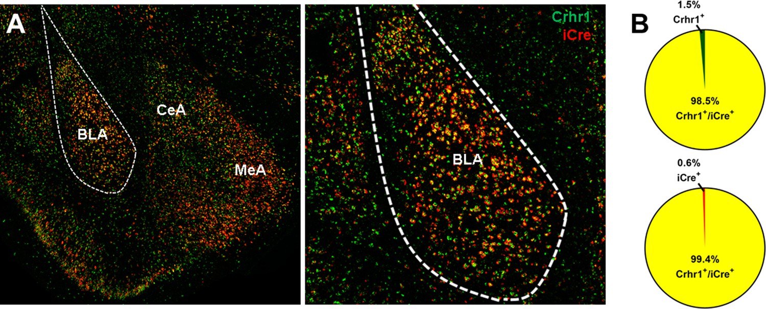
Crhr1 and iCre mRNA expression in BLA of CRF1-Cre-tdTomato rats.
(A) Crhr1 (green) and iCre (red) mRNA are highly co-expressed in BLA and surrounding areas. (B) Within the BLA, 98.5% of Crhr1+ + co-express iCre (top), and 99.4% of iCre+ + co-express Crhr1 (bottom).
Figure 9
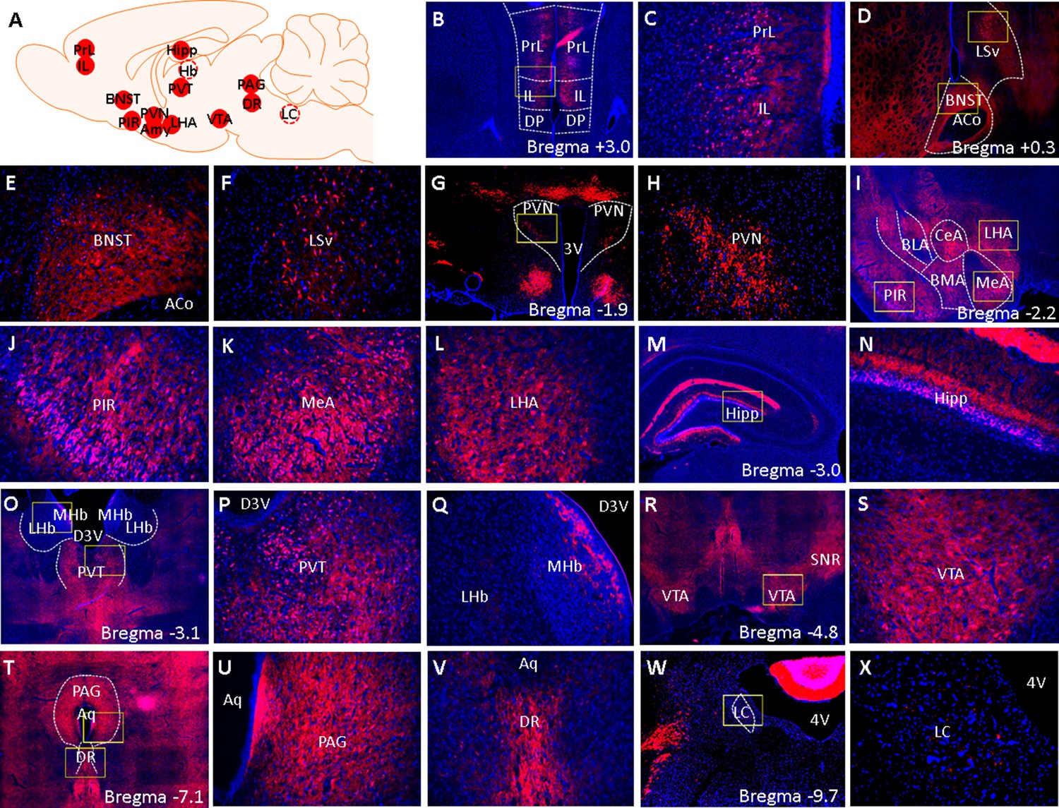
Fluorescent images of brainwide tdTomato expression.
(A) A schematic summary of brain areas that were surveyed for tdTomato expression. Solid red circles indicate brain areas that contain tdTomato cells and dashed circles indicate brain areas that were devoid of tdTomato cells. (B–X) Low (4 x) and high-magnification (×20 ) images of tdTomato (red) and DAPI (blue) fluorescent signals. Yellow boxes demarcate areas from which high-magnification (×20 ) images were acquired. (B) 4 x image of the PrL, IL, and surrounding landmarks. (C) 20 x image of PrL and IL. (D) 4 x of BNST and LSv. (E) 20 x image of BNST and (F) LSv. (G) 4 x and (H) 20 x image of PVN. (I) 4 x image of PIR, MeA, and LHA. (J) 20 x image of PIR, (K) MeA, and (L) LHA. (M) 4 x and (N) 20 x image of hippocampus. (O) 4 x image PVT, LHB, and MHb. (P) 20 x image of PVT and (Q) LHb/MHb. (R) 4 x and (S) 20 x image of VTA. (T) 4 x image of PAG and DR. (U) 20 x image of PAG and (V) DR. (W) 4 x and (X) 20 x image of LC. 3 V: 3rd ventricle, 4 V: 4th ventricle, D3V: dorsal 3rd ventricle, Aq: cerebral aqueduct.
Tables
Table 1
Comparison of CRF1-Cre-tdTomato cell densities with Crhr1 mRNA expression levels in rats and CRF1-GFP cell densities in mice in several areas across the rostrocaudal axis of the brain.
+: Some cells are positive, ++: A substantial number of cells are positive, +++: Most cells are positive.
| Brain area | Density of tdTomato cells in CRF1-Cre-tdTomato rats | Expression level of Crhr1 mRNA in wildtype rats (Van Pett et al., 2000) | Density of GFP cells in CRF1-GFP Mice (Justice et al., 2008) |
|---|---|---|---|
| PrL | ++ | ++ | ++ |
| IL | ++ | ++ | ++ |
| BNST | +++ | +++ | +/++ |
| LSv | + | + | - |
| PVN | + | + | ++ |
| PIR | +++ | +++ | ++/+++ |
| MeA | ++ | + | +/++ |
| LHA | + | + | ++ |
| Hipp | + (subiculum, CA3)/+++ (CA1) | ++ (CA1, CA3, subiculum) | + (subiculum, CA3)/+++ (CA1) |
| PVT | ++ | ++ | - |
| LHb | - | - | ++ |
| MHb | + | - | - |
| VTA | ++ | ++ | ++ |
| PAG | ++ | + | + |
| DR | ++ | + | ++ |
| LC | - | - | +/- |
Key resources table
| Reagent type (species) or resource | Designation | Source or reference | Identifiers | Additional information |
|---|---|---|---|---|
| Gene (Rattus norvegicus) | BAC containing the Crfr1 genomic locus | Riken Gene Engineering Division | RNB2-336H12 | |
| Transfected construct (Rattus norvegicus) | iCre-2a-tdTomato BAC transgene | This paper | See Results, Design of CRF1-Cre BAC; Contact Justice Lab | |
| Genetic reagent (Rattus norvegicus) | CRF1-Cre-tdTomato rat | This paper | See Results, Generation of Transgenic Rats; Contact Gilpin Lab | |
| Recombinant DNA reagent | AAV8-hSyn-DIO-HA-hM3D(Gq)-IRES-mCitrine | Addgene | Cat# 50454-AAV8 | |
| Recombinant DNA reagent | AAV5-hSyn-DIO-EGFP | Addgene | Cat# 50457-AAV5 | |
| Antibody | Anti-c-Fos (Rabbit polyclonal) | Abcam | Cat# ab190289, RRID:AB_2737414 | (1:1000) |
| Antibody | Anti-HA-Tag (Rabbit monoclonal) | Cell Signaling | Cat# 3724, RRID:AB_1549585 | (1:250) |
| Antibody | Anti-RFP (Rabbit monoclonal) | Abcam | Cat# ab34771, RRID:AB_777699 | (1:500) |
| Antibody | Anti-CRF (Rabbit monoclonal) | The Salk Institute | Rc-68 | (1:2000) |
| Commercial assay or kit | RNAscope Multiplex Fluorescent Kit v2 | ACD Bio | iCre and Crfr1 probes | |
| Commercial assay or kit | TSA Detection Kit | Akoya Biosciences | Cat# NEL701A001KT | |
| Commercial assay or kit | Prolong Gold Antifade Reagent with DAPI | Invitrogen | Cat# P36935 | |
| Chemical compound, drug | Clozapine-n-oxide | NIH Drug Supply Program | ||
| Chemical compound, drug | Corticotropin-releasing factor | Tocris | Cat# 1,607 | |
| Software, algorithm | Mini Analysis | Synaptosoft Inc. | RRID:SCR_002184 | |
| Software, algorithm | Clampfit 10.6 | Molecular Devices | ||
| Software, algorithm | Prism 7.0 | GraphPad | RRID:SCR_002798 | |
| Software, algorithm | SPSS 25 | IBM SPSS | RRID:SCR_019096 |
Additional files
-
Transparent reporting form
- https://cdn.elifesciences.org/articles/67822/elife-67822-transrepform1-v2.docx
-
Source data 1
Slice electrophysiology data.
- https://cdn.elifesciences.org/articles/67822/elife-67822-data1-v2.xlsx
-
Source data 2
Behavioral data.
- https://cdn.elifesciences.org/articles/67822/elife-67822-data2-v2.xlsx
Download links
A two-part list of links to download the article, or parts of the article, in various formats.
Downloads (link to download the article as PDF)
Open citations (links to open the citations from this article in various online reference manager services)
Cite this article (links to download the citations from this article in formats compatible with various reference manager tools)
Generation of a CRF1-Cre transgenic rat and the role of central amygdala CRF1 cells in nociception and anxiety-like behavior
eLife 11:e67822.
https://doi.org/10.7554/eLife.67822


