Molecular structures and conformations of protocadherin-15 and its complexes on stereocilia elucidated by cryo-electron tomography
- Vollum Institute, Oregon Health and Science University, United States
- RNA Therapeutics Institute, UMass Chan Medical School, United States
- Howard Hughes Medical Institute, United States
Figures
Figure 1
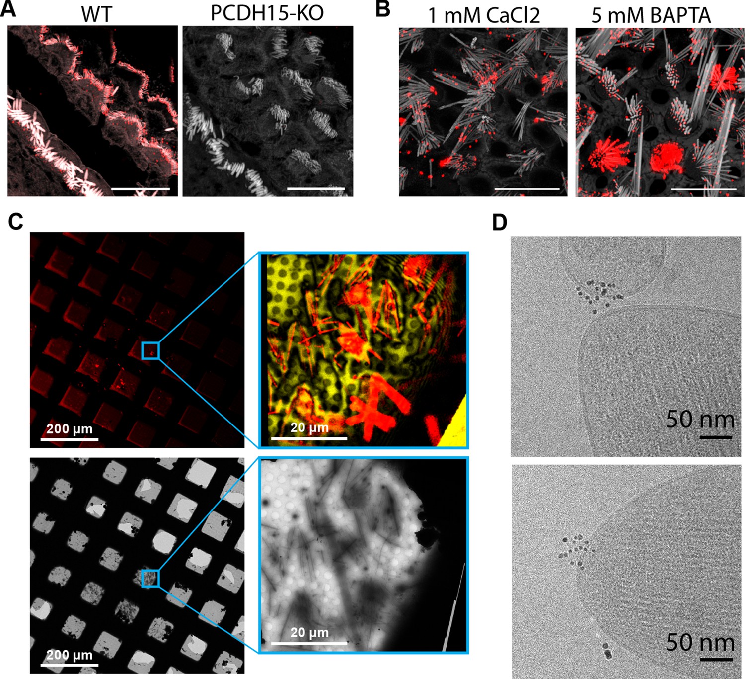
Cryo-EM data collection of antibody-stained stereocilia tips supported by cryo-CLEM.
(A) Immunostaining of WT cochlea or PCDH15 KO with polyclonal antibody raised in rabbits against PCDH15 extracellular domain. Scale bars correspond to 10 µm. (B) Immunostaining of WT utricle with polyclonal antibody in media containing 1 mM CaCl2 or with the addition of 5 mM BAPTA. Scale bars correspond to 10 µm. (C) CLEM-based screening to identify squares with thin-ice supported stereocilia. The top row shows low- and medium magnification views acquired using a cryo-light microscope. In both views, SiR-actin fluorescence is shown in red. In the medium-magnification view, laser reflection is shown in gold. The bottom rows show the same areas as acquired in a cryo-TEM. (D) High-magnification cryo-TEM micrographs of stereocilia tips stained with the anti-PCDH15 rabbit polyclonal antibody, detected with a 5 nm gold coupled secondary antibody. CLEM, correlative light and electron microscopy; WT, wild-type.
Figure 2 with 1 supplement
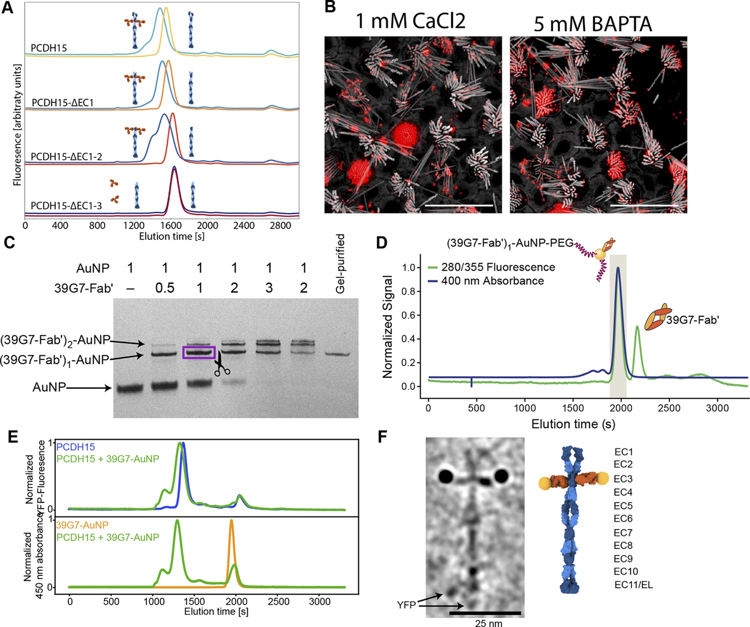
Stoichiometric gold staining of PCDH15 using a monoclonal Fab AuNP conjugate.
(A) FSEC analysis of binding of 39G7 to the PCDH15 extracellular domain. 39G7 addition leads to earlier elution of PCDH15 in full-length constructs and after deletion of EC1 and EC2. Deletion of EC3 abolished 39G7 binding. (B) Immunostaining of WT utricle with 39G7 in media containing 1 mM CaCl2 or with the addition of 5 mM BAPTA. The qualitatively similar staining in both conditions suggests that binding can occur in the context of a PCDH15/CDH23 complex (in the presence of calcium) or in the context of unbound PCDH15 (after disruption of tip links by BAPTA). (C) PAGE analysis and purification of conjugation between 39G7 Fab’ and a 2 nm AuNP. Higher ratios of 39G7-Fab’ to AuNP lead to lower amount of free AuNP and higher amounts of AuNP bound to multiple copies of 39G7-Fab’. The fastest migrating 39G7-Fab’-AuNP band was assumed to correspond to a 1:1 complex and purified by cutting it out from the PAGE gel. (D) SEC purification of PEG-coated 39G7-Fab’-AuNP-PEG conjugates. By only collecting fractions corresponding to 39G7-Fab’-AuNP-PEG conjugates free Fab’, which would disrupt labeling, is removed. (E) FSEC analysis of 39G7-Fab’-AuNP-PEG conjugate binding to PCDH15 extracellular domain. Mixing of 39G7-Fab’-AuNP-PEG and PCDH15 extracellular domain leads to faster elution of both molecules at identical times, indicating that they formed a complex. (F) Cryo-TEM image of PCDH15 extracellular domain bound to 39G7-Fab’-AuNP-PEG conjugate together with model of the complex. In the model, PCDH15 is shown in blue, 39G7 Fab in orange, and AuNPs as golden spheres.
-
Figure 2—source data 1
Original image of polyacrylamide gel shown in Figure 2C.
- https://cdn.elifesciences.org/articles/74512/elife-74512-fig2-data1-v2.zip
Figure 2—video 1
Tomogram of recombinant PCDH15 extracellular domain in complex with 39G7-AuNP conjugate.
Figure 3 with 4 supplements
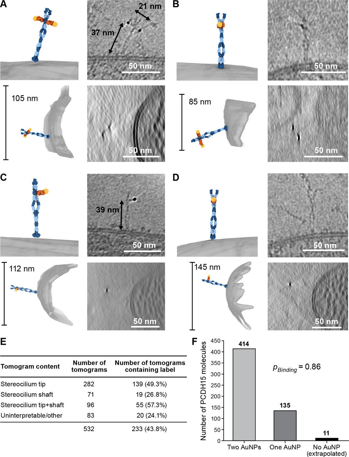
39G7-AuNP conjugate labels PCDH15 dimer in stereocilia.
(A–D) Representative examples of PCDH15 dimers with two bound AuNPs (A, B) or one bound AuNP (C, D) image in stereocilia. On the left-hand side of each panel, a manually annotate model is shown in a view roughly corresponding to the electron beam (top) and from the side (bottom). On the right-hand side are corresponding slices through the electron density. Due to the missing-wedge effect, the resolution of the side-view at the bottom is lower, blurring AuNP and precluding direct visualization of PCDH15. For panels (B) and (D), it is therefore impossible to show AuNPs and PCDH15 in a single projection as the AuNPs are situated above and below PCDH15 in relation to the electron beam. In panels (C) and (D), there are dimeric features in the PCDH15 density, suggesting that these molecules are PCDH15 dimers with one epitope not bound to 39G7. (E) Table detailing numbers and content of collected tomograms. (F) Bar chart quantifying the ratio of PCDH15 molecules labeled by one or two AuNPs. Assuming that AuNPs bind independently to the two epitopes in the PCDH15 dimer results suggests that 86% of all epitopes were labeled and only 2% of PCDH15 molecules were unlabeled.
Figure 3—video 1
.Tomogram of stereocilium displaying a PCDH15 dimer with two bound 39G7-AuNP conjugates, also depicted in Figure 3A.
Figure 3—video 2
Tomogram of stereocilium displaying a PCDH15 dimer with two bound 39G7-AuNP conjugates, also depicted in Figure 3B.
Figure 3—video 3
Tomogram of stereocilium displaying a PCDH15 dimer with one bound 39G7-AuNP conjugates also depicted in Figure 3C.
Figure 3—video 4
Tomogram of stereocilium displaying a PCDH15 dimer with one bound 39G7-AuNP conjugates, also depicted in Figure 3D.
Figure 4 with 5 supplements
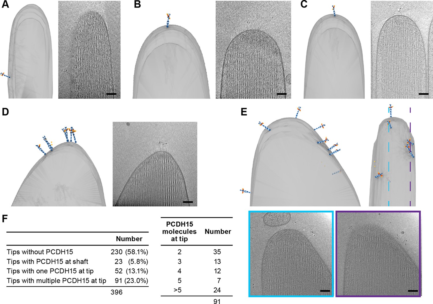
Stereocilia tips frequently harbor more than one copy of PCDH15.
(A) Representative example of stereocilia tip with PCDH15 molecule only found in adjacent shaft. (B, C) Representative examples of stereocilia tips with one copy of PCDH15 at tip. (D, E) Representative examples of stereocilia tips with multiple copies of PCDH15 at tip. (F) Table detailing number of imaged stereocilia tips and distribution of PCDH15 molecules found at the tip.
Figure 4—video 1
Tomogram of stereocilium tip with a single PCDH15 dimer in the shaft region of the tip, also depicted in Figure 4A.
Figure 4—video 2
Tomogram of stereocilium tip with a single PCDH15 dimer at the apex of the tip, also depicted in Figure 4B.
Figure 4—video 3
Tomogram of stereocilium tip with a single PCDH15 dimer at the apex of the tip, also depicted in Figure 4C.
Figure 4—video 4
Tomogram of stereocilium tip with a cluster of multiple PCDH15 dimers at the apex of the tip, also depicted in Figure 4D.
Figure 4—video 5
Tomogram of stereocilium tip with multiple PCDH15 dimers scattered around the tip, also depicted in Figure 4E.
Figure 5 with 4 supplements

PCDH15 complexes are found in clusters.
On the left side of each panel, the tomogram annotation is shown from the side and from top. On the sideview ice thickness is indicated. To the right of the annotation is a projection of the cluster density in the context of the stereocilium. Farther to the right are detailed slices of the electron density. The angle and position of each slice are indicated in the sideview of the tomogram annotation. Scale bars correspond to 50 nm. (A, B) Representative tomograms showing PCDH15 complexes with PCDH15 at the tip of a stereocilium and the complex partner in a lipid fragment. (C, D) Representative tomograms showing PCDH15 complexes with complex partner inserted in the side of a stereocilium and PCDH15 in a lipid fragment.
Figure 5—video 1
Tomogram of stereocilium tip containing multiple PCDH15 dimers connected to putative CDH23 filaments in a lipid vesicle, also depicted in Figure 5A.
Figure 5—video 2
Tomogram of stereocilium tip containing multiple PCDH15 dimers connected to putative CDH23 filaments in a lipid membrane fragment, also depicted in Figure 5B.
Figure 5—video 3
Tomogram of a stereocilium tip containing multiple putative CDH23 filaments connected to PCDH15 dimers in a lipid vesicle, also depicted in Figure 5C.
Figure 5—video 4
Tomogram of a stereocilium shaft containing multiple putative CDH23 filaments connected to PCDH15 dimers in a lipid vesicle, also depicted in Figure 5D.
Figure 6 with 1 supplement
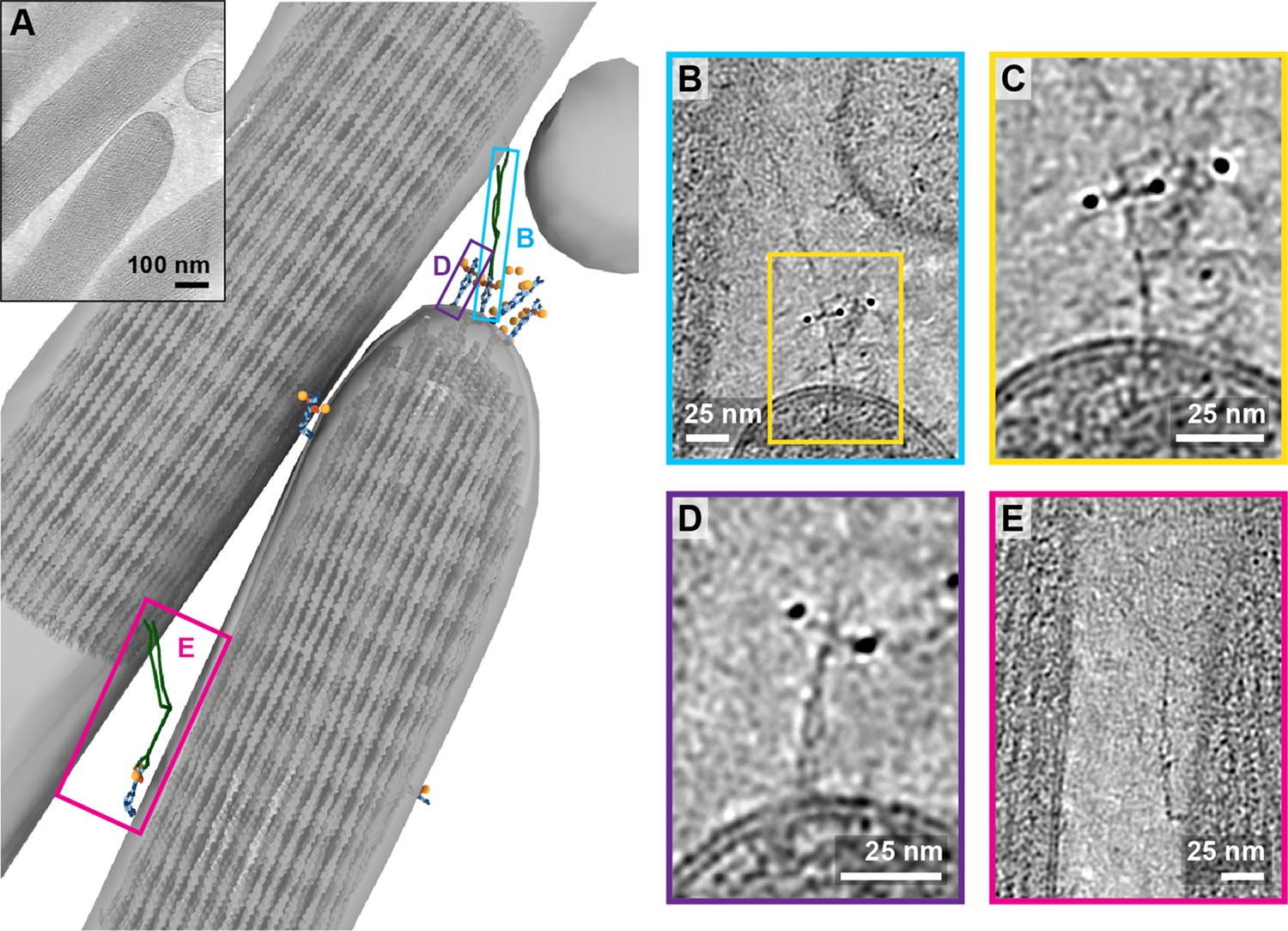
Tomogram containing intact tip link.
(A) Annotation of tomogram showing a putative intact tip link. The inset at the top left shows a projection of the tomogram. (B) Close-up view of putative tip link density in the tomogram. (C) Close-up of PCDH15 density in tip link. (D) Close-up of PCDH15 molecule at tip not bound to a putative CDH23. (E) Close-up of PCDH15-complex in the stereocilia shaft region. PCDH15 has a 90° bend near the EC9/EC10 interface.
Figure 6—video 1
Tomogram of a stereocilium tip and a stereocilium shaft containing a PCDH15 dimer in the stereocilium tip that is connected to a putative CDH23 filament in the neighboring stereocilium shaft.
The tip also contains PCDH15 dimers not bound to a complex partner. An additional PCDH15-complex is connecting the two stereocilia in the shaft region. Also depicted in Figure 6.
Figure 7 with 1 supplement
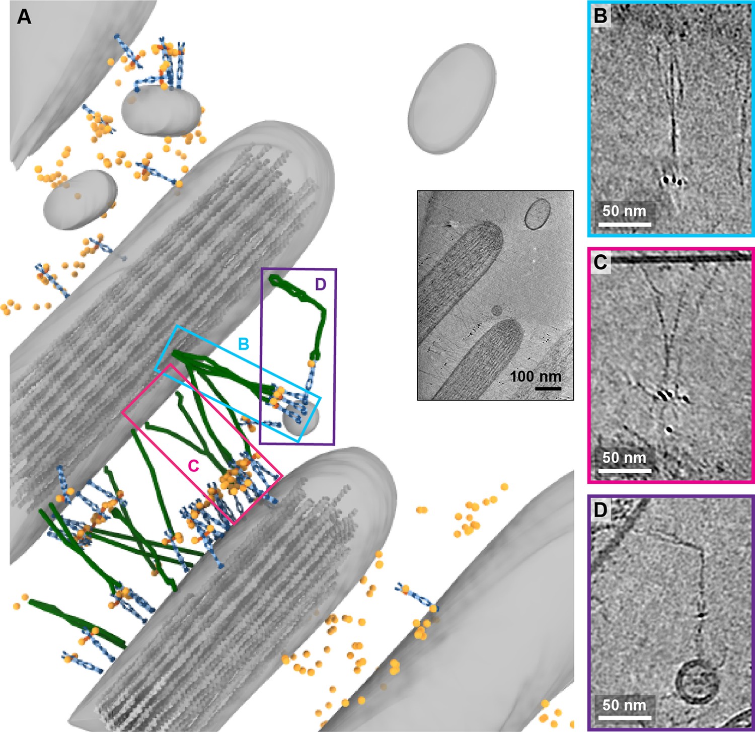
Tomogram depicting lateral links containing PCDH15 in small stereocilia.
(A) Annotation of tomogram showing developing stereocilia including PCDH15-containing ‘lateral links.’ The inset at the bottom right shows a projection of the tomogram. (B, C) Close-up of putative CDH23 molecules clustering together into single strand. (D) Close-up of a putative CDH23 molecule with distinct bend halfway between N- and C-terminal end.
Figure 7—video 1
Tomogram of stereocilia connected by tens of PCDH15-containing complexes.
Noteworthy complexes that include apparent interaction between putative CDH23 dimers, as well as bends in the putative CDH23 dimer are highlighted. Also depicted in Figure 7.
Tables
Key resources table
| Reagent type (species) or resource | Designation | Source or reference | Identifiers | Additional information |
|---|---|---|---|---|
| Gene (Mus musculus) | Pcdh15 | Uniprot | Q99PJ1 | |
| Cell line (Spodoptera frugiperda) | Sf9 | Thermo Fisher Scientific | 12659017 RRID:CVCL_0549 | |
| Cell line (Homo sapiens) | HEK293 tsa 201 | ATCC | CRL-11268RRID:CVCL_1926 | |
| Recombinant DNA reagent | PCDH15 EC1-EL-YFP expression plasmid | This paper | Created using pEG BacMam (Addgene plasmid # 160451) | |
| Recombinant DNA reagent | 39G7 Fab’ expression plasmid | This paper (created by VectorBuilder) | Backbone described in d oi:10/fhwrn3 | |
| Antibody | Anti-PCDH15 (Rabbit polyclonal) | This paper (created by Genscript) | IF: 10 µg/ml | |
| Antibody | Anti-PCDH15 39G7 (Rabbit monoclonal) | This paper (created by Genscript) | IF: 10 µg/ml | |
| Biological sample (M. musculus) | Inner ear | P6–P9 mice | ||
| Chemical compound, drug | 10 nm gold fiducials | Ted Pella | 90010 | |
| Chemical compound, drug | HAuCL4 | Sigma-Aldrich | 520918 | |
| Chemical compound, drug | 3-MBA | Sigma-Aldrich | 451436 | |
| Chemical compound, drug | mPEG-550-SH | Creative PEGworks | PLS-607 | |
| Software, algorithm | SerialEM | 10.1016 /j.jsb.2005.07.007 | RRID:SCR_017293 | |
| Software, algorithm | IMOD | doi.org.10.1006/jsbi.1996.0013 | RRID:SCR_003297 | |
| Software, algorithm | Topaz | doi.org.10.1038/s41467-020-18952-1 | ||
| Software, algorithm | TomoAlign | doi.org.10.1016/j.jsb.2019.01.005 | ||
| Software, algorithm | Motioncor2 | doi.org.10.1038/nmeth.4193 | RRID:SCR_016499 | |
| Software, algorithm | ChimeraX | doi.org.10.1002/pro.3943 | RRID:SCR_015872 |
Additional files
Download links
A two-part list of links to download the article, or parts of the article, in various formats.
Downloads (link to download the article as PDF)
Open citations (links to open the citations from this article in various online reference manager services)
Cite this article (links to download the citations from this article in formats compatible with various reference manager tools)
Molecular structures and conformations of protocadherin-15 and its complexes on stereocilia elucidated by cryo-electron tomography
eLife 10:e74512.
https://doi.org/10.7554/eLife.74512


