Oxytocin signaling in the posterior hypothalamus prevents hyperphagic obesity in mice
- RIKEN Center for Biosystems Dynamics Research, Japan
- Laboratory of Molecular Biology, Department of Molecular and Cell Biology, Graduate School of Agricultural Science, Tohoku University, Japan
- Division of Brain and Neurophysiology, Department of Physiology, Jichi Medical University, Japan
- Department of Obesity and Inflammation Research, Fukushima Medical University, Japan
- CREST, Japan Science and Technology Agency, Japan
Figures
Figure 1 with 1 supplement
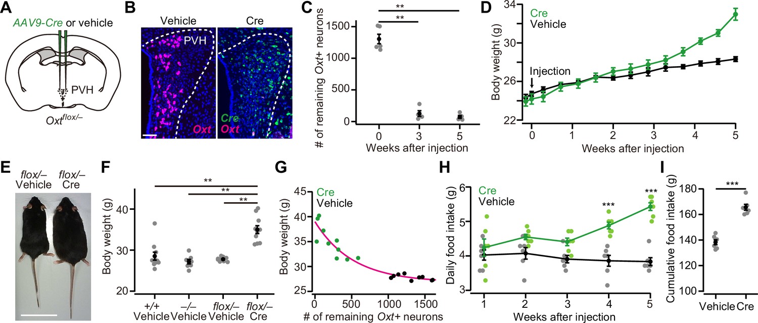
Oxytocin (Oxt) conditional knockout (cKO) in paraventricular hypothalamic nucleus (PVH) induces an increase in body weight and food intake.
(A) Schematic of the virus injection. AAV-Cre or vehicle was injected into the bilateral PVH of Oxtflox/– male mice. (B) Representative coronal sections of the PVH from Oxtflox/– mice received vehicle (left) or AAV-Cre (right) injection. Data were obtained at 5 weeks after the injection. Magenta and green represent Oxt and Cre in situ stainings, respectively. Blue, DAPI. Scale bar, 50 μm. (C) The number of remaining Oxt+ neurons in the PVH of mice that received AAV-Cre injection. **p<0.01, one-way ANOVA with post hoc Tukey’s HSD. N=5 each. (D) Time course of body weight after AAV-Cre or vehicle injection. N=6 each. (E) Representative photos of Oxtflox/– mice that received either vehicle (left) or AAV-Cre injection (right). Five weeks after the injection. Scale bar, 5 cm. (F) Body weight of wild-type (+/+), Oxt KO (–/–), and Oxt cKO (flox/–) mice. The weight was measured at 5 weeks after injection of either vehicle or AAV-Cre. Note that this time point corresponds to 13 weeks of age. **p<0.01, one-way ANOVA with post hoc Tukey’s HSD. N=10, 7, 9, and 10 for +/+, –/–, flox/– vehicle, and flox/– Cre, respectively. (G) Relationship between the number of remaining Oxt+ neurons in the PVH and the body weight of Oxtflox/– mice shown in (F). Magenta, exponential fit for the data from both Cre and vehicle. (H) Time course of daily food intake, defined as the average food intake in each week after AAV-Cre or vehicle injection. ***p<0.001, Student’s t-test with post hoc Bonferroni correction. N=6 each. (I) Cumulative food intake during the 5 weeks after the injection. ***p<0.001, Student’s t-test. N=6 each.
Error bars, standard error of mean (SEM).
Figure 1—figure supplement 1

AAV-Cre injection into the paraventricular hypothalamic nucleus (PVH) of wild-type mice does not induce hyperphagic obesity.
(A) Schematic of the virus injection. AAV-Cre or vehicle was injected into the bilateral PVH of wild-type male mice. (B) Representative coronal section of the left PVH from wild-type mice that had received AAV-Cre injection. Data were obtained at 5 weeks after the injection. Green, Cre in situ staining, blue, DAPI. Scale bar, 50 μm. (C) Body weight measured at 5 weeks after the injection was not statistically different (p>0.82, Student’s t-test). N=10 each. (D) Daily food intake measured at 5 weeks after the injection (p>0.59, Student’s t-test). Error bars, SEM.
Figure 2
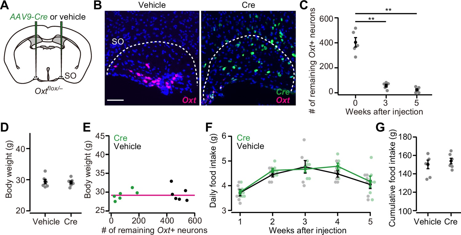
Oxytocin (Oxt) conditional knockout (cKO) in supraoptic nucleus (SO) has a negligible effect on food intake and body weight.
(A) Schematic of the virus injection. AAV-Cre or vehicle was injected into the bilateral SO of Oxtflox/– male mice. (B) Representative coronal sections of left SO from Oxtflox/– mice received vehicle (left) or AAV-Cre (right) injection. Five weeks after the injection. Magenta and green represent Oxt and Cre in situ stainings, respectively. Blue, DAPI. Scale bar, 50 μm. (C) The number of remaining Oxt+ neurons in the SO of mice that received AAV-Cre injection. **p<0.01, one-way ANOVA with post hoc Tukey’s HSD. N=5 each. (D) The body weight of Oxtflox/– mice did not differ between vehicle or AAV-Cre (Student’s t-test). N=6 each. Data were obtained at 5 weeks after the injection. (E) Relationship between the number of remaining Oxt+ neurons in the SO and the body weight of Oxtflox/– mice shown in (D). Magenta, exponential fit for the data from both Cre and vehicle. (F) The time course of daily food intake was not statistically different (Student’s t-test with post hoc Bonferroni correction). N=6 each. (G) Cumulative food intake during the 5 weeks after the injection. N=6 each.
Error bars, SEM.
Figure 3

Weight of viscera and the blood constituents.
(A) Schematic of the virus injection. AAV-Cre or vehicle was injected into the bilateral paraventricular hypothalamic nucleus (PVH) of Oxtflox/– male. Data were obtained at 5 weeks after the injection. (B) The weight of the stomach was not statistically different (p>0.5, Student’s t-test. N=9 and 10 for vehicle and Cre, respectively). (C) The weight of the liver was significantly heavier in AAV-Cre-injected mice (***p<0.001, Student’s t-test). (D) Plasma glucose (left), triglyceride (middle), and leptin (right) measured in the non-fasted Oxtflox/– mice. **p<0.01, ***p<0.001, Student’s t-test. N=9 and 8 mice for vehicle and Cre, respectively.
Error bars, SEM.
Figure 4 with 1 supplement
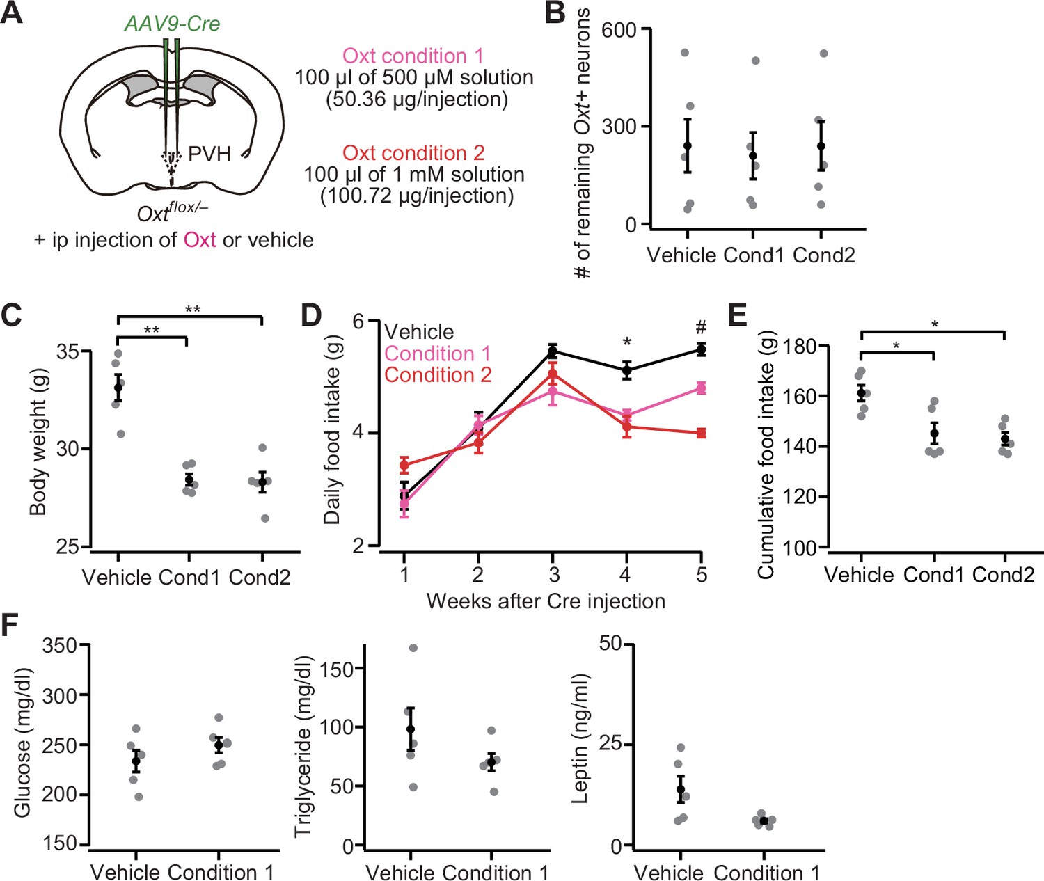
Intraperitoneal (ip) injection of oxytocin (Oxt) partially rescues paraventricular hypothalamic nucleus (PVH) Oxt conditional knockout (cKO) phenotypes.
(A) Schematic of the experiments. AAV-Cre was injected into the bilateral PVH of Oxtflox/– male mice. Data were obtained 5 weeks after the virus injection. Once every 3 days, the mice received ip injection of vehicle, 50.36 μg of Oxt (condition 1) or 100.72 μg of Oxt (condition 2) (see Materials and methods). (B) The number of remaining Oxt+ neurons was not statistically different (p>0.9, one-way ANOVA). Cond, condition. N=5 each (same mice across panels B–E). (C) Ip injection of Oxt significantly decreased body weight. **p<0.01, one-way ANOVA with post hoc Tukey’s HSD. Cond, condition. (D) Time course of daily food intake. Asterisks (*) denote significant differences for vehicle versus condition 1 and vehicle versus condition 2 (p<0.05, Tukey’s HSD), and hashes (#) denote significant differences for vehicle versus condition 1, vehicle versus condition 2, and condition 1 versus condition 2 (p<0.05, Tukey’s HSD). (E) Cumulative food intake during the 5 weeks after the virus injection was decreased in the mice that received ip injection of Oxt (*p<0.05, one-way ANOVA with post hoc Tukey’s HSD). Cond, condition. (F) Plasma glucose (left), triglyceride (middle), and leptin (right) measured in the non-fasted Oxtflox/– mice. Decreases in triglyceride and leptin on average were found in Oxt-treated mice but did not reach the level of statistical significance (p=0.314 and 0.065 for triglyceride and leptin, respectively, Student’s t-test). N=5 each.
Error bars, SEM.
Figure 4—figure supplement 1
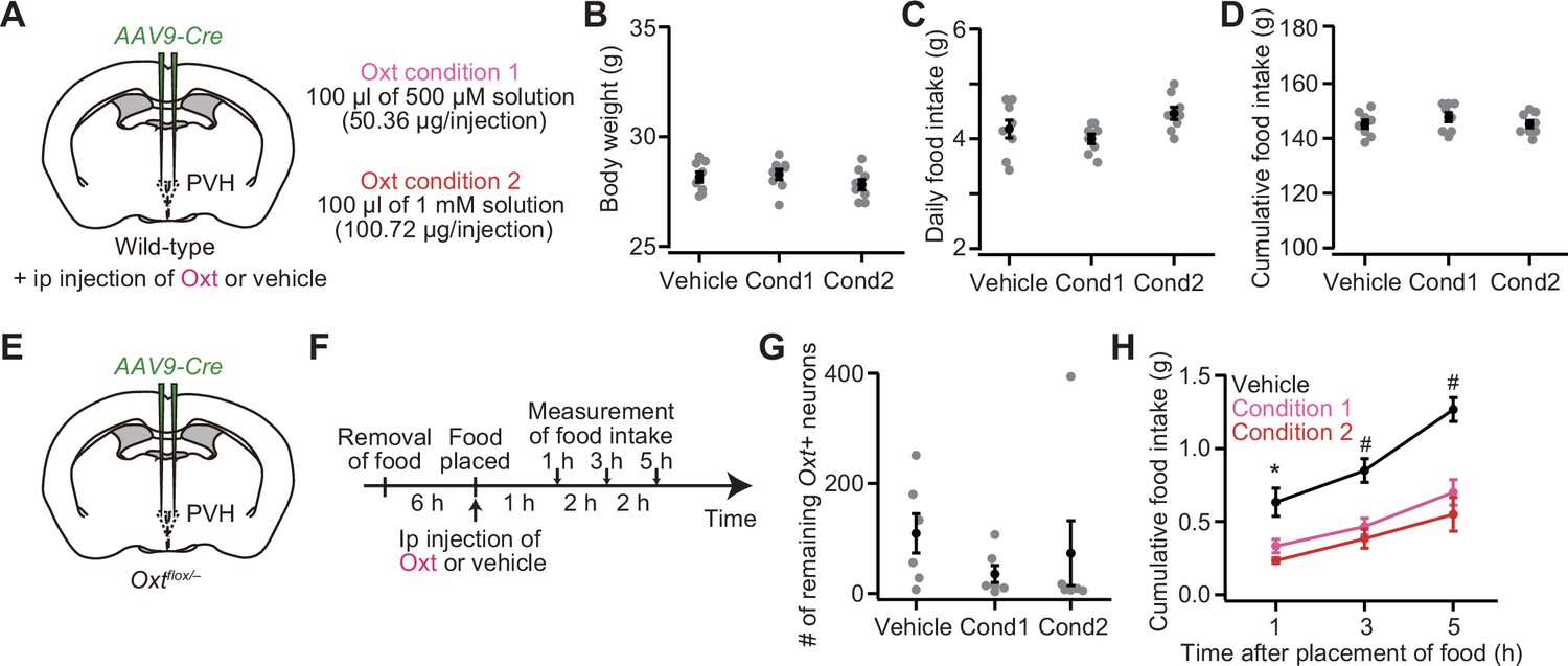
Intraperitoneal (Ip) injection of oxytocin (Oxt) does not affect either body weight or food intake in wild-type mice, but reduces the hourly food intake of mice with Oxt conditional knockout (cKO) in the paraventricular hypothalamic nucleus (PVH).
(A) Schematic of the experiments. AAV-Cre was injected into the bilateral PVH of wild-type male mice. The procedure used for the ip injection of Oxt solution was the same as that in Figure 4. (B) Body weight measured at 5 weeks after the injection was not statistically different (p>0.41, one-way ANOVA). N=8 each. (C) Daily food intake measured at 5 weeks after the injection (p>0.068, one-way ANOVA). N=8 each. (D) Cumulative food intake during the 5 weeks after the injection was not statistically different (p>0.46, one-way ANOVA). N=8 each. (E) Schematic of the experiments. AAV-Cre was injected into the bilateral PVH of Oxtflox/– male mice. Data were obtained at 5 weeks after the virus injection. (F) Schematic of the timeline of the experiment. Each mouse fasted for 6 hr. Food intake was measured at 1, 3, and 5 hr after the placement of food and ip injection of Oxt or vehicle. (G) The number of remaining Oxt+ neurons in the PVH was not statistically different (p>0.52, one-way ANOVA). Cond, condition. N=6 each (same mice across panels G and H). (H) Cumulative food intake. Asterisks (*) denote significant differences for vehicle versus condition 1 (p<0.05, one-way ANOVA with post hoc Tukey’s HSD) and vehicle versus condition 2 (p<0.01), and hashes (#) denote significant differences for vehicle versus condition 1 (p<0.01) and vehicle versus condition 2 (p<0.01).
Error bars, SEM.
Figure 5 with 2 supplements
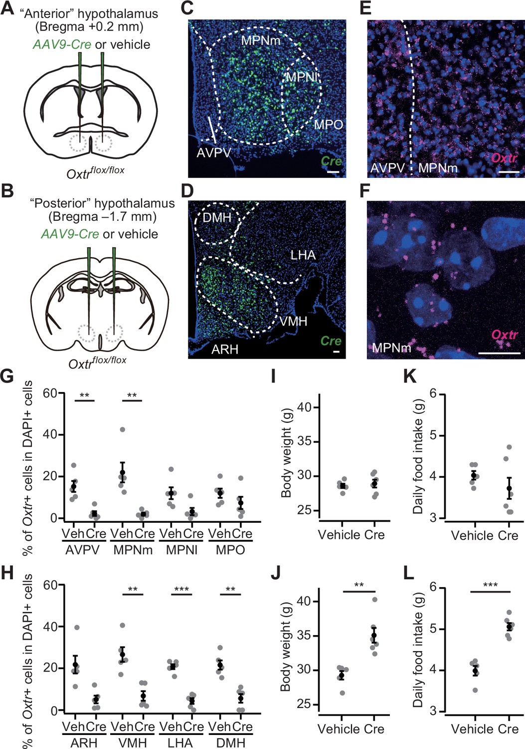
Oxtr conditional knockout (cKO) in the posterior hypothalamus induces increases in body weight and food intake.
(A, B) Schematic of the virus injection. AAV-Cre or vehicle was injected into the bilateral anterior or posterior hypothalamus (see Materials and methods) of Oxtrflox/flox male mice. (C, D) Representative coronal section showing Cre mRNA (green). Blue, DAPI. Scale bar, 50 μm. (E) A representative coronal section showing anteroventral periventricular nucleus (AVPV) and medial preoptic nucleus medial part (MPNm) from a vehicle-injected mouse. Oxtr mRNA was visualized by RNAscope (magenta). Blue, DAPI. Scale bar, 30 μm. (F) Projection of a confocal stack in MPNm from a vehicle-injected mouse. Magenta, Oxtr mRNA. Blue, DAPI. Scale bar, 5 μm. (G, H) Fraction of DAPI+ cells expressing Oxtr. Cells showing three or more RNAscope dots were defined as Oxtr+ (Figure 5—figure supplement 2). Veh, vehicle. N=5 each. **p<0.01, ***p<0.001, Student’s t-test with Bonferroni correction. Decreases in the MPN lateral part (MPNl), medial preoptic area (MPO), and arcuate hypothalamic nucleus (ARH) on average were found in AAV-Cre-injected mice but did not reach the level of statistical significance in Student’s t-test with Bonferroni correction (p=0.045, 0.289, and 0.012, respectively). (I, J) Body weight measured at 5 weeks after the injection. **p<0.01, Student’s t-test. Anterior hypothalamus, N=5 and 6 for vehicle and Cre, respectively, and posterior hypothalamus, N=5 and 6 for vehicle and Cre, respectively. (K, L) Daily food intake measured at 5 weeks after the injection. ***p<0.001, Student’s t-test. Anterior hypothalamus, N=5 and 6 for vehicle and Cre, respectively, and posterior hypothalamus, N=5 and 6 for vehicle and Cre, respectively. Error bars, SEM.
Figure 5—figure supplement 1
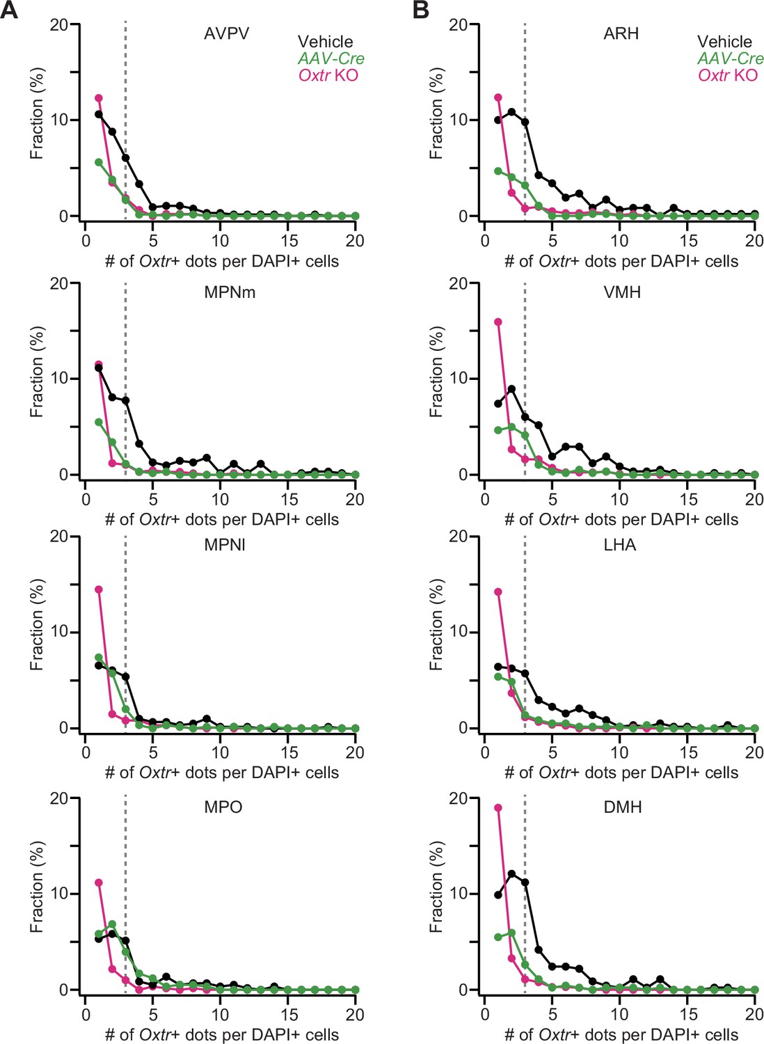
Distribution of oxytocin receptor (Oxtr)+ RNAscope dots.
(A, B) Fraction of the number of Oxtr+ RNAscope dots in each DAPI+ cell in the nuclei in the anterior hypothalamus (A) or posterior hypothalamus (B). Black and green represent vehicle and AAV-Cre injected into the anterior hypothalamus (A) or posterior hypothalamus (B) in Oxtrflox/flox mice, respectively. Data from the same mice shown in Figure 5G and H. Magenta represents Oxtr knockout (KO) mice (N=3 and 5 for anterior and posterior, respectively). Dotted lines indicate three Oxtr+ dots in each DAPI+ cell.
Figure 5—figure supplement 2
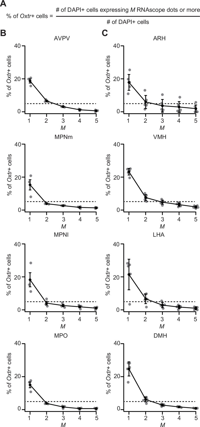
Testing the stringency in the definition of oxytocin receptor (Oxtr)+ cell by different numbers of RNAscope dots.
(A) Calculation of the fraction of Oxtr+ cells as defined in this equation: we counted the number of RNAscope dots in each DAPI+ cell and defined a cell with M or more dots as an Oxtr-expressing cell in this figure. (B, C) Because we observed RNAscope dots in the Oxtr knockout (KO) mice in which no Oxtr mRNA should be expressed, we evaluated the fraction of pseudo-positive detection with varying M values. Relationship between M and the fraction of Oxtr+ cells in the anterior hypothalamus (B) and posterior hypothalamus (C). Given that the pseudo-positive ratio on average was less than 5% (dotted line) in M≥3, we defined a DAPI+ cell with three or more dots as an Oxtr+ cell in Figures 5 and 6. N=3 and 5 mice for anterior and posterior, respectively.
Error bars, SEM.
Figure 6
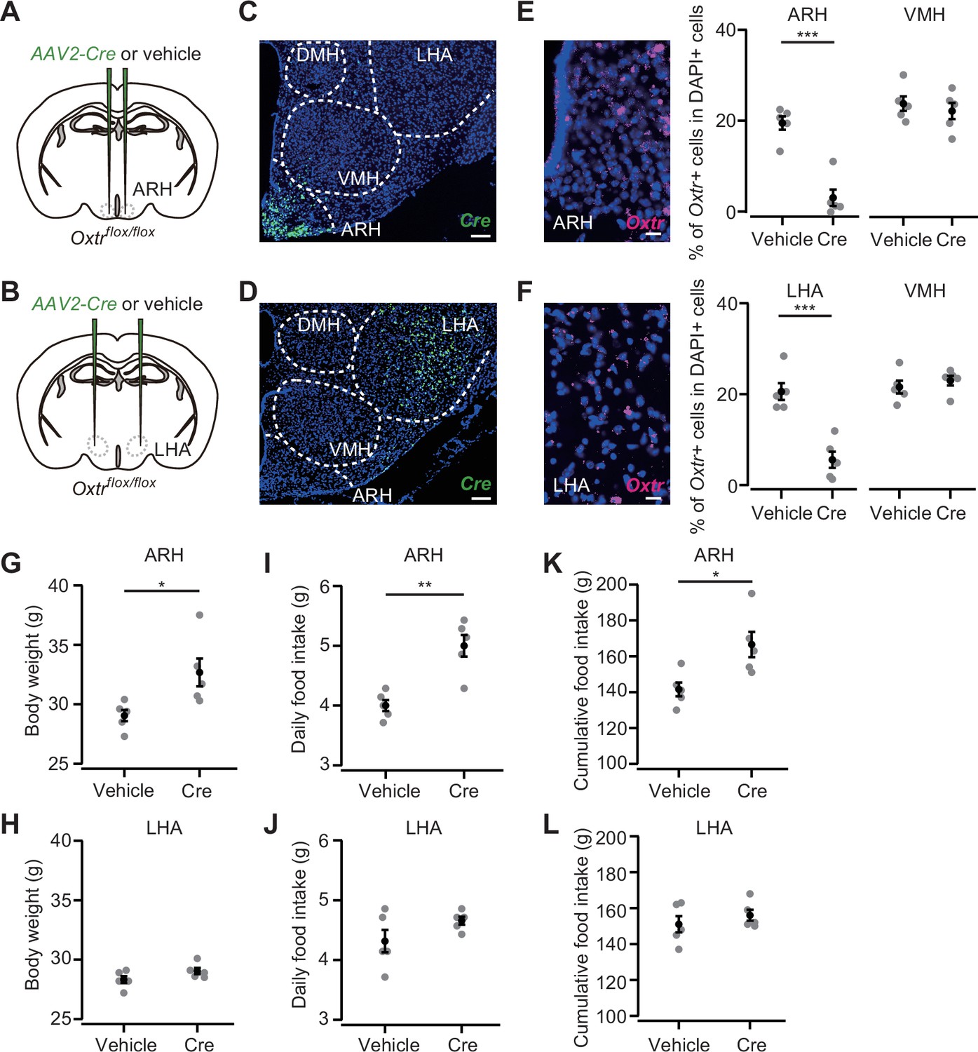
Oxytocin receptor (Oxtr) expression in the arcuate hypothalamic nucleus (ARH) suppresses body weight and food intake.
(A, B) Schematic of the virus injection. AAV-Cre or vehicle was injected into the bilateral ARH or lateral hypothalamic area (LHA) of Oxtrflox/flox male mice. (C, D) Representative coronal section showing Cre mRNA (green). Blue, DAPI. Scale bar, 50 μm. (E, F) Left, representative coronal section showing the ARH or LHA from a vehicle-injected mouse. Oxtr mRNA was visualized by RNAscope (magenta). Blue, DAPI. Scale bar, 5 μm. Right, fraction of DAPI+ cells expressing Oxtr in the ARH (E) or LHA (F) and ventromedial hypothalamic nucleus (VMH), a neighboring nucleus of the ARH and LHA. Cells showing three or more RNAscope dots were defined as Oxtr+. N=5 each. ***p<0.001, Student’s t-test with Bonferroni correction. (G, H) Body weight measured at 5 weeks after the injection. *p<0.05, Student’s t-test. N=5 each. (I, J) Daily food intake measured at 5 weeks after the injection. **p<0.01, Student’s t-test. N=5 each. (K, L) Cumulative food intake during the 5 weeks after the injection. *p<0.05, Student’s t-test. N=5 each.
Error bars, SEM.
Tables
Key resources table
| Reagent type (species) or resource | Designation | Source or reference | Identifiers | Additional information |
|---|---|---|---|---|
| Strain, strain background (mouse, male) | Oxt KO | Inada et al., 2022 | #CDB0204E | |
| Strain, strain background (mouse, male) | Oxt cKO (floxed) | Inada et al., 2022 | #CDB0116E | |
| Strain, strain background (mouse, male) | Oxtrflox/flox | Takayanagi et al., 2005 | ||
| Recombinant DNA reagent | AAV9-hSyn-Cre | Addgene | RRID:Addgene_105555-AAV9 | |
| Recombinant DNA reagent | AAV2-CMV-Cre-GFP | University of North Carolina viral core | https://www.med.unc.edu/genetherapy/vectorcore/in- stock-aav-vectors/reporter-vectors/ | |
| Commercial assay or kit | RNAscope Multiplex Fluorescent Reagent Kit | Advance Cell Diagnostics | 323110 | |
| Commercial assay or kit | RNAscope Mm-OXTR | Advance Cell Diagnostics | 412171 | |
| Software, algorithm | Igor Pro | Wavemetrics | RRID: SCR_000325 | |
| Software, algorithm | ImageJ | NIH | RRID: SCR_003070 |
Additional files
Download links
A two-part list of links to download the article, or parts of the article, in various formats.
Downloads (link to download the article as PDF)
Open citations (links to open the citations from this article in various online reference manager services)
Cite this article (links to download the citations from this article in formats compatible with various reference manager tools)
Oxytocin signaling in the posterior hypothalamus prevents hyperphagic obesity in mice
eLife 11:e75718.
https://doi.org/10.7554/eLife.75718


