Cohesin-independent STAG proteins interact with RNA and R-loops and promote complex loading
- Research Department of Cancer Biology, Cancer Institute, University College London, United Kingdom
- Centre for Genomic Regulation (CRG), Barcelona Institute of Science and Technology, Spain
- Regulatory Genomics Group, Cancer Institute, University College London, United Kingdom
- Proteomics Research Translational Technology Platform, Cancer Institute, University College London, United Kingdom
- Universitat Pompeu Fabra (UPF), Spain
- Institució Catalana de Recerca i Estudis Avançats (ICREA), Spain
Figures
Figure 1 with 1 supplement
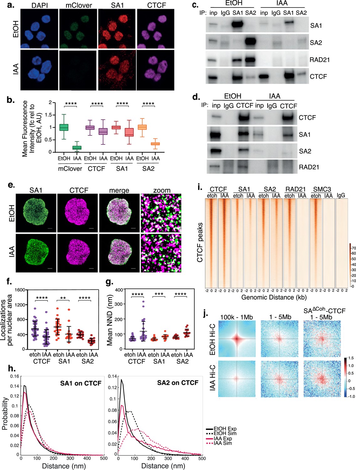
SA interacts with CTCF in the absence of cohesin.
(a) Representative confocal images of SA1 and CTCF IF in RAD21mAC cells treated with ethanol (EtOH) as a control or Auxin (IAA) for 4 hr. Nuclei were counterstained with DAPI. (b) Imaris quantification of the relative mean fluorescence intensity (MFI) of mClover, CTCF, SA1 and SA2 in EtOH and IAA-treated RAD21mAC cells. Whiskers and boxes indicate all and 50% of values, respectively. Central line represents the median. Asterisks indicate a statistically significant difference as assessed using two-tailed t-test. **** p<0.0001. n>50 cells/condition from three biological replicates. Chromatin coIP of (c) SA1, SA2, and IgG with RAD21 and CTCF or (d) CTCF and IgG with RAD21, SA1, and SA2 in RAD21mAC cells treated with EtOH or IAA for 4 hr. Input represents (c) 2.5% and (d) 1.25% of the material used for immunoprecipitation. (e) Dual-color STORM images of SA1 (green) and CTCF (magenta) in EtOH and IAA-treated RAD21mAC cells. Representative full nuclei and zoomed nuclear areas are shown. Line denotes 2 microns and 200 nm for full nuclei and zoomed areas respectively. See figure supplements for SA2 STORM images. (f) Mean CTCF, SA1 and SA2 localization densities (localizations normalized with nuclear area) in EtOH and IAA-treated RAD21mAC cells (n = >30, >17, and>15 nuclei for CTCF, SA1, and SA2, respectively). Mean and SD are plotted, Mann Whitney test. ** p<0.005, *** p<0.0005, **** p<0.0001. (g) Mean Nearest Neighbor Distance (NND) of CTCF, SA1, and SA2 clusters in nanometers in EtOH and IAA-treated cells (n = >38, >14. and>23 nuclei for CTCF, SA1, and SA2, respectively). Mean and SD are plotted, Mann Whitney test. **** p<0.0001. (h) NND distribution plot of the distance between CTCF and SA1 (left panel) or SA2 (right panel) clusters in EtOH and IAA-treated cells. Experimental data are shown as continuous lines, random simulated data are displayed as dotted lines. (i) ChIP–seq deepTools heat map of CTCF, SA1, SA2, Rad21 and SMC3 binding profiles in control (EtOH) and IAA-treated RAD21mAC cells. Selected regions are bound by CTCF in control conditions. (j) Analysis of contact frequency hotspots from Hi-C libraries generated from EtOH-treated (top row) and IAA-treated (bottom row) RAD21mAC cells. Contact frequencies were calculated in two distance ranges of 100 kb – 1 Mb and 1–5 Mb. The last column includes contact frequencies specifically at SA-CTCFΔCoh binding sites.
-
Figure 1—source data 1
Original, unedited western blots corresponding to Figure 1.
- https://cdn.elifesciences.org/articles/79386/elife-79386-fig1-data1-v2.zip
Figure 1—figure supplement 1
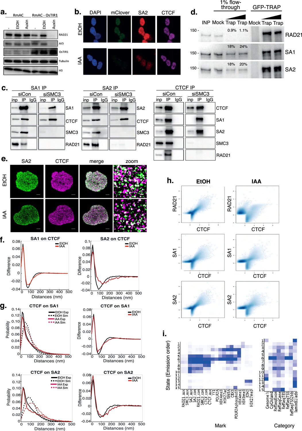
SA interacts with CTCF in the absence of cohesin.
(a) Immunoblot analysis of RAD21 levels in whole cell lysates from in RAD21mAC cells with and without CMV-OsTIR1 integration in the genome. Cells were untreated (-), treated with ethanol (EtOH) or with Auxin (IAA) for 4 hr. The IAA affect was also assessed with antibody to the AID tag. Tubulin and H3 serve as loading controls. (b) Representative confocal images of SA2 and CTCF IF in RAD21mAC cells treated with ethanol (EtOH) or Auxin (IAA) for 4 hr. Nuclei were counterstained with DAPI. (c) Chromatin coIP of SA1, SA2, and CTCF, together with Mock IgG controls in HeLa cells treated with siRNA to SMC3 for 48 hr. (d) GFP-TRAP in RAD21mAC cells immunoblotted for RAD21, SA1, and SA2. TRAP was performed with two concentrations of beads. Shown also is the % residual protein in the flow-through material (post-TRAP) relative to IP and as determined by ImageJ densitometry. (e) Dual-color STORM images of SA2 (green) and CTCF (magenta) in EtOH and IAA-treated RAD21mAC cells. Representative full nuclei and zoomed nuclear areas are shown. Line denotes 2 microns and 200 nm for full nuclei and zoomed areas respectively. (f) Nearest Neighbor Distance (NND) distribution plot of the distance difference between the experimental and random simulated data for SA1 (left) or SA2 (right) at CTCF localizations in EtOH (black) and IAA (red)-treated cells. (g) NND distribution plot of the distance between CTCF and SA1 (top left) or SA2 (bottom left) clusters in EtOH and IAA-treated cells. Experimental data are shown as continuous lines, random simulated data are displayed as dotted lines. Shown also are the NND distribution plots of the distance difference between the experimental and random simulated data for CTCF at SA1 (top right) or CTCF at SA2 (bottom right) localizations in EtOH (black) and IAA (red)-treated cells. (h) Pairwise comparisons of global CTCF ChIP-seq data (from merged biological replicates) compared to RAD21, SA1 and SA2 ChIP-seq in EtOH and IAA-treated RAD21mAC cells. (i) ChromHMM analysis of our ChIP-seq data as well as publicly available ChIP data in HCT116 cells as shown (Methods). Marks enriched within a given state (left) and enriched genomic features (right) are shown. We note that state 6 includes enrichments for ChIP data from RAD21 and SMC3 controls as well as SA1, SA2, CTCF in both control and IAA. This state is also enriched for active marks such as Polr2a, H3K4me3, and H3K27ac.
-
Figure 1—figure supplement 1—source data 1
Original, unedited western blots for Figure 1—figure supplement 1.
- https://cdn.elifesciences.org/articles/79386/elife-79386-fig1-figsupp1-data1-v2.zip
Figure 2 with 1 supplement
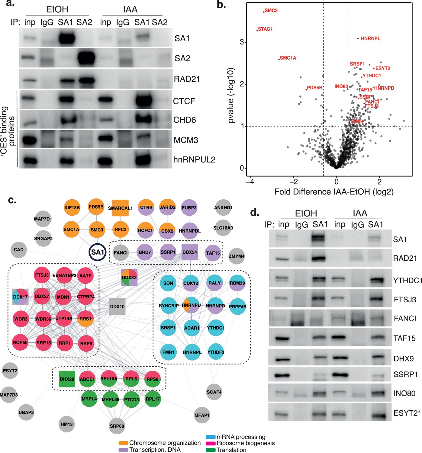
Characterization of SA1 protein-protein interaction network in RAD21-depleted cells.
(a) Chromatin coIP of SA1, SA2, and IgG with four predicted CES-binding proteins in RAD21mAC cells treated with EtOH or IAA for 4 hr. Input represents 1.25% of the material used for immunoprecipitation. (b) Volcano plot displaying the statistical significance (-log10 p-value) versus magnitude of change (log2 fold change) from SA1 IP-MS data produced from untreated or IAA-treated RAD21mAC cells (n=3). Vertical dashed lines represent changes of 1.5-fold. Horizontal dashed line represents a pvalue of 0.1. Cohesin complex members and validated high-confidence proteins have been highlighted. (c) SA1ΔCoh interaction network of protein–protein interactions identified in RAD21mAC cells using STRING. Node colors describe the major enriched categories, with squares denoting helicases. Proteins within each enrichment category were subset based on p-value change in B. See figure supplements for full network. Dashed boxes indicate the proteins and categories which were specifically enriched in IAA-treatment compared to the SA1 interactome. (d) Chromatin IP of SA1 and IgG in RAD21mAC cells treated with EtOH or IAA and immunoblotted with antibodies to validate the proteins identified by IP-MS. Input represents 1.25% of the material used for immunoprecipitation. * We note that ESYT2 is a F/YXF-motif containing protein.
-
Figure 2—source data 1
Original, unedited Western blots for Figure 2.
- https://cdn.elifesciences.org/articles/79386/elife-79386-fig2-data1-v2.zip
-
Figure 2—source data 2
MS stats values used in Figure 2.
- https://cdn.elifesciences.org/articles/79386/elife-79386-fig2-data2-v2.xlsx
Figure 2—figure supplement 1
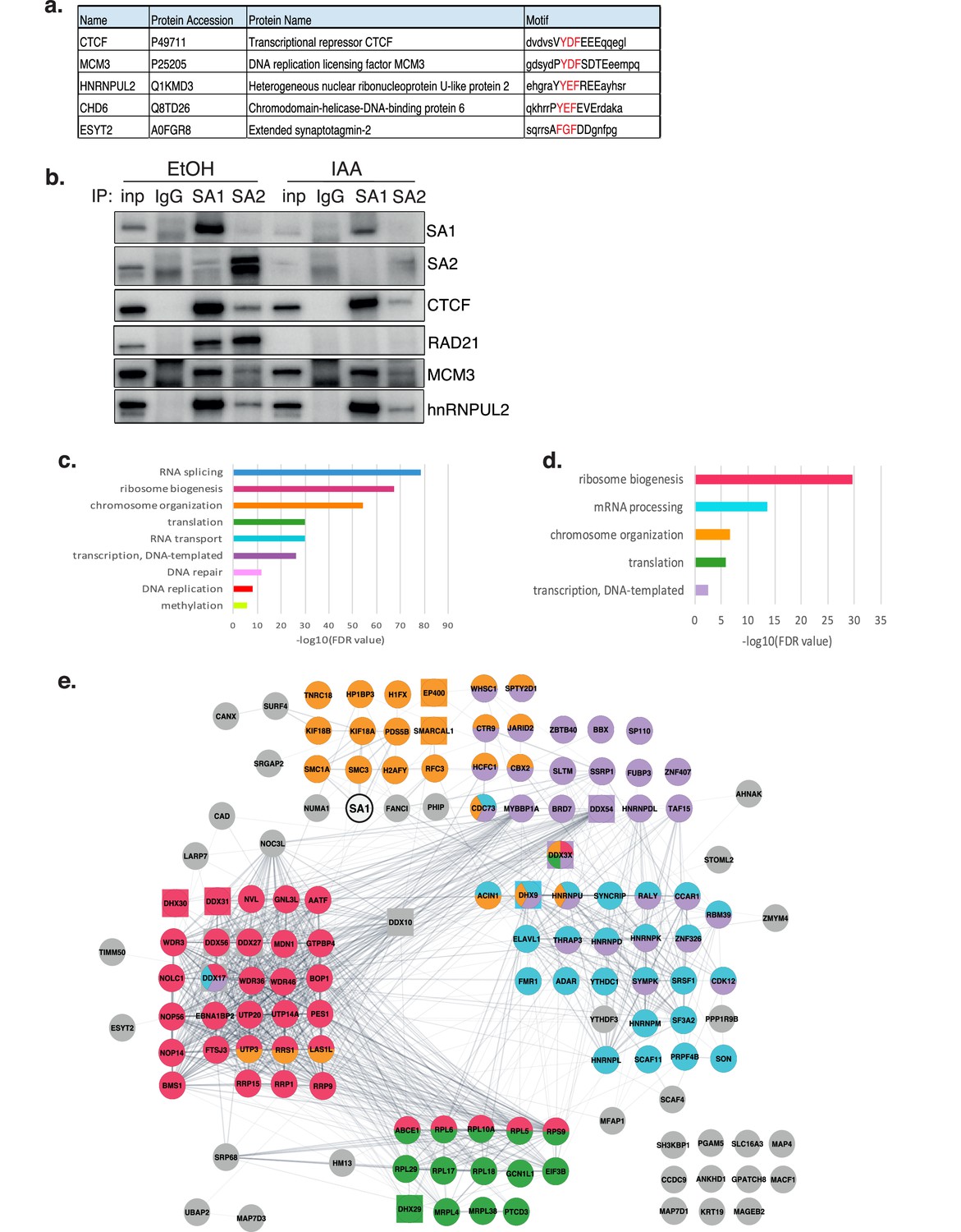
Characterization of SA1 protein-protein interaction network in RAD21-depleted cells.
(a) SlimSearch motifs for the shown ‘CES’ binding proteins. The CES-binding motif is capitalized and the F/YXF-motif within is highlighted in red. (b) Replicate chromatin coIP of SA1, SA2, and IgG with predicted CES-binding proteins in RAD21mAC cells treated with EtOH or IAA for 4 hr. Input represents 1.25% of the material used for immunoprecipitation. Enriched biological processes for the (c) SA1 interactome and (d) SA1∆Coh interactome compared to the whole genome. (e) Full SA1ΔCoh interaction network of protein–protein interactions identified in RAD21mAC cells using STRING. Colors and annotations as in Figure 2c.
-
Figure 2—figure supplement 1—source data 1
Original, unedited western blots for Figure 2—figure supplement 1.
- https://cdn.elifesciences.org/articles/79386/elife-79386-fig2-figsupp1-data1-v2.zip
-
Figure 2—figure supplement 1—source data 2
Sequence motifs for proteins tested in Figure 2a.
- https://cdn.elifesciences.org/articles/79386/elife-79386-fig2-figsupp1-data2-v2.xlsx
Figure 3 with 1 supplement
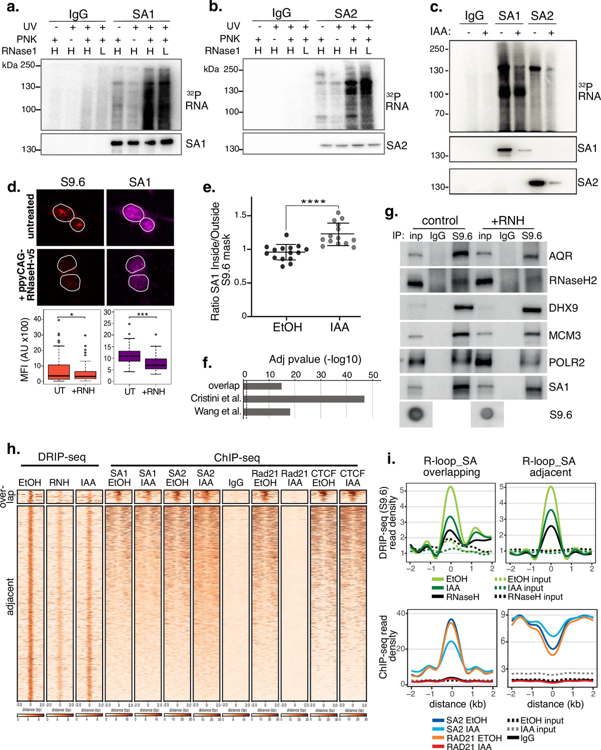
SA proteins bind to RNA and localize to R-loops.
CLIP for (a) SA1, (b) SA2 and IgG controls. Autoradiograms of crosslinked 32P-labeled RNA are shown at the top and the corresponding immunoblots, below. CLIP was performed with and without UV crosslinking and polynucleotide kinase (PNK) and with high (H; 1/50 dilution) or low (L; 1/500 dilution) concentrations of RNase I. (c) CLIP for (a) SA1, SA2 and IgG control in EtOH (-) or IAA-treated (+) Rad21mAC cells. 32P-labeled RNA and the corresponding immunoblots are shown as above. (d) Top, Representative confocal images of S9.6 and SA1 IF in RAD21mAC cells untreated or overexpressing ppyCAG-RNaseH-v5. Expressing cells were identified with v5 staining. Nuclear outlines (white) are derived from DAPI counterstain. Bottom, Imaris quantification of the relative mean fluorescence Intensity (MFI) of S9.6 and SA1. Data are from two biological replicates with >75 cells counted/condition. Quantifications and statistical analysis were done as above. (e) STORM analysis of localization density for SA1 in S9.6 masks in EtOH and IAA. Ratio of SA1 localizations inside and outside S9.6 masks is shown. Ratio of above 1 represents an enrichment within the S9.6 domain. Mean and SD are plotted, statistics based on One-Way Anova test. Data are from two biological replicates. (f) -log10 transformed adjusted p-value (FDR) for enrichment of S9.6 interactome data from Cristini et al. and Wang et al., with the SA1ΔCoh interactome. Overlap indicates the proteins identified in both of the S9.6 interactome datasets, representing a high confidence R-loop interactome list. (g) Chromatin coIP of S9.6 and IgG in RAD21mAC cells treated with RNase H and immunoblotted with antibodies representing known R-loop proteins, as well as SA1. Input represents 1.25% of the material used for immunoprecipitation. Bottom, S9.6 dot blot of lysates used in coIP. (h) deepTools heatmap of DRIP-seq and ChIP-seq from RAD21mAC cells. DRIP-seq was carried out in control (EtOH), RNase H (RNH), and IAA-treated cells. ChIP-seq was carried out for SA1, SA2, RAD21, CTCF, and IgG in EtOH and IAA-treated cells. Regions were selected based on DRIP-seq sensitivity to RNH and proximity with SA1 ChIP-seq. BEDTools identified regions of overlap or adjacent SA1 co-binding. (i) Summary plots showing mean DRIP-seq (top) or ChIP-seq (bottom) read density across the regions from (h), including sites of R-loop and SA ‘overlap’ (Left) or ‘adjacent’ (right) regions. Input samples are indicated with dotted lines.
-
Figure 3—source data 1
Original, unedited western blots for Figure 3.
- https://cdn.elifesciences.org/articles/79386/elife-79386-fig3-data1-v2.zip
Figure 3—figure supplement 1
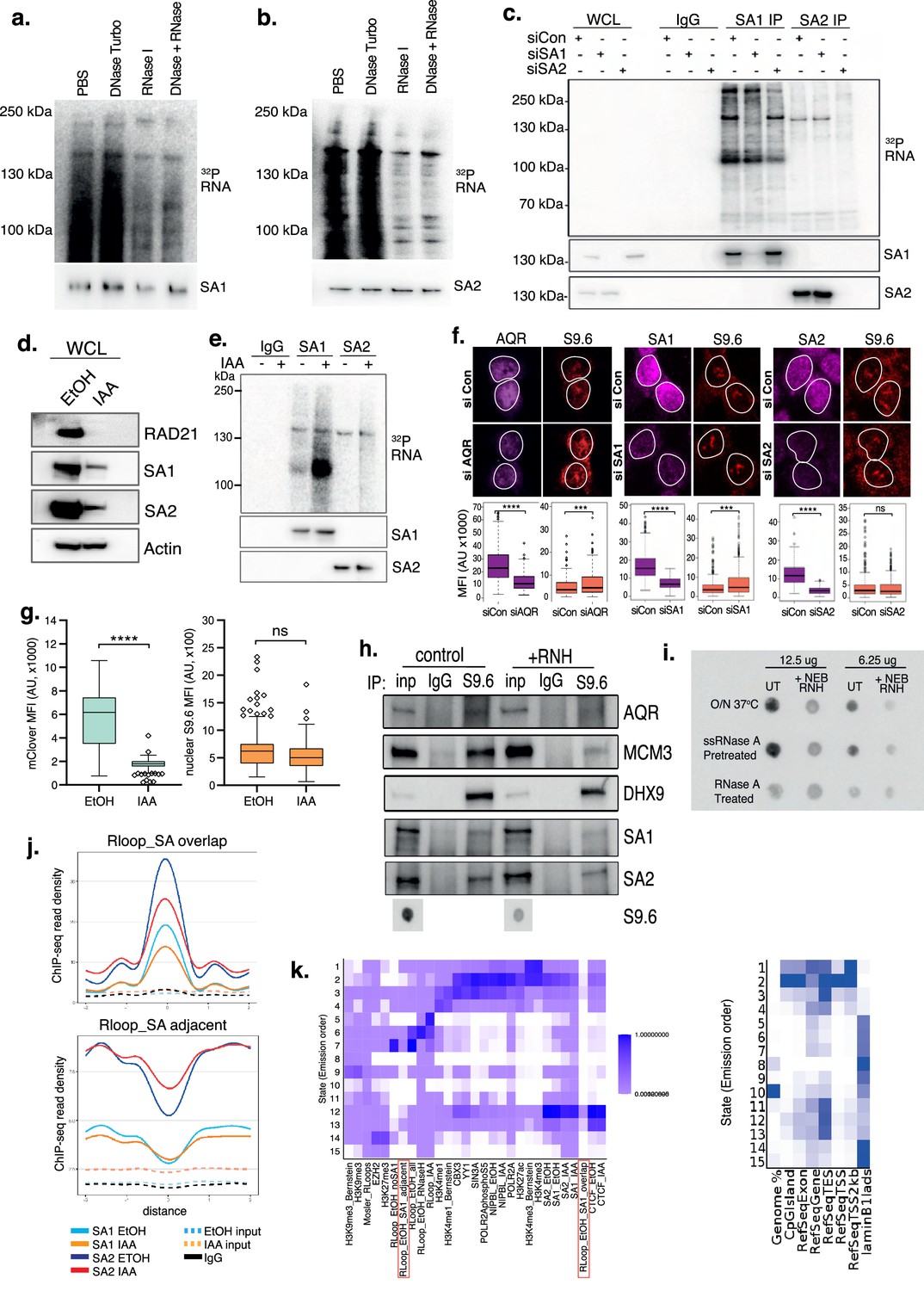
SA proteins bind to RNA and localize to R-loops.
CLIP for (a) SA1 and (b) SA2 treated with various controls as shown. Autoradiograms of crosslinked 32P-labeled RNA are shown at the top and the corresponding immunoblots, below. (c) CLIP for SA1, SA2, and IgG control in cells treated with siRNAs to scramble control (siscr), SA1 (siSA1), or SA2 (siSA2). 32P-labeled RNA and the corresponding immunoblots are shown as above. Input material (WCL) is included on the blots. Note the loss of the RNA signal upon siRNA KD. (d) Immunoblot analysis of RAD21, SA1 and SA2 levels in whole cell lysates (WCL) from RAD21mAC cells treated with EtOH or IAA. Actin serves as a loading control. (e) CLIP for SA1, SA2, and IgG control in EtOH (-) or IAA-treated (+) samples from (d). 32P-labeled RNA and the corresponding immunoblots are shown as above. Note SA1 and SA2 samples were loaded to show proportional amounts of the proteins in the IAA condition. (f) Representative confocal images of S9.6 and AQR (leftmost panel), SA1 (middle panel), and SA2 (rightmost panel) IF in RAD21mAC cells treated with scramble control siRNA (si Con) or siRNA to the protein of interest. Nuclear outlines (white) are derived from DAPI counterstain. Mean fluorescence Intensity (MFI) shown below images. Data are from three biological replicates with >50 cells counted/condition. Quantifications and statistical analysis were done as previously stated. (g) Imaris quantification of (left) mClover or (right) S9.6 signal by IF in RAD21mAC cells treated with EtOH or IAA for 4 hr. Data are from three biological replicates with >50 cells counted/condition. NB We detected a change in S9.6 signal upon IAA treatment by IF; however, this did not reach statistical significance. Quantifications and statistical analysis were done as indicated previously. (h) Replicate chromatin coIP of S9.6 and IgG in RAD21mAC cells treated with RNase H enzyme (RNH) and immunoblotted with antibodies representing known R-loop proteins and both SA1 and SA2. Input represents 1.25% of the material used for immunoprecipitation. Bottom, S9.6 dot blot of lysates used in coIP. (i) Representative S9.6 dot blot of two dilutions of chromatin samples used in the S9.6 coIPs and treated with enzymes as shown. A positive control for the digestion of RNA:DNA hybrids was included with a high concentration and temperature global RNase A digestion sample. (j) Summary plots showing mean ChIP-seq read density across the regions from Figure 3h including the SA1 ChIP-seq data.(k) ChromHMM analysis as in Figure 1—figure supplement 1i and now including our DRIP-seq data from HCT116 cells. The ‘R-loop_SA overlap’ and ‘R-loop_SA adjacent’ sites are marked in red. Marks enriched within a given state (left) and enriched genomic features (right) are shown. We note that ‘R-loop_SA overlap’ sites enrich genes and active marks such as Polr2a, H3K4me3, and H3K27ac in states 2/3/12 while the ‘R-loop_SA adjacent’ sites cluster separately with repressive chromatin marks such as Lamin B1 LADs in states 7/8.
-
Figure 3—figure supplement 1—source data 1
Original, unedited western blots for Figure 3—figure supplement 1.
- https://cdn.elifesciences.org/articles/79386/elife-79386-fig3-figsupp1-data1-v2.zip
Figure 4 with 1 supplement
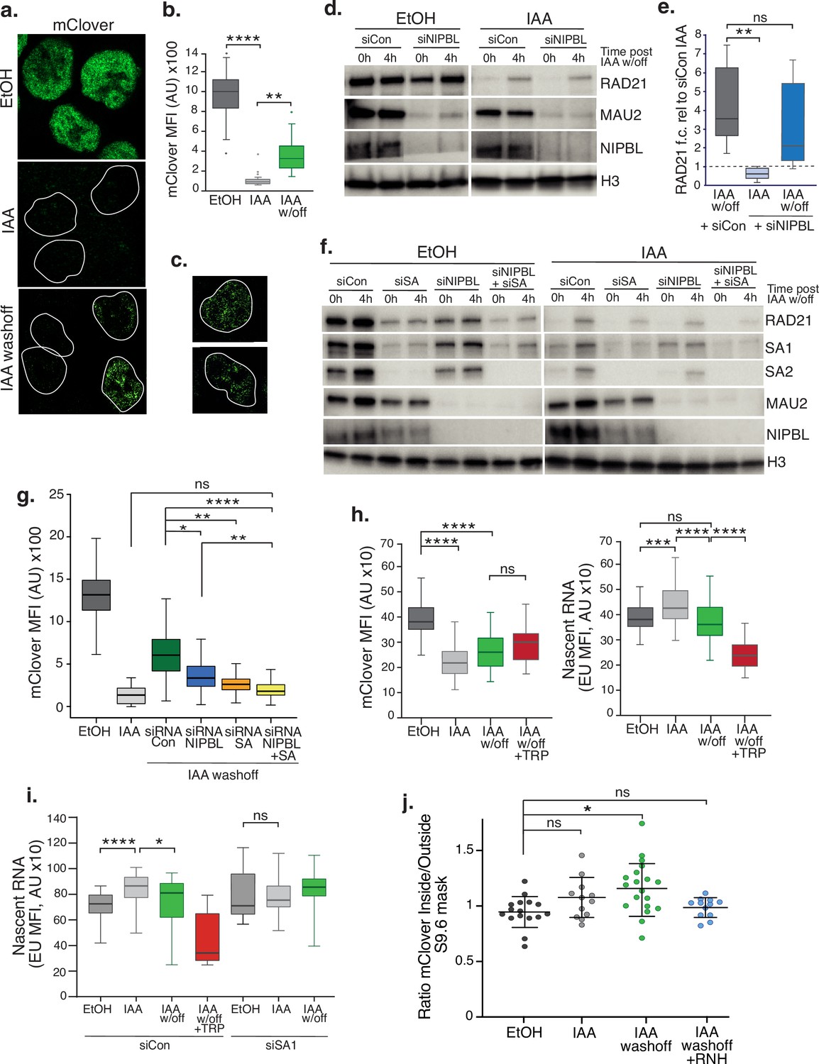
SA proteins contribute to cohesin loading.
(a) Representative confocal images of immunofluorescence for mClover in EtOH, IAA and IAA washoff conditions. White lines denote nuclei based on DAPI staining. (b) Imaris quantification of the mean fluorescence intensity (MFI) of mClover in EtOH, IAA-treated and IAA washoff RAD21mAC cells. Analysis and statistics as before. n>50 cells/condition from two biological replicates. (c) Examples of individual cells 4 hr post IAA washoff showing different distributions of mClover signal within the nucleus. White lines denote nuclei based on DAPI staining. (d) Representative immunoblot analysis of chromatin-bound RAD21, MAU2 and NIPBL levels in RAD21mAC cells treated with scramble control siRNA (si Con) or siRNA to NIPBL followed by EtOH or IAA treatment. 0 h and 4 h represent no wash-off of IAA or a sample taken 4 hr after washoff of IAA (the ‘reloading timepoint). H3 was used as a loading control. NB The full blots are in Figure 4—figure supplement 1c. (e) Western blot densitometry quantification. RAD21 fold change relative to siCon samples at the 0 h timepoint in siCon 4 h (grey), siNIPBL 0 hr (light blue), and siNIPBL 4 hr (dark blue). Whiskers and boxes indicate all and 50% of values, respectively. Central line represents the median. Statistical analysis as assessed using a two-tailed t-test. Data is from eight biological replicates. (f) Representative immunoblot analysis of chromatin-bound RAD21, SA1, SA2, MAU2, and NIPBL levels in RAD21mAC cells treated according to the schematic described in Figure 4—figure supplement 1b and including samples treated with siRNA to SA1 and SA2 together (siSA) and siRNA to NIPBL +siSA. H3 was used as a loading control. Quantifications can be seen in in Figure 4—figure supplement 1d. (g) Imaris quantification of the mClover MFI in EtOH, IAA-treated and IAA washoff RAD21mAC cells treated with siRNA to NIPBL, SA1/2 and siRNA to NIPBL +siSA. Asterisks indicate a statistically significant difference as assessed using two-tailed T-test. Data is from two biological replicates with >50 cells per experiment. (h) Imaris quantification of the mClover MFI (left) and RNA (based on EU incorporation (right) in EtOH and IAA-treated RAD21mAC cells. Whiskers and boxes indicate all and 50% of values, respectively. Central line represents the median. Asterisks indicate a statistically significant difference as assessed using one-way ANOVA. n>50 cells/condition from two biological replicates.(i) Analysis of mClover MFI as in h) above, this time treated with siRNA to SA1/2 or scrambled controls. Analysis and statistics as above. n>50 cells/condition from two biological replicates. (j) STORM analysis of localization density for RAD21-mClover in S9.6 masks in EtOH (black), IAA (grey), and IAA washoff (green) conditions. Ratio of mClover localizations inside and outside S9.6 masks is shown. Ratio of above 1 represents an enrichment within the S9.6 domain. Mean and SD are plotted, statistics based on One-Way ANOVA test. Data are from two biological replicates.
-
Figure 4—source data 1
Original, unedited western blots for Figure 4.
- https://cdn.elifesciences.org/articles/79386/elife-79386-fig4-data1-v2.zip
Figure 4—figure supplement 1
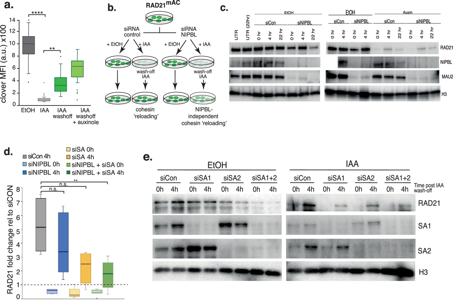
SA proteins contribute to cohesin loading.
(a) mClover MFI shown as in Figure 4b and including a IAA-washoff sample treated with auxinole (an Auxin inhibitor) which leads to increased recovery of mClover signal in cells.(b) Schematic of experimental set-up. RAD21mAC cells expressing mClover (green cells in dishes) were treated with scramble siRNAs or siRNA to NIPBL. Prior to collection, cells were cultured in EtOH or IAA for 4 hr to degrade RAD21 (0 h timepoints). The EtOH or IAA treatment was washed-off and the cells were left to recover for 4 hr (4 h timepoints). Chromatin fractions were prepared from all samples and used in immunoblot analysis. (c) Full immunoblot analysis of the data shown in main Figure 4d. Chromatin-bound RAD21, NIPBL, and MAU2 levels in RAD21mAC cells treated according to the schematic shown in Figure 4—figure supplement 1b as well as a longer timepoint of ‘re-loading’ (22 hr, which we found to be too stressful to cells). H3 was used as a loading control. (d) Quantification of the RAD21 fold change relative to siCon samples at the 0 hr timepoint in siCon, siNIPBL, siSA, and siNIPBL +siSA from immunoblot analysis of chromatin-bound RAD21 in main Figure 4f. Asterisks indicate a statistically significant difference as assessed using two-tailed T-test. p-Values as before. Data is from five biological replicates. (e) Immunoblot analysis of chromatin-bound RAD21, SA1, SA2, and H3 levels in RAD21mAC cells treated according to the schematic shown in Figure 4—figure supplement 1c and including samples treated with siRNA to SA1, SA2, and SA1 and SA2 together. H3 was used as a loading control.
-
Figure 4—figure supplement 1—source data 1
Original, unedited western blots for Figure 4—figure supplement 1.
- https://cdn.elifesciences.org/articles/79386/elife-79386-fig4-figsupp1-data1-v2.zip
Figure 5 with 1 supplement
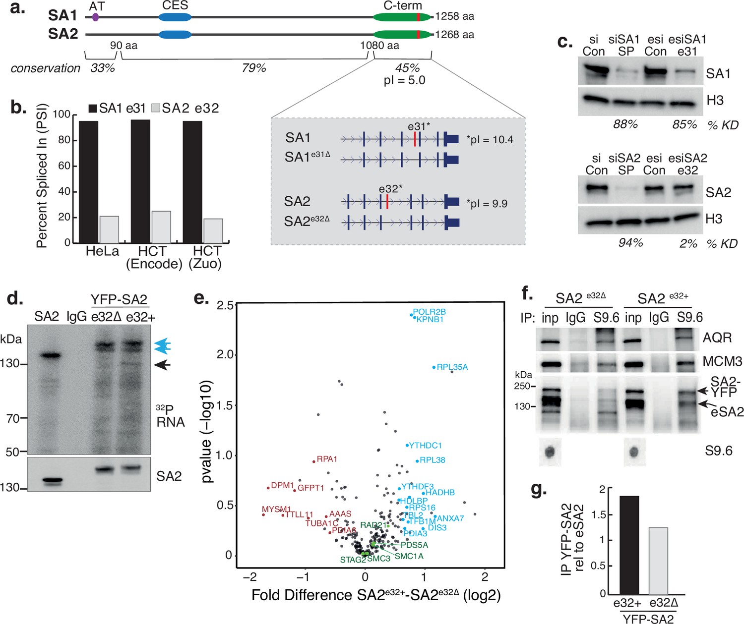
A basic exon in SA2 influences RBP stability.
(a) Schematic of the SA1 and SA2 proteins showing the SA1-specific AT-hook, the conserved CES domain (blue) and the acidic C-terminus (green) which contains the basic alternatively spliced exon (red). Right-hand zoom-in indicates the spliced exons for SA1 (top) and SA2 (bottom) and the pI for each. The conservation scores for the divergent N- and C-termini and the middle portion of the proteins which contains the CES domain are shown. (b) Percent Spliced In (PSI) calculations for SA1 exon 31 (black) and SA2 exon 32 (grey) based on VAST-Tools analysis of RNA-seq from multiple datasets (see Methods). (c) (top) Immunoblot analysis of SA1 levels in chromatin lysates after treatment with scrambled siRNAs (siCon), SmartPool SA1 siRNAs (siSA1 SP), control esiRNAs (esiCon) and esiRNA designed to target SA1 exon 31 for 48 hr in RAD21mAC cells. (bottom) Immunoblot analysis of SA2 levels in chromatin lysates after treatment with scrambled siRNAs (siCon), SmartPool SA2 siRNAs (siSA2 SP), control esiRNAs (esiCon) and siRNA designed to target SA2 exon 32 for 48 hr in RAD21mAC cells. H3 serves as a loading control. The percentage of knockdown (KD) after SA signal is normalized to H3 is shown. (d) CLIP for endogenous SA2 and IgG control in cells in which either YFP-tagged SA2 containing exon 32 (e32+) or YFP-tagged SA2 lacking exon 32 were expressed for 48 hrs. CLIP reveals RNA associated with SA2 (blue arrows) and RBPs which specifically associate with exon-32 containing SA2 (black arrow). (e) Volcano plot displaying the statistical significance (-log10 p-value) versus magnitude of change (log2 fold change) from IP-MS of HCT116 cells expressing either YFP-SA2e32+ or YFP-SA2e32∆ (n=3 biological replicate IP). Cohesin complex members are highlighted in green and the two most enriched functional categories of RNA-binding proteins in blue or Post-translational modification in red. (f) Chromatin coIP of S9.6 and IgG in RAD21mAC cells expressing the YFP-SA2 isoforms and immunoblotted with antibodies representing known R-loop proteins, as well as endogenous SA2 (eSA2). Input represents 1.25% of the material used for immunoprecipitation. Bottom, S9.6 dot blot of lysates used in coIP. NB the shift in SA2 signal representing overexpressed protein (SA2-YFP). (g) Quantification of the immunoblot signal from (f) of SA2 in the YFP-SA2 isoform band relative to input and to eSA2 signal. YFP-SA2e32+ is more enriched by S9.6 IP compared to YFP-SA2e32∆.
-
Figure 5—source data 1
Original, unedited western blots for Figure 5.
- https://cdn.elifesciences.org/articles/79386/elife-79386-fig5-data1-v2.zip
-
Figure 5—source data 2
MS stats values used in Figure 5.
- https://cdn.elifesciences.org/articles/79386/elife-79386-fig5-data2-v2.xlsx
Figure 5—figure supplement 1
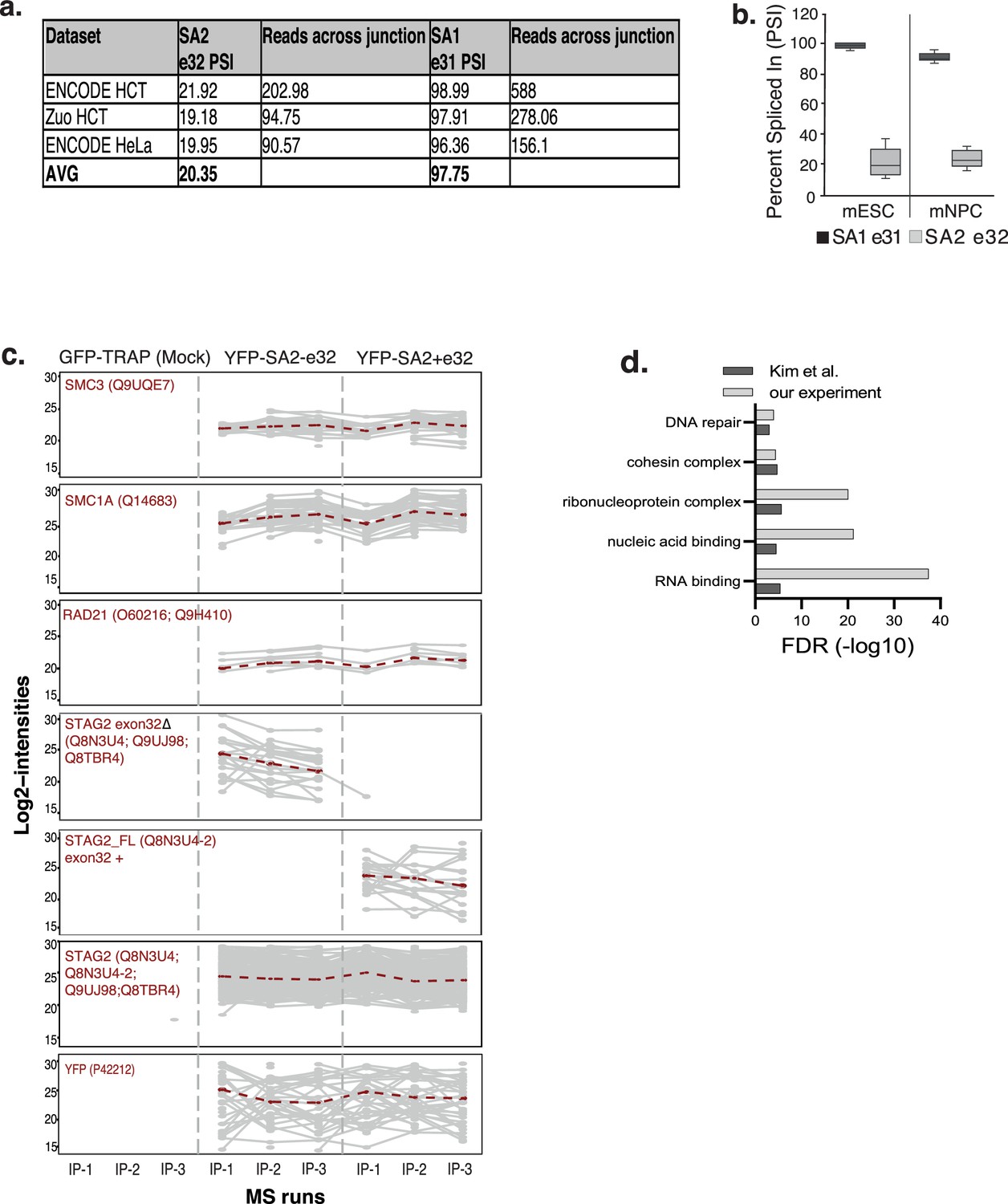
A basic exon in SA2 influences RBP stability.
(a) Percent Spliced In (PSI) calculations for SA1 exon 31 and SA2 exon 32 based on VAST-Tools analysis of RNA-seq in three separate datasets and the average (AVG) reported in the manuscript. Reads crossing exon junctions from each sample are also shown. (b) PSI distributions for SA1 exon 31 and SA2 exon 32 based on VAST-Tools analysis of RNA-seq from mouse ESCs and neural progenitors (NPC). (c) Peptide profile plots from YFP-SA2 overexpression CLIP-MS experiment from Figure 5e. Shown are the Log2 peptide intensities for each protein in each biological replicate IP from GFP-TRAP alone (mock IP), YFP-SA2-e32 and YFP-SA2-FL. Core cohesin components, YFP and STAG2 are robustly IP’d in all samples. (d) Enriched biological processes for the YFP-SA2 interactome compared to published SA2 mass spec study in Kim et al. NB. RNA-binding proteins are enriched in both.
-
Figure 5—figure supplement 1—source data 1
Excel document of the PSI values for Figure 5—figure supplement 1.
- https://cdn.elifesciences.org/articles/79386/elife-79386-fig5-figsupp1-data1-v2.xlsx
-
Figure 5—figure supplement 1—source data 2
Excel document of intensities of cohesin proteins for Figure 5—figure supplement 1c.
- https://cdn.elifesciences.org/articles/79386/elife-79386-fig5-figsupp1-data2-v2.xlsx
Author response image 1
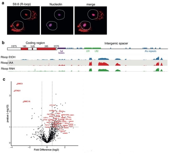
(a) RAD21mAC cells stained for S9.
6 representing R-loops and Nucleolin. White line denotes the nucleus based on DAPI. NB. the expected nucleolar R-loop signal. (b) Top, cartoon of the consensus hg19 ribosomal DNA (rDNA), showing the ribosomal genes and the intergenic spacer (IGS) region which contains several Alu elements (blue). Bottom, S9.6 DRIP-seq from RAD21mAC cells treated with EtOH, IAA and RNaseH (RNH) and aligned to the consensus rDNA. NB. R-loops within the coding region are sensitive to RNH and IAA while those within the IGS are less affected. (C) Volcano plot displaying the statistical significance (-log10 p-value) versus magnitude of change (log2 fold change) from SA1 IPMS data produced from EtOH or IAA-treated RAD21mAC cells. Data as in Figure 2b from the manuscript except the proteins that are part of the enriched ‘Ribosome biogenesis’ functional category are highlighted in red.
Tables
Table 1
Published datasets used in this study.
| Accession no. | Analysis description | Publication DOI or Ref | Figure Reference |
|---|---|---|---|
| GSE104334 | Long-range contact analysis of Hi-C datasets | 10.1016 /j.cell.2017.09.026 | 1j |
| GSE89729 | Percent Spliced In (PSI) analysis of RNA-seq datasets | 10.1172/jci.insight.91419 | 5d, “HCT Zuo” |
| GSM958749 | Percent Spliced In (PSI) analysis of RNA-seq datasets | ENCODE HCT116 RNAseq | 5b, “HCT ENCODE” |
| GSM958735 | Percent Spliced In (PSI) analysis of RNA-seq datasets | ENCODE HeLa RNAseq | 5b, “HeLa” |
Table 2
siRNAs used in this study.
| siRNA name | Company | Target | Catalogue no. | custom siRNA sequence |
|---|---|---|---|---|
| si scramble control | Dharmacon | Smartpool | D-001810-10-05 | |
| siSA1 | Dharmacon | Smartpool | L-010638-01-0010 | |
| siSA2 | Dharmacon | Smartpool | L-021351-00-0010 | |
| siNIPBL | Dharmacon | Smartpool | L-012980-00-0010 | |
| siAQR | Dharmacon | Smartpool | L-022214-01-0005 | |
| esi control | Sigma | Luciferase | EHUFLUC | |
| esi SA1 | Sigma | exon 31 | custom esiRNA | TCCTCAGATGCAGATCTCTTGGTTAGGCC AGCCGAAGTTAGAAGACTTAAATCGGAAG GACAGAACAGGAATGAACTACATGAAAGTG AGAACTGGAGTGAGGCATGCTGT |
| esi SA2 | Sigma | exon 32 | custom esiRNA | CACGCAGGTAACATGGATGTTAGCTCAAAG ACAACAAGAGGAAGCAAGGCAACAGCAGG AGAGAGCAGCAATGAGCTATGTTAAACTG CGAACTAATCTTCAGCATGCCAT |
Table 3
Antibodies used in this study.
| Protein | Company | Catalogue No. | Species | Main Figure Reference |
|---|---|---|---|---|
| SA1 | Abcam | ab4455 | Mouse | 1 a, c, d, e, 2 a, b, d, 3 a, b, c, d, e, g, h 4f,g, 5b, c, f, h, i |
| SA1 | Abcam | ab4457 | Mouse | 1i |
| SA2 | Bethyl | A300-159 | Goat | 1b, c, d, , 2 a, 3 a, b, c, f, 4 g, e, f, 5 c, d, e, f, g |
| SA2 | Bethyl, AbVantage Pack | A310-941A | Goat | 1i |
| CTCF | Diagenode | C15410210 | Rabbit | 1 c, d, i, 2 a |
| CTCF | Cell signalling | 2899 s | Rabbit | 1 a, e |
| RAD21 | Abcam | ab992 | Rabbit | 1 c, d, i, 2 a, d, 4d, e, g, j |
| GFP-TRAP | ChromoTek | gtd-20 | 1i, 5f | |
| GFP | Invitrogen | A11122 | Rabbit | 1 a, e |
| mAID | MBL | M214-3 | Mouse | Figure 1—figure supplement 1a |
| OsTIR | MBL | PD048 | Rabbit | Figure 1—figure supplement 1a |
| SMC3 | Abcam | ab9263 | Rabbit | 1i |
| CHD6 | Bethyl | A301-221A | Rabbit | 2 a |
| MCM3 | Bethyl | A300-124A | Goat | 2 a, 3g, 5f |
| HNRNPUL2 | Abcam | ab195338 | Rabbit | 2 a |
| YTHDC1 | Abcam | ab122340 | Rabbit | 2d |
| FTSJ3 | Bethyl | A304-199A-M | Rabbit | 2d |
| FANCI | Bethyl | A301-254A-M | Rabbit | 2d |
| TAF15 | Abcam | ab134916 | Rabbit | 2d |
| DHX9 | Abcam | Ab26271 | Rabbit | 2d |
| SSRP1 | Abcam | ab26212 | Mouse | 2d |
| INO80 | Proteintech | 18810–1-AP | Rabbit | 2d |
| ESYT2 | Sigma-Aldrich | HPA002132 | Rabbit | 2d |
| S9.6 | Kerafast | ENH001 | Mouse | 3d, g, h, 5f |
| RNASE H2 | Novus | NBP1-76981 | Rabbit | 3g |
| AQR | Bethyl | A302-547A | Rabbit | 3g, 5f, e |
| POLR2 | Covance | MMS-1289 | Mouse | 3g |
| MAU2 | Abcam | ab183033 | Rabbit | 4d, f |
| NIPBL | Abbiotec | 250133 | Rat | 4d, f, |
| H3 | Abcam | ab1791 | Rabbit | 4d, f, 5c |
| Name(SecondaryAbs) | Fluorophore | Company | Catalogue No. | Figure Reference |
| Donkey anti-Rabbit | Cy3_AF647 | Home made from Jackson Immunoresearch IgG | Home made from 711-005-152 | 1e, Figure 1—figure supplement 1 |
| Donkey anti-Goat | AF405_AF647 | Home made from Jackson Immunoresearch IgG | Home made from 705-005-147 | 1e, Figure 1—figure supplement 1 |
| Donkey anti-mouse | AF647 | Invitrogen | A31570 | 1a, e, Figure 1—figure supplement 1b, e, Figure 3—figure supplement 1f |
| Donkey anti-rabbit | AF488 | Invitrogen | A21206 | 1a, e, Figure 1—figure supplement 1b, e, Figure 3—figure supplement 1f |
| Donkey anti-rabbit | AF647 | Invitrogen | A31573 | 1a, e, Figure 1—figure supplement 1b, e, Figure 3—figure supplement 1f |
| Donkey anti-goat | AF555 | Invitrogen | A21432 | 1a, e, Figure 1—figure supplement 1b, e, Figure 3—figure supplement 1f |
| Donkey anti-goat | AF647 | Invitrogen | A21447 | 1a, e, Figure 1—figure supplement 1b, e, Figure 3—figure supplement 1f |
| Goat anti-Mouse | AF568 | ThermoFisher Scientific | A-11031 | 1a, e, Figure 1—figure supplement 1b, e, Figure 3—figure supplement 1f |
| Goat anti-Rabbit | AF647 | ThermoFisher Scientific | A-21244 | 1a, e, Figure 1—figure supplement 1b, e, Figure 3—figure supplement 1f |
| Rabbit anti- Goat | AF647 | ThermoFisher Scientific | A-21446 |
Table 4
Published ChIP-seq datasets used for ChromHMM.
| Protein | Accession no. | Publication | Matched input |
|---|---|---|---|
| NIPBL (EtOH- and IAA-treated) | GSE104334 | Rao et al., 2017 | - |
| CBX1 | GSM1010758 | Gertz et al., 2013 | |
| EZH2 | GSM3498250 | Dunham et al., 2012 | GSM2308475;GSM2308476 |
| POLR2A | GSM935426 | Dunham et al., 2012 | GSM2308422 |
| POLR2AphosphoS5 | GSM803474 | Gertz et al., 2013 | GSM803475 |
| SIN3A | GSM1010905 | Gertz et al., 2013 | |
| YY1 | GSM803354 | Gertz et al., 2013 | GSM803475 |
| H3K4me1 | GSM945858 | GSM2308475; GSM2308476 | |
| H3K4me1 | GSM2527549 | Dunham et al., 2012 | GSM2308422 |
| H3K4me3 | GSM2533929 | Dunham et al., 2012 | GSM2308475; GSM2308476 |
| H3K4me3 | GSM945304 | Thurman et al., 2012 | GSM945287 |
| H3K9me3 | GSM2527565 | Dunham et al., 2012 | |
| H3K9me3 | GSM2308431 | Dunham et al., 2012 | |
| H3K27ac | GSM2534277 | Dunham et al., 2012 | GSM2308422 |
| H3K27me3 | GSM2308612 | Dunham et al., 2012 |
Additional files
Download links
A two-part list of links to download the article, or parts of the article, in various formats.
Downloads (link to download the article as PDF)
Open citations (links to open the citations from this article in various online reference manager services)
Cite this article (links to download the citations from this article in formats compatible with various reference manager tools)
Cohesin-independent STAG proteins interact with RNA and R-loops and promote complex loading
eLife 12:e79386.
https://doi.org/10.7554/eLife.79386


