Ectodermal Wnt signaling, cell fate determination, and polarity of the skate gill arch skeleton
- Department of Zoology, University of Cambridge, United Kingdom
- School of Biological Sciences, University of Aberdeen, United Kingdom
- The Babraham Institute, United Kingdom
- Department of Genetics, Rutgers University, United States
- Josephine Bay Paul Center for Comparative Molecular Biology and Evolution, Marine Biological Laboratory, United States
Figures
Figure 1
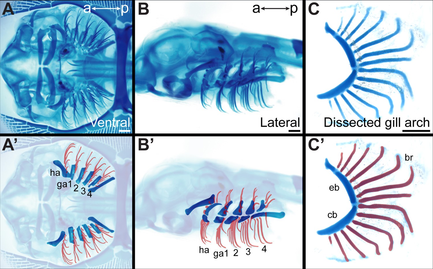
Overview of skate gill arch skeletal anatomy.
Skeletal preparation of an S33 skate embryo in (A) ventral and (B) lateral view. In (A’) and (B’) the gill arches and branchial rays are highlighted, with branchial rays false-colored red. Note that branchial rays project from the posterior margin of the hyoid and first four gill arches. (C) A dissected gill arch with the (C’) dorsal epibranchial and ventral ceratobranchial gill arch cartilages indicated, and with branchial rays false-colored red. a–p indicates anterior–posterior axis. br: branchial ray, cb: ceratobranchial, eb: epibranchial, ga1-4: gill arches 1–4, ha: hyoid arch, ma: mandibular arch. Scale bars: 1 mm.
Figure 2 with 1 supplement
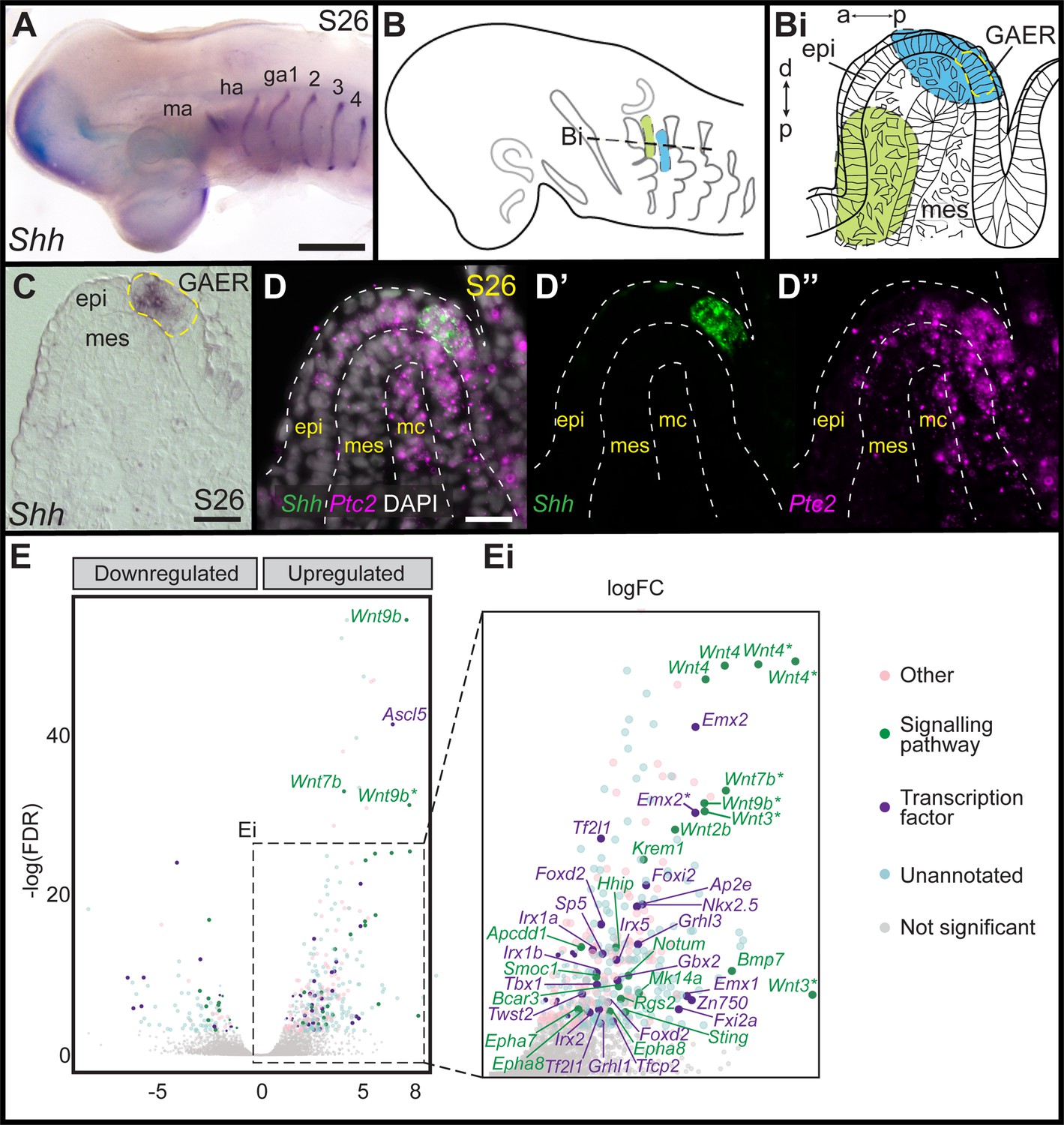
Differential gene expression analysis of gill arch epithelial ridge (GAER) and non-GAER gill arch tissues in S26 skate embryos.
(A) Sonic hedgehog (Shh) is expressed in the GAER of the hyoid and first four gill arches of skate embryos. (B) Schematic of dissection from gill arch one of (Bi) the non-GAER region (control, green) and the GAER region (blue, with GAER location indicated by yellow dashed outline), as guided by (C) mRNA in situ hybridization for Shh on paraffin section. (D) The dissected GAER region includes the Shh-expressing GAER, as well as adjacent Shh-responsive (i.e. Ptc2+) tissues, as indicated by multiplexed mRNA in situ hybridization by chain reaction (HCR) for Shh and Ptc2 on paraffin section. (E, Ei) Volcano plot illustrating differential gene expression between GAER and non-GAER tissues. Genes with >2 logFC and <0.05 false discovery rate (FDR) are highlighted and assigned functional categories using color coding as per key. Genes with >2 logFC are shown in larger point sizes. Signaling molecules and transcription factors with >2 logFC and <0.05 FDR are labeled (* denotes manual annotation of sequence). Differential expression was determined using edgeR with a general linear model and likelihood ratio test, corrected for multiple testing using the Benjamin-Hochberg method to control the FDR. a–p indicates anterior–posterior axis, p–d indicates proximal–distal axis. epi: epithelium, ga1-4: gill arches 1–4, ha: hyoid arch, ma: mandibular arch, mc: mesodermal core, mes: mesenchyme. Scale bars; A: 500 μm C-D: 50 μm.
Figure 2—figure supplement 1
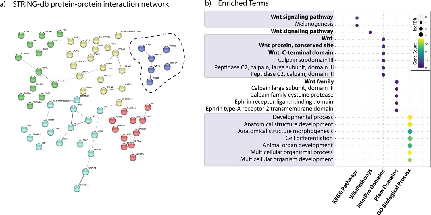
Genes significantly upregulated in gill arch epithelial ridge (GAER) vs. non-GAER gill arch tissues in S26 skate embryos.
(A) All significantly upregulated genes (logFC >2, FDR <0.05) tested for interaction using STRING db v11.5, with a cluster of genes encoding Wnt signaling components highlighted. (B) Most significantly enriched terms in GAER region tissues (GO process, KEGG Pathway, InterPro, Pfam, and WikiPathway screened).
Figure 3
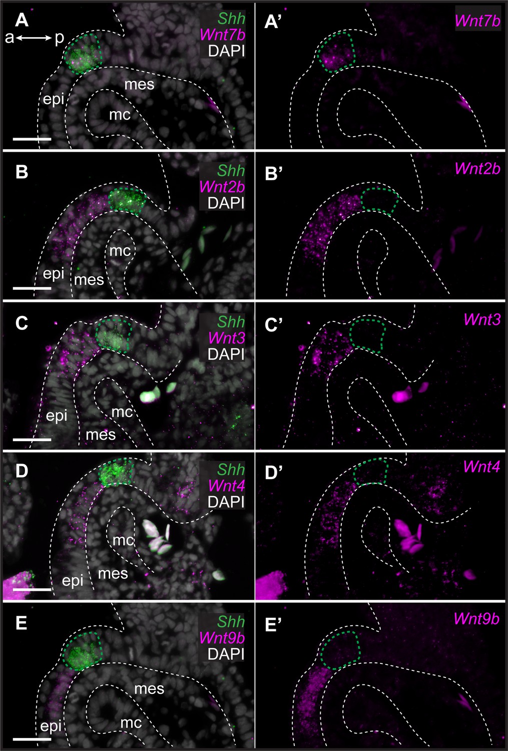
Genes encoding Wnt ligands are expressed in and around the gill arch epithelial ridge (GAER) in skate.
In S26 skates, (A, A’) Wnt7b is co-expressed with Sonic hedgehog (Shh) in the GAER. (B) Wnt2b, (C) Wnt3, (D) Wnt4 and (E), Wnt9b are predominantly expressed in the epithelium immediately adjacent to the GAER. For ease of visualization in A’–E’, Shh expression is not shown but GAER cells are outlined with a green dashed line. Arch tissues are outlined with white dashed lines. a–p indicates anterior–posterior axis. epi: epithelium, mes: mesenchyme, mc: mesodermal core. Scale bar: 50 μm.
Figure 4 with 1 supplement
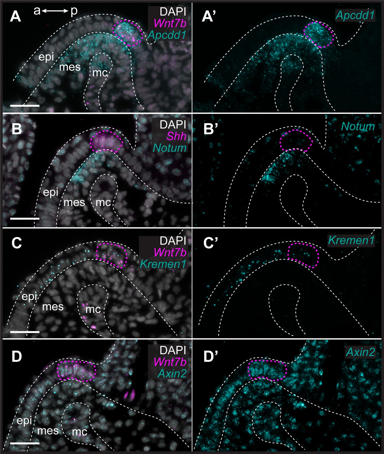
Transduction of canonical Wnt signaling in the anterior gill arch territory in skate.
In S26 skates, (A, A’) Apcdd1 is expressed in the gill arch epithelial ridge (GAER) and in the anterior-distal mesenchyme underlying the GAER. (B) Notum is expressed in anterior-distal arch mesenchyme. (C) Kremen1 is expressed in the GAER and in GAER-adjacent epithelium. (D) Broad Axin2 expression in the vicinity of Wnt-expressing epithelium indicates signaling through the canonical Wnt signaling pathway. ISH by in situ hybridization by chain reaction (HCR) for Sonic hedgehog (Shh) or Wnt7b was performed alongside each gene of interest as a marker of the GAER (outlined with a magenta dashed line). a–p indicates anterior–posterior axis. epi: epithelium, mes: mesenchyme, mc: mesodermal core. Scale bar: 50 μm.
Figure 4—figure supplement 1
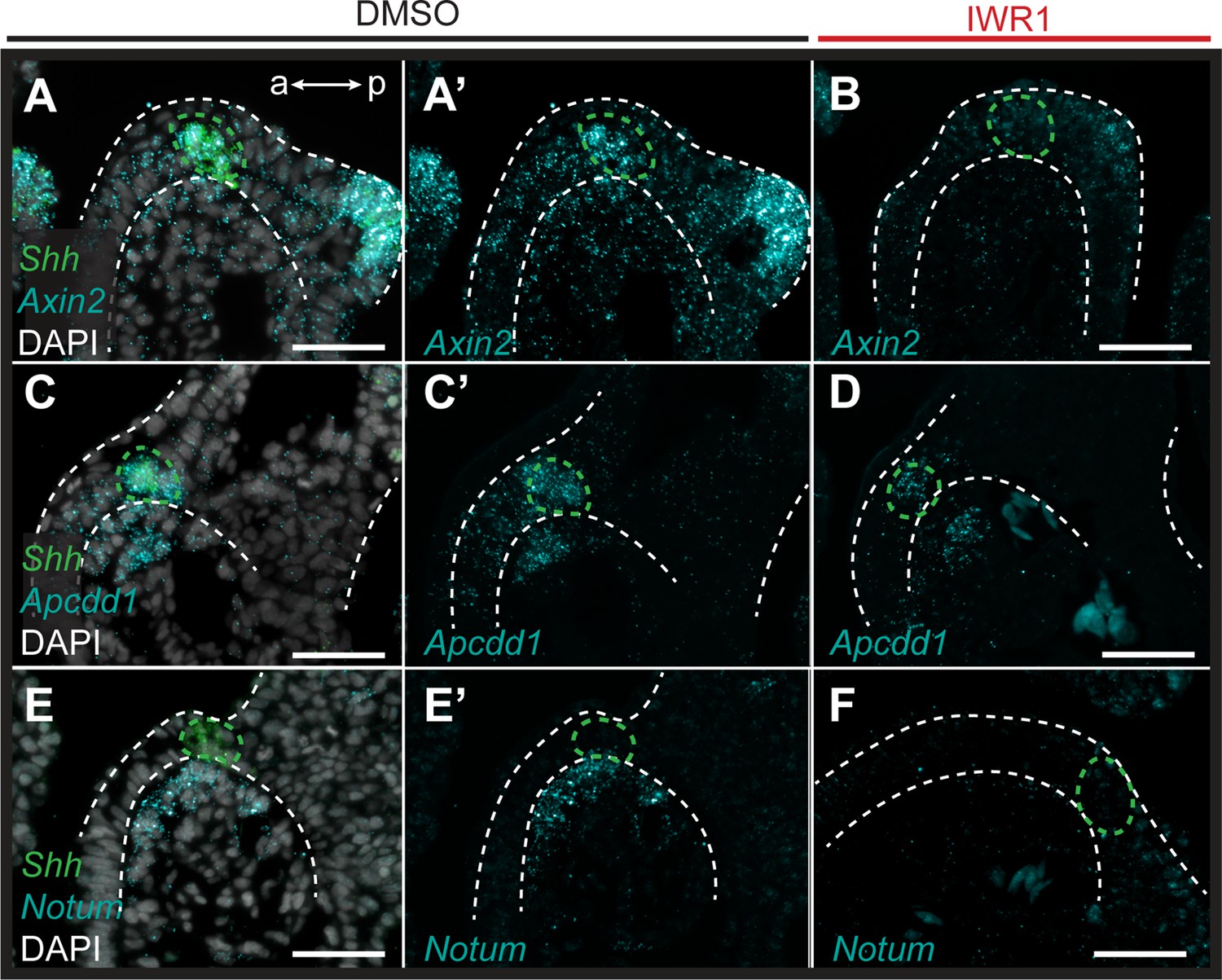
Pharmacological inhibition of Wnt signaling in skate leads to downregulation of Wnt pathway genes.
ISH by in situ hybridization by chain reaction (HCR) was performed on sections of S26 skate embryos treated for 72 hr with either DMSO (control) or the canonical Wnt inhibitor IWR1. Sonic hedgehog (Shh) was performed alongside each gene of interest as a marker of the gill arch epithelial ridge (GAER). (A–A’) Axin2 is broadly expressed throughout the distal arch epithelium and mesenchyme in control embryos, with particularly strong expression in the cells of the GAER and in developing gill buds. (B) Axin2 expression is substantially depressed with IWR1 treatment. (C–C’) Apcdd1 is expressed in the cells of the GAER, and within anterior–distal mesenchyme in control embryos, and (D) both of these expression domains are reduced with IWR1 treatment. (E–E’) Notum is expressed within the anterior-distal arch mesenchyme in control embryos, and (F) expression is completely lost in the anterior–distal arch mesenchyme with IWR1 treatment. All images are representative of experiments performed in triplicate. For ease of visualization in (A’, B, C’, D, E’, F) Shh expression is not shown but GAER cells are outlined with green dashed lines and arch tissues are outlined with white dashed lines. a–p indicates anterior–posterior axis in all panels. Scale bar: 50 μm.
Figure 5
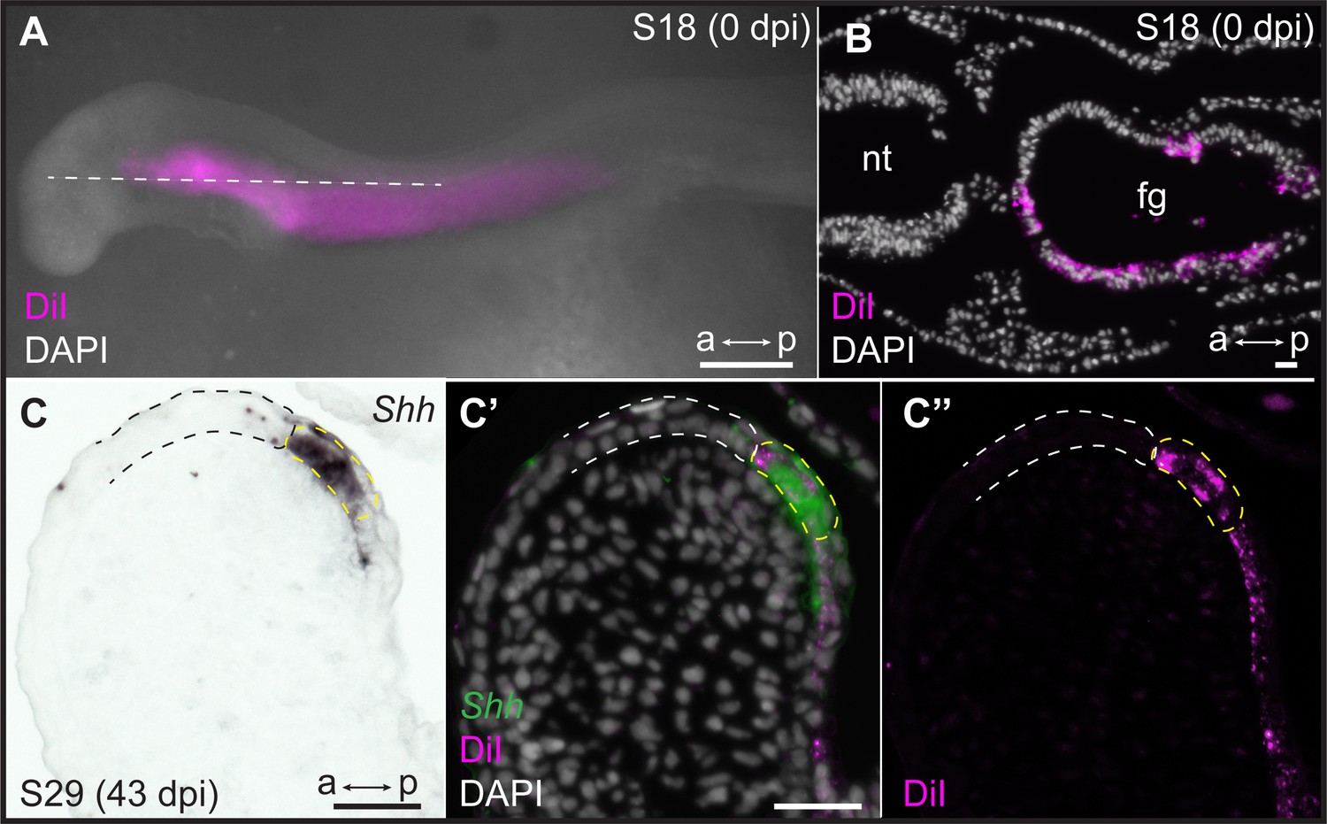
Endodermal origin of the skate gill arch epithelial ridge (GAER).
(A) Microinjection of CM-DiI into the pharyngeal cavity of skate embryos at S18 results in (B) specific labeling of the pharyngeal endoderm. (C) Sonic hedgehog (Shh) is a marker of the GAER (dashed yellow line) in CM-DiI-labeled embryos, and (C’, C’’) co-localization of CM-DiI and Shh expression indicates that the cells of the GAER are of endodermal origin. Dashed white outline demarcates GAER-adjacent (ectodermal) epithelium, which is never labeled with CM-DiI. (C), (C’), and (C’’) are the same section, imaged sequentially for Shh expression and CM-DiI. a–p indicates anterior–posterior axis. dpi: days post-injection, fg: foregut, nt: neural tube. Scale bars; A: 500 μm, B–C: 40 μm.
Figure 6
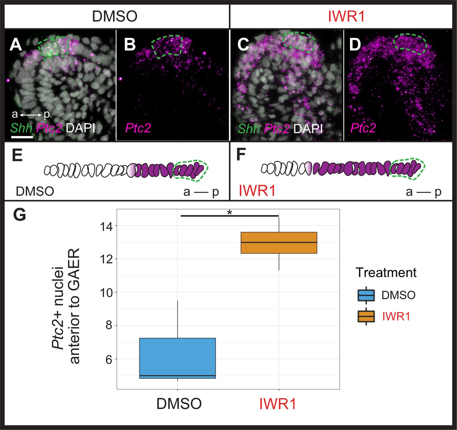
Wnt signaling restricts transduction of gill arch epithelial ridge (GAER) Sonic hedgehog (Shh) signaling to the posterior gill arch environment.
ISH by in situ hybridization by chain reaction (HCR) on sections of skate embryos treated for 72 hr with (A,B) DMSO or (C,D) IWR1 shows no difference in Shh expression but an expansion of Ptc2 expression within the epithelium anterior to the Shh-expressing GAER. (E) Schematic illustration of the mean number of Ptc2+ nuclei anterior to the GAER in DMSO control (n=3 embryos; mean = 6.3 cells) or (F) IWR1 treated (n=4 embryos; mean = 12.94 cells) embryos, using the mean cell count of two or three sections at equivalent positions in the gill arch from each embryo. (G) We find a significant increase in Ptc2+ nuclei in the epithelium anterior to the GAER in IWR1-treated embryos compared to control (p=0.036; t-test, t=–3.87, df = 2.74 - box and whisker plot shows the interquartile range and spread of the data). In A–F, Shh expression is indicated by a green dashed line. Images in A–D were taken using identical exposure settings. a–p indicates anterior–posterior axis. Scale bar: 20 μm.
Figure 7

Ectopic branchial ray formation upon inhibition of Wnt signaling.
(A) In control (DMSO) skate embryos, branchial rays (magnified in Ai) articulate exclusively with the posterior margin of the gill arches. (B) Embryos reared from S25 to S31/32 in the Wnt inhibitor IWR1 possessed ectopic branchial rays (magnified in Bi) that were embedded within the connective tissue of the anterior gill arch (ectopic rays present in n=7/8 embryos examined; indicated by a red asterisk). a–p indicates anterior–posterior axis. Scale bars: 500 μm.
Figure 8
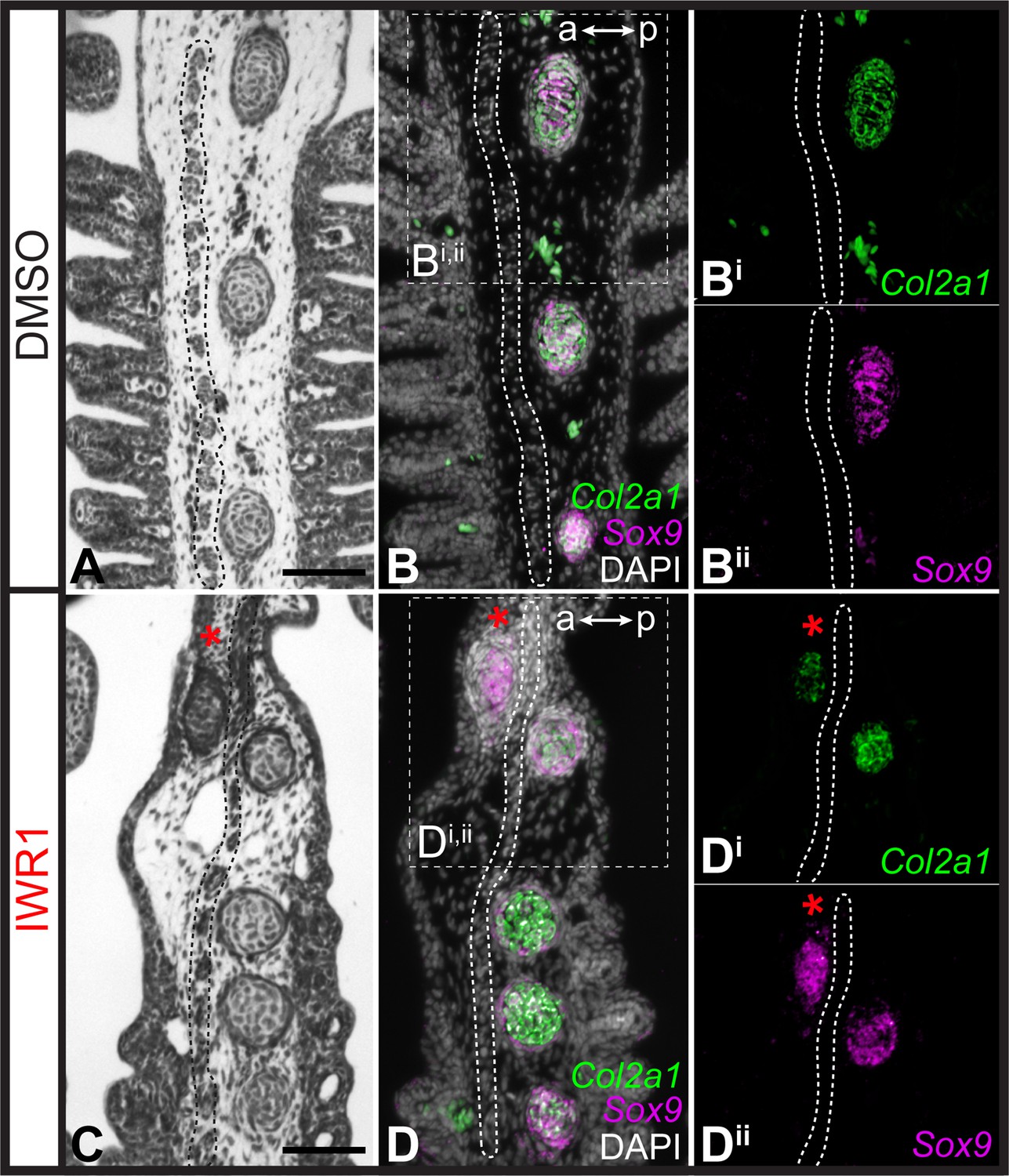
Repression of cartilage development in the anterior gill arch by Wnt signaling.
(A) Section through the gill arch of control (DMSO) S31 skate embryo, showing that branchial rays develop exclusively on the posterior side of the interbranchial muscle plate, as is evident from histochemical staining and (B, Bi, Bii) ISH by in situ hybridization by chain reaction (HCR) for the chondrocyte markers Sox9 and Cola2a1. (C) Section through the gill arch of a S31 embryo reared in the canonical Wnt inhibitor IWR1 reveals that ectopic branchial rays form anterior to the interbranchial muscle plate, as evident from histological analysis and (D, Di, Dii) ISH by HCR for Sox9 and Col2a1. Red asterisks indicate ectopic branchial rays, and the interbranchial muscle plate is outlined with a dashed line. a–p indicates anterior–posterior axis. Scale bars: 50 μm.
Figure 9
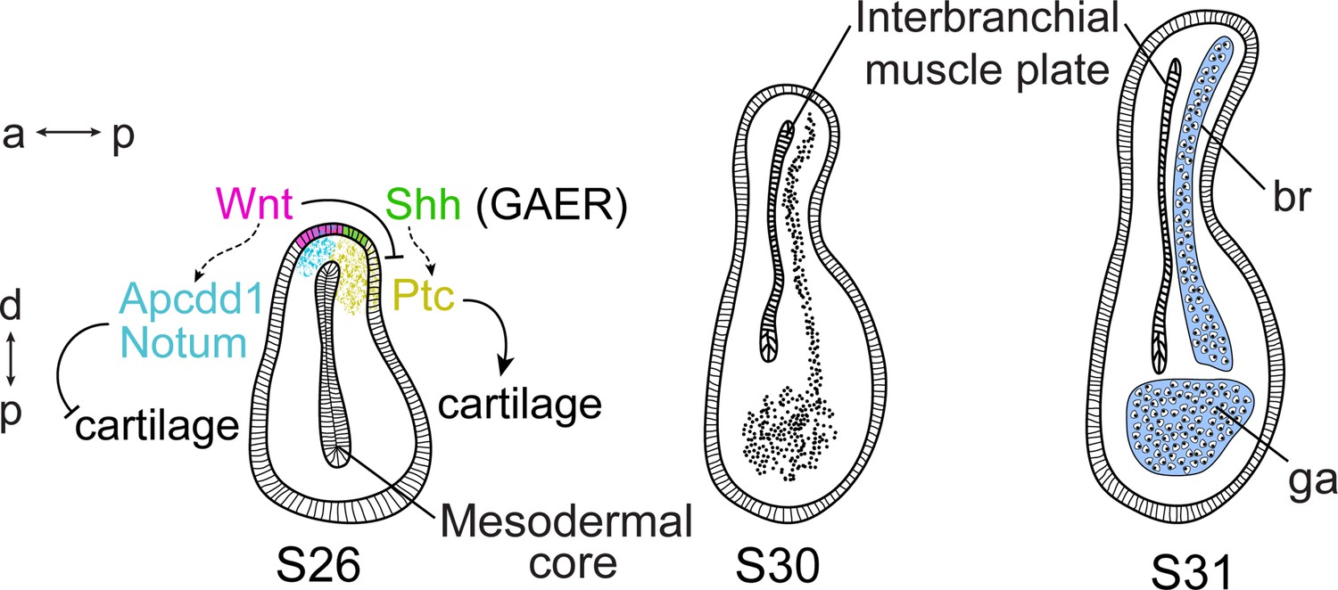
Wnt and Sonic hedgehog (Shh) signaling and cell fate determination in skate gill arches.
Shh signaling from the endodermal-derived gill arch epithelial ridge (GAER) is transduced within the posterior-distal gill arch (ga) environment, where it promotes the differentiation of cartilaginous branchial rays (br). Wnt signals from ectoderm adjacent to the GAER are transduced within the anterior–distal gill arch environment, where they repress Shh signal transduction and inhibit cartilage formation in the anterior gill arch. a–p indicates the anterior–posterior axis and p–d indicates the proximal–distal axis.
Additional files
Download links
A two-part list of links to download the article, or parts of the article, in various formats.
Downloads (link to download the article as PDF)
Open citations (links to open the citations from this article in various online reference manager services)
Cite this article (links to download the citations from this article in formats compatible with various reference manager tools)
Ectodermal Wnt signaling, cell fate determination, and polarity of the skate gill arch skeleton
eLife 12:e79964.
https://doi.org/10.7554/eLife.79964


