Activin A marks a novel progenitor cell population during fracture healing and reveals a therapeutic strategy
- Translational Research Program in Pediatric Orthopaedics, Division of Orthopaedic Surgery, Children’s Hospital of Philadelphia, United States
- Department of Orthopaedics, The First Hospital of China Medical University, China
- Department of Orthopaedic Surgery, Perelman School of Medicine, University of Pennsylvania, United States
- Department of Orthopaedic Surgery, Michigan Medicine, University of Michigan, United States
Figures
Figure 1 with 1 supplement
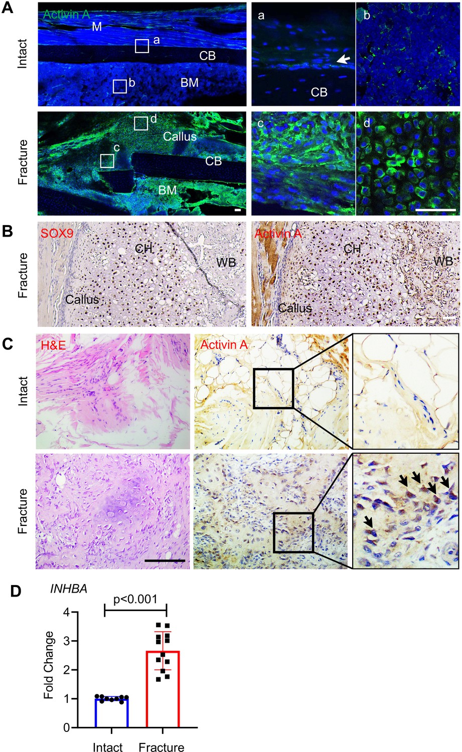
Activin A becomes more abundant during fracture repair.
(A) Whole mount immunofluorescence images of endogenous Activin A distribution in intact and day 5 fractured mouse tibia. The boxed areas in the left panel are shown as enlarged images at the right. M: muscle, CB: cortical bone; BM: bone marrow. Scale bar, 50 μm. (B) Immunohistochemistry (IHC) of fracture sections indicates that many Sox9+ chondrocytes within fracture callus produce Activin A. CH: chondrocytes; WB: woven bone. Scale bar, 50 μm. (C) IHC of human specimens (right panels) shows that Activin A is scanty in intact bone tissue but becomes much more prominent at the fracture site. Left panel: H&E staining. Scale bar, 200 μm. (D) Quantative RT-PCR analysis of INHBA mRNA in intact and fractured human periosteal tissue samples. n = 9–12 specimens/group. Data are expressed as means ± standard deviation (SD) and analyzed by unpaired two-tailed t-test.
-
Figure 1—source data 1
Source data for Figure 1D.
- https://cdn.elifesciences.org/articles/89822/elife-89822-fig1-data1-v2.xlsx
Figure 1—figure supplement 1
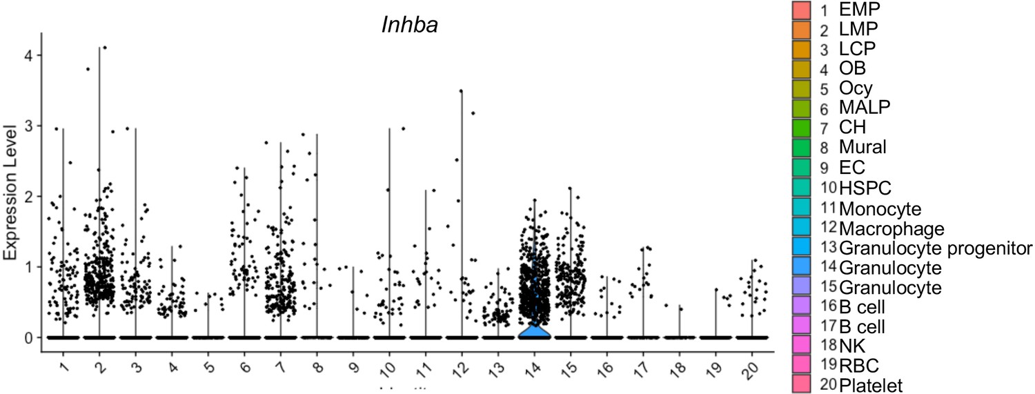
Inhba gene expression in mouse bone marrow cell populations.
Violin plot of Inhba expression in bone marrow cells from 1-month-old mice based on our previous published single-cell RNA-sequencing (scRNA-seq) data. EMP: early mesenchymal progenitors; LMP: late mesenchymal progenitors; LCP: lineage committed progenitors; OB: osteoblasts; Ocy: osteocytes; MALP: marrow adipose lineage precursor cells; CH: chondrocytes; EC: endothelial cells; HSPC: haemopoietic stem and progenitor cells; NK: natural killer cells; RBC: red blood cells.
Figure 2 with 6 supplements

Single-cell transcriptomics analyses reveal identities and developmental trajectories of periosteal mesenchymal populations.
(A) The uniform manifold approximation and projection (UMAP) plot of 13,040 Td+ mesenchymal lineage cells isolated from tibial periosteum of 2-month-old Col2/Td mice. Datasets from cells isolated from intact periosteum (day 0) and fracture site on days 5 and 10 post-surgery were merged and combined into a single plot. (B) UMAP plots of those cells shown at individual time point. (C) Violin plots of cluster-specific makers of mesenchymal lineage cells. MPC: mesenchymal progenitor cell; PPC: proliferative progenitor cell; CH: chondrocyte; HCH: hypertrophic chondrocyte; EOB: early osteoblast; OB: osteoblast. (D) Cell cycle phase of periosteal mesenchymal lineage cells at days 0, 5, and 10. (E) The percentage of proliferative cells (S/G2/M phase) in each cell cluster at days 0, 5, and 10 was computationally quantified. (F) Slingshot trajectory plots of periosteum mesenchymal lineage cells at days 0, 5, and 10.
-
Figure 2—source data 1
Source data for Figure 2E.
- https://cdn.elifesciences.org/articles/89822/elife-89822-fig2-data1-v2.xlsx
Figure 2—figure supplement 1
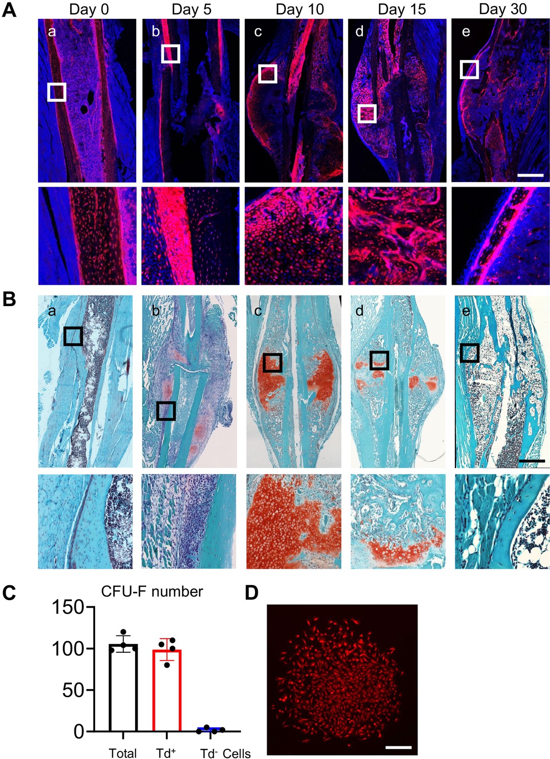
Col2/Td labels periosteal mesenchymal progenitors in intact and fractured tibiae.
(A) Fluorescence images of intact (day 0) and fractured tibiae from 2-month-old Col2/Td mice. Fractured samples were collected at days 5, 10, 15, and 30 post injury. Boxed areas in the top panel are shown at a higher magnification in the lower panel. Scale bar, 1 mm. (B) Safranin-O staining of intact and fractured tibiae at each time point. Boxed areas in the top panel are shown at a higher magnification in the lower panel. Scale bar, 1 mm. (C) Colony-forming unit fibroblast (CFU-F) assay of unsorted (total) and sorted periosteal cells. Periosteal cells isolated from intact Col2/Td tibiae were sorted into Td+ and Td− cells. 1 × 10⁶ total cells, 3 × 10⁴ Td+ cells, and 1 × 10⁶ Td− cells were seeded per flask to determine CFU-F number after 7 days of culture. n = 4 mice/group. Data are expressed as means ± SD. (D) Only Td+ cells form CFU-F colonies. Scale bar, 200 μm.
-
Figure 2—figure supplement 1—source data 1
Source data for Figure 2—figure supplement 1C.
- https://cdn.elifesciences.org/articles/89822/elife-89822-fig2-figsupp1-data1-v2.xlsx
Figure 2—figure supplement 2
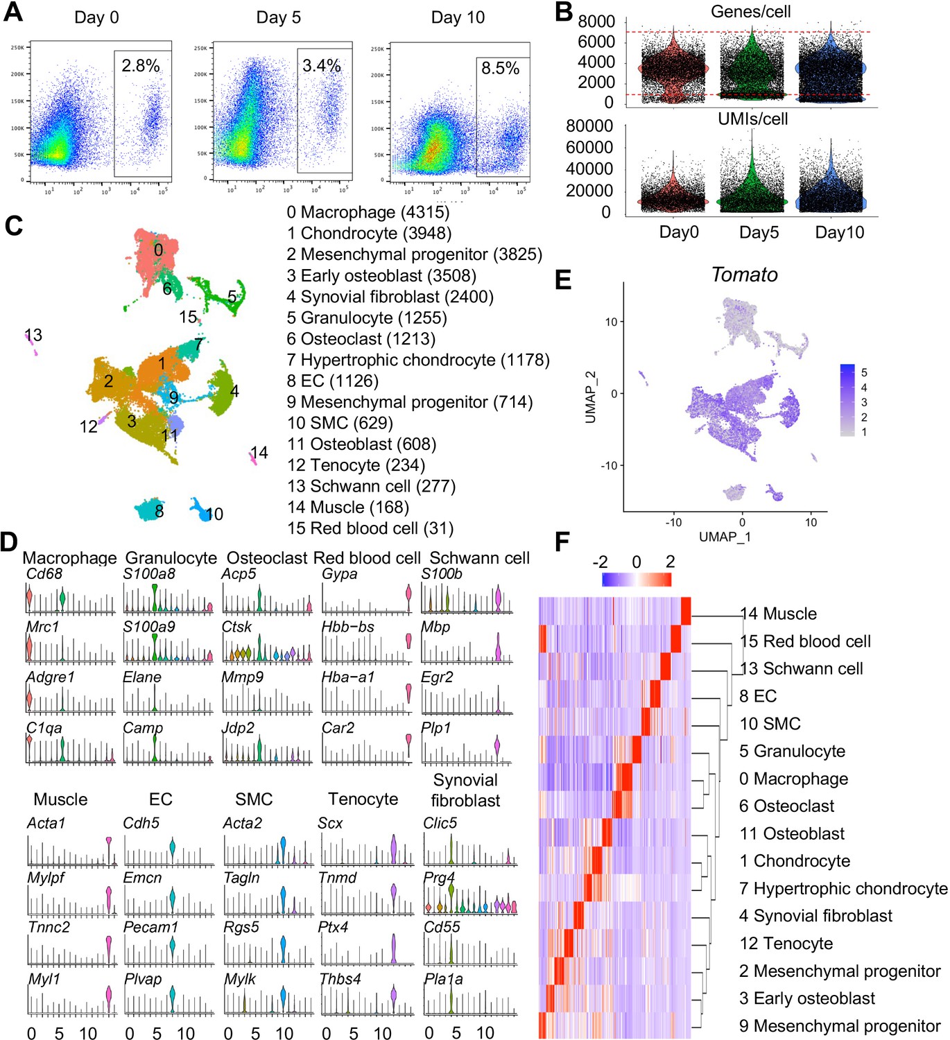
Large-scale single-cell RNA-sequencing (scRNA-seq) analysis of Td+ cells sorted from tibial periosteum of 2-month-old Col2/Td mice.
(A) Flow assay of enzymatically released periosteal cells from 2-month-old Col2/Td mouse tibiae with or without fracture. Periosteal cells from intact tibiae (day 0) and injured tibiae at days 5 and 10 post fracture were collected to measure the percentage of Td+ cells. n = 5–6 mice/time point. (B) Violin plots show the number of genes and unique molecular identifiers (UMIs) per cell in days 0, 5, and 10 scRNA-seq datasets. Red box indicates cells within the selection criteria of quality controls. (C) The uniform manifold approximation and projection (UMAP) plot of 25,429 periosteal cells in the merged scRNA-seq dataset. Cell numbers in each cell type cluster are listed in parenthesis. (D) Violin plots of cluster-specific makers of hematopoietic cells (macrophages and granulocytes), osteoclasts, red blood cells, Schwann cells, muscle cells, endothelial cells (ECs), smooth muscle cells (SMCs), tenocytes, and synovial fibroblasts. (E) The expression pattern of Tomato in the UMAP plot. (F) Hierarchy clustering and heatmap of all cell clusters. Color bar on the top indicates scaled gene expression level.
Figure 2—figure supplement 3
PPCs are greatly and rapidly expanded after fracture.
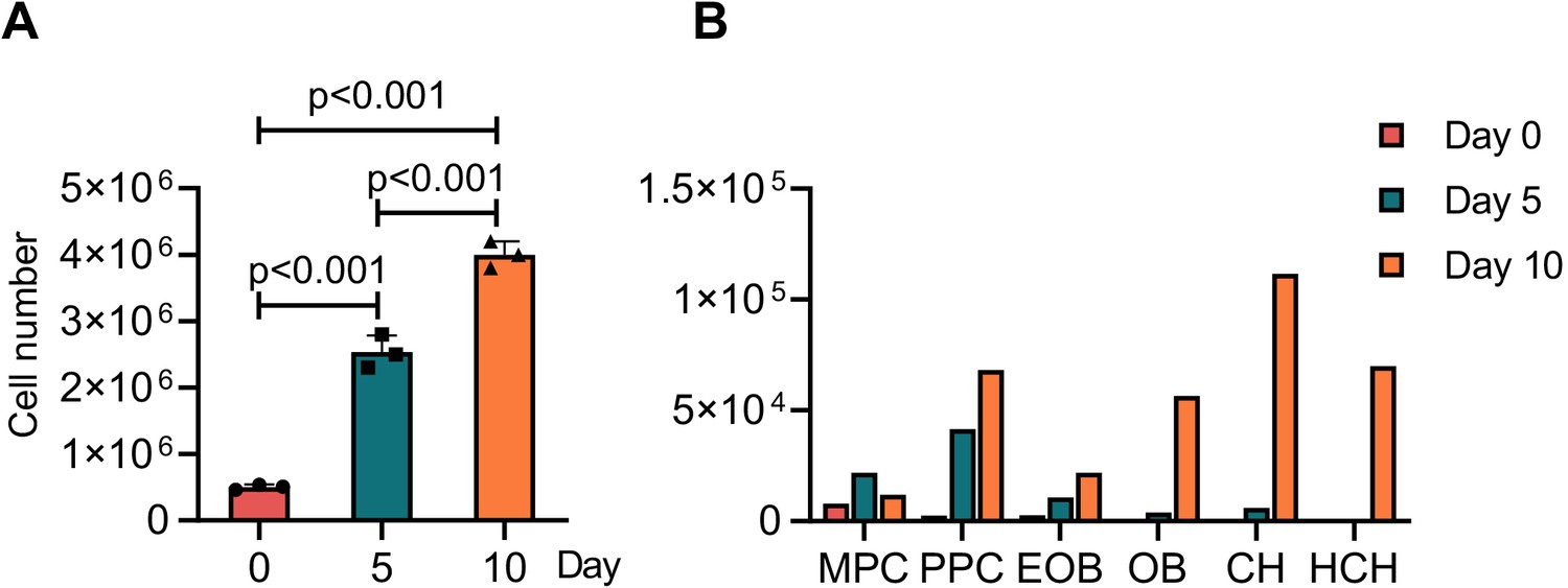
(A) Cell numbers of total enzymatically dissociated periosteal cells from intact (day 0) and injured (days 5 and 10 post fracture) 2-month-old tibiae were. n = 3 mice/time point. Data are expressed as means ± SD and analyzed by one-way ANOVA with Tukey post-hoc test. (B) Based on this information, the number of cells in each cell cluster at different time points was estimated. MPC: mesenchymal progenitor cells; PPC: proliferative progenitor cells; CH: chondrocytes; HCH: hypertrophic chondrocytes; EOB: early osteoblasts; OB: osteoblasts.
-
Figure 2—figure supplement 3—source data 1
Source data for Figure 2—figure supplement 3A.
- https://cdn.elifesciences.org/articles/89822/elife-89822-fig2-figsupp3-data1-v2.xlsx
-
Figure 2—figure supplement 3—source data 2
Source data for Figure 2—figure supplement 3B.
- https://cdn.elifesciences.org/articles/89822/elife-89822-fig2-figsupp3-data2-v2.xlsx
Figure 2—figure supplement 4
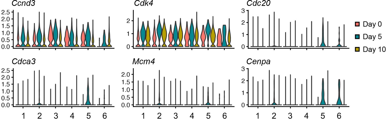
Violin plots of proliferation makers in mesenchymal lineage cells within the single-cell RNA-sequencing (scRNA-seq) datasets.
Figure 2—figure supplement 5
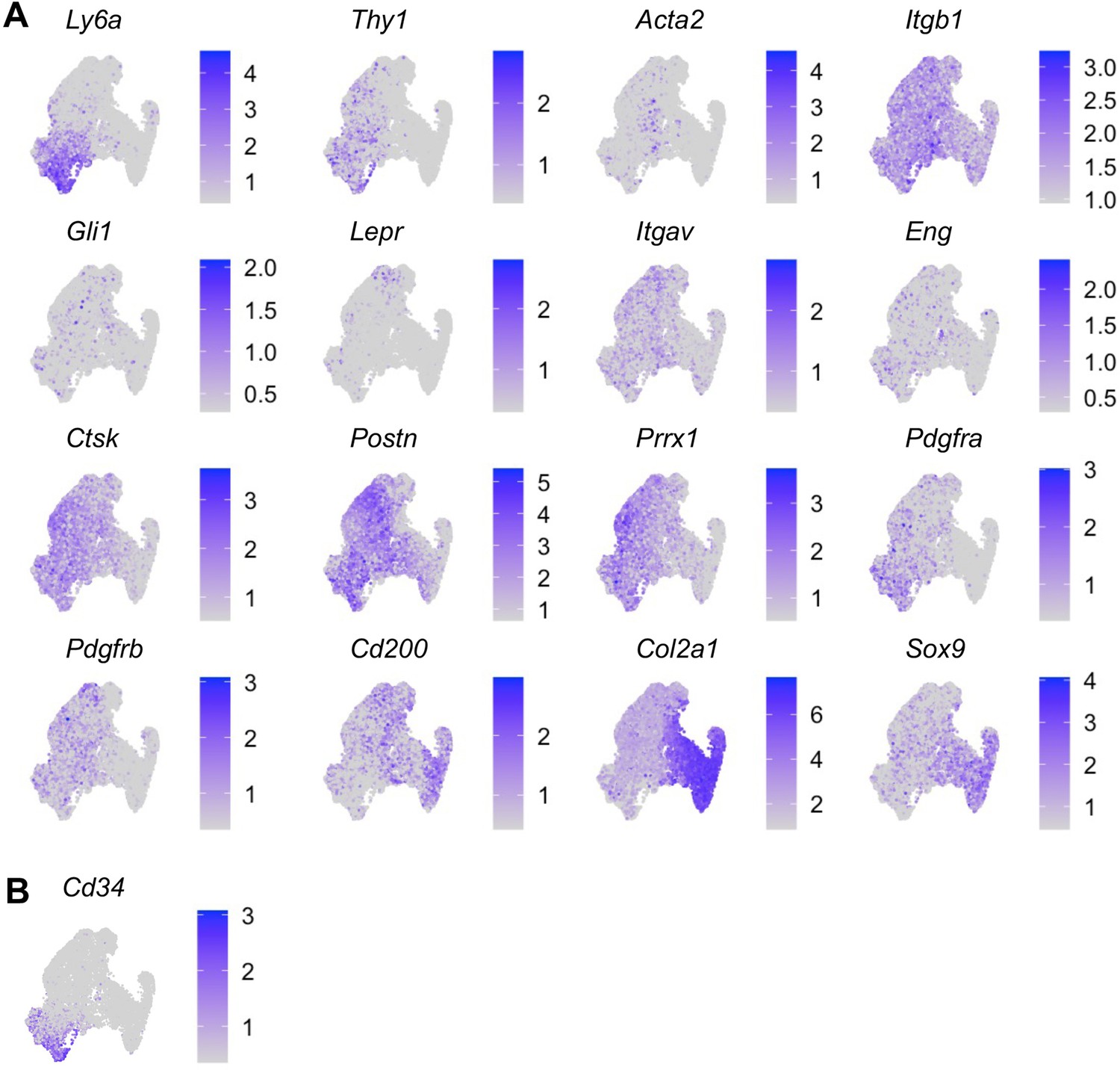
The expression patterns of previously reported periosteal mesenchymal progenitor markers are shown in uniform manifold approximation and projection (UMAP) plots.
(A) Previously reported periosteal mesenchymal progenitor markers. (B) Expression pattern of Cd34 shown in UMAP plot.
Figure 2—figure supplement 6
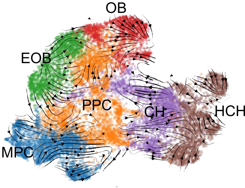
RNA velocity analysis predicts differentiation routes of periosteal mesenchymal cells during fracture healing.
MPC: mesenchymal progenitor cells; PPC: proliferative progenitor cells; CH: chondrocytes; HCH: hypertrophic chondrocytes; EOB: early osteoblasts; OB: osteoblasts.
Figure 3 with 1 supplement
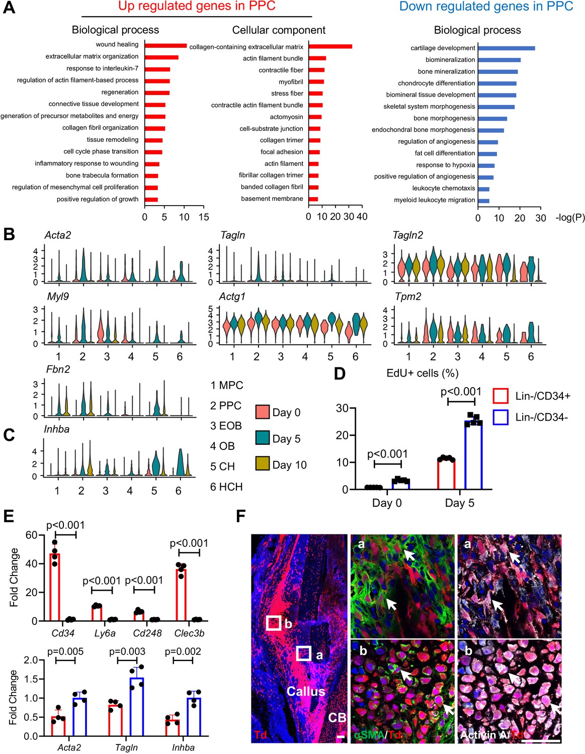
Proliferative progenitor cells (PPCs) have a myofibroblast-like phenotype and express Inhba.
(A) GO term analysis of genes up- or down-regulated in the PPCs (cluster 2) compared to other periosteal mesenchymal cell clusters. (B) Violin plots of myofibroblastic cell marker gene expression. (C) Violin plots of Inhba gene expression. (D) Flow analysis of EdU+ cells in mesenchymal progenitor cells (MPCs; Lin−Cd34+) and PPCs (Lin−Cd34−) from the periosteum of intact and fractured (day 5) mouse bones. n = 4 mice/group. (E) qRT-PCR analyses of stem cell markers (top), myofibroblast markers, and Inhba (bottom) in MPCs and PPCs at day 5 post fracture. n = 4 mice/group. Data are expressed as means ± SD and analyzed by unpaired two-tailed t-test. (F) Whole mount immunofluorescence images of αSMA and Activin A distribution in mouse callus at day 5 post fracture. Boxed areas in the left panel are shown enlarged on the right. Arrows point to representative Td+ cells that are co-stained with both αSMA and Activin A antibodies. CB: cortical bone. Scale bar, 50 μm.
-
Figure 3—source data 1
Source data for Figure 3D.
- https://cdn.elifesciences.org/articles/89822/elife-89822-fig3-data1-v2.xlsx
-
Figure 3—source data 2
Source data for Figure 3E.
- https://cdn.elifesciences.org/articles/89822/elife-89822-fig3-data2-v2.xlsx
Figure 3—figure supplement 1
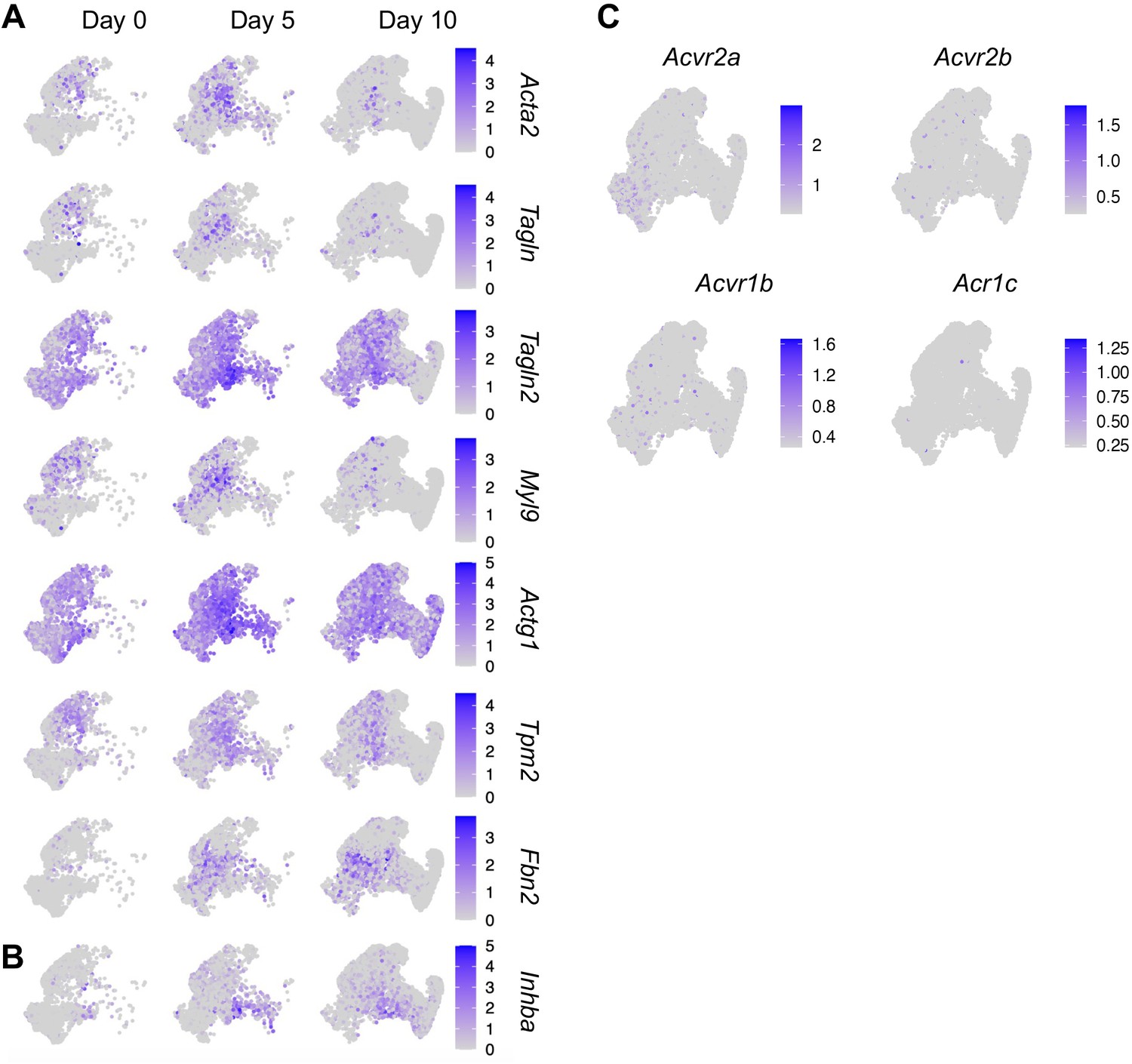
The expression patterns of myofibroblast-like cell markers, Inhba, and its receptors in fracture healing.
(A) Myofibroblast-like marker genes were examined for expression in cell clusters during the fracture healing process on day 0 (prior to fracture) and days 5 and 10 after fracture. (B) Inhba gene expression patterns are shown for comparison. (C) Expression pattern of Activin A receptors in mesenchymal lineage cells.
Figure 4
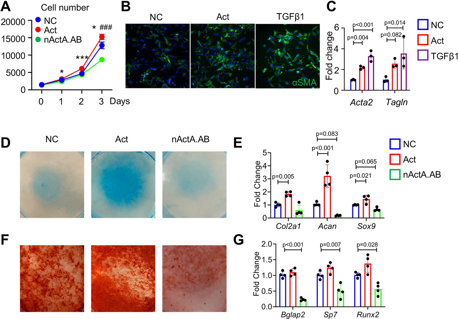
Activin A regulates proliferation and differentiation of periosteal mesenchymal progenitors in vitro.
(A) Proliferation assay of periosteal mesenchymal progenitors treated with recombinant Activin A (Act) or neutralizing monoclonal antibody (nActA.AB) versus control antibody (NC). * p<0.05, *** p<0.001 Act vs NC. ### p<0.001 nActA.AB vs NC. (B) Immunofluorescence images of αSMA in periosteal mesenchymal progenitors treated with Activin A (100 ng/ml) or TGF-β1 (10 ng/ml) for 3 days. (C) qRT-PCR analysis of myofibroblast-like marker expression. (D) Alcian blue staining of periosteal mesenchymal progenitors in micromass cultures undergoing chondrogenic differentiation in the presence of Activin A (100 ng/ml) or nActA.AB (100 μg/ml) for 2 weeks. (E) qRT-PCR analyses of chondrogenic markers. (F) Alizarin red staining of periosteal mesenchymal progenitors undergoing osteogenic differentiation in the presence of Activin A or nActA.AB for 2 weeks. (G) qRT-PCR analyses of osteogenic markers. Data are expressed as means ± SD and analyzed by one-way ANOVA with Tukey post-hoc test.
-
Figure 4—source data 1
Source data for Figure 4A.
- https://cdn.elifesciences.org/articles/89822/elife-89822-fig4-data1-v2.xlsx
-
Figure 4—source data 2
Source data for Figure 4C.
- https://cdn.elifesciences.org/articles/89822/elife-89822-fig4-data2-v2.xlsx
-
Figure 4—source data 3
Source data for Figure 4E.
- https://cdn.elifesciences.org/articles/89822/elife-89822-fig4-data3-v2.xlsx
-
Figure 4—source data 4
Source data for Figure 4G.
- https://cdn.elifesciences.org/articles/89822/elife-89822-fig4-data4-v2.xlsx
Figure 5 with 2 supplements

Systemic administration of Activin A antibody delays mouse fracture healing.
(A) Representative Safranin O/Fast green staining images of fracture calluses at days 5, 7, 10, and 14 post fracture. Mice received subcutaneous injections of control IgG2b isotype or neutralizing monoclonal antibody against Activin A (nActA.AB, 10 mg/kg) twice a week after fracture. Scale bar, 1 mm. (B) Callus area, cartilage area, and bone area were quantified at indicated time points. n = 4–7 mice/time point. (C) Measurement of fracture healing scores at 6 weeks post fracture. n = 10 mice/group. (D) Mechanical testing was performed on bones at 6 weeks post fracture. n = 10 mice/group. (E) Immunofluorescence images of pSMAD2 and αSMA in fracture calluses of control (isotype) and nActA.AB-treated mice at day 7 post fracture. White arrows point to pSMAD2+αSMA+ cells and yellow arrows point to pSMAD2+αSMA− cells. Scale bar, 500 μm (low mag), 50 μm (high mag). (F) Percentages of pSMAD2+ and αSMA+ cells in fracture calluses and pSMAD2+ cells within the αSMA+/− populations were quantified. n = 3 mice/group. (G) qRT-PCR analyses of Acta2 and Inhba expression in day 7 callus from 2-month-old mice treated with nActA.AB versus isotype control. n = 4 mice/group. Data are expressed as means ± SD and analyzed by unpaired two-tailed t-test.
-
Figure 5—source data 1
Source data for Figure 5B.
- https://cdn.elifesciences.org/articles/89822/elife-89822-fig5-data1-v2.xlsx
-
Figure 5—source data 2
Source data for Figure 5C.
- https://cdn.elifesciences.org/articles/89822/elife-89822-fig5-data2-v2.xlsx
-
Figure 5—source data 3
Source data for Figure 5D.
- https://cdn.elifesciences.org/articles/89822/elife-89822-fig5-data3-v2.xlsx
-
Figure 5—source data 4
Source data for Figure 5F.
- https://cdn.elifesciences.org/articles/89822/elife-89822-fig5-data4-v2.xlsx
-
Figure 5—source data 5
Source data for Figure 5G.
- https://cdn.elifesciences.org/articles/89822/elife-89822-fig5-data5-v2.xlsx
Figure 5—figure supplement 1
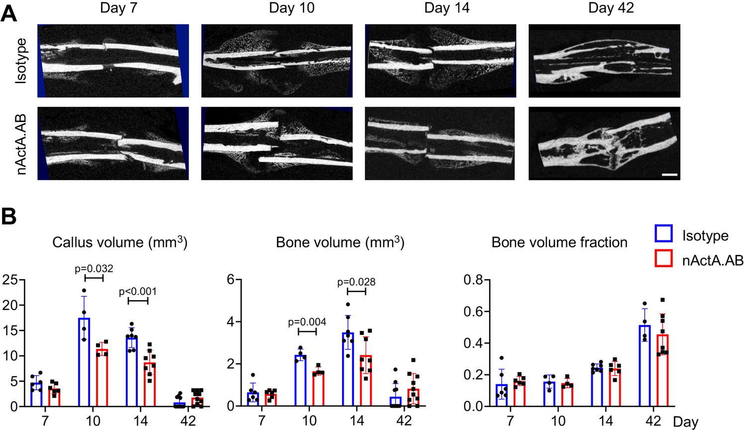
Immunological inhibition of Activin A impedes fracture healing.
(A) Representative micro-computed tomography (μCT) images of fracture calluses on days 7, 10, 14, and 42 post fracture. Mice received injections of control IgG2b isotype antibody or neutralizing monoclonal antibody against Activin A (nActA.AB, 10 mg/kg) twice a week after fracture. Scale bar, 1 mm. (B) Callus volume, bone volume and bone volume fraction were measured. n = 4–10 mice/time point/group. Data are expressed as means ± SD and analyzed by unpaired two-tailed t-test.
-
Figure 5—figure supplement 1—source data 1
Source data for Figure 5—figure supplement 1B.
- https://cdn.elifesciences.org/articles/89822/elife-89822-fig5-figsupp1-data1-v2.xlsx
Figure 5—figure supplement 2
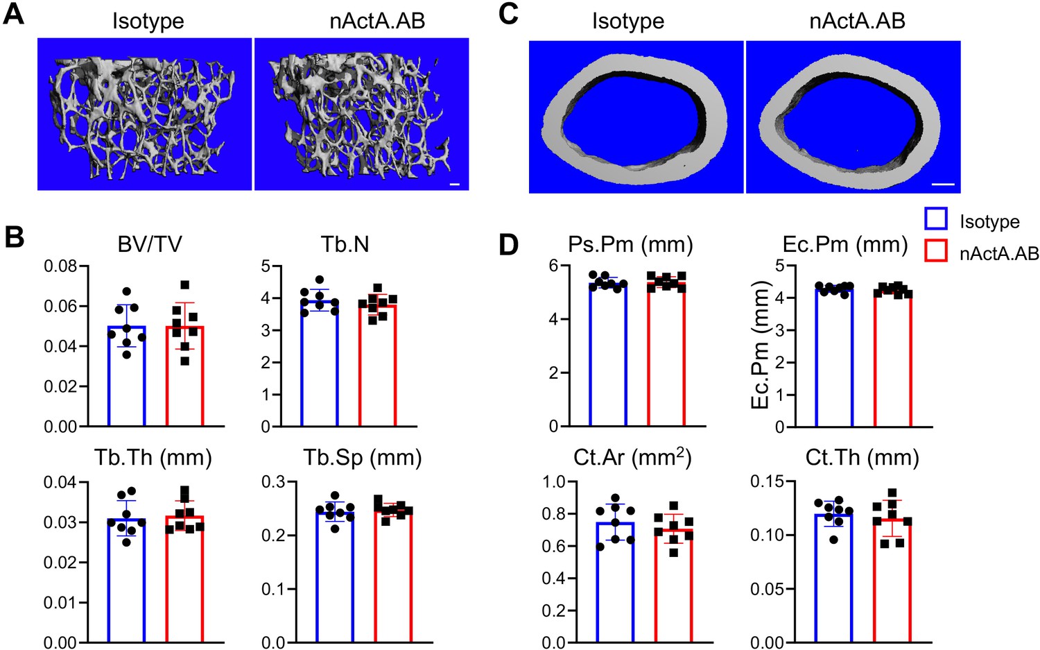
Immunological inhibition of Activin A does not affect normal bone homeostasis.
(A) Representative 3D micro-computed tomography (μCT) images of trabecular bone of contralateral uninjured femur at 6 weeks post fracture. Mice received injections of control IgG2b isotype antibody or neutralizing monoclonal antibody against Activin A (nActA.AB, 10 mg/kg) twice a week after fracture. Scale bar, 100 μm. (B) μCT measurement of femoral trabecular bone structural parameters: bone volume fraction (BV/TV), trabecular number (Tb.N), trabecular thickness (Tb.Th), and trabecular separation (Tb.Sp). n = 8 mice/group. (C) Representative 3D μCT images of cortical bone of contralateral uninjured femur at 6 weeks post fracture. Scale bar, 100 μm. (D) μCT measurement of femoral cortical bone structural parameters: periosteal perimeter (Ps.Pm), endosteal perimeter (Ec.Pm), cortical bone thickness (Ct.Th), and cortical bone area (Ct.Ar). n = 8 mice/group. Data are expressed as means ± SD and analyzed by unpaired two-tailed t-test.
-
Figure 5—figure supplement 2—source data 1
Source data for Figure 5—figure supplement 2B.
- https://cdn.elifesciences.org/articles/89822/elife-89822-fig5-figsupp2-data1-v2.xlsx
-
Figure 5—figure supplement 2—source data 2
Source data for Figure 5—figure supplement 2D.
- https://cdn.elifesciences.org/articles/89822/elife-89822-fig5-figsupp2-data2-v2.xlsx
Figure 6 with 1 supplement

Local Activin A implantation promotes fracture healing.
(A) Representative Safranin O/Fast green staining histochemical images of fracture calluses at days 5, 14, and 28 post fracture. Two-month-old (2M) or 20-month-old (20M) mice were implanted with a 50-μl Matrigel aliquot containing vehicle (Veh) or Activin A (ACT) (1 μg) at the fracture site immediately after surgery. Scale bar, 1 mm. (B) Callus area, cartilage area, and bone area were quantified at indicated time points. n = 4 mice/group. (C) Immunofluorescence images of pSMAD2 and αSMA in fracture calluses of control and ACT-implanted mice at day 5 post fracture. White arrows point to pSMAD2+αSMA+ cells and yellow arrows point to pSMAD2+αSMA− cells. Scale bar, 500 μm (low mag), 50 μm (high mag). (D) Percentage of pSMAD2+, αSMA+ cells in fracture calluses and pSMAD2+ cells within αSMA+/− populations were quantified. n = 3 mice/group. (E) Fracture healing scores were quantified in bones of 2- and 20-month-old mice at 6 weeks post fracture. n = 10 mice/group. (F) Mechanical testing was performed on bones of 2- and 20-month-old mice at 6 weeks post fracture. n = 10 mice/group. Data are expressed as means ± SD and analyzed by unpaired two-tailed t-test.
-
Figure 6—source data 1
Source data for Figure 6B.
- https://cdn.elifesciences.org/articles/89822/elife-89822-fig6-data1-v2.xlsx
-
Figure 6—source data 2
Source data for Figure 6D.
- https://cdn.elifesciences.org/articles/89822/elife-89822-fig6-data2-v2.xlsx
-
Figure 6—source data 3
Source data for Figure 6E.
- https://cdn.elifesciences.org/articles/89822/elife-89822-fig6-data3-v2.xlsx
-
Figure 6—source data 4
Source data for Figure 6F.
- https://cdn.elifesciences.org/articles/89822/elife-89822-fig6-data4-v2.xlsx
Figure 6—figure supplement 1
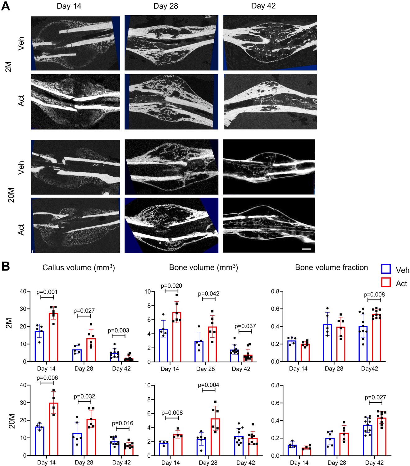
Activin A treatment accelerates fracture healing.
(A) Representative micro-computed tomography (μCT) images of fracture calluses on days 14, 28, and 42 post fracture. Mice at 2 or 20 months of age were implanted with 50 μl Matrigel containing vehicle or Activin A (1 μg) at the fracture site at the time of surgery. Scale bar, 1 mm. (B) Callus volume, bone volume, and bone volume fraction were measured. n = 4–10 mice/time point/group. Data are expressed as means ± SD and analyzed by unpaired two-tailed t-test.
-
Figure 6—figure supplement 1—source data 1
Source data for Figure 6—figure supplement 1B.
- https://cdn.elifesciences.org/articles/89822/elife-89822-fig6-figsupp1-data1-v2.xlsx
Figure 7
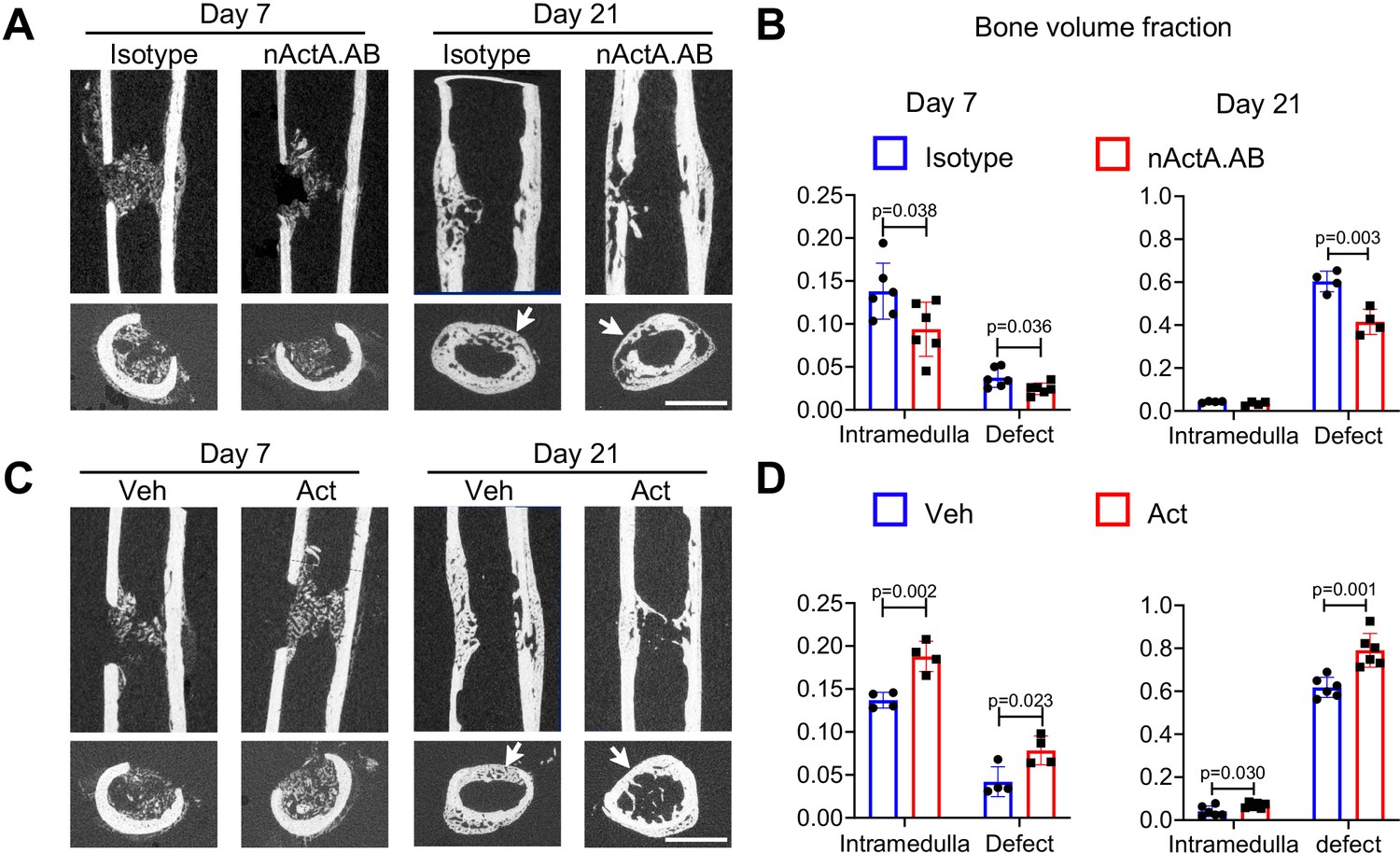
Activin A promotes intramembranous bone repair of unicortical drill holes.
(A) Representative sagittal (top) and transverse (bottom) cross-sections of micro-computed tomography (μCT) images of Activin A blocking antibody (nActA.AB)-treated drill-hole defects. Mice received injections of control IgG2b isotype or neutralizing monoclonal antibody against Activin A (nActA.AB, 10 mg/kg) twice a week after drill-hole injury. Arrows point to the defect region. Scale bar, 1 mm. (B) Bone volume fraction of intramedullary and cortical defect regions at days 7 and 21 post-injury. n = 4–6 mice/group. (C) Representative sagittal (top) and transverse (bottom) cross-sections of μCT images of recombinant Activin A (Act)-treated drill-hole defects. Two-month-old mice were implanted with a 50-μl Matrigel aliquot containing vehicle (Veh) or Activin A (Act) (1 μg) at the drill-hole site immediately after surgery. Arrows point to the defect region. Scale bar, 1 mm. (D) Bone volume fraction of intramedullary and cortical defect regions at days 7 and 21 post-injury. n = 4–6 mice/group. Data are expressed as means ± SD and analyzed by unpaired two-tailed t-test.
-
Figure 7—source data 1
Source data for Figure 7B.
- https://cdn.elifesciences.org/articles/89822/elife-89822-fig7-data1-v2.xlsx
-
Figure 7—source data 2
Source data for Figure 7D.
- https://cdn.elifesciences.org/articles/89822/elife-89822-fig7-data2-v2.xlsx
Figure 8 with 1 supplement
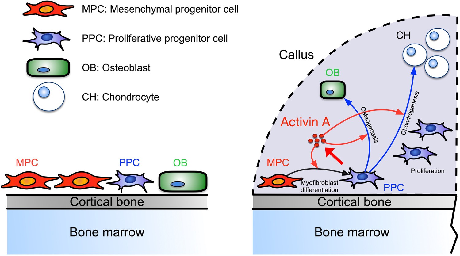
Working model of Activin A roles in callus during progression of fracture healing.
Figure 8—figure supplement 1
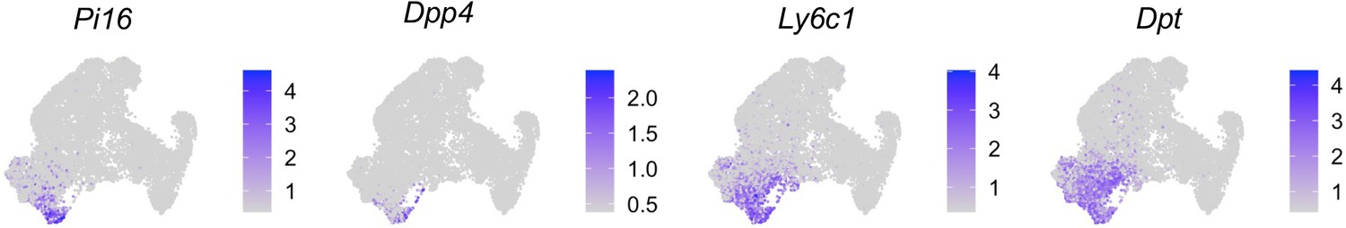
Markers of Pi16+ cells from a recent single-cell RNA-sequencing (scRNA-seq) study of fibroblasts are specifically expressed in the mesenchymal progenitor cell (MPC) cluster.
Author response image 1
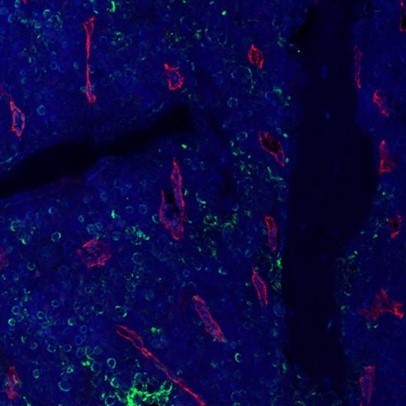
Fluorescent Activin A (green) and Emcn (red) staining of bone marrow in a WT mouse femur.
Author response image 2

Fluorescent image of a Col2/Td tibia shows no Td signal in the neighboring muscle tissue.
Author response image 3

Adipoq+ cells are absent at mouse periosteum.
(A) Immunofluorescent image of an intact tibiae from Adipoq-Cre Td mice stained with Endomucin (Emcn, an endothelial cell marker). (B) Expression pattern of Adipoq in mesenchymal lineage cells in the fracture scRNA-seq datasets.
Author response image 4
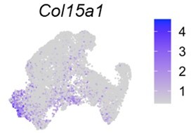
UMAP plot of Col15a1 in the merged fracture dataset.
Tables
Key resources table
| Reagent type (species) or resource | Designation | Source or reference | Identifiers | Additional information |
|---|---|---|---|---|
| Genetic reagent (Mus musculus) | Col2a1-Cre | Jackson Laboratory | Stock #: 003554 | |
| Genetic reagent (Mus musculus) | Rosa26LSL-tdTomato | Jackson Laboratory | Stock #: 007909 | |
| Genetic reagent (Mus musculus) | C57BL/6 | Jackson Laboratory | Stock #: 000664 | |
| Antibody | Mouse monoclonal neutralizing antibody against Activin A | Biolegend | Cat #: 693604 | 10 mg/kg |
| Antibody | Rabbit monoclonal anti-mouse pSMAD2 | Cell Signaling | Cat #: 3108S | 1:200 |
| Antibody | Mouse monoclonal anti-mouse αSMA | Sigma | Cat #: A5228 | 1:200 |
| Antibody | Goat polyclonal anti-mouse Activin A | R&D Systems | Cat #: AF338 | 1:200 |
| Antibody | Alexa Fluor 647 donkey polyclonal anti-goat | Invitrogen | Cat #: A-21447 | 1:200 |
| Antibody | Alexa Fluor 488 donkey polyclonal anti-rabbit | Invitrogen | Cat #: A-21206 | 1:200 |
| Antibody | Alexa Fluor 488 donkey polyclonal anti-mouse | Invitrogen | Cat #: A-21202 | 1:200 |
| Antibody | Alexa Fluor 555 donkey polyclonal anti-mouse | Invitrogen | Cat #: A-31570 | 1:200 |
| Antibody | Rat monoclonal anti-mouse Ter119 FITC | Biolegend | Cat #: A-116205 | 1:100 |
| Antibody | Rat monoclonal anti-mouse CD31 FITC | Biolegend | Cat #: A-102509 | 1:100 |
| Antibody | Rat monoclonal anti-mouse CD45 FITC | Biolegend | Cat #: A-147709 | 1:100 |
| Antibody | Rat monoclonal anti-mouse CD34 BV421 | BD Biosciences | Cat #: A-562608 | 1:100 |
| Commercial kit | Click-iT Plus EdU Alexa Fluor 647 Flow Cytometry Assay Kit | Thermo Fisher | Cat #: A-C10424 | |
| Peptide, recombinant protein | Recombinant Activin A | R&D Systems | Cat #: 338-AC-010 | 10 µg |
| Software | Cellranger | https://support.10xgenomics.com | Version 6.0.1 | |
| Software | ImageJ software | ImageJ (http://imagej.nih.gov/ij/) | ||
| Software | GraphPad Prism software | GraphPad Prism (https://graphpad.com) |
Additional files
-
Supplementary file 1
Supplementary tables.
(a) Cell numbers and percentages are listed for cell clusters at day 0 before fracture and days 5 and 10 after fracture. (b) Cell numbers and percentages are listed for cell clusters of periosteal mesenchymal lineage cells at day 0 before fracture or days 5 and 10 after fracture. (c) Mouse real-time RT-PCR primer sequences used in this study.
- https://cdn.elifesciences.org/articles/89822/elife-89822-supp1-v2.docx
-
MDAR checklist
- https://cdn.elifesciences.org/articles/89822/elife-89822-mdarchecklist1-v2.pdf
Download links
A two-part list of links to download the article, or parts of the article, in various formats.
Downloads (link to download the article as PDF)
Open citations (links to open the citations from this article in various online reference manager services)
Cite this article (links to download the citations from this article in formats compatible with various reference manager tools)
Activin A marks a novel progenitor cell population during fracture healing and reveals a therapeutic strategy
eLife 12:e89822.
https://doi.org/10.7554/eLife.89822


