β-catenin inhibition disrupts the homeostasis of osteogenic/adipogenic differentiation leading to the development of glucocorticoid-induced osteonecrosis of the femoral head
- Institute of Orthopedics and Traumatology, The First Affiliated Hospital of Zhejiang Chinese Medical University, Zhejiang Provincial Hospital of Chinese Medicine, China
- Department of Orthopedic Surgery, the Affiliated Lihuili Hospital of Ningbo University, China
- The First College of Clinical Medicine, Zhejiang Chinese Medical University, China
- School of Basic Medical Sciences, Zhejiang Chinese Medical University, China
- Department of Orthopedic Surgery, the First Affiliated Hospital of Zhejiang Chinese Medical University, China
- Faculty of Pharmaceutical Sciences, Shenzhen Institute of Advanced Technology, China
Figures
Figure 1
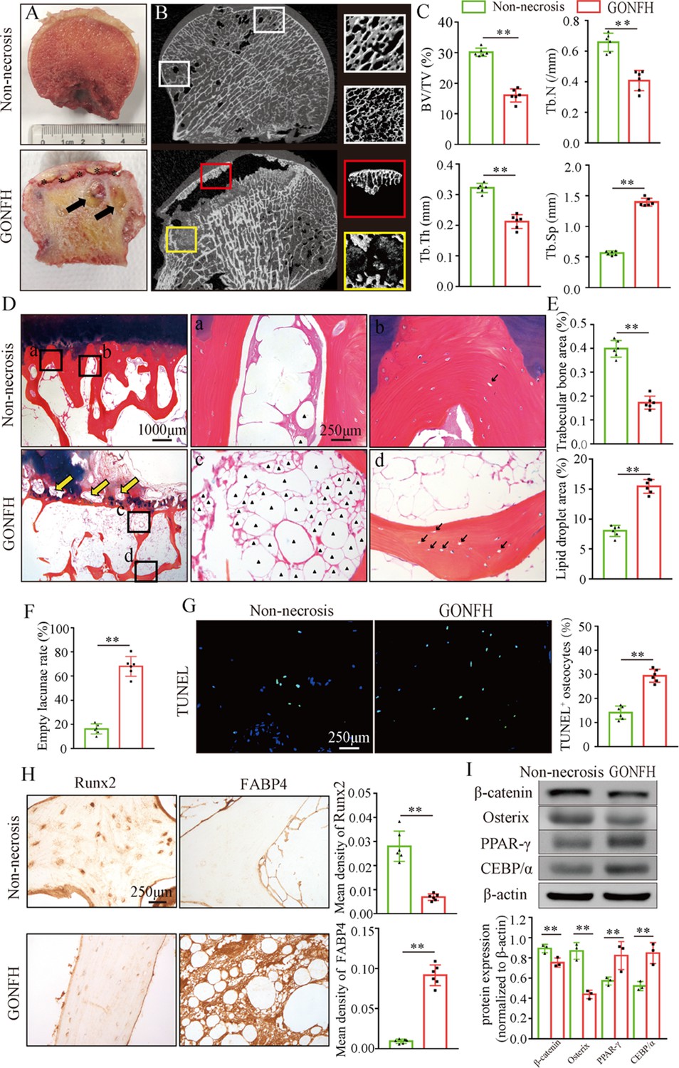
Abnormal osteogenesis and adipogenesis with decreased β-catenin signaling in the necrotic femoral heads of glucocorticoid-induced osteonecrosis of the femoral head (GONFH) patients.
Necrotic (n = 15) and non-necrotic (n = 10) femoral head samples were obtained from GONFH patients or femoral neck fracture patients, respectively. (A) Gross anatomy analysis of human necrotic and non-necrotic femoral head samples. Black asterisks: subchondral collapsed region. Black arrows: liquefied necrotic region. (B) μCT images of human necrotic and non-necrotic femoral heads. Red and yellow boxed areas: 3D images of subchondral collapsed region and liquefied necrotic region, respectively. White boxed areas: 3D images of corresponding regions in the non-necrotic femoral heads. (C) Quantitative analysis of BV/TV, Tb.N, Tb.Th, and Tb.Sp on the necrotic regions. (D) Alcian Blue Hematoxylin (ABH) staining of human necrotic and non-necrotic femoral heads. (a, c) High-magnification images of bone marrow; (b, d) high-magnification images of bone trabeculae; yellow arrows: subchondral bone destruction; black arrows: empty lacunae of osteocytes; black triangles: fat droplets. (E) Histomorphological quantitative analysis of trabecular bone area and fat droplet area. (F) Histomorphological quantitative analysis of empty lacunae rate. (G) TUNEL staining of osteocytes in human necrotic and non-necrotic femoral heads. (H) Immunohistochemistry (IHC) staining of Runx2 and FABP4 expressions in human necrotic and non-necrotic femoral heads. (I) Western blot of β-catenin, Osterix, PPAR-γ, and CEBP/α in human necrotic and non-necrotic femoral heads.
-
Figure 1—source data 1
Raw data for Figure 1.
- https://cdn.elifesciences.org/articles/92469/elife-92469-fig1-data1-v1.zip
-
Figure 1—source data 2
Labeled uncropped western blots for Figure 1.
- https://cdn.elifesciences.org/articles/92469/elife-92469-fig1-data2-v1.zip
-
Figure 1—source data 3
Raw unedited blots for Figure 1.
- https://cdn.elifesciences.org/articles/92469/elife-92469-fig1-data3-v1.zip
Figure 2
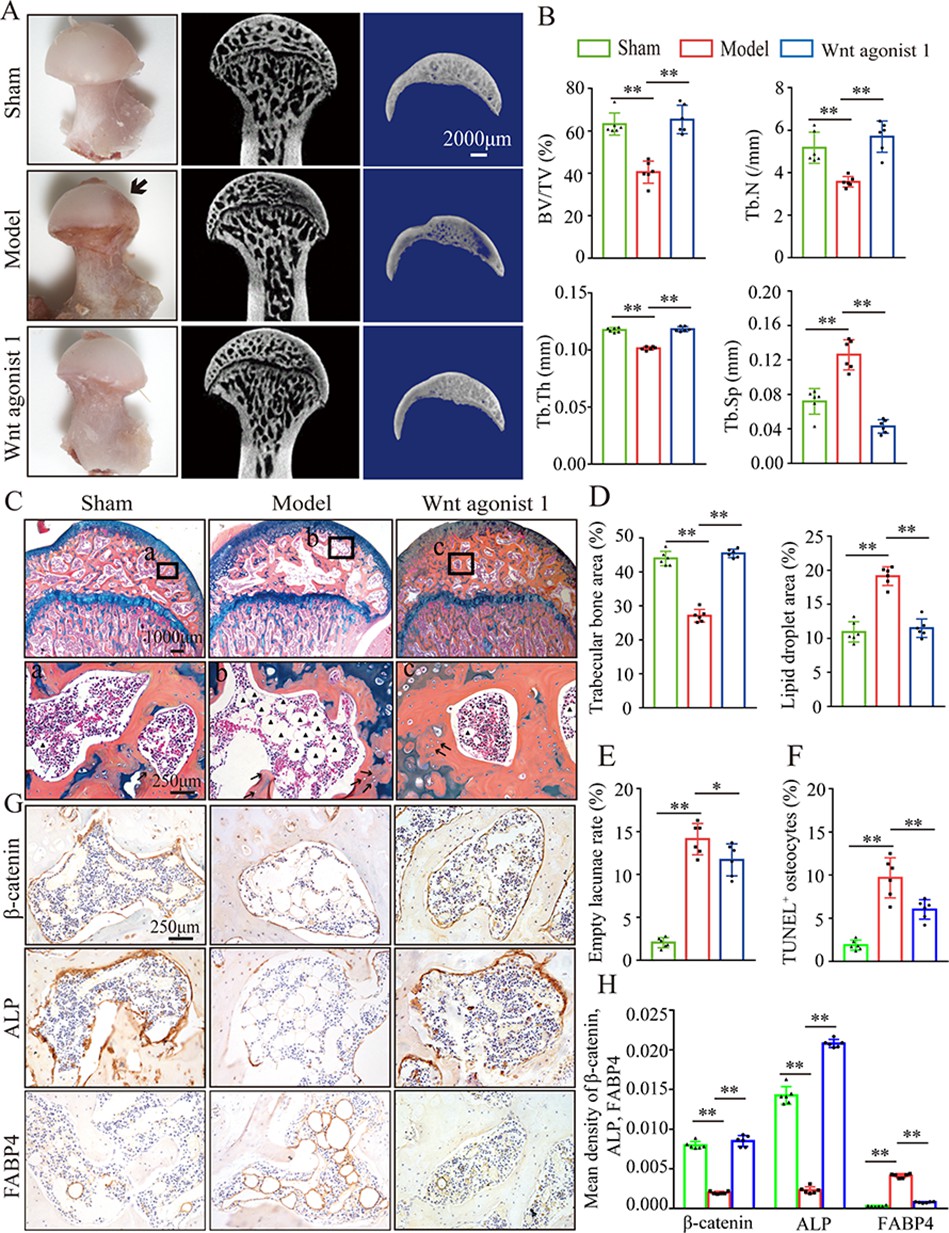
Systemic injection of Wnt agonist 1 alleviates abnormal osteogenesis and adipogenesis in rat glucocorticoid-induced osteonecrosis of the femoral head (GONFH).
The rat model of GONFH was established by a co-induction of lipopolysaccharide and methylprednisolone (MPS). The femoral head samples were harvested after GONFH rats were intravenously injected with Wnt agonist 1 for 6 wk. (A) Gross anatomy analysis and μCT images of femoral heads in sham, model, and Wnt agonist 1-treated groups. (B) μCT quantitative analysis of BV/TV, Tb.N, Tb.Th, and Tb.Sp in each group. (C) Alcian Blue Hematoxylin (ABH) staining of femoral heads in each group. (a–c) High-magnification images of the representative region; black arrows: empty lacunae of osteocytes; black triangles: fat droplets. (D) Histomorphological quantitative analysis of trabecular bone area and fat droplet area in each group. (E) Histomorphological quantitative analysis of empty lacunae rate in each group. (F) TUNEL staining of osteocytes in the femoral heads of GONFH rats. (G, H) Immunohistochemistry (IHC) staining and quantitative analysis of β-catenin, ALP, and FABP4 in rat femoral heads of each group.
-
Figure 2—source data 1
Raw data for Figure 2.
- https://cdn.elifesciences.org/articles/92469/elife-92469-fig2-data1-v1.zip
Figure 3
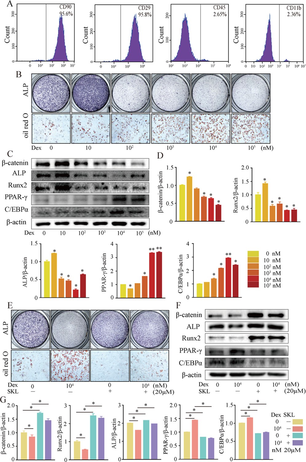
Activation of β-catenin redirected dexamethasone (Dex)-induced imbalanced osteogenic/adipogenic differentiation of bone marrow mesenchymal cells (BMSCs).
Primary BMSCs were extracted from the proximal femur of 4-week-old Sprague–Dawley (SD) rats and cultivated to passage 3 for subsequent experiments. (A) Flow cytometry analyzing the surface markers of rat BMSCs. (B) ALP staining and oil red O staining of BMSCs at the increasing concentrations of Dex. (C, D) Western blot and quantitative analysis of β-catenin, Runx2, ALP, PPAR-γ, and CEBP/α in BMSCs at the increasing concentrations of Dex. (E) ALP staining and oil red O staining of rat BMSCs at the condition with or without 104 nM Dex and 20 μM SKL2001 (an agonist of β-catenin). (F, G) Western blot and quantitative analysis of β-catenin, Runx2, ALP, PPAR-γ, and CEBP/α in rat BMSCs at the condition with or without 104 nM Dex and 20 μM SKL2001.
-
Figure 3—source data 1
Raw data for Figure 3.
- https://cdn.elifesciences.org/articles/92469/elife-92469-fig3-data1-v1.zip
-
Figure 3—source data 2
Labeled uncropped western blots for Figure 3.
- https://cdn.elifesciences.org/articles/92469/elife-92469-fig3-data2-v1.zip
-
Figure 3—source data 3
Raw unedited blots for Figure 3.
- https://cdn.elifesciences.org/articles/92469/elife-92469-fig3-data3-v1.zip
Figure 4
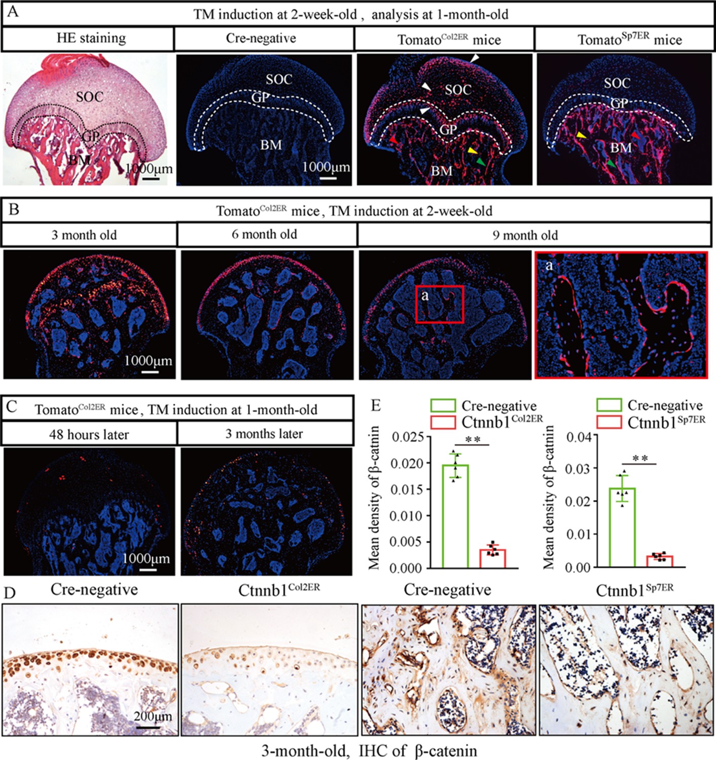
Fate mapping of Col2+ cells and Sp7+ cells in the femoral head.
TomatoCol2ER mice and TomatoSp7ER mice continuously received five doses of tamoxifen (TM) injections (1 mg/10 g body weight) at the age of 2 wk for fate mapping analysis. (A) Distributions of Col2+ and Sp7+ cells in the femoral heads of 1-month-old TomatoCol2ER mice and TomatoSp7ER mice. White arrowheads: chondrocytes; green arrowheads: osteoblasts; yellow arrowheads: osteocytes; red arrowheads: bone marrow stromal cells; GP: growth plate; SOC: second ossification center; BM: bone marrow. (B) Lineage trace of Col2+ cells in the femoral head for 9 mo. a: high-magnification image of bone marrow region. (C) Poor Cre-recombinase efficiency in the femoral heads of TomatoCol2ER mice with TM induction at the age of 1 mo. (D, E) Femoral heads were harvested from 3-month-old Ctnnb1Col2ER mice and Ctnnb1Sp7ER mice to detect expression of β‐catenin. Immunohistochemistry (IHC) staining and quantitative analysis of β-catenin in the femoral heads of 3-month-old Ctnnb1Col2ER mice and Ctnnb1Sp7ER mice.
-
Figure 4—source data 1
Raw data for Figure 4.
- https://cdn.elifesciences.org/articles/92469/elife-92469-fig4-data1-v1.zip
Figure 5
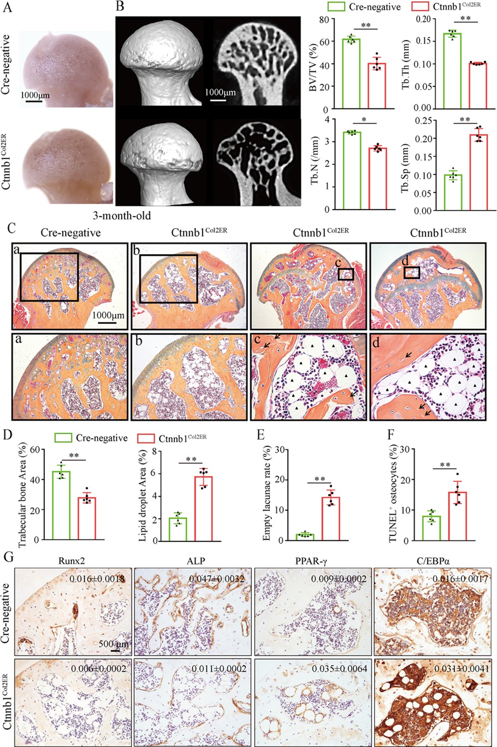
Deletion of Ctnnb1 in Col2+ cells leads to a glucocorticoid-induced osteonecrosis of the femoral head (GONFH)-like phenotype.
Femoral heads were harvested from 3-month-old Ctnnb1Col2ER mice and Cre-negative littermates with five continuous dosages of tamoxifen (TM) injections (1 mg/10 g body weight) at the age of 2 wk. (A) Gross anatomy analysis of femoral heads in Ctnnb1Col2ER mice and Cre-negative littermates. (B) Representative μCT images and quantitative analysis of BV/TV, Tb.N, Tb.Th, and Tb.Sp in the femoral heads of Ctnnb1Col2ER mice. (C) Alcian Blue Hematoxylin (ABH) staining of femoral heads in Ctnnb1Col2ER mice. (a–d) High-magnification images of representative subchondral bone region; black triangle arrowheads: fat droplets; black arrows: empty lacunae of osteocytes. (D) Histomorphological quantitative analysis of trabecular bone area and fat droplet area in the femoral heads of Ctnnb1Col2ER mice. (E) Histomorphological quantitative analysis of empty lacunae rate in the femoral heads of Ctnnb1Col2ER mice. (F) TUNEL staining of osteocytes in the femoral heads of Ctnnb1Col2ER mice. (G) Immunohistochemistry (IHC) staining and quantitative analysis of Runx2, ALP, PPAR-γ, and CEBP/α in the femoral heads of Ctnnb1Col2ER mice.
-
Figure 5—source data 1
Raw data for Figure 5.
- https://cdn.elifesciences.org/articles/92469/elife-92469-fig5-data1-v1.zip
Figure 6
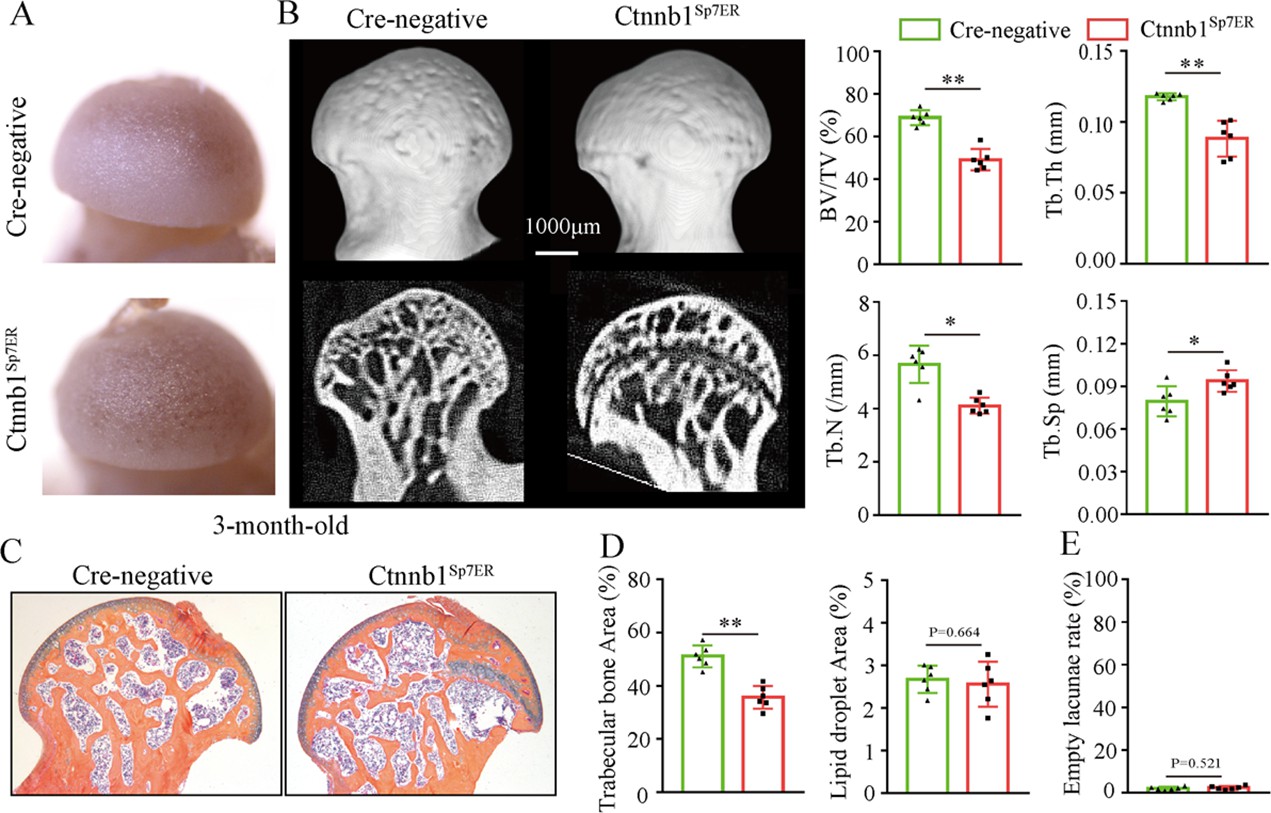
Loss of function of Ctnnb1 in Sp7+ cells causes bone loss in the femoral heads.
Femoral heads were harvested from 3-month-old Ctnnb1Sp7ER mice and Cre-negative littermates with five continuous dosages of tamoxifen (TM) injections (1 mg/10 g body weight) at the age of 2 wk. (A) Gross anatomy analysis of femoral heads in Ctnnb1Sp7ER mice and Cre-negative littermates. (B) μCT images and quantitative analysis of BV/TV, Tb.N, Tb.Th, and Tb.Sp in the femoral heads of Ctnnb1Sp7ER mice. (C) Alcian Blue Hematoxylin (ABH) staining of femoral heads in Ctnnb1Sp7ER mice. (D) Histomorphological quantitative analysis of trabecular bone area and fat droplet area in the femoral heads of Ctnnb1Sp7ER mice. (E) Histomorphological quantitative analysis of empty lacunae rate in the femoral heads of Ctnnb1Sp7ER mice.
-
Figure 6—source data 1
Raw data for Figure 6.
- https://cdn.elifesciences.org/articles/92469/elife-92469-fig6-data1-v1.zip
Figure 7
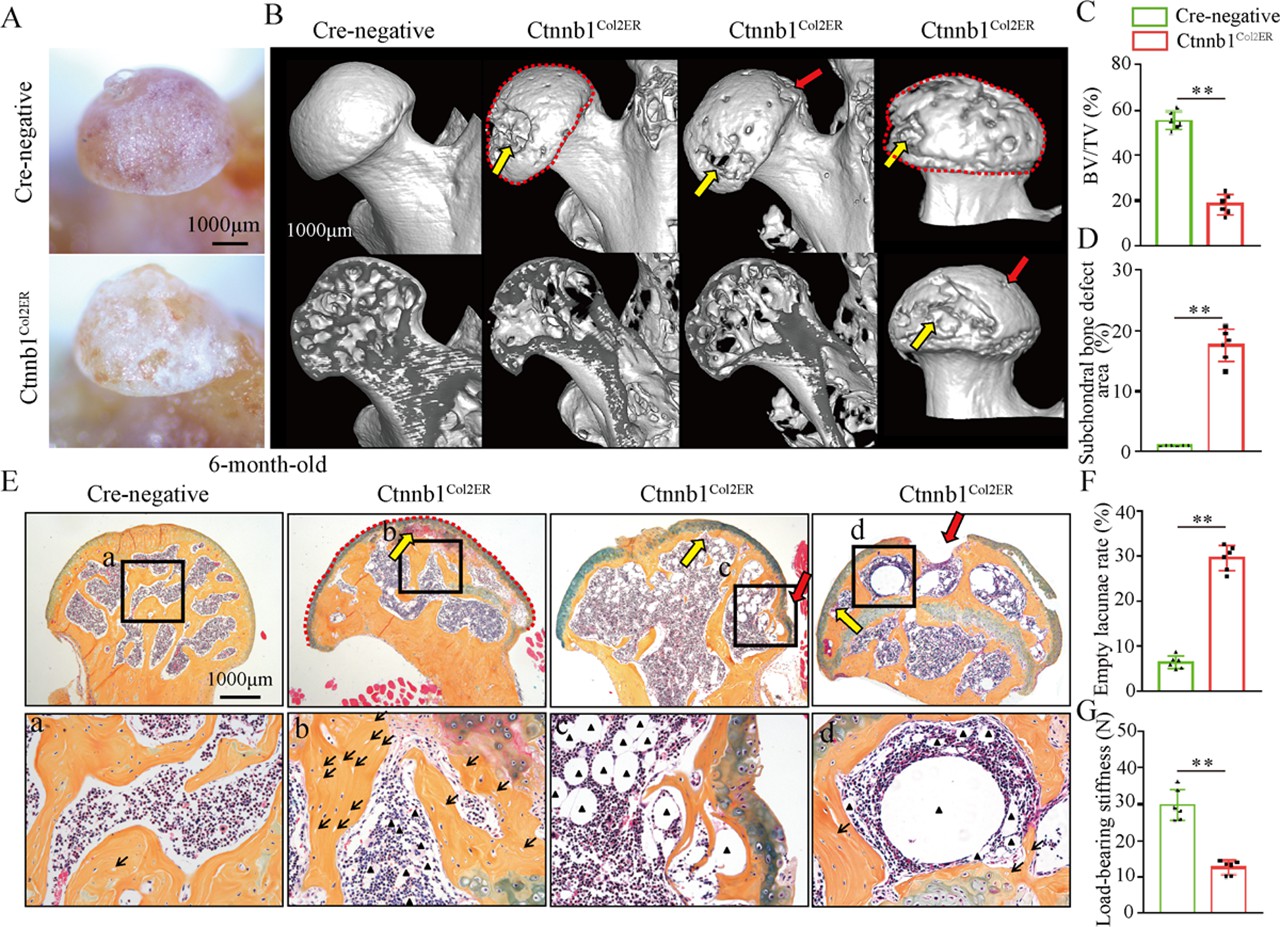
Older Ctnnb1Col2ER mice display subchondral bone destruction and collapse tendency in femoral heads.
Femoral heads were harvested from 6-month-old Ctnnb1Col2ER mice and Cre-negative littermates with five continuous dosages of tamoxifen (TM) injections (1 mg/10 g body weight) at the age of 2 wk. (A) Gross anatomy analysis of femoral heads in 6-month-old Ctnnb1Col2ER mice. (B) Representative μCT images of femoral heads in 6-month-old Ctnnb1Col2ER mice. Red dotted lines: integral deformity. Red arrows: local collapse. Yellow arrows: subchondral bone destruction. (C) Quantitative analysis of BV/TV in the femoral heads in 6-month-old Ctnnb1Col2ER mice. (D) Quantitative analysis of subchondral bone defect area on the femoral head surface in older Ctnnb1Col2ER mice. (E) Alcian Blue Hematoxylin (ABH) staining of femoral heads in 6-month-old Ctnnb1Col2ER mice. (a–c) High-magnification images of representative subchondral bone region. Red dotted lines: integral deformity. Red arrows: local collapse. Black arrows: empty lacunae of osteoctyes. Black triangles: fat droplets. (F) Histomorphological quantitative analysis of empty lacunae rate. (G) Loading-bearing stiffness of the femoral heads in 6-month-old Ctnnb1Col2ER mice.
-
Figure 7—source data 1
Raw data for Figure 7.
- https://cdn.elifesciences.org/articles/92469/elife-92469-fig7-data1-v1.zip
Figure 8
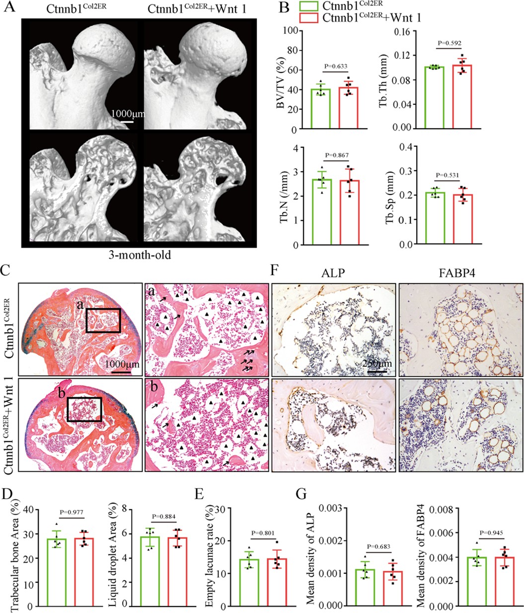
Systemic injection of Wnt agonist 1 cannot alleviate the glucocorticoid-induced osteonecrosis of the femoral head (GONFH)-like phenotype in Ctnnb1Col2ER mice.
Ctnnb1Col2ER mice with five continuous dosages of tamoxifen (TM) injections (1 mg/10 g body weight) were treated with Wnt agonist 1 three times once a week until sacrifice at the age of 3 mo. (A) Representative μCT images of femoral heads in Wnt agonist 1-treated Ctnnb1Col2ER mice. (B) Quantitative analysis of BV/TV, Tb.N, Tb.Th, and Tb.Sp in the femoral heads of Wnt agonist 1-treated Ctnnb1Col2ER mice. (C) Alcian Blue Hematoxylin (ABH) staining of the femoral heads in Wnt agonist 1-treated Ctnnb1Col2ER mice. (a, b) High-magnification images of subchondral bone region. Black triangles: fat droplets. (D) Histomorphological quantitative analysis of trabecular bone area and fat droplet area in the femoral heads of Wnt agonist 1-treated Ctnnb1Col2ER mice. (E) Histomorphological quantitative analysis of empty lacunae rate in the femoral heads of Wnt agonist 1-treated Ctnnb1Col2ER mice. (F, G) Immunohistochemistry (IHC) staining and quantitative analysis of ALP and FABP4 in the femoral heads of Wnt agonist 1-treated Ctnnb1Col2ER mice.
-
Figure 8—source data 1
Raw data for Figure 8.
- https://cdn.elifesciences.org/articles/92469/elife-92469-fig8-data1-v1.zip
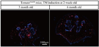
Author response image 1
Additional files
-
Supplementary file 1
The information of patients who provided femoral head samples.
- https://cdn.elifesciences.org/articles/92469/elife-92469-supp1-v1.docx
-
Supplementary file 2
The primer sequences of mouse genotype.
- https://cdn.elifesciences.org/articles/92469/elife-92469-supp2-v1.docx
-
MDAR checklist
- https://cdn.elifesciences.org/articles/92469/elife-92469-mdarchecklist1-v1.docx
Download links
A two-part list of links to download the article, or parts of the article, in various formats.
Downloads (link to download the article as PDF)
Open citations (links to open the citations from this article in various online reference manager services)
Cite this article (links to download the citations from this article in formats compatible with various reference manager tools)
β-catenin inhibition disrupts the homeostasis of osteogenic/adipogenic differentiation leading to the development of glucocorticoid-induced osteonecrosis of the femoral head
eLife 12:RP92469.
https://doi.org/10.7554/eLife.92469.3


