Proteasomes: Nrf1 to the rescue
- University of Texas Southwestern Medical Center, United States
Proteasomes are protein complexes that play an important role in cells by breaking down proteins that are damaged or are not needed, thus allowing the amino acids in the proteins to be recycled to make new proteins. Sometimes the proteins that need to be broken down are in the cytosol, and sometimes they are bound to the membrane of the endoplasmic reticulum—the organelle inside which strings of amino acids are folded to make proteins. The process through which the latter proteins are broken down is known as endoplasmic reticulum-associated degradation.
Now, in eLife, Senthil Radhakrishnan, Willem den Besten and Raymond Deshaeis of the California Institute of Technology report that endoplasmic reticulum-associated degradation is also involved in the production of proteasomes to begin with (Radhakrishnan et al., 2014). This involvement happens via a transcription factor called Nrf1 that controls the transcription of the genes that encode the various subunits within the proteasome. The Caltech group shows that, under normal circumstances, this transcription factor is always degraded by the proteasomes. However, when the level of proteasomal activity within the cell drops, the transcription factor can travel to the nucleus of the cell and kick start production of more proteasomes—a phenomenon known as ‘bounce back’.
Endoplasmic reticulum-associated degradation begins with enzymes on this organelle adding small proteins called ubiquitins to the proteins that need to be broken down. However, the proteasomes are found in the cytosol of the cell, not the endoplasmic reticulum, so the proteins that have been ubiquitinated must somehow be brought together with the proteasomes to allow the degradation process to take place. To make this happen the parts of the proteins that are located inside the endoplasmic reticulum undergo a process called retrotranslocation—which is catalyzed by a protein called p97—that moves them to the cytosol (Vembar and Brodsky, 2008). Once the ubiquitinated proteins are exposed to the proteasomes in the cytosol, the process of breaking them down can begin.
The transcription factor Nrf1 is also produced within the endoplasmic reticulum and contains an NH2-terminal domain at one end and a COOH-terminal basic leucine zipper (bZip) domain at the other end (Figure 1). The bZip domain is the part of Nrf1 that activates transcription of the genes that encode the various subunits within the proteasome.
Figure 1
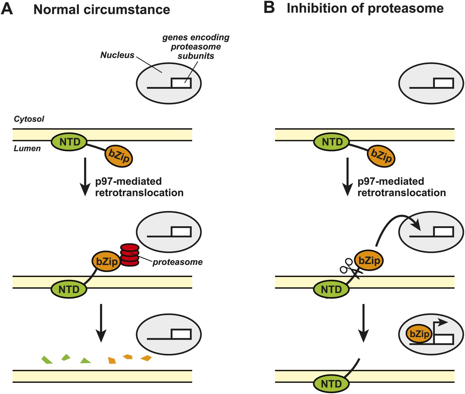
Nrf1 and the regulation of proteasome synthesis.
The transcription factor Nrf1 contains an NH2-terminal domain (NTD) that directs it into the lumen of the endoplasmic reticulum (ER) and allows it to interact with the membrane of the ER (shown in yellow). Nrf1 also contains a COOH-terminal basic leucine zipper domain (bZip) that can activate the transcription of certain genes. (A) Under normal circumstances, the enzyme p97 catalyzes the retrotranslocation of Nrf1 so that the bZip domain is in the cytosol, which results in the whole protein being rapidly degraded by proteasomes. (B) When proteasome inhibitors are added to the cell, the retrotranslocated Nrf1 is cleaved by an unidentified enzyme. This allows the bZip domain to leave the membrane and enter the nucleus, where it activates transcription of the genes that encode the various subunits within the proteasome.
Figure credit: Nancy Heard and Jin Ye
The Caltech group showed that, under normal conditions, the bZip domain of the protein was retrotranslocated from the endoplasmic reticulum to the cytosol, where it was rapidly degraded by the proteasomes. This process happened continuously, which meant that the bZip domain never reached the target genes in the nucleus of the cell (Figure 1A). However, when the cells were treated with a proteasome inhibitor, the amount of retrotranslocated Nrf1 (and hence the amount of bZip in the cytosol) increased, which allowed an unidentified enzyme to cleave the transcription factor between the NH2-terminal domain and the bZip domain. Since the bZip domain was no longer attached to the membrane of the endoplasmic reticulum, it was able to enter the nucleus and activate transcription of genes encoding the proteasome subunits (Figure 1B). This mechanism allows cells to sense any reduction in the activity of proteasomes and to compensate for this by increasing the synthesis of new proteasomes.
The work of the Caltech group is significant as it reveals the signaling mechanism that regulates proteasome synthesis. This knowledge should be useful in developing novel strategies to treat diseases in which abnormal proteasomal activity is involved. For example, neurodegenerative diseases such as Parkinson’s disease and various Prion diseases are characterized by the accumulation of ubiquitinated protein aggregates in neurons, which suggests that there too little proteasomal activity within these cells (Lehman, 2009). Is the expression or activation of Nrf1 impaired in these neurons? If so, will activation of the Nrf1 signaling pathway help to eliminate these aggregates? Future studies to address these questions may yield desperately needed novel strategies to combat these diseases by increasing proteasome synthesis.
Treating certain cancers, on the other hand, will require the opposite approach. Proteasome inhibitors have been used to treat multiple myeloma by blocking the degradation of proteins that inhibit the proliferation of cancer cells (Mahindra et al., 2012). Although this treatment is effective at first, nearly all patients eventually relapse because the increased expression of proteasome subunits in the cancer cells leads to drug resistance. Therefore, inhibiting the activation of Nrf1 might make such treatment more effective. This could be done by targeting p97, the protein that catalyzes the retrotranslocation process: however, p97 performs multiple functions inside cells (Halawani and Latterich, 2006), so a better target would be the unidentified enzyme that cleaves Nrf1. Identification of this enzyme is, therefore, a clear priority for future research.
References
-
p97: the cell’s molecular purgatory?Molecular Cell 22:713–717.https://doi.org/10.1016/j.molcel.2006.06.003
-
The ubiquitin proteasome system in neuropathologyActa Neuropathology 118:329–347.https://doi.org/10.1007/s00401-009-0560-x
-
Latest advances and current challenges in the treatment of multiple myelomaNature Reviews Clinical Oncology 9:135–143.https://doi.org/10.1038/nrclinonc.2012.15
-
One step at a time: endoplasmic reticulum-associated degradationNature Reviews Molecular Cell Biology 9:944–957.https://doi.org/10.1038/nrm2546
Article and author information
Author details
Publication history
Copyright
© 2014, Ye
This article is distributed under the terms of the Creative Commons Attribution License, which permits unrestricted use and redistribution provided that the original author and source are credited.
Download links
A two-part list of links to download the article, or parts of the article, in various formats.
Downloads (link to download the article as PDF)
Open citations (links to open the citations from this article in various online reference manager services)
Cite this article (links to download the citations from this article in formats compatible with various reference manager tools)
Proteasomes: Nrf1 to the rescue
eLife 3:e02062.
https://doi.org/10.7554/eLife.02062
Further reading
-
- Cell Biology
- Immunology and Inflammation
The endothelial blood-brain barrier (BBB) strictly controls immune cell trafficking into the central nervous system (CNS). In neuroinflammatory diseases such as multiple sclerosis, this tight control is, however, disturbed, leading to immune cell infiltration into the CNS. The development of in vitro models of the BBB combined with microfluidic devices has advanced our understanding of the cellular and molecular mechanisms mediating the multistep T-cell extravasation across the BBB. A major bottleneck of these in vitro studies is the absence of a robust and automated pipeline suitable for analyzing and quantifying the sequential interaction steps of different immune cell subsets with the BBB under physiological flow in vitro. Here, we present the under-flow migration tracker (UFMTrack) framework for studying immune cell interactions with endothelial monolayers under physiological flow. We then showcase a pipeline built based on it to study the entire multistep extravasation cascade of immune cells across brain microvascular endothelial cells under physiological flow in vitro. UFMTrack achieves 90% track reconstruction efficiency and allows for scaling due to the reduction of the analysis cost and by eliminating experimenter bias. This allowed for an in-depth analysis of all behavioral regimes involved in the multistep immune cell extravasation cascade. The study summarizes how UFMTrack can be employed to delineate the interactions of CD4+ and CD8+ T cells with the BBB under physiological flow. We also demonstrate its applicability to the other BBB models, showcasing broader applicability of the developed framework to a range of immune cell-endothelial monolayer interaction studies. The UFMTrack framework along with the generated datasets is publicly available in the corresponding repositories.
-
- Cell Biology
- Chromosomes and Gene Expression
We investigated the role of the nucleolar protein Treacle in organizing and regulating the nucleolus in human cells. Our results support Treacle’s ability to form liquid-like phase condensates through electrostatic interactions among molecules. The formation of these biomolecular condensates is crucial for segregating nucleolar fibrillar centers from the dense fibrillar component and ensuring high levels of ribosomal RNA (rRNA) gene transcription and accurate rRNA processing. Both the central and C-terminal domains of Treacle are required to form liquid-like condensates. The initiation of phase separation is attributed to the C-terminal domain. The central domain is characterized by repeated stretches of alternatively charged amino acid residues and is vital for condensate stability. Overexpression of mutant forms of Treacle that cannot form liquid-like phase condensates compromises the assembly of fibrillar centers, suppressing rRNA gene transcription and disrupting rRNA processing. These mutant forms also fail to recruit DNA topoisomerase II binding protein 1 (TOPBP1), suppressing the DNA damage response in the nucleolus.


