Peer review process
Revised: This Reviewed Preprint has been revised by the authors in response to the previous round of peer review; the eLife assessment and the public reviews have been updated where necessary by the editors and peer reviewers.
Read more about eLife’s peer review process.Editors
- Reviewing EditorAshley Tyrer
- Senior EditorChristian BüchelUniversity Medical Center Hamburg-Eppendorf, Hamburg, Germany
Reviewer #2 (Public Review):
Summary:
The authors have conducted an exceptionally informative series of studies investigating the neural basis of interoception in transdiagnostic psychiatric symptoms. By comparing differential and overlapping neural activation during 'top-down' and 'bottom-up' interoceptive tasks, they reveal convergent activation largely localised to the ventral dysgranular subregion ('mid-insula'), which differs in extent between patients and controls, replicating and extending previous suggestions of this region as a central locus of disruption in psychiatric disorders. Their work also reveals different extents of divergent activation in the anterior insula during anticipation of interoceptive disruption. This substantially advances our previous knowledge of the anatomy of interoception, and confirms theoretical predictions of the roles of different cytoarchitectural subregions of the insula in interoceptive dysfunction in mental health conditions.
Strengths:
The work is exceptional in terms of breadth and depth, making use of multiple imaging and analysis techniques which are non-standard and go well beyond what is known today. The study is statistically well-powered and the tasks are well-validated in the literature. To my knowledge, these functions of the insula in interoception and mental health have never been compared directly before, so the results are novel and informative for both basic science and psychiatry. The work is strongly theory-driven, building on and directly testing results from influential theories and previous studies. It is likely that the results will strengthen our theoretical models of interoception and advance psychiatric studies of the insula.
Weaknesses:
The study has three limitations. (1) The interpretation of the resting-state isoproterenol data could potentially represent fluctuations over time rather than following interoception specifically; future studies should investigate test-retest reliability of this measure. Note this does not preclude the strong conclusions which can be drawn from the authors' task-based data. (2) The transdiagnostic patient sample was almost entirely female, and many were currently taking psychotropic medications; future studies should replicate these effects in unmedicated, sex-balanced samples (3) As the authors point out, there may have been task-specific preprocessing/analysis differences that influenced results, for example due to physiological correction in one but not both tasks; however, there are also merits to this analysis approach, such as comparability with previous studies.
Reviewer #3 (Public Review):
Summary:
Adamic and colleagues present fMRI data from ADE patients and a healthy control group acquired during two interoceptive tasks (attention and perturbation) from the same session. They report convergent activity within the granular and dysgranular insular cortex during both tasks, with a patient group-specific lateralisation effect. Furthermore, insular functional connectivity was found to be linked to disease severity.
Strengths:
The study is well-designed and - despite some limitations noted by the authors - provides much-needed insight into the functional pathways of interoceptive processing in health and disease. The manuscript is clear, concise, and well-written.
Weaknesses:
None remain after the authors' revision.
Reviewer #4 (Public Review):
Summary:
In the manuscript titled "Hemispheric Divergence of Interoceptive Processing Across Psychiatric Disorders", the authors analyzed a subset of data collected for a larger project investigating interoception in anorexia nervosa and generalized anxiety disorder (ClinicalTrials.gov Identifier: NCT02615119). This study utilized fMRI and various analyses with a special focus on the insula and its connectivity to map the neural commonalities and differences in both top-down and bottom-up interoceptive processing.
The primary aim was to compare whether these neural activations were quantitatively and qualitatively different in a sample of healthy controls (HC) versus patients diagnosed with anxiety, depression, and/or eating disorders (ADE).
The study initially recruited 70 patients with primary diagnoses of ADE and 57 HC. After applying exclusion criteria, the final sample consisted of 46 ADE patients and 46 matched HC. Participants underwent task-related and resting-state fMRI scan sessions.
Specifically, participants performed 2 tasks in fMRI: i) a bottom-up interoceptive (ISO) task involving intravenous infusions of isoproterenol (a peripherally-acting beta-adrenergic receptor agonist) administered in a double-blind, placebo-controlled fashion to alter cardiovascular activity where participants were asked about their visceral awareness; and ii) a top-down interoceptive attention (VIA) task where participants were asked to focus on their visceral sensations triggered by words indicating specific body parts (e.g., STOMACH, HEART, LUNGS) or to pay attention to color changes of the word TARGET during an exteroceptive control task.
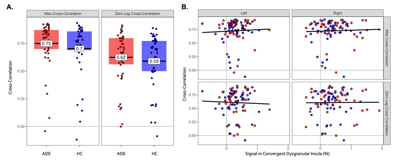
Main results show overlapping patterns of neural activation within the dysgranular mid-insula during top-down and bottom-up interoceptive processing with hemispheric differences. The patterns of dysgranular activation distinguished individuals with ADE compared to HC. Also differences in the activation of the anterior agranular insula during periods of interoceptive uncertainty differentiate ADE patients from HC.
Strengths:
- This is a very nice study that aligns with modern Clinical Neuroscience approaches, as recommended by NIH policy (i.e. RDoC initiative), which puts emphasis describing clinical conditions via transdiagnostic dimensions measured on psychological processes, behaviors, and neural processes rather than merely identifying a series of symptoms.
I appreciated very much the different analyses that authors performed to characterize differences at the qualitative and quantitative regarding the insular activity and its connectivity during bottom-up and top-down interoceptive processes.
These findings may open avenues for new studies that will explain the mechanisms underlying these phenomena and provide useful insights for developing novel interventions.
Weaknesses:
Weakness/Requests of additional clarifications
(1) The sample
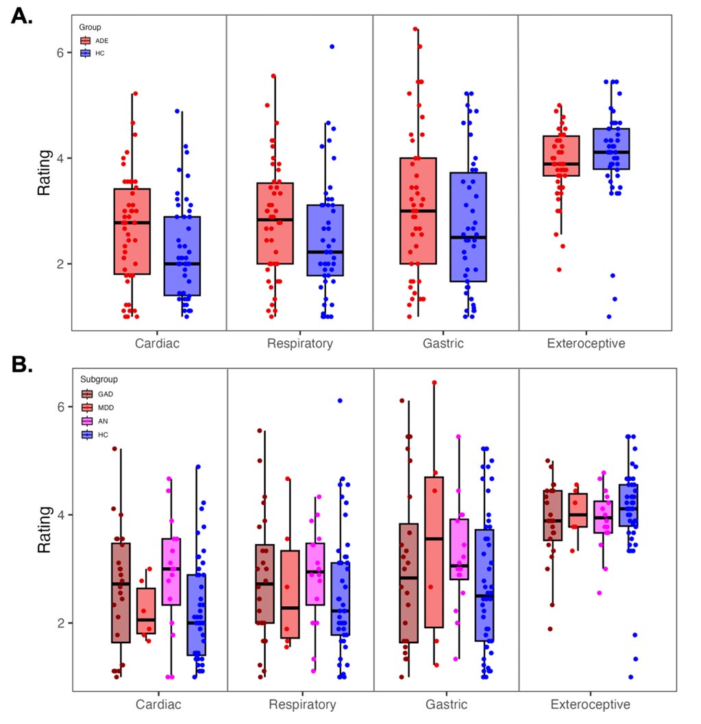
(1.1) The authors describe the patient's group as having a primary diagnosis of anxiety, depression, and/or eating disorders. However, Table 1 shows that the majority had Anxiety disorders, some Major Depression (it is not clear which are the percentages of patients that at the time of the study had a concurred problem of major depression, please clarify), and very few had a diagnosis of Anorexia Nervosa. The leftward activation asymmetry and distinct activation patterns in the left dysgranular mid-insula across both the ISO and VIA tasks found on ADE did not correlate with symptoms measured by the SCOFF questionnaire, but correlated with anxiety and depressive symptoms. It would be nice if the authors can comment on these results in relation to eating disorders.
(1.2) Furthermore, the sample consisted of 5 males and 41 females in the HC group and 1 male and 45 females in the ADE group. In order to generalize these findings, the authors should acknowledge this gender imbalance and discuss whether they expect similar results in a predominantly male sample.
(2) The procedure
While the fixed order of tasks reflects the primary emphasis on acquiring data from the infusion (ISO) task, this could introduce confounding order effects. The authors should acknowledge this as a limitation of this study.
(3) The rationale behind the study
- The authors recognized that there was a broader aim behind this data collection. It would be important to clarify a little bit more how the differences in insular areas mapping both (or specifically) bottom-up and top-down interoceptive processes and insular connectivity, recorded in ADE patients compared to healthy controls (HC), contribute to psychiatric diagnoses (hypothesis 3).
For example, they should explain the psychopathological dimensions common to the three patient groups. Are disturbances in bottom-up and top-down interoceptive processing common traits in these patients, reflected in the asymmetric interhemispheric dysgranular mid-insular activation? The link between these disturbances and anatomical evidence of convergence/divergence of top-down vs. bottom-up interoceptive processes should be clearly stated.
(4) Operationalization of Convergence / Divergence maps underlying top-down and bottom-up interoceptive processes in HC vs ADE patients
It is not clear to me the concept of Convergence / Divergence maps underlying top-down and bottom-up interoceptive processes. The authors want to compare, in HCs and ADE patients, the neural structures that are co-activated (convergence maps) vs those that are uniquely involved (divergence maps) in top-down and bottom-up interoceptive processes in the two groups. Thus, I would expect that these two different analyses would have been performed on similar portions of data, instead different moments of the tasks (= different bottom-up / top-down interoceptive processes) have been analyzed.
Specifically, the convergence maps have been identified by comparing active voxels recorded when participants were focusing on the heart and the lungs (compared to when they were focused on the exteroceptive features of the target) in the VIA task, and during infusions (Peak period) of 2mcg isoproterenol (compared to baseline) in the ISO task. The divergence maps have been identified by comparing voxels uniquely active during the anticipatory phases of both isoproterenol and saline infusions (compared to baseline) and during the peak period of saline dose of the ISO task with respect to when participants focused their attention on the heart and the lungs (compared to when they were focuses on the exteroceptive features) in the VIA task.
I understand the idea of mapping interoceptive uncertainty, however I think that these two analyses do not show commonalities and differences in the neural structures involved in bottom up vs top down processes (in ADE vs HC), but also neural correlates underlying different types of interoceptive processes involving or nor top-down expectations.
According to the authors, which is the most important neural marker that differentiates the ADE group: the difference in hemispheric activations within the left and right dysgranular insula or the less granular anterior insular activation during periods of interoceptive uncertainty? Also, do they reflect different transdiagnostic dimensions?
(5) Collected physiological measures
The authors speak about cardiorespiratory interoceptive processes, but they only included cardiac measures. Including respiratory changes could provide a more comprehensive comparison between bottom-up signals and top-down attentional processes. Also, I guess that the "STOMACH" trials of the VIA task were not analyzed in this study since those are used in the bigger study and since no gastric measures were collected? Please clarify this point.
(6) ISO task instructions
To better understand the task and participants' expectations, could the authors clarify the instructions given to participants regarding the isoproterenol and saline infusions. Did the participants have two types of expectations?
(7) Title of the study
I understand that the term "divergence" in the title refers to the different hemispheric activations characterizing ADE patients compared to HC. However, it also suggests an analysis based on convergence/divergence maps, which might be ambiguous. Could the authors make some small modifications to the title to make it clearer?
(8) Caption of Figure 7
The caption of Fig.7 notes that no difference in HR was found during the Saline infusion between the HC and ADE groups. However, it would be fair to mention the significant difference in dial ratings observed during the Saline infusion. How do the authors explain this difference?
Typos
Figure 3 In Figure 3, "Hemispheric divergence", I think, should be corrected to "Hemispheric convergence."
I believe that by addressing these points, the manuscript will provide a clearer and more comprehensive understanding of the rationale, methods, and findings underlying this study.


