Inflammasomes: A tale of two caspases
Macrophages control intracellular pathogens like Salmonella by using two caspase enzymes at different times during infection.
- Department of Microbiology and Immunology, Stanford University School of Medicine, United States
Salmonella bacteria are well-known (and feared) for causing countless foodborne illnesses. After entering the body through contaminated food, Salmonella target the intestinal tract, where they invade and replicate in host cells, including frontline defenders called macrophages (Santos and Bäumler, 2004).
The bacteria use a syringe and needle-like structure to inject molecules into the host cell that help reprogram it into a bacterial haven where the pathogen can replicate (Galán, 2021; Dos Santos et al., 2020). Enclosed in a specialized compartment called the Salmonella-containing vacuole (SCV), the bacteria can replicate while evading many immune defenses (Li et al., 2023). If Salmonella escape the vacuole and invade the cytosol they ultimately expose themselves to the full fury of the host’s immune arsenal.
At the heart of the immune response lies the inflammasome, a molecular sensor that detects when the cell’s inner sanctum is breached. This, in turn, sets off an explosive immune chain reaction. In macrophages, inflammasomes activate inflammatory enzymes, specifically caspase-1 and caspase-4. Caspase-1 triggers the release of alarm signals, such as interleukin-1β (IL-1β), which alert neighboring cells. In tandem, it activates gasdermin D (GSDMD), a pore-forming protein central to initiating a type of programmed cell death known as pyroptosis (Egan et al., 2023a). How exactly inflammasomes restrict bacterial replication has so far been unclear. Now, in eLife, Sunny Shin and colleagues at the University of Pennsylvania – including Marisa Egan as first author – report how two caspases play distinct roles in the battle against Salmonella enterica serovar Typhimurium (Egan et al., 2023b).
Using chemical inhibitors and genetic knockouts in human macrophages, Egan et al. showed that, early in the battle, caspase-1 was the frontline warrior (Figure 1). Blocking this enzyme caused IL-1β secretion and pyroptosis to be significantly reduced, allowing Salmonella to multiply rapidly within macrophages. As the infection advanced, caspase-4 became more important. In its absence, cells had a higher number of bacteria 24 hours following infection. This finding hints at a strategic, time-sensitive role for caspase-4 – perhaps targeting Salmonella that persist within vacuoles or those that have shed their protective layer and moved into the cytosol.
Figure 1
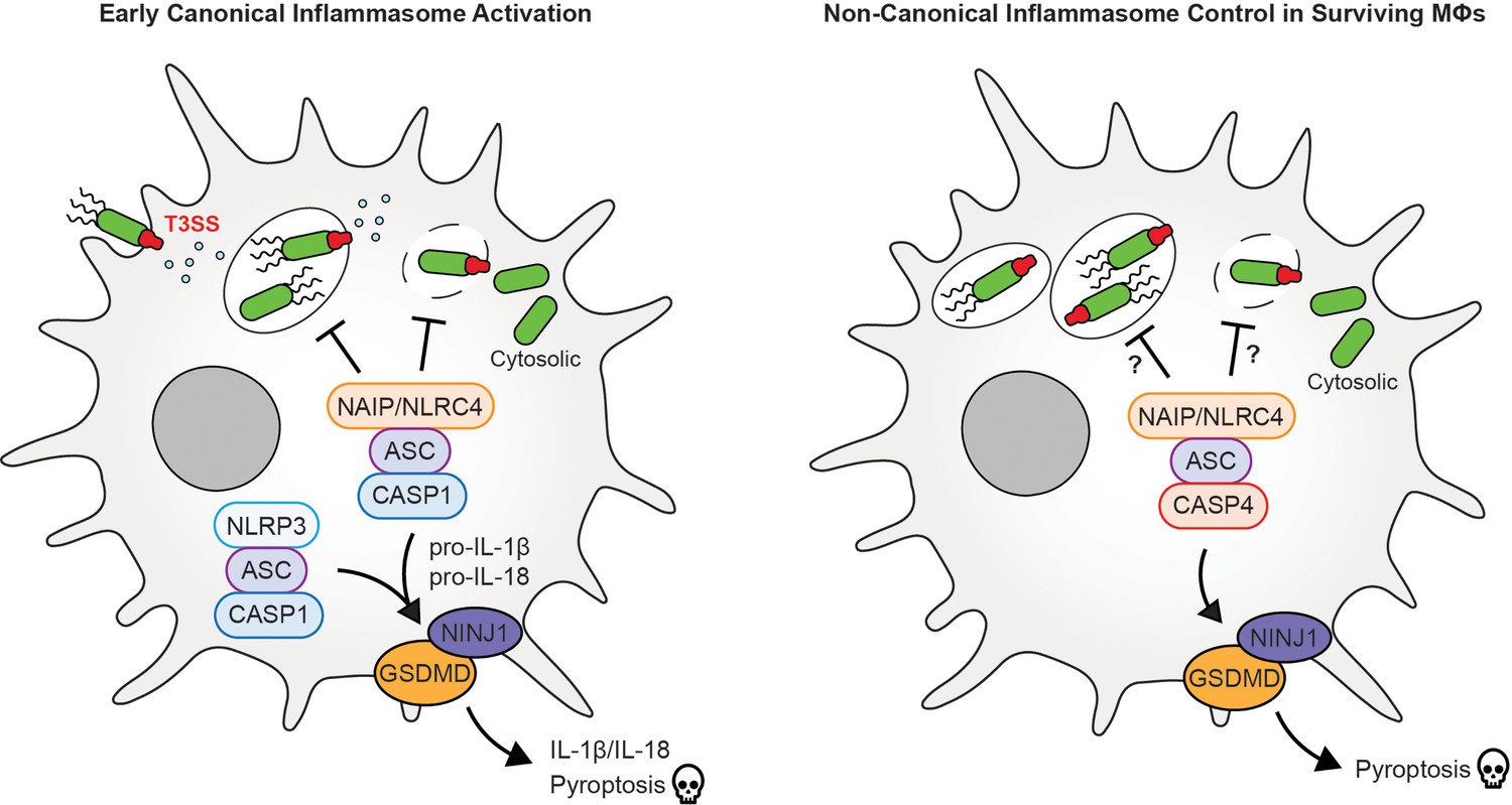
Early and late immune responses of macrophages to Salmonella bacteria.
(Left) Early inflammasome activation: During the early stages of infection, Salmonella bacteria use a type III secretion system (T3SSs; green/red structure) to inject virulence factors into the host cell to help it replicate. This, in turn, activates immune responses, such as a molecular sensor called the NAIP/NLRC4 inflammasome, which recognizes components of the T3SS. This leads to the recruitment of an adaptor protein apoptosis-associated speck-like protein containing CARD, called ASC, which is crucial for the formation of various inflammasomes and the activation of the caspase-1 (CASP1) enzyme, which transforms inactive cytokine precursors, notably interleukin-1β (IL-1β) and IL-18, into their active forms, which alert neighboring cells. CASP1 further cleaves gasdermin D (GSDMD), a pore-forming protein, allowing cytokines to be released and leading to pyroptosis, a lytic form of inflammatory cell death. NINJ1, a membrane protein, facilitates the final rupture of the cell membrane. Additional activation of NLRP3, another inflammasome sensor, further amplifies this inflammatory response. (Right) Late inflammasome control: During a later stage of infection, caspase-4 (CASP4) takes the lead, and similar to CASP1, cleaves GSDMD and triggers pyroptosis. More research is needed to find out if there is a specific control of Salmonella replication in the vacuole and cytosol at this point during infection.
Image credit: Denise Monack, with special thanks to Jeff Hsiao for providing help in making the figure (CC BY 4.0).
The researchers then looked at GSDMD and another protein involved in the final stages of pyroptosis, ninjurin-1 (NINJ1; Fu et al., 2024). When GSDMD was rendered inactive, cytokines were also inactive and cell death was stalled, enabling Salmonella to thrive. Similarly, experiments deactivating NINJ1 revealed that even a partial loss of this defense system leads to more bacteria, underscoring its vital role in curbing infection.
One of the most impactful discoveries was that inflammasomes are especially potent at limiting Salmonella replication in the cytosol. In a series of cutting-edge experiments – ranging from chemical assays to single-cell microscopy and electron microscopy – Egan et al. uncovered that when inflammasome components like caspase-1 were disabled, the number of Salmonella bacteria within the cytosol exploded, and the replication of Salmonella within vacuoles also increased significantly. In contrast, intact inflammasome signaling kept the bacterial numbers in check, ensuring their cytosolic presence remained minimal. Their findings underscore the pivotal role of inflammatory caspases and pyroptosis in steering inflammasome responses that confine Salmonella replication to specific subcellular niches within human macrophages, a crucial battleground where the pathogen must be contained.
These insights redefine our understanding of host-pathogen warfare and catapult us into the heart of a microscopic war, where inflammasomes orchestrate a ruthless defense against Salmonella. The differential roles of caspase-1 and caspase-4 and the concerted actions of GSDMD and NINJ1 provide a detailed map of how our cells fight back against intracellular invaders. This battle provides a fascinating glimpse into the complexities of the human immune response.
By mimicking or enhancing these natural defense mechanisms, we may one day devise treatments that better protect against Salmonella and similar pathogens. Human macrophages prove to be a force to be reckoned with, and the striking contrast between human and murine macrophages – where the latter seem less capable of curbing Salmonella replication – underscores the importance of studying human cells directly (Thurston et al., 2016). The detailed dissection of these mechanisms offers a thrilling promise for targeted medicine, where boosting the right component of the immune response could tip the scales in favor of the host.
References
-
Human and mouse NAIP/NLRC4 inflammasome responses to bacterial infectionCurrent Opinion in Microbiology 73:102298.https://doi.org/10.1016/j.mib.2023.102298
-
Mechanistic insights from inflammasome structuresNature Reviews Immunology 24:518–535.https://doi.org/10.1038/s41577-024-00995-w
-
Salmonella typhimurium and inflammation: a pathogen-centric affairNature Reviews Microbiology 19:716–725.https://doi.org/10.1038/s41579-021-00561-4
-
Strategies adopted by Salmonella to survive in host: a reviewArchives of Microbiology 205:362.https://doi.org/10.1007/s00203-023-03702-w
-
Cell tropism of Salmonella entericaInternational Journal of Medical Microbiology 294:225–233.https://doi.org/10.1016/j.ijmm.2004.06.029
Article and author information
Author details
Publication history
Copyright
© 2025, Monack
This article is distributed under the terms of the Creative Commons Attribution License, which permits unrestricted use and redistribution provided that the original author and source are credited.
Metrics
-
- 469
- views
-
- 29
- downloads
-
- 0
- citations
Views, downloads and citations are aggregated across all versions of this paper published by eLife.
Download links
A two-part list of links to download the article, or parts of the article, in various formats.
Downloads (link to download the article as PDF)
Open citations (links to open the citations from this article in various online reference manager services)
Cite this article (links to download the citations from this article in formats compatible with various reference manager tools)
Inflammasomes: A tale of two caspases
eLife 14:e106581.
https://doi.org/10.7554/eLife.106581


