Huntingtin’s spherical solenoid structure enables polyglutamine tract-dependent modulation of its structure and function

  1. Ravi Vijayvargia
  2. Raquel Epand
  3. Alexander Leitner
  4. Tae-Yang Jung
  5. Baehyun Shin
  6. Roy Jung
  7. Alejandro Lloret
  8. Randy Singh Atwal
  9. Hyeongseok Lee
  10. Jong-Min Lee
  11. Ruedi Aebersold
  12. Hans Hebert
  13. Ji-Joon Song  Is a corresponding author
  14. Ihn Sik Seong  Is a corresponding author
  1. Massachusetts General Hospital, United States
  2. Harvard Medical School, United States
  3. McMaster University, Canada
  4. Eidgenössische Technische Hochschule Zürich, Switzerland
  5. Korea Advanced Institute of Science and Technology, Republic of Korea
  6. Karolinska Institute, Sweden
  7. KTH Royal Institute of Technology, Sweden
  8. University of Zurich, Switzerland

Abstract

The polyglutamine expansion in huntingtin protein causes Huntington’s disease. Here, we investigated structural and biochemical properties of huntingtin and the effect of the polyglutamine expansion using various biophysical experiments including circular dichroism, single-particle electron microscopy and cross-linking mass spectrometry. Huntingtin is likely composed of five distinct domains and adopts a spherical α-helical solenoid where the amino-terminal and carboxyl-terminal regions fold to contain a circumscribed central cavity. Interestingly, we showed that the polyglutamine expansion increases α-helical properties of huntingtin and affects the intramolecular interactions among the domains. Our work delineates the structural characteristics of full-length huntingtin, which are affected by the polyglutamine expansion, and provides an elegant solution to the apparent conundrum of how the extreme amino-terminal polyglutamine tract confers a novel property on huntingtin, causing the disease.

https://doi.org/10.7554/eLife.11184.001

eLife digest

Huntington’s disease is an inherited disorder that occurs in adulthood and sometimes in children. It causes progressive damage to the brain and people with the condition develop memory loss, movement difficulties, confusion, and other symptoms of mental decline. Eventually, the disease leads to death. Mutations in the gene that encodes a protein called huntingtin cause Huntington’s disease. Individuals who inherit just one copy of the mutated gene develop the condition. No treatments currently exist that can slow or stop disease progression.

Genetic and molecular studies are beginning to shed light on how mutations in the gene encoding huntingtin cause the disease. Normally, the protein has a section near its tail end made up of the amino acid glutamine repeated around 23 times. Mutations that increase the number of glutamines to more than 38 cause Huntington’s disease. The more extra glutamines there are in this region of the protein, the earlier in life the disease symptoms begin. But it was not clear how these extra glutamines near the tail of huntingtin affect the structure and behavior of a protein that is more than 3,000 amino acids long.

Now, Vijayvargia et al. have revealed why the tail end of huntingtin is so important. Several biophysical methods were used to determine the three-dimensional structure of the huntingtin protein. These methods revealed that the protein folds up into a hollow sphere and that its tail end is able to interact with the entire length of the protein and physically touches its opposite end.

To see this in more detail, Vijayvargia et al. used another experimental technique called crosslinking mass spectrometry to confirm which parts of the huntingtin protein are in close contact with each other. Together with the structural data, these experiments suggest that the stretch of glutamines is in the position to bring about subtle, but widespread, changes throughout the huntingtin protein. That is to say, that having more glutamines slightly changes the curve of the sphere and alters the way different parts of the protein interact.

Together the new findings explain why mutations that alter the tail of huntingtin affect the rest of the protein. Further work will now aim to provide a more-detailed structure of the huntingtin protein and to investigate what other roles of huntingtin are affected by the increased number of glutamines in the protein’s tail. These insights may help scientists understand how the mutated protein causes brain decline.

https://doi.org/10.7554/eLife.11184.002

Introduction

Huntingtin is the entire large protein product (>350 kDa MW) of the Huntingtin gene (HTT previously HD). Huntingtin has a segment of polyglutamine near its amino terminus (Amino-terminal) that is encoded by a polymorphic CAG trinucleotide repeat. If expanded above 38-residues, this mutation causes Huntington’s disease (HD), a dominant neurodegenerative disorder (Huntington's Disease Collaborative Research Group, 1993). The strong correlation between the size of the expanded repeat and the age at diagnosis of HD motor, cognitive and psychiatric symptoms shows that CAG repeat-size is the primary determinant of the rate of the disease progression (Brinkman et al., 1997; Snell et al., 1993). This biological relationship also provides a human patient-based rationale for delineating the HD disease-trigger in studies with an allelic series designed to determine the effects of progressively increasing the size of the mutation. Consistent with genetic studies in distinct CAG-expansion neurodegenerative disorders and CAG knock-in mice that replicate the HD mutation, the mechanism that triggers the disease process that leads to the characteristic vulnerability of striatal neurons in HD is thought to involve a novel gain of function that is conferred on mutant huntingtin by the expanded polyglutamine segment (Gusella and MacDonald, 2000; Nucifora et al., 2001; Trettel et al., 2000).

By analogy with other members of the HEAT/HEAT-like (Huntingtin, Elongation factor 3, protein phosphatase 2A, Target of rapamycin 1) repeat family (Andrade and Bork, 1995; Perry and Kleckner, 2003), huntingtin is likely a HEAT domain solenoid that functions as a mechanical scaffold for multi-member complexes (Grinthal et al., 2010; Takano and Gusella, 2002). Huntingtin’s large size and predicted predominant HEAT/HEAT-like repeat domain structure is well conserved through 500 million years of evolution (Seong et al., 2010). The polyglutamine region is not conserved in some huntingtin orthologues (Seong et al., 2010), implying a role as an extra feature that fine-tunes huntingtin structure and function. Indeed, testing this idea, we have previously demonstrated, with purified recombinant human huntingtins in a cell-free assay, that lengthening the polyglutamine tract quantitatively enhances the basal function of huntingtin in stimulating Polycomb repressive complex 2 (PRC2) histone methyltransferase (Seong et al., 2010).

The structures of smaller HEAT/HEAT-like repeat solenoid scaffold proteins, such as PR65/A and Importin β, have been solved to high-resolution, and each has been shown to assume a distinctive extended curvilinear shape determined by the specific stacking characteristics of its HEAT/HEAT-like repeats (Cingolani et al., 1999; Groves et al., 1999). The topology of the huntingtin solenoid is expected to reflect the specific stacking characteristics of α-helical HEAT/HEAT-like repeats that span the molecule. The shape imparted by intramolecular stacking cannot be predicted because of the idiosyncratic nature of HEAT/HEAT-like repeats, which are loosely conserved ~34 amino acid bipartite α-helical units (Takano and Gusella, 2002). Nevertheless this shape must enable modulation by the amino-terminal polyglutamine segment. It seems reasonable that this may involve some structural feature that is critical to huntingtin function. One possibility is structure-dependent post-translational modification. Human huntingtin is phosphorylated, at more than seventy modified serine, threonine and tyrosine residues (Hornbeck et al., 2012). Indeed, a comparison of lines of transgenic modified HTT BAC mice has implicated unique amino-terminal serine phosphorylation in protection against deleterious effects of mutant huntingtin (Gu et al., 2009) and the polyglutamine expansion at the amino-terminal causes a trend of hypo-phosphorylation in all sites, including sites near the carboxyl-terminus (Anne et al., 2007; Schilling et al., 2006; Warby et al., 2005), indirectly implying a long-range impact of the amino-terminal region on huntingtin structure and function.

In order to solve the apparent puzzle of how huntingtin’s solenoid structure may enable quantitative or qualitative (or both) modulation of huntingtin function, according to the size of the amino-terminal polyglutamine tract, we have extended initial observations showing a likely flexible α-helical structure by conducting systematic biophysical and biochemical analyses of members of a panel of highly purified human recombinant huntingtins, with varied lengths of polyglutamine tracts (Fodale et al., 2014; Huang et al., 2015; Li et al., 2006).

Results

Huntingtin α-helical structure is quantitatively altered with polyglutamine tract size

It has been reported previously that purified huntingtin exhibits a predominantly α-helical secondary structure but among studies the impact of polyglutamine size has been inconsistent (Fodale et al., 2014; Huang et al., 2015; Li et al., 2006). To carry out a standardized evaluation, we performed circular dichroism (CD) analysis of a series of recombinant human huntingtins with different polyglutamine tract lengths (Q2-, Q23-, Q46-, Q67-, Q78-huntingtin, respectively) purified to homogeneity (Figure 1—figure supplement 1). The CD spectra (Figure 1B) of all of the huntingtins are consistent with a predominant α-helical secondary structure (Liu and Rost, 2003; Rost et al., 1994) (Figure 1A), with typical minima at 222 and 208 nm and a positive peak at 195 nm, and all exhibited the same irreversible thermal denaturation pattern, with secondary structure stable up to ~38–40°C, a gradual slow denaturation as the temperature is increased to 65–70°C, followed by aggregation and some precipitation (Figure 1C). These results imply the same basic core structure and stability regardless of the size of the expanded polyglutamine segment. Plotting an average of the Mean Residue Ellipticity (MRE) at 222 nm (characteristic of an α-helix) reveals an incremental quantitative effect of lengthening the polyglutamine tract at the amino terminus on the α-helicity of the entire molecule (Figure 1D).

Figure 1 with 1 supplement see all
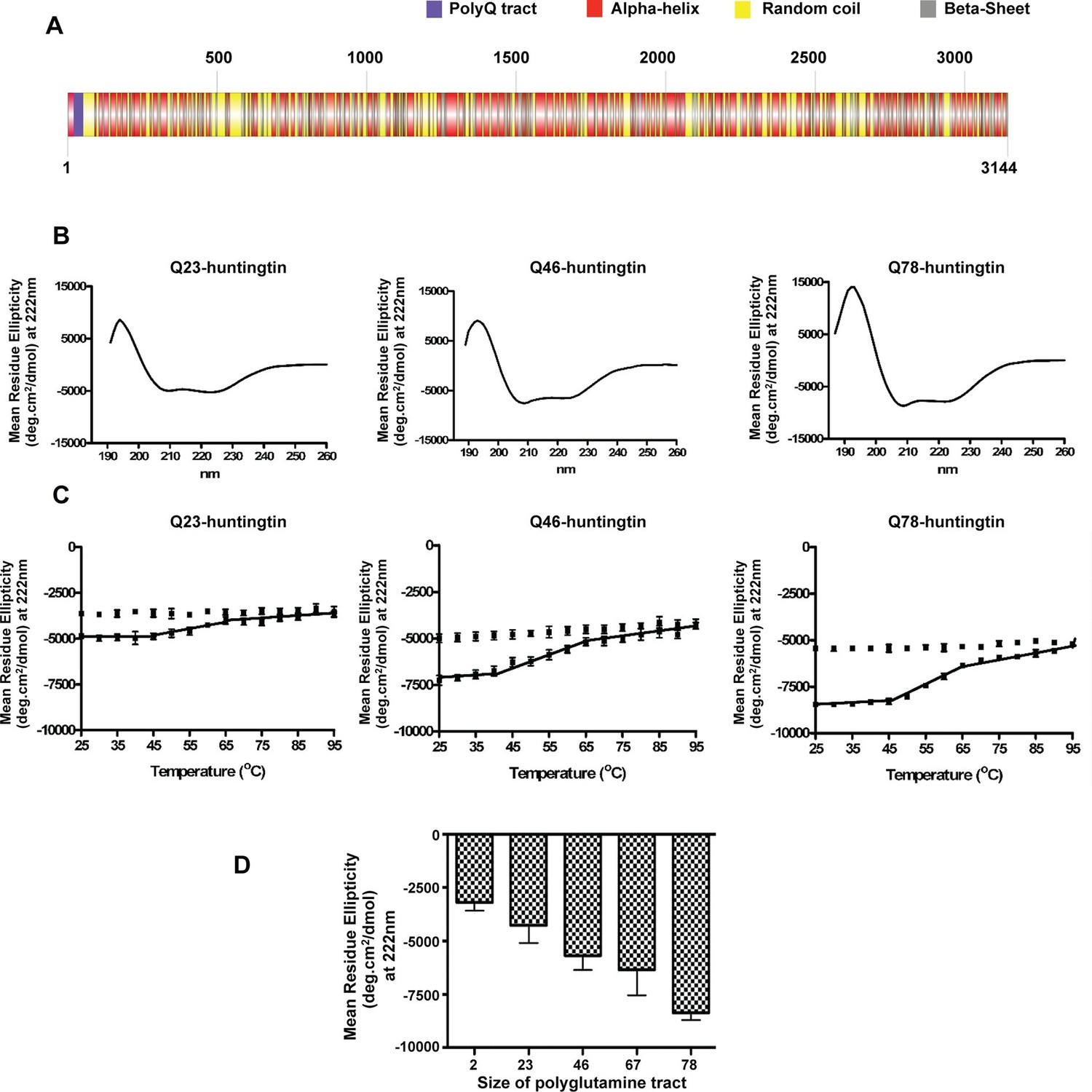
Huntingtin secondary structure is modulated by the length of polyglutamine tract.

(A) Human huntingtin amino acid sequence (Homo sapiens; NP_002102) was analyzed for predicted secondary structure using: NORSp (Liu and Rost, 2003) and PROF (Profile network prediction Heidelberg) (Rost et al.,1994). Stick model of human huntingtin protein (3144 amino acids) was generated depicting the predicted alpha helical (red), random coil (yellow) and beta sheet (grey) regions. The polyglutamine tract in the amino-terminus is indicated in purple. (B) The far UV-wavelength scan at 25°C of these purified huntingtin proteins generates a curve typical of α-helical proteins. (C) Thermal behavior of Q23-, Q46- and Q78-huntingtin. The heat denaturation curves, from 25 to 95°C, of all proteins showed the similar pattern of irreversible thermal denaturation starting their denaturation above 40°C by MRE values at 222 nm. Due to inherent variation caused by inefficient mixing in the cuvette with taking readings every five degrees of heating, their heat denaturation curves were acquired in duplicates. Solid line represents heating to 95°C; dotted line represents cooling from 95°C. (D) An average (MRE) in units of deg.cm2/dmol, at 222 nm wavelength characteristic of an α-helix), from two independent experiments, was plotted against the length of the polyglutamine tract of huntingtin proteins (bars represent mean ± SEM). Temperature was 25°C.

https://doi.org/10.7554/eLife.11184.003

3D EM analysis reveals a spherical shape with a central cavity and overlying Amino-terminus

We then investigated the proposal that huntingtin’s shape may enable a structural impact of the amino-terminal polyglutamine tract, by performing single-particle electron microscopy (EM) of recombinant human huntingtins with polyglutamine tract lengths of 23- and 78-residues. These were purified to high homogeneity using a gradient purification method with mild crosslinking (GraFix) (Kastner et al., 2008), collecting only the monomer fraction for analysis to eliminate potential contributions from oligomeric structures that would confound interpretation of the results (Figure 2—figure supplement 1). Negative-stained micrographs of these proteins confirmed that the samples were highly homogeneous (Figure 2—figure supplement 2). A total of 10,169 particles were chosen for generating 2D class averages and 30 class averages were used for reconstructing a 3D EM map of Q23-huntingtin (Figure 2—figure supplement 3A). The EM map of Q23-huntingtin at about 30 Å resolution, estimated from Fourier shell correlation analysis, shows that the molecule adopts an overall spherical shape with 130 Å height and 100 Å width (Figure 2A and Figure 2—figure supplement 4). The overall shape of huntingtin was not apparently affected either through crosslinking or by the tag as 2D class averages of huntingtin with no cross linker or without tag also showed a similar spherical shape (Figure 2—figure supplement 3). The outer volume of the structure can be roughly estimated as 861,829 Å3 and a Mathew’s coefficient (VM) of 2.48, assuming that Q23-huntingtin is a sphere with 115 Å diameter with 348 kDa molecular weight. Considering the VM=1.23 for the protein itself, Q23-huntingtin appears to contain a large solvent cavity (up to 50% by volume). Consistent with this estimation, the 3D EM reconstruction of Q23-huntingtin shows a large cavity in the core (Figure 2B). The analysis of negatively-stained Q78-huntingtin (Figure 2C and Figure 2—figure supplement 3B) disclosed a 3D map showing a similar overall spherical shape, with a large cavity in the core (Figure 2D). Although 3D maps were reconstructed de novo without other experimental methods such as random conical tilt, the high similarity of the shapes between Q23- and Q78-huntingtin 3D maps attests that huntingtin has the spherical structure with a cavity (Figure 2—figure supplement 5). Notably, manual superimposition of the 3D maps of Q23-huntingtin and Q78-huntingtin reveals potential differences in the two structures (Figure 2—figure supplement 5), which may reflect technical variation (image processing, stain distribution, sample heterogeneity), in addition to the structural effects of the lengthened polyglutamine segment that were foreshadowed by the altered CD spectra (Figure 1D).

Figure 2 with 5 supplements see all
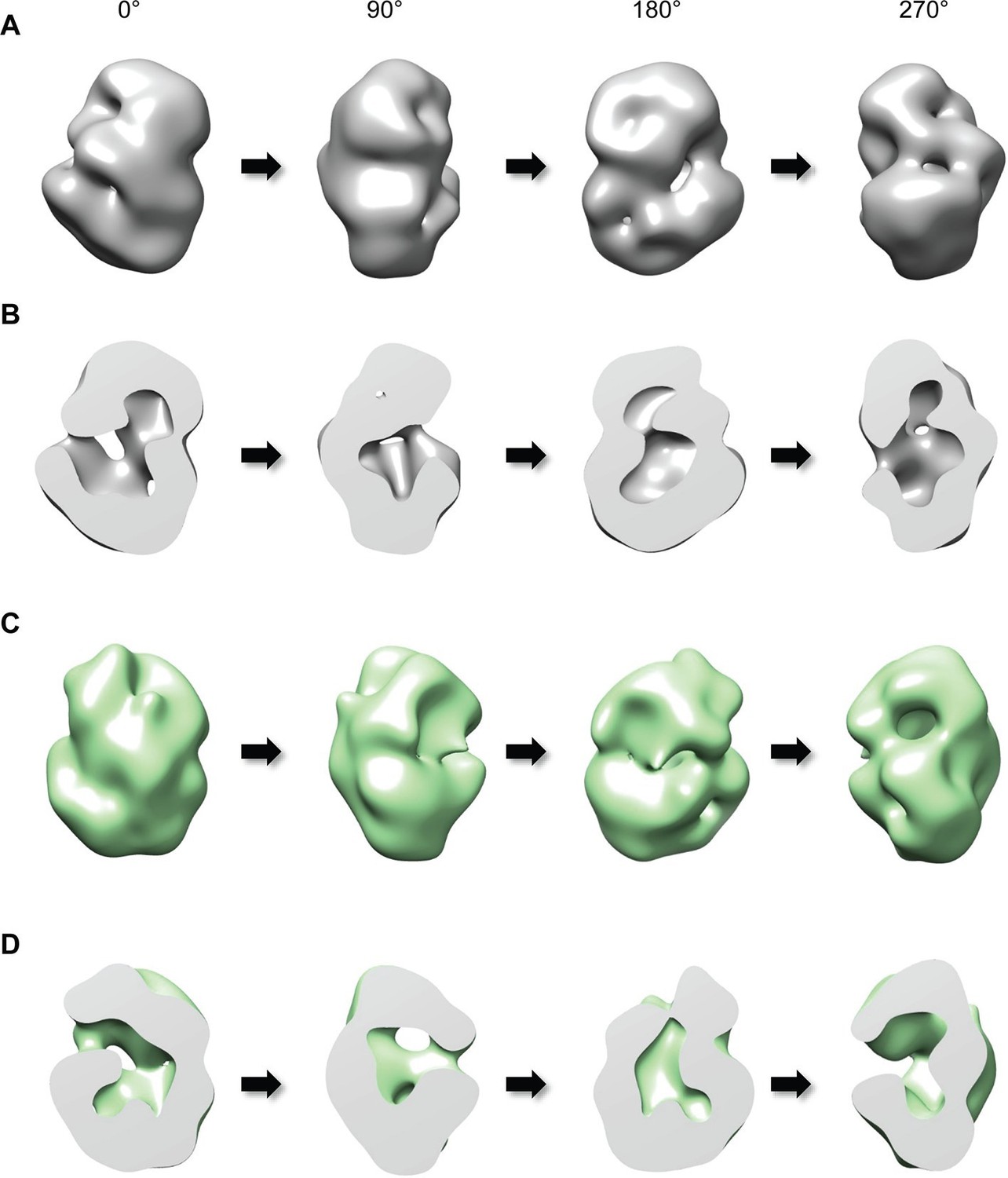
Three-dimensional reconstruction of negatively-stained Q23- and Q78-huntingtin.

(A) 3D EM map of Q23-huntingtin was reconstructed from negatively-stained particles of Q23 monomer separated by GraFix. The resolution was estimated as 33.5 Å at 0.5 FSC. 3D map of Q23-huntingtin is shown in different orientation rotated about the y axis (0°, 90°, 180°, 270°). (B) Sectioned view of 3D EM map of Q23-huntingtin in the same orientation as in A revealing a large cavity inside of Q23-huntingtin. (C) 3D EM map of Q78-huntingin (32.0 Å at 0.5 FSC) was reconstructed as for Q23-huntingtin and shown in different angles rotated about the y axis (0°, 90°, 180°, 270°). (D) Sectioned view of 3D EM map of Q78-huntingtin in the same orientation as in C also showing a large cavity inside of Q78-huntingtin. This figure has additional supplement files: Figure supplement 1, 2, 3, 4, and 5

https://doi.org/10.7554/eLife.11184.005

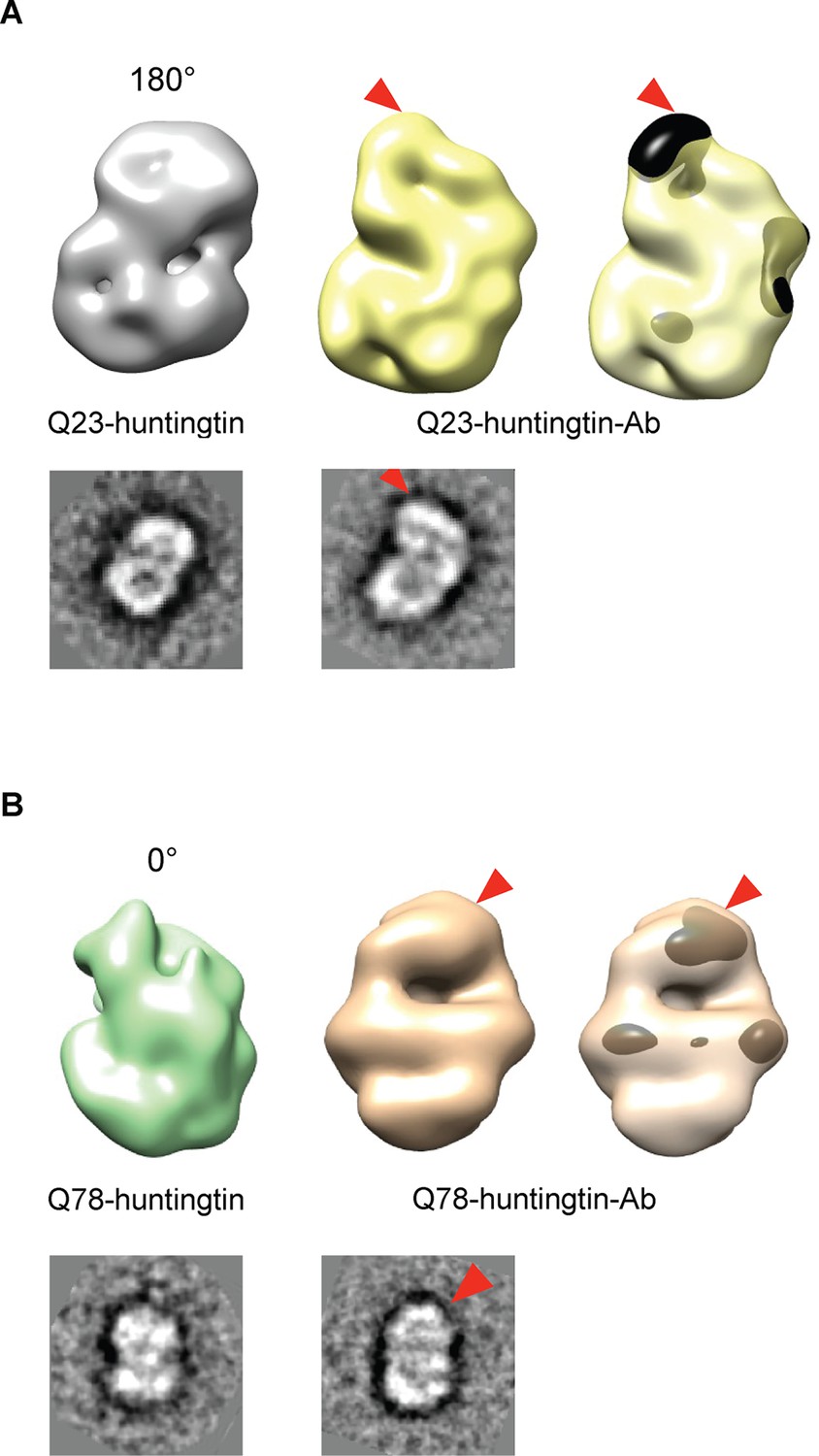
We then attempted to locate the amino-terminus (17 residues adjacent to the polyglutamine tract) of huntingtin in the EM maps, by collecting images of negatively-stained purified complexes of antibody-bound amino-terminal FLAG-tags of the Q23- and Q78-huntingtin (Figure 3 and Figure 3—figure supplement 1 and 2). We also proceeded to reconstitute 3D structure of Q23- and Q78-huntingtin-antibody complexes. Comparisons of the 2D class averages and 3D reconstituted structures between huntingtin alone and the huntingtin-FLAG-antibody complex pairs clearly reveals an extra density at the top of the structure (in the view shown) for both Q23- and Q78-huntingtin (Figure 3). These observations strongly imply that the extreme N-terminus, and by inference the adjacent polyglutamine tract, is folded back, forming a spherically shaped solenoid with an internal cavity, but is accessible at the outside surface, regardless of its length.

Figure 3 with 2 supplements see all
Detection of the amino-terminus region of huntingtin by electron microscopy.

3D reconstitutions of Q23-huntingtin and Q23-huntingtin antibody complex (A) or Q78-huntingtin and Q78-huntingtin antibody complex (B) are shown in gray and yellow or in green and brown, respectively. 2D class averages corresponding to each huntingtin and its antibody complex are shown below the 3D reconstituted EM map and the extra density is marked with a red triangle. The extra-density indicating antibody on 3D EM map is colored in black (A) or dark brown (B) with red triangles.

https://doi.org/10.7554/eLife.11184.011

Cross-linking-MS analysis reveals a modulated network of intramolecular contacts

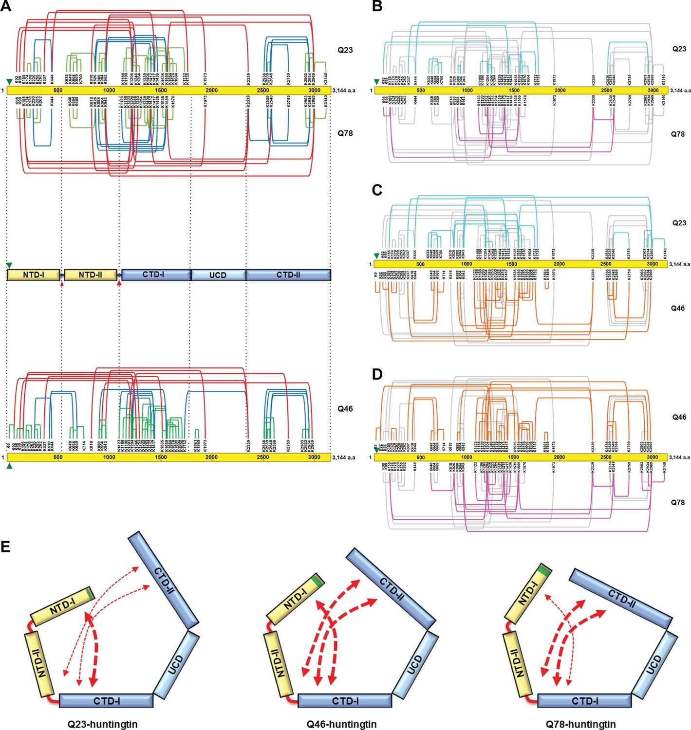
To further examine structural characteristics such as folding, we then assessed intramolecular interactions within huntingtin as estimated from the spatial proximity of lysine residues in sucrose-gradient-selected monomeric Q23- and Q78-huntingtin, determined by disuccinimidyl suberate (DSS) cross-linking mass spectrometry (DSS XL-MS) analysis (Leitner et al., 2014) (Figure 4—figure supplement 1, Supplementary file 1). Based upon the spacing between the DSS-cross-linked lysine residues in the primary sequence, the interactions that were detected for either huntingtin can be grouped into three categories, depicted in Figure 4A. These comprise: #1) short-range interactions (within a 200 amino acid interval), which seem likely to occur within the same secondary structure element, including the contacts between pairs of adjacent lysines (e.g. K174-K178 and K664-K669), and the interactions between K220, K255 and K262; #2) mid-range interactions (201 to 1000 amino acid interval), such as K826-K1559, K943-K1559 and K2548-K2934; and #3) long-range contacts (interval of >1000 amino acids), including between the carboxyl-terminal K2969 and amino-terminal K943 residue. Inspection of the depiction of the short-range cross-link contact sites relative to the location of the protease-sensitive sites (Seong et al., 2010) indicates that huntingtin is likely to be composed of five distinct domains (Figure 4A upper panel). The location of the protease-sensitive major hinge region located at residues 1184–1254 defines the 150 kDa amino-terminal domain (NTD) and the 200 kDa carboxyl-terminal domain (CTD). A minor-protease sensitive site located at ~ residue 500 demarcates the NTD into NTD-1 and NTD-II. Interestingly, a region centrally located within the CTD showed strikingly few crosslinks, despite the presence of several lysine residues, strongly implying a distinct sub-domain that we call ‘uncrosslinked’ sub-domain (UCD), which, given the paucity of short range intramolecular contacts, may adopt a largely unfolded structure. The UCD is flanked by regions with numerous intramolecular contacts; the CTD-I in proximity to the major proteolysis site and the carboxyl-terminal CTD-II (Figure 4A). Consistent with the hypothesis of five discernable huntingtin sub-domains, the results of hydrophobicity analysis show a transition in hydrophobicity prediction at the edge of each sub-domain (data not shown).

Figure 4 with 1 supplement see all
Cross-linking mass spectrometry analysis shows the intra-molecular interactions of Q23-, Q78- and Q46-huntingtin.

The 3,144 amino acid primary huntingtin sequence (by convention Q23-huntingtin) is depicted as a yellow bar with the location of the polyglutamine tract indicated by the green arrowhead (A). The short-, mid- and long-range Lys-Lys cross-links by DSS identified in Q23-huntingtin (above the bar, Q23) and Q78-huntingtin (below the bar, Q78) by XL-MS are depicted by the green, blue and red-colored lines, respectively. Below that is a schematic view of huntingtin with five sub-domains delineated by the shared patterns of intra-molecular interactions; two amino-terminal (NTD-I, NTD-II) and three carboxyl-terminal (CTD-I, UCD and CTD-II), as defined relative to the landmark major protease-sensitive site at ~ residue 1200 identified previously (Seong et al., 2010), which is denoted by the large red arrowhead, while the secondary minor cleavage site is denoted by the small red arrowhead. The cross-links of Q46 huntingtin (Q46) identified in XL-MS analysis are also shown under the five sub-domains schematic. Lys-Lys cross-links by DSS unique to Q23-huntingtin, Q78-huntingtin and Q46-huntingtin are shown in cyan, pink, and orange, respectively in each pair-wise comparison of Q23 vs Q78 (B), Q23 vs Q46 (C) or Q46 vs Q78 (D). The amino-terminal (yellow) and carboxyl-terminal (blue) sub-domains are depicted in cartoons (E) to show more substantial interactions (red thicker dashed arrows) between NTD-I and CTD-I in Q23-huntingtin (left) and between CTD-II and NTD-II or CTD-I in Q78-huntingtin (right) and throughout both amino- and carboxyl-terminal sub-domains in Q46-huntingtin (middle). In all three huntingtins (all red dashed arrows), NTD-I folds to contact CTD-I and CTD-II also contacts CTD-I as well as NTD-II, implying that the physical impact of the polyglutamine tract at the very amino-terminal end has the opportunity to subtly but globally alter the entire huntingtin structure and function in a polyglutamine length-dependent manner.

https://doi.org/10.7554/eLife.11184.014

The mid- and long-range interactions occur between these sub-domains in a pattern that indicates close-proximity of the extreme amino- and carboxyl-terminal sub-domains. Specifically, NTD-I mainly interacts with CTD-I, while CTD-II interacts with NTD-II and notably with CTD-1. Thus, the pattern of mid- and long-range contacts supports a view of huntingtin folding such that the extreme amino-terminal subdomain (NTD-I), with its polyglutamine tract, and the extreme carboxyl-terminal subdomain (CTD-II) are close to each other by virtue of contacts that each makes with the NTD-II and CTD-I sub-domains that flank the major cleavage site.

Notably, the overall contact-patterns for the Q23- and Q78-huntingtin were similar, supporting observations of a generally similar core-stability (Figure 1) and shape (Figures 2 and 3). However, subtraction of the 38 crosslinks common to both proteins highlight networks of contacts that are relatively specific for either Q23-huntingtin (13 crosslinks) or Q78-huntingtin (8 crosslinks), as depicted in Figure 4B. To further examine the patterns of the internal interaction depending on its polyglutamine length, we also performed XL-MS analysis of Q46-huntingtin. First, the overall contact-patterns of Q46-huntingtin were similar and consistent with the five distinct domains (Figure 4A lower panel). Compared with those of Q23- and Q78-huntingtin, the unique contacts of Q46 reveal widespread interactions across the entire region of the protein (Figure 4C and D). These unique, largely mid- and long-range contacts disclose that Q23-huntingtin exhibits more unique interactions of the NTD-I with the CTD-I, and Q78-huntingtin displays more unique contacts between the CTD-II and the CTD-I and on occasion with NTD-II. On the other hand, the unique crosslinks of Q46-huntingtin reveal that CTD-I seems to interact with both NTD-I and CTD-II as if Q46-huntingtin posits an intermediate conformation between Q23- and Q78-huntingtin. (Figure 4E). These observations imply a subtle but detectable 3-dimensional structural impact of polyglutamine tract length as it increases.

Discussion

We applied a systematic structure-function approach to delineate the features of huntingtin that conspire with its polyglutamine tract to comprise, in conjunction with some as yet unknown target, the dominant gain of function mechanism that triggers the pathogenic process in patients with HD. Our biophysical analyses of an allelic series of native recombinant human huntingtins now provide a satisfying solution to the mystery of how the amino-terminal polyglutamine tract may be in a position to modulate huntingtin structure and function. The results of EM and XL-MS analyses provide coherent support for a HEAT/HEAT-like repeat solenoid comprising a major hinge that delimits two large nearly equal-sized domains that fold such that the ends of each arm are in close proximity and the whole circumscribes an extensive internal cavity. Other HEAT repeat proteins such as nuclear importin and exportins have functional protein-protein binding interfaces located at the inner side of the solenoid structure (Chook and Blobel, 2001; Cingolani et al., 1999). Considering that the size of huntingtin is much bigger than other HEAT repeat proteins, we can imagine that the HEAT repeat domains can be folded back to form a closed structure that we have observed in huntingtin, having functional sites located in the internal cavity. This shape classifies huntingtin as a closed helical solenoid, contrasting with the shorter open curvilinear HEAT/HEAT-like repeat solenoids whose native structures have been solved at high resolution (Cingolani et al., 1999; Groves et al., 1999). Huntingtin’s distinctive shape is predicted to provide both internal and external surface topologies that may mediate the binding of proteins or nucleic acids, as befitting a mechanical HEAT/HEAT-like repeat interaction-scaffold (Takano and Gusella, 2002).

Our biophysical analyses also provide basic insights for future higher-resolution studies that will be needed to more precisely delineate huntingtin structure and its modulation by the polyglutamine tract. The DSS-XL-MS intramolecular cross-linking data, together with our previously reported limited proteolysis analysis (Seong et al., 2010) provide a general sense of how rod-like α-helical HEAT/HEAT-like repeat domains, may fold to delimit the closed shape that we observe. The pattern of proteolysis, regardless of polyglutamine tract length, revealed a single major cleavage-sensitive site at ~ residue 1200, strongly predicting a major hinge or pivot that roughly parses the protein into two nearly equal ‘arms’, a 150 kDa NTD and a 200 kDa CTD. This is supported by the patterns of short- and mid-range intramolecular DSS-XL-MS delineated intramolecular contacts that are shared by the Q23-, Q46- and Q78-huntingtin, which are mainly located within and between each of the regions that immediately flank this location, implying extensive local internal folding and close proximity of the ‘arms’ around the pivot-point. Multiple short- and mid-range interactions are also detected at the ends of the NTD-I and CTD-II. These contacts imply internal folding near the ends of each arm. Limited proteolysis of the amino-terminal domain did reveal an internal cleavage site, located at ~ residue 500, which is consistent with internal pivot points, along with other internal folding, that may explain the short- and mid-range contacts detected by XL-MS near the terminus. However, the carboxyl-terminal domain lacked internal cleavage-accessible sites (Seong et al., 2010), implying a paucity of accessible hinge-points. Consistent with this, XL-MS fails to detect short- or mid-range cross-linked lysine residues in the sub-region spanning amino acids ~1800 to ~2300, except one long distance contact and one short distance contact only in Q46-huntingtin. This sub-region contains 13 lysine residues. It is possible that these residues are not DSS-accessible although we did observe DSS modified, but not cross-linked peptides in this region (data not shown), implying instead a more extended 3D structure that maximizes the surface area available for interaction with binding-partners, as in other HEAT repeat solenoid proteins (Cingolani et al., 1999; Grinthal et al., 2010; Groves et al., 1999). By contrast, the adjacent extreme C-terminus does display some internal short- and mid-range cross-linking interactions that suggest internal folding, though apparently without a well-defined proteolysis-accessible hinge-point. However, perhaps the most striking finding is the multiple long-range interaction cross-links between the CTD-II and CTD-I or NTD-II, as well as the long-range contacts of the NTD-I with the CTD-I close to the major hinge-pivot, which places the ends of each arm in close proximity to each other on the carboxyl-terminal domain arm, above the major hinge. Of note, EM analysis of the amino-terminal FLAG-antibody-huntingtin complex strongly suggests accessibility of the extreme N-terminus, and likely the adjacent polyglutamine tract, at the external surface. Thus, folding of the two main HEAT/HEAT-like domains forms an extensive internal cavity consistent with the shape that we observe in EM analysis, while providing an elegant explanation for the conundrum of how the polyglutamine tract located at the end of the amino-terminal arm may affect change throughout the entire protein.

All of the huntingtins, regardless of polyglutamine tract length, appear to have the same basic core structure. Our CD data demonstrates that they are all α-helical and have the same pattern of thermal stability, denaturing over the same temperature range. They have similar shapes in EM analysis and display mostly shared DSS-XL-MS intramolecular interactions, as discussed above. Nevertheless the differences among huntingtins with different polyglutamine lengths (Q23, Q46 and Q78) are telling. Each displays a unique intramolecular interaction pattern that is most evident in the long-range contacts detected by DSS-XL-MS analysis. Q23-huntingtin features unique additional contacts between the end of the amino-terminal domain arm with the carboxyl-terminal arm, near the hinge region, whereas Q78-huntingtin is characterized by additional interactions of the end region of the carboxyl-terminal arm with itself near the hinge region, in proximity to the location of the contacts made by the end region of the amino-terminal arm, or with the amino-terminal arm near the hinge. It is intriguing that most of these unique normal and mutant huntingtin folding characteristics involve contacts at the locations on the CTD-I near the major hinge where many other contacts that are common to both proteins converge. While Q23- and Q78-huntingtin show unique crosslinks skewed toward either amino-terminal or carboxyl-terminal region respectively, the wide-spread intramolecular interactions of Q46-huntingtin prompt us to hypothesize that the polyglutamine tract expansion induces subtle but progressive structural changes in huntingtin. This implies that polyglutamine tract length may subtly alter the basic structure by influencing the degree to which the end of the carboxyl-terminal arm is folded back upon itself onto the region around the major hinge: the longer the polyglutamine tract, the more are the contacts. This suggests a location of torsion or tension on the major hinge region that may be exerted by a balance between the positions of the amino-terminal end region contacts and carboxyl-terminal end region contacts. Consistent with our data, particularly including long-range interaction affected by the polyglutamine expansion, a few studies previously implied and reported the global structural and functional influence by the polyglutamine tract size: the interaction with HAP40 at the extreme carboxyl-terminus of huntingtin was influenced by the polyglutamine expansion (Pal et al., 2006); the proteolytically cleaved amino-terminal fragment interacted with the carboxyl-terminal fragment of huntingtin (El-Daher et al., 2015); the proximity between the first 17 residues and the polyproline region has been shown to change by the polyglutamine tract size both in exon 1 fragment and endogenous full-length huntingtin (Caron et al., 2013). Perhaps huntingtin binding involves internal surface features that are accessed by a spring-loaded action of the major hinge-region that entails the end-region contacts of each arm. Importin HEAT/HEAT-like solenoid proteins undergo substantial conformational change around a hinge-pivot region upon cargo binding (Cingolani et al., 1999). The impact of the polyglutamine tract on huntingtin basic structural features strongly suggests that it is possible that huntingtin may also undergo dramatic conformational change upon interaction with its binding partners.

The success of our structural analysis using an allelic series of huntingtins with different polyglutamine tract lengths suggests that this approach applied to high-resolution analyses will continue to yield insights into the huntingtin disease trigger-mechanism.

Materials and methods

Human FLAG-huntingtin insect vector expression clones

Request a detailed protocol

All recombinant human FLAG-huntingtin cDNA used in this study were cloned in insect expression vector systems that were modified as previously described (Seong et al., 2010). Essentially, the original polylinker region of pFASTBAC1 vector (Invitrogen, Carlsbad, CA) was swapped with the modified polylinker containing 1X FLAG, 6X histidine tag, TEV protease recognition site, and several restriction enzyme sites, including NcoI, XhoI and SacII, using BamHI-KpnI sites. Full-length HTT cDNA was cloned in two steps. First, the NcoI-XhoI HTT cDNA fragment (Faber et al., 1998; Seong et al., 2010), encoding huntingtin amino acids 1–171 with varying polyglutamine tracts (Q2, 23, 46, 67, 78), was inserted between the unique NcoI and XhoI restriction sites in the modified linker. Second, the 9,046 bp XhoI-SacII HTT cDNA fragment from a full huntingtin cDNA clone, pBS-HD1-3144Q23 (Faber et al., 1998; Seong et al., 2010), encoding huntingtin amino acids 172–3,144 was inserted in frame using XhoI-SacII into the linker region. We confirmed by sequencing that this XhoI-SacII cDNA differs from the reference cDNA (Genbank accession number L12392) in two locations, reflecting polymorphisms: Lys1240Arg and the Delta2642 polymorphism (Ambrose et al., 1994) encoding Glu amino acids 2640–2645 in a run of five rather than six residues. The SacII site in the linker was unique because the original SacII site in pFASTBac1 vector had been removed before adding the linker. All final clones, namely pALHDQ2, pALHDQ23, pALHDQ46, pALHDQ67 and pALHDQ78 encoding full-length human FLAG-Q2-, 23-, 46-, 67- and Q78- huntingtin, respectively, were verified using full DNA sequence analysis. By convention, the amino acid numbering throughout the text follows the numbering of L12392 (Q23) regardless of the length of the polyglutamine tract.

Full-length human huntingtin purification

Request a detailed protocol

Purification of FLAG-tag huntingtin was carried out as previously described (Seong et al., 2010). Briefly, FLAG-tag huntingtin was expressed from pALHD(Q2,23,46,67,78) in the Baculovirus Expression system (Invitrogen, Carlsbad, CA). The Sf9 cell lysate, obtained by freezing/thawing in buffer A (50 mM Tris-HCl pH 8.0, 500 mM NaCl, and 5% glycerol) containing complete protease inhibitor cocktail and PhosSTOP phosphatase inhibitor cocktail (Roche Applied Science, Branford, CT), was spun at 25,000 xg (2 hr). The supernatant was incubated with M2 anti-FLAG beads (Sigma-Aldrich, St. Louis, MO) (2 hr, 4°C). The non-specifically bound proteins were removed by washing extensively with buffer A. FLAG-huntingtin was eluted with buffer (50 mM Tris-HCl pH 8.0, 300 mM NaCl, 5% glycerol) containing 0.4 mg/ml FLAG peptide and loaded onto a calibrated Superose 6 10/300 column (GE Healthcare, Little Chalfont, UK) equilibrated with 50 mM Tris-HCl pH 8.0 and 150 mM NaCl. FLAG-huntingtin eluted discretely and was estimated to be at least 90% pure by Coomassie staining. To generate non-FLAG-tagged huntingtin, M2-bead bound huntingtin proteins were resuspended in buffer (20 mM HEPES, 150 mM NaCl, 0.5 mM EDTA, 0.25 mM DTT) and incubated with AcTEV protease (Invitrogen) for 5 hr at 25°C. The huntingtin proteins without FLAG-tag were released from the M2-bead and further purified using the same procedure as mentioned above.

Comparative analyses of huntingtin proteins with varying polyglutamine sizes were performed with an equal amount of each protein, verified by Bio-Rad DC protein assay (Bio-Rad Laboratories Inc, Hercules, CA) and R-250 Coomassie blue staining of bands on 10% SDS PAGE to control for potential differences in protein purity and amount. The molarity for all huntingtins was calculated using a molecular weight of 350 kDa deduced from the human cDNA sequence.

Immunoblot analysis

Request a detailed protocol

50–100 ng of purified protein was run on a 10% Bis-Tris gel and transferred onto nitrocellulose membranes. All antibodies were blocked with 5% milk/TBST. Anti-huntingtin antibodies were used at dilutions of 1:2000 (mAB2166) and 1:5000 (HF-1). mAb1F8 antibody targeting the polyglutamine region was used at 1:10,000 dilution. After washing, the blots were probed with anti-Rabbit HRP secondary antibodies and developed using the ECL system. mAb2166 was purchased from Millipore (EMD Millipore, Darmstadt, Germany), whereas rabbit polyclonal antibodies HF-1 (against amino acids 1981–2580) were generated in the laboratory against the fusion protein, as previously reported (Persichetti et al., 1995; Persichetti et al., 1996). mAb1F8 antibody was also generated in the laboratory as previously reported (Persichetti et al., 1999). Streptavidin-HRP was obtained from Cell Signaling Technology (Danvers, MA).

Circular dichroism

Request a detailed protocol

Purified full-length human huntingtins with different polyglutamine tract lengths (0.2 mg/ml) were dialyzed against 100 mM phosphate buffer pH 7.2 before CD analysis. Far-UV CD spectra were obtained by scanning from 260 nm to 185 nm at 25°C on a 410 AVIV spectropolarimeter (Lakewood, NJ) using a 1 mm quartz cuvette (Hellma, Plainview, NY) placed in a thermally controlled cell holder. The machine was equipped with a Peltier junction thermal device and a Thermo Neslab M25 circulating bath. Spectra were obtained with a wavelength step of 1 nm, an averaging time of 3 s for each data point and 30 s equilibration time between points. The data were calculated and plotted with Graphpad Prism software v.4.01. Concentrations of proteins were checked by absorbance at 280 nm prior to the experiment. The CD data was normalized for concentration to allow for a comparative analysis, and presented in the units of deg. cm2/dmole. The thermal dependence of the CD was carried out for each protein by heating in 5°C steps from 25 to 95°C, with the wavelength set at 222 nm. The deconvolution of the CD curves to estimate secondary structure is not presented, as there is currently no reference database of HEAT-repeat proteins with known X-ray structure to make an accurate evaluation.

Electron microscopy

Request a detailed protocol

Full-length FLAG-tag huntingtins were applied to ultracentrifugation at 74,329 xg for 16 hr with a 5–20% sucrose gradient in presence of a 0–0.2% glutaraldehyde gradient. A fraction containing only the monomeric huntingtin was collected and the protein was then negatively-stained with 2% (w/v) uranyl acetate for 2 min on 400 mesh carbon grids. Images were collected at 50,000x magnification with a defocus value of 0.5–1.5 μm on a 4x4K CCD camera (Tietz Vieo and imaging Processing System) attached to a Jeol JEM2100F filed emission gun transmission electron microscope at 200 kV. Data were processed using EMAN2 program (Thakur et al., 2009). For the huntingtin-FLAG Antibody complexes, FLAG-antibody (Sigma-Aldrich, St. Louis, MO) and huntingtin were incubated overnight at 4°C and only the antibody bound monomer of huntingtin was isolated as for huntingtin alone. Total 10,169, 9368, 4714, and 3239 particles were selected for Q23-huntingtin, Q78-huntingtin, Q23-huntingtin FLAG-antibody, and Q78-huntingtin FLAG-antibody, respectively. The selected particles were used for further processing to generate reference-free class-averages. The models were further iteratively refined with a low-pass-filter (cutoff=0.033). Four refined models were superimposed and difference maps between Q23-huntingtin, Q78-huntingtin alone and Q23-huntingtin-, Q78-huntingtin-Flag antibody complex, were calculated by Chimera (Pettersen et al., 2004), respectively.

Cross-linking mass spectrometry analysis

Request a detailed protocol

In order to prepare the cross-linked huntingtins, Q23-, Q46- or Q78-huntingtin (200 µg, 1.0 mg/ml) were incubated with 1 mM of DSS-H12/D12 (Creative Molecules Inc.) for 20 min at 37°C with mild shaking. The cross-linking reaction was stopped by adding ammonium bicarbonate to a final concentration of 50 mM. Then, each cross-linked huntingtin was separated by ultracentrifugation at 111,541 xg for 16 hr with 10–30% sucrose gradient in 20 mM HEPES and 100 mM NaCl buffer. Only the monomeric population was collected (Figure 4—figure supplement 1) and evaporated to dryness for XL-MS analysis.

Approximately 50 μg of cross-linked huntingtins (Q23, Q46 and Q78) forms were separately redissolved in 75 µl 8 M urea. Potential disulfide bonds were reduced by addition of 5 µl of 50 mM tris(2-carboxyethyl)phosphine, followed by incubation for 30 min at 37°C, and free thiol groups were subsequently alkylated by the addition of 5 µl of a 100 mM iodoacetamide solution and incubation for 30 min at 23°C in the dark. For the two-step protease digestion, the samples were first diluted with 50 µl of 150 mM ammonium bicarbonate solution and 0.6 µg of endoproteinase Lys-C (Wako, Richmond, VA) was added. Lys-C digestion was carried out for 3 hr at 37°C. The samples were then further diluted by addition of 640 µl of 50 mM ammonium bicarbonate solution (final urea concentration = 1 M) and 1.2 µg of sequencing-grade trypsin (Promega, Madison, WI) was added. Trypsin digestion proceeded overnight at 37°C.

Enzymatic digestion was stopped by addition of pure formic acid to 2%, v/v, and samples were purified by solid-phase extraction (SPE) using 50 mg Sep-Pak tC18 cartridges (Waters, Milford, MA) using standard procedures. The SPE eluates were evaporated to dryness in a vacuum centrifuge. Digests of cross-linked huntingtins were fractionated by size exclusion chromatography (SEC) as described (Leitner et al., 2012; 2014). Three fractions were collected and subjected to LC-MS/MS analysis on a Thermo Orbitrap Elite mass spectrometer as described previously (Greber et al., 2014). Cross-linked peptides were identified from the MS/MS spectra using xQuest (Walzthoeni et al., 2012) with the following settings: Enzyme = trypsin, maximum number of missed cleavages = 2, cross-linking site = K and mass shifts for the cross-linking reagent DSS-d0/d12. A sequence database was constructed from an independent search of an unfractionated sample against the UniProt/SwissProt database with Mascot (Perkins et al., 1999). The final database contained the huntingtin sequence and 13 identified low-level contaminants. xQuest search results were filtered according to the following criteria: mass error < 4 ppm, minimum peptide length = 6 residues, delta score < 0.9,% TIC ≥ 0.1, minimum number of bond cleavages per peptides = 4. An xQuest score cut-off of 17 was selected, corresponding to an estimated false discovery rate of < 5%. In addition to the cross-links on huntingtin, only one cross-link on a contaminant protein (HSP7C_DROME) was identified.

References

    1. Brinkman RR
    2. Mezei MM
    3. Theilmann J
    4. Almqvist E
    5. Hayden MR
    (1997)
    The likelihood of being affected with Huntington disease by a particular age, for a specific CAG size
    American Journal of Human Genetics 60:1202–1210.
    1. Persichetti F
    2. Ambrose CM
    3. Ge P
    4. McNeil SM
    5. Srinidhi J
    6. Anderson MA
    7. Jenkins B
    8. Barnes GT
    9. Duyao MP
    10. Kanaley L
    (1995)
    Normal and expanded Huntington's disease gene alleles produce distinguishable proteins due to translation across the CAG repeat
    Molecular Medicine 1:374–383.

Decision letter

  1. Bart De Strooper
    Reviewing Editor; VIB Center for the Biology of Disease, KU Leuven, Belgium

In the interests of transparency, eLife includes the editorial decision letter and accompanying author responses. A lightly edited version of the letter sent to the authors after peer review is shown, indicating the most substantive concerns; minor comments are not usually included.

[Editors’ note: this article was originally rejected after discussions between the reviewers, but the authors were invited to resubmit after an appeal against the decision.]

Thank you for submitting your interesting work entitled "Huntingtin's spherical solenoid structure enables polyglutamine tract-dependent modulation of its structure and function" for consideration by eLife. Your article has been reviewed by three peer reviewers, and the evaluation has been overseen by a Reviewing Editor and a Senior Editor. Our decision has been reached after consultation between the reviewers. Based on these discussions and the individual reviews below, we regret to inform you that your work will not be considered further for publication in eLife.

As you can see the three referees found your work interesting but also raised a lot of questions that will require considerable work to be addressed. We anticipate that this will take too much time beyond the two months we allow for revisions and therefore we reject the paper in its current version. I hope that the reports are helpful and will give you guidance to improve your work. If you feel that you can address all questions with additional experimentation, you are allowed to resubmit the work to eLife but the paper will be considered as a new submission and reviewed as such.

Reviewer #1:

Protein structure is an important area of polyQ proteins/diseases where data are lacking. This study uses several approaches to examine structural features of recombinant human huntingtin. The most novel and informative studies are those using 3D EM and cross-linking followed by mass spec. While relatively crude these date provide the most informative structural data to date. Most of the work is done using huntingtin with either 23 or 78 glutamines – in a sense a range representing a broad range of allele lengths from wt to a rather long expanded allele, one that would cause juvenile HD. The extensive internal cavity is intriguing regarding potential functions of this protein. What is telling is the high degree of similarity on structure found for huntingtins with these two polyQ tract lengths. Yet subtle structural differences were seen that the authors speculate on in regards to polyQ effect on function and pathogenesis. What the authors fail to acknowledge is that the subtle changes were seen in comparing 23Q with 78Q. Much less data are provided for an intermediate allele of 46Qs – an allele representing a considerably more frequent mutant allele that leads to disease. Overall it seems that the structural effect of polyQ expansion is quite subtle, which in itself is an important finding. It would be interesting for the authors to comment on what the HTT work might imply for the other polyQ disorders.

Lastly, the fact that the NTD-1 region is in contact with essentially all of the others regions of HTT strongly indicates, as the authors mention, expansion of the polyQ in this region has the potential of impacting the rest of HTT. One suggestion would be to move this point from Figure 7 to Figure 4A so that Figure 7 would focus on interactions impacted by polyQ expansion.

Reviewer #2:

I appreciate the efforts in this study to generate three-dimensional (3D) models of full-length huntingtin (HTT) proteins with different Q lengths. Additionally, 16 novel phospho-specific huntingtin antibodies were produced in order to detect potential changes in the phosphorylation patterns in polyQ-containing huntingtin proteins. The study provides important information about potential intramolecular interactions in the full-length HTT protein. However, it is hard to judge the quality of the generated 3D models and the potential physiological relevance of the identified intramolecular interactions. A confirmation of the results with alternative methods is missing. Furthermore, there are several additional points that have to be addressed before the paper is suitable for publication in eLife.

1) The authors describe that the recombinant human huntingtin proteins with different polyglutamine lengths exhibit a very similar thermal stability (Results, first paragraph, Figure 1C). Judging from the results presented in Figure 1C I would conclude that Q46-huntingtin is less thermally stable than the Q23- and the Q78-huntingtin proteins. The Q46-huntingtin protein loses its secondary structure already at 35 °C whereas the Q23- and Q78-huntingtin proteins seem to be stable up to 45 °C. This needs to be clarified.

2) In the attempt to locate the N-terminus in Q23- and Q78 huntingtin the authors applied negative stain electron microscopy (subsection “3D EM analysis reveals a spherical shape with a central cavity and overlying N-terminus”, last paragraph, Figure 3). By comparing the huntingtin-FLAG-antibody complexes with the huntingtin protein alone I find it very hard to identify an extra density, which should indicate the FLAG-antibody bound to the N-terminus of huntingtin. I would highly appreciate additional experiments to support these results.

3) Further, the FLAG-tag might influence the location of the extreme N-terminus. As a TEV-cleavage site has been introduced the authors should remove the FLAG-tag from the HTT protein and apply a specific anti-HTT antibody (e.g. an antibody recognizing N17) to detect the N-terminus of huntingtin.

4) In Figure 4A the authors showed a hydrophobicity plot in order to support their choice of domain subdivision according to short rage intramolecular contacts. In my opinion this plot does not obviously support their choice. A different domain pattern might be conceivable judging from the hydrophobicity plot (for instance subdivision at aa 1201 and aa 2050).

5) The authors examined the differences of phosphorylation patterns in HTT with different polyglutamine lengths (subsection “The pattern of phosphorylated residues is altered with polyglutamine tract size“, Figure 5). It should be mentioned in the main text (not only in the Methods section) that these proteins are expressed and purified from Sf9 insect cells. Furthermore, I would like the authors to comment on the biological relevance of the identified phosphorylation patterns, as the proteins have not been purified from a mammalian system and only 14 out of 70 previously reported phosphorylation sites were confirmed/found.

6) In Figure 5—figure supplement 1A fifteen phosphopeptides are presented that were used for antibody generation. Although I highly appreciate the effort of generating and testing 16 different phospho-specific antibodies, I have concerns about phosphopeptide 11 and the consequent antibodies (α-Htt-p2114 and α-Htt-p2116). I would like the authors to explain, how they were able to purify two antibodies binding to different phospho-epitopes by the use of only one peptide comprising both of these epitopes.

7) Furthermore, in Figure 5—figure supplement 1B, 16 phosphopeptides are used for antibody testing. However, the numbers do not match the labeling in Figure 5—figure supplement 1A. Please clarify.

8) When analyzing Q-length dependent differences of phosphorylation (Figure 5—figure supplements 2 and 3) it is critical to assess whether the changes are significant. For better comparability, I suggest to display the quantification of all data sets using the same scale on the ordinate.

9) In the first paragraph of the subsection “Phosphorylation status distinguishes a novel property of mutant huntingtin “the authors stated that they analyzed pairs of phosphorylated and hypophosphorylated recombinant Q2-, Q23-, Q46- and Q78 huntingtin proteins by immunoblotting with phospho-epitope specific antibodies. Data are not shown for Q2- and Q46 huntingtin. Please clarify or show the missing data.

10) In Figure 6B, the quantification of H3 methylation does not seem to mirror the changes displayed in the autoradiogram above. This is especially true for measurements that have been done after CIP treatment. In comparison to their first publication in which the assay was introduced (Seong et al. 2010) the changes of H3 methylation, displayed in Figure 6B, are scarcely recognizable.

11) The authors stated that the phosphorylation status does not affect the secondary structure of either normal or mutant huntingtin (Figure 6—figure supplement 2). I would like to ask the authors to plot the MRE at 222 nm (in order to be able to compare the results to Figure 1B) or comment why normalization is necessary for this data set.

Reviewer #3:

This manuscript presents data on the structure of full-length huntingtin and the results of polyglutamine expansion seen in Huntington's disease on that structure, and the effects on post-translational modifications of huntingtin. This work could be highly relevant and innovative to the HD community and any research focus on large proteins with repeated HEAT structures. This information fills a large gap in HD research due to the technical difficulties of studying a protein of 350Kda, but highlights the important observations of polyglutamine effects on huntingtin far beyond small fragments that have been used in mouse models of HD. The effects on post-translational modifications have direct relevance to HD therapeutic development.

The manuscript has significant problems in the writing, poor context to published work in this field, and some significant concerns about the biochemical systems used and the interpretations to this reviewer.

The paper needs a major writing revision, in the Abstract and title to reflect the mechanism outlined in the data. As it stands, the title and Abstract do not reflect the contents of the paper, i.e. the PRC complex data. The manuscript is poorly referenced on data concepts that have been reported in the past by others, but are being presented here as novel.

Structures for determination of the amino -terminal location used a FLAG tag. This peptide is commonly used for purification and immuno-tagging, but it is fused here to a short α-helical leader region before the polyglutamine tract and FLAG peptide is DYDDDDK, which now confers a huge charge on a region with a neutral charge. Thus, there is a good chance of artifactual effects from electrostatic interactions. FLAG tags are manipulated in biochemistry experiments to enhance the solubility of proteins, but they obviously cannot be innocuous with that run of charged residues to protein structure. It's not clear if the additionally charged polyhistidine tag and TEV protease site remain on the proteins or not.

From that work, "These observations strongly imply that the extreme N-terminus, and by inference the adjacent polyglutamine tract, is folded back,"

This concept was previously reported in PNAS in 2013 (Proc Natl Acad Sci U S A. 2013 Sep 3;110(36):14610-5.) but was not referenced in this manuscript. That manuscript also discusses the conformational change in the amino-terminus of huntingtin impacting total huntingtin conformation, which are presented here as novel concepts.

Similarly, the concept of the amino and carboxyl termini interacting is not novel, and has in fact been shown in vivo, with implications of huntingtin function at ER integrity. Again, not referenced.

(EMBO J. 2015 Sep 2;34(17):2255-71. doi: 10.15252/embj.201490808. Epub 2015 Jul 12.)

The data in Figure 3 is very difficult to interpret. They know polyglutamine expanded huntingtin has a tendency to precipitate, but while the data is the result of averaging of many images by EM, how to we know this isn't just precipitate versus soluble protein? I cannot distinguish the extra density of the FLAG-tag that the arrows are pointing to.

I'm really confused by the data in Figure 5. This is human recombinant huntingtin purified from insect Sf9 cells. How are any of the modifications in this figure therefore relevant in a mammalian context? For this to be true, then all of the modification in insects would have to be identical, despite over-expression of this protein, while we know stoichiometry of huntingtin is important. This is a problem with the manuscript, and the concept of the amino-terminus being important to total structure via PTMs, as the amino-terminus of mammalian huntingtin proteins has no homology to the gene annotated in insect species as huntingtin. For all we know, those PTMs may be relevant for proper folding, then removed, but the purification is in the context of phosphatase inhibitor cocktails. This leaves me with significant concerns about the validity of a data in Figure 5 to mammalian context.

While the PRC2 complex was assembled in equimolar concentration, I am surprised that this complex in vitro can be considered relevant to biology when neither DNA nor chromatin is present. I don't think the exact minimalist nature of this experiment has been outlined with the inherent caveats. They are describing a polycomb repressor complex that acts on chromatin and DNA, in the absence of chromatin or DNA. To stay within this manuscript, they will need cell data.

The Discussion needs revision. The term "function" is not appropriate in the second paragraph. They show a disrupted interaction with EZH2, but at no point in this manuscript is actual function described, and they cannot conclude functional information for reasons outlined above.

They examined 16 phospho-sites across huntingtin, but have not tested the most studied site with the first 17 amino acids, which has been shown by genetics and small molecule effects to affect the disease phenotype in mouse models (and by genetic modification, to thus be the most critical site). They have not tested every PTM in regions of huntingtin that are known as fragments to cause phenotype in the mouse. This is a major omission in this data. They did reference this work by Gu et al. The problem is that they have no data in a region of huntingtin that is known to cause a disease phenotype in trans in a polyglutamine -length dependent manner.

How many of those phospho-antibodies have been fully validated? The data is not shown, and no figure supplements 1,2 and 3 were uploaded. I can only access the table. The full gels should be supplemental data, not just the cropped images.

"Thus folding of the two main HEAT/HEAT-like domains forms an extensive internal cavity consistent with the shape that we observe in EM analysis, while providing an elegant explanation for the conundrum of how the polyglutamine tract located at the end of the N-terminal arm may affect change throughout the entire protein.": I fail to see this elegant explanation from Figure 7. They need a clearer model. Most HEAT importins show a super-helical structure with the internal face interacting with proteins to induce allosteric effects on the HEAT protein to modify the scaffold that are transduced along the scaffold (as described by Kleckner in one of the references). This has been done very well by Yuh-min Chook on the analysis of karyopherin Beta2, a huntingtin interactor and HEAT-rich protein.

Reviewer #3 (Additional data files and statistical comments):

Need to see phospho-antibody validation data. This would include: dot blots to gauge affinity, full western blots to gauge specificity, as well as blots on extracts with either no or reduced huntingtin, and IF studies with antigen peptide competition.

[Editors’ note: what now follows is the decision letter after the authors submitted for further consideration.]

Thank you for resubmitting your work entitled "Huntingtin's spherical solenoid structure enables polyglutamine tract-dependent modulation of its structure and function" for further consideration at eLife. Your revised article has been favorably evaluated by a Senior editor, a Reviewing editor, and three reviewers. The study presents the results of structural studies on full-length huntingtin using CD, 3D EM, and a mass spec/cross-linking approach. In this revised manuscript the investigators include additional data on huntingtin with 46 repeats, a repeat tract found frequently on affected alleles. Finding that huntingtin folds into a structure that consists of a large central cavity, regardless of polyQ tract length, is intriguing regarding potential functions of this large protein and effect of polyQ expansion on function. Overall this work is an important contribution to HD research and polyQ diseases in general, field where structural data are lacking. However, the manuscript still needs important revisions. We feel sorry that we have to delay further acceptance, but important advice given in the first round of revision is insufficiently addressed to allow publication without further important adaptations of the text.

The referees raised mainly doubts with regard to the phosphorylation experiments and in particular the physiological relevance of these data. There are about 100 kinases in fly, around 200 in worm, and over 500 in humans, with 13 human kinase families not seen in fly or worm (Manning et al., 2002. TIBS 27(10) pp.514-520). Thus, there is a high chance of different phospho-PTMs of a human protein expressed in insect cells. A useful reference of a study that showed only 38% similarity of PTMs from human to Drosophila cells: Krishnamoorthy, S. PLoS ONE. 2008; 3(8): e2877. Positively one could argue that even if these phosphorylation events are physiologically not validated, they provide proof of concept that changes in the N-terminal extension can affect posttranslational modification (PTM). This needs however follow up studies in a mammalian expression system for further validation. We suggest to the authors to consider to remove this part of the manuscript or to be at least much more careful in the claims. They should clearly mention that insect cells are not a full model for PTM in mammalian cells and that they only cover a fraction of the kinome in mammals. They also should explicitly mention the possibility of artefacts. Finally, we provide several references of papers that investigated the topic before, these papers need to be discussed in a final version. Alternatively, this part can be deleted as there remained also still serious doubts about the quality controls of the phosphoantibodies which were provided in Figure 5—figure supplement 5 but for which no controls on reactivity in cell extracts were provided.

There are, as indicated already, also additional serious problems with the representation of past research. We suggest to discuss and compare previously published data with the data/conclusions in the current paper using the papers and indications provided in additional comments.

Additional comments:

1) In Figure 1C the meaning of the dotted line is not explained. Please include one sentence for clarification as you did in the original version of the paper.

2) In the figure legend of Figure 1C exchange duplicated for duplicates.

3) From the Introduction: "Indeed, a comparison of lines of transgenic modified HTT BAC mice has implicated unique amino-terminal serine phosphorylation in mutant huntingtin gain of toxic function, indirectly implying a long-range impact of the amino-terminal region on huntingtin structure and function (Gu et al., 2009)." The Gu et al. manuscript clearly demonstrates the protective effect of serine 13 and 16 phospho-mimetic mutations in a Q84 context, and no effect of S13AS16A mutations. This is the opposite of this statement. Furthermore, another group showed protection in the YAC128 model by small molecules that induced this PTM. The proper reference for polyglutamine effects at distal regions of huntingtin is likely from Zerial's work on full-length huntingtin and HAP40 interactions at the carboxyl-terminus affected by the polyglutamine expansion. (Pal et al., J Cell Biol. 2006 Feb 13;172(4):605-18).

Another paper that deserves discussion in this contest is Schilling B, Gafni J, Torcassi C, Cong X, Row RH, LaFevre-Bernt MA, Cusack MP, Ratovitski T, Hirschhorn R, Ross CA, Gibson BW, Ellerby LM. J Biol Chem. 2006 Aug 18;281(33):23686-97. Epub 2006 Jun 16. This work mapped out phosphorylation sites across huntingtin, and showed a trend of all sites being hypo-phosphorylated due to polyglutamine expansion, even near the carboxyl-terminus.

4) The authors claim in the Abstract that they provide the first glimpse into the structural properties of huntingtin and an elegant solution to the apparent conundrum of how the extreme amino-terminal polyglutamine tract confers a novel property on huntingtin, causing the disease. Some qualifiers or reformulation are indicated: this is not the first glimpse, a recent huntingtin structural paper demonstrated the α-solenoid structure of huntingtin. There are across a few labs many papers on huntingtin PTMs in full-length huntingtin and conclusions of reduced phosphorylation at well characterized sites. Many publications suggest a loss of function of mutant huntingtin in events post-development. Some are included above and below for the reference of the authors:

Pal A, Severin F, Lommer B, Shevchenko A, Zerial M. J Cell Biol. 2006 Feb 13;172(4):605-18. Here they showed that interactions with HAP40 at the

extreme c-terminus was influenced by the polyglutamine expansion on the carboxyl-terminus.

El-Daher MT, Hangen E, Bruyère J, Poizat G, Al-Ramahi I, Pardo R, Bourg N, Souquere S, Mayet C, Pierron G, Lévêque-Fort S, Botas J, Humbert S, Saudou F. EMBO J. 2015 Sep 2;34(17):2255-71. doi: 10.15252/embj.201490808. Epub 2015 Jul 12. This work showed that Amino-terminal proteolytic fragments could interact with Carboxy-terminal fragments of huntingtin, as well as toxicity by the carboxyl terminal fragments of huntingtin.

Caron NS, Desmond CR, Xia J, Truant R. Proc Natl Acad Sci U S A. 2013 Sep 3;110(36):14610-5. doi: 10.1073/pnas.1301342110. Epub 2013 Jul 29. This work showed the folding back of huntingtin amino terminus to carboxyl-distal regions of huntingtin in both fragment and full-length endogenous huntingtin contexts.

5) One of the referees questioned the data in Figure 3. S/he suggested "that the images were selected with the nanogold particles on the top in B (which are not as specific as gold-labelled antibodies), but it is difficult to gauge this relative to the rest of the structures, which all look very different. In my opinion, the data is not consistent with the 2D class average in 3D structures, and could just be the selection of images of a different orientation with the epitope up in the Z plane.

In 3A, I see obvious differences in the 2D-3D class average with or without the antibody in the top half of the structure."

Please address this concern in the new revision of the paper.

6) Some discussion of the structural data comparing with other HEAT-rich proteins would be beneficial. Most of these HEAT proteins have large, solvent exposed central cavity, this is very common in the nuclear transport field. So, in a large HEAT protein, this would be anticipated.

7) Figure 5: Mab2166 is a good choice of antibody, as it is one of the few fully validated anti-human huntingtin antibodies. They need a loading control, or proper loading that has the Mab2166 signal within the linear range of the assays. This is a problem in 5C, the bands are too intense.

8) In Figure 6A, the first 2166 blots are also all darker than the range of the assay, and in the bottom blot comparing to pS2550, the levels of pS2550 clearly track with more or less total huntingtin in the 2166 blot (lanes 1 and 3). In both figures, they would be far more convincing with some additional cell biological imaging data in addition to the biochemical assays. This would give the reader more confidence.

https://doi.org/10.7554/eLife.11184.017

Author response

[Editors’ note: the author responses to the first round of peer review follow.]

Reviewer #1:

[…]

Much less data are provided for an intermediate allele of 46Qs – an allele representing a considerably more frequent mutant allele that leads to disease. Overall it seems that the structural effect of polyQ expansion is quite subtle, which in itself is an important finding. It would be interesting for the authors to comment on what the HTT work might imply for the other polyQ disorders.

We fully agreed and performed XL-MS analysis on Q46-huntingtin. The pair-comparison of the XL-MS data from Q23-, Q46- and Q78-huntingtin show that while Q23-and Q78-huntingtin show unique crosslinks skewed at either N-terminal or C-terminal region respectively, Q46-huntingtin shows widespread intramolecular interaction throughout the entire region of huntingtin as if Q46-huntingtin posits an intermediate conformation between Q23- and Q78-huntingtin. These analyses suggest that the polyglutamine expansion located at the very N-terminal induces progressive but subtle structural change in full-length huntingtin in a polyglutamine length dependent manner. We incorporated the new data in a reformatted form of Figure 4, its figure supplement, Figure 4—figure supplement 1 and Supplementary file 1, and describe the results in a revised Results, Discussion and Methods.

Lastly, the fact that the NTD-1 region is in contact with essentially all of the others regions of HTT strongly indicates, as the authors mention, expansion of the polyQ in this region has the potential of impacting the rest of HTT. One suggestion would be to move this point from Figure 7 to Figure 4A so that Figure 7 would focus on interactions impacted by polyQ expansion.

We agree and moved the cartoon in Figure 7 including a new Q46-huntingtin schematic to Figure 4E showing the differential intramolecular interactions as the polyglutamine tract expands. We removed Figure 7 because we can describe the point about the global impact of polyglutamine expansion on huntingtin structure and function in the revised Results and Discussion with Figure 4E.

Reviewer #2:

1) The authors describe that the recombinant human huntingtin proteins with different polyglutamine lengths exhibit a very similar thermal stability (Results, first paragraph, Figure 1C). Judging from the results presented in Figure 1C I would conclude that Q46-huntingtin is less thermally stable than the Q23- and the Q78-huntingtin proteins. The Q46-huntingtin protein loses its secondary structure already at 35 °C whereas the Q23- and Q78-huntingtin proteins seem to be stable up to 45 °C. This needs to be clarified.

We agree and have clarified by explicitly stating that there are variations caused by inefficient mixing in the cuvette, inherent in taking readings every five degrees of heating, and stating that the heat denaturation curves with the huntingtin proteins were duplicated, providing confidence that their profiles are not significantly different, starting their denaturation above 40 oC. We modified Figure 1C to enlarge the error bars by changing the scale of y-axis and revised the legend of Figure 1C in the revised manuscript.

2) In the attempt to locate the N-terminus in Q23- and Q78 huntingtin the authors applied negative stain electron microscopy (subsection “3D EM analysis reveals a spherical shape with a central cavity and overlying N-terminus”, last paragraph, Figure 3). By comparing the huntingtin-FLAG-antibody complexes with the huntingtin protein alone I find it very hard to identify an extra density, which should indicate the FLAG-antibody bound to the N-terminus of huntingtin. I would highly appreciate additional experiments to support these results.

3) Further, the FLAG-tag might influence the location of the extreme N-terminus. As a TEV-cleavage site has been introduced the authors should remove the FLAG-tag from the HTT protein and apply a specific anti-HTT antibody (e.g. an antibody recognizing N17) to detect the N-terminus of huntingtin.

We agree and have now performed several experiments to locate the N-terminal region of huntingtin. In the revised manuscript, we collected for huntingtin alone (no antibody) more micrograph images and reprocessed to reconstitute 3D EM structures of huntingtin alone which by comparison with the results for the huntingtin-antibody complexes clearly show the extra-density of the FLAG antibodies in the latter but similar shape for huntingtin alone and huntingtin-antibody complexes, indicating that the FLAG-antibody does not evidently alter full-length huntingtin EM structure. Thus, we have updated Figure 2 and added the 3D reconstituted images of huntingtin-antibody complexes with their 2D averages to compare with ones of huntingtin alone in the revised Figure 3.

The reviewer suggests using an ‘N17 antibody’ to locate the N-terminal region of full-length native huntingtin. We have attempted this experiment with the N17 antibody (from Dr. Ray Truant) and have repeatedly found that this reagent does not bind to native (undenatured) full-length huntingtin, either with or without the FLAG-tag, although it does bind well when full-length huntingtin proteins are denatured and analyzed by immunoblot or by immunocytochemistry. Dr, Truant had not tried to detect native huntingtin with this reagent (personal communication) but our results suggest either that the N17-epitope is buried in the native full-length huntingtin structure or, alternatively that the N17-antibody epitope is formed only when the protein is denatured and assumes a non-native structure. The latter explanation seems more likely, given the accessibility of the FLAG-epitope in native FLAG-full-length huntingtin and the fact that the N17 antibody was generated to denatured N-17 peptide. In any case, the inability of the N17 antibody to bind to native full-length huntingtin precluded our ability to utilize the N17 antibody to locate the N-terminal of full-length ‘native’ huntingtin. Instead, as the reviewer suggested, we applied an additional experimental method to locate the N-terminal of huntingtin. We utilized the Ni-NTA-Nanogold for labeling His-tag located at the N-termini of Q23- and Q78-full-length huntingtins, and observed that the gold nanoparticle labeled Q23- and Q78-huntingtins, permitting the N-termini to be visualized. These data are also included in the revised Figure 3. With these updated and new data, we have clearly described the location near the N-terminal of huntingtin in our EM image in the revised Result section.

4) In Figure 4A the authors showed a hydrophobicity plot in order to support their choice of domain subdivision according to short rage intramolecular contacts. In my opinion this plot does not obviously support their choice. A different domain pattern might be conceivable judging from the hydrophobicity plot (for instance subdivision at aa 1201 and aa 2050).

We agree. As stated in the text, the sub-domains are delineated based on the short-range contacts. We were attempting to make the point that at each sub-domain-edge there is a transition in hydrophobicity. We have removed the hydrophobicity figure from Figure 4A and simply mentioned the observation of the transition in hydrophobicity prediction at the edge of each subdomain (data not shown) in the revised manuscript.

5) The authors examined the differences of phosphorylation patterns in HTT with different polyglutamine lengths (subsection “The pattern of phosphorylated residues is altered with polyglutamine tract size“, Figure 5). It should be mentioned in the main text (not only in the Methods section) that these proteins are expressed and purified from Sf9 insect cells. Furthermore, I would like the authors to comment on the biological relevance of the identified phosphorylation patterns, as the proteins have not been purified from a mammalian system and only 14 out of 70 previously reported phosphorylation sites were confirmed/found.

This comment indicates that we did not properly contextualize the utility of the insect cell system, for which there is deep precedent for studying mammalian PTMs. We have therefore now emphasized the insect cell system as an excellent surrogate for mammalian cell biology in the revised Results section by explicitly explaining the conservation of mammalian kinase families in insect cells with a reference (Busconi, L and Michel,T (1995) “Recombinant endothelial nitric oxide synthase: post-translational modifications in a baculovirus expression system” Mol Pharmacol 47(4): 655-659). We have now also explicitly mentioned in the revised manuscript that 14 out of the 16 phosphorylation sites that we have identified are also found in mammalian cell studies. The number ‘70’ noted by the Reviewer is the sum total of sites reported in more than 10 different studies, each of which each reports 10 ~ 15 sites, not all of which are consistent from study-to-study. To clarify this point, we added “collectively” in the first paragraph of the subsection “The pattern of phosphorylated residues is altered with polyglutamine tract size“.

6) In Figure 5—figure supplement 1A fifteen phosphopeptides are presented that were used for antibody generation. Although I highly appreciate the effort of generating and testing 16 different phospho-specific antibodies, I have concerns about phosphopeptide 11 and the consequent antibodies (α-Htt-p2114 and α-Htt-p2116). I would like the authors to explain, how they were able to purify two antibodies binding to different phospho-epitopes by the use of only one peptide comprising both of these epitopes.

7) Furthermore, in Figure 5—figure supplement 1B, 16 phosphopeptides are used for antibody testing. However, the numbers do not match the labeling in Figure 5—figure supplement 1A. Please clarify.

We appear not to have written clearly, leading to confusion. Figure 5—figure supplement 1A reports a list of phosphopeptides that were detected by mass spectrometry, as stated in the figure legend, not the peptides used for immunization for production of phosphor-site specific antibodies. As stated in the Methods, 16 different phosphor-antibodies against 16 identified sites (not phosphopeptides) were generated to validate each of the 16 phosphorylation sites shown in Figure 5B. To clarify further, we have revised “~ the respective phosphopeptides” into “~ the 16 respective phosphopeptides having single phosphorylation site (e.g. either pS2114 or pS2116)” in Figure 5—figure supplement 1B legend.

8) When analyzing Q-length dependent differences of phosphorylation (Figure 5—figure supplements 2 and 3) it is critical to assess whether the changes are significant. For better comparability, I suggest to display the quantification of all data sets using the same scale on the ordinate.

We agree and have changed the scales for the quantification data in Figure 5 and Figure 5—figure supplement 3 so that the same scale is used in both.

9) In the first paragraph of the subsection “Phosphorylation status distinguishes a novel property of mutant huntingtin “the authors stated that they analyzed pairs of phosphorylated and hypophosphorylated recombinant Q2-, Q23-, Q46- and Q78 huntingtin proteins by immunoblotting with phospho-epitope specific antibodies. Data are not shown for Q2- and Q46 huntingtin. Please clarify or show the missing data.

We agree and to be accurate we have now replaced “The latter huntingtins” with “Two representative hypophosphorylated Q23- and Q78-huntingtin proteins” in the revised manuscript.

10) In Figure 6B, the quantification of H3 methylation does not seem to mirror the changes displayed in the autoradiogram above. This is especially true for measurements that have been done after CIP treatment. In comparison to their first publication in which the assay was introduced (Seong et al. 2010) the changes of H3 methylation, displayed in Figure 6B, are scarcely recognizable.

The autoradiogram is one representative from 3 independent experiments that were quantified and summarize in the graph below the autoradiogram. We have included all three autoradiograms with each band quantification value including the other two as a supplementary figure (Figure 6—figure supplement 3).

11) The authors stated that the phosphorylation status does not affect the secondary structure of either normal or mutant huntingtin (Figure 6—figure supplement 2). I would like to ask the authors to plot the MRE at 222 nm (in order to be able to compare the results to Figure 1B) or comment why normalization is necessary for this data set. We agree. Figure 6—figure supplement 2 cannot be properly compared with Figure 1B because we found that the additional steps, such as the phosphatase treatment, required to prepare hypophosphorylated huntingtin (described in Methods) exert effects on MRE at 222nm, as well as the sensitivity of CD measurement to batch effects. Thus, in Figure 6—figure supplement 2 legend in the revised manuscript we have added the comment, “To examine a subtle potential difference, each hypophosphorylated huntingtin was prepared together with its counterpart from the beginning to the end of purification including the additional phosphatase treatment step (described in Method) and the results were normalized by each counterpart’s MRE value.”

Reviewer #3: The manuscript has significant problems in the writing, poor context to published work in this field, and some significant concerns about the biochemical systems used and the interpretations to this reviewer. The paper needs a major writing revision, in the Abstract and title to reflect the mechanism outlined in the data. As it stands, the title and Abstract do not reflect the contents of the paper, i.e. the PRC complex data. The manuscript is poorly referenced on data concepts that have been reported in the past by others, but are being presented here as novel.

We are perplexed by these comments and other related comments (below), but speculate from the emphasis placed on the exon 1-fragment literature, which is not relevant to our study, that the Reviewer may have: 1) been misled by the ‘huntingtin literature’ which in reality reports only on ‘huntingtin-fragment which is inaccurately called ‘huntingtin’; 2) not fully understood our genetics-based biochemical strategy for full-length huntingtin, which is a classic structure-function approach; 3) thought that we were evaluating the standard fragment-hypothesis rather than the specific genetic hypothesis that we are evaluating (i.e. the fundamental HD mutational mechanism entails an impact of the polyQ tract at the N-terminus of full-length huntingtin that can be observed to modulate the structure and activity of the full-length huntingtin protein in a polyQ length-dependent manner), and finally 4) not appreciated that our biochemical PRC2 nucleosome array histone H3K27me3 activity assay has previously been validated as a readout for full-length huntingtin functional activities in murine development in vivoand in cell culture, as reported in two previous publications (Seong et al., Hum. Mol. Genet. 2010;19:573-583 and Biagioli et al., Hum Mol Genet. 2015; 24:2375-2389), as referenced in our manuscript. Our observations on full-length huntingtin are novel and we do not cite reports studying exon 1-fragment because the data has not been demonstrated to be predictive of the structure of the polyQ region in full-length huntingtin structure or of the normal functional activity of full-length huntingtin. Indeed, the fragment lacks 97% of all full-length huntingtin residues, has none of full-length huntingtin’s HEAT repeats, and does not have the normal functional activities of full-length huntingtin. Thus, we strongly rebut most comments of this reviewer but also respond to the comments which are feasible and strengthen our manuscript as much as we can (detailed below in each point).

Structures for determination of the amino -terminal location used a FLAG tag. This peptide is commonly used for purification and immuno-tagging, but it is fused here to a short α-helical leader region before the polyglutamine tract and FLAG peptide is DYDDDDK, which now confers a huge charge on a region with a neutral charge. Thus, there is a good chance of artifactual effects from electrostatic interactions. FLAG tags are manipulated in biochemistry experiments to enhance the solubility of proteins, but they obviously cannot be innocuous with that run of charged residues to protein structure. It's not clear if the additionally charged polyhistidine tag and TEV protease site remain on the proteins or not.

We agree that it is possible that the FLAG tag may introduce ‘artifact’ relative to full-length huntingtin without the tag. To address this (as discussed above Reviewer 2 comments), we have generated 2D class-averages of huntingtin without the tag and added in the revised Figure 2—figure supplement 3 which shows similar structural properties as FLAG-huntingtin, thereby demonstrating that the FLAG-tag does not introduce significant artifact.

From that work, "These observations strongly imply that the extreme N-terminus, and by inference the adjacent polyglutamine tract, is folded back," This concept was previously reported in PNAS in 2013 (Proc Natl Acad Sci U S A. 2013 Sep 3;110(36):14610-5.) but was not referenced in this manuscript. That manuscript also discusses the conformational change in the amino-terminus of huntingtin impacting total huntingtin conformation, which are presented here as novel concepts.

In fact, this study reports neither. In that study, FRET assay was designed to estimate the distance between the N17 region and the polyproline region, which flank the polyglutamine stretch, in huntingtin-exon 1-fragment. They first found that in the exon 1 fragment, the polyglutamine region folds so that the two flanking regions come into contact in a polyQ length dependent manner. They also showed that the FRET signal from this contact between the N17 region and the polyproline region in exon 1 is different in HD fibroblast compared to control fibroblast. Thus, this work did not show or discuss the possibility that the extreme N-terminus of full-length huntingtin is physically folded back to contact to the C-terminal-region of full-length huntingtin. Instead, what we mean by ‘folding back’ is based on our observations of the closed spherical shape of 3D EM images and the long-range interactions of the NTD-1 region of full-length huntingtin with a region of full-length huntingtin that is 1000 amino acids away in the primary amino acid sequence. Moreover, we did not present our model as a novel concept because folding back of HEAT-repeat domains is a concept already in the literature, but we do state that the model that we present for full-length huntingtin that is based on our empirical findings is novel, because it is the first report of a structure-based analysis of the full-length huntingtin protein. We have in the Discussion cited the PNAS paper as the local interaction changes by the polyQ tract size in fragment, while stating that this is quite distinct from our long-range interactions affected by the polyQ tract of the full-length protein.

Similarly, the concept of the amino and carboxyl termini interacting is not novel, and has in fact been shown in vivo, with implications of huntingtin function at ER integrity. Again, not referenced. (EMBO J. 2015 Sep 2;34(17):2255-71. doi: 10.15252/embj.201490808. Epub 2015 Jul 12.)

What this study actually showed was the folding structure of a large carboxyl terminal fragment (~2,500 amino acids), which after cleavage from full-length huntingtin is toxic. The interaction shown was between C-terminal fragment and a specific N-terminal fragment (~500 amino acids), but not a small N-terminal (~ 100 amino acids). The study did not show this interaction (or any other) in the context of full-length huntingtin, which would be required to make the observations relevant to our full-length huntingtin study. However, to clarify for the readers, we have in the revised Discussion cited this paper stating how as fragments which have different structural features than the regions as found in full-length huntingtin, can interact, but the interactions are not found in our full-length huntingtin data.

The data in Figure 3 is very difficult to interpret. They know polyglutamine expanded huntingtin has a tendency to precipitate, but while the data is the result of averaging of many images by EM, how to we know this isn't just precipitate versus soluble protein? I cannot distinguish the extra density of the FLAG-tag that the arrows are pointing to.

First, it is important to note that the full-length huntingtin protein does not have the same strong propensity to aggregate as N-terminal-fragment does. We clearly demonstrate that the EM analysis of full-length huntingtin is done after the separation of antibody bound monomer fraction by GraFix (legend Figure 3) and confirm this with micrographs in the Figure 2—figure supplement 3, which show disperse particles of the homogenous monomer population separated by GraFix. We fully agree with the second point and clarified the extra density of the FLAG-tag location with the new experiment using Ni-NTA-Nanogold in the revised Figure 3 (See response to Reviewer #2).

I'm really confused by the data in Figure 5. This is human recombinant huntingtin purified from insect Sf9 cells. How are any of the modifications in this figure therefore relevant in a mammalian context? For this to be true, then all of the modification in insects would have to be identical, despite over-expression of this protein, while we know stoichiometry of huntingtin is important. This is a problem with the manuscript, and the concept of the amino-terminus being important to total structure via PTMs, as the amino-terminus of mammalian huntingtin proteins has no homology to the gene annotated in insect species as huntingtin.

To address the concern of PTM sites identified in insect cells we have now added a reference to a study that demonstrates the fundamental conservation of the insect phosphorylation system with the mammalian system and emphasized in our text that the majority of full-length human huntingtin PTMs identified in our study have previously been reported from studies of full-length mammalian huntingtin (see response to Reviewer #2). Our study is of human full-length huntingtin, not of insect huntingtin, so the comment on lack of homology is not relevant to our study.

For all we know, those PTMs may be relevant for proper folding, then removed, but the purification is in the context of phosphatase inhibitor cocktails. This leaves me with significant concerns about the validity of a data in Figure 5 to mammalian context.

We are not sure that the Reviewer means with this comment. There are cases that PTM is necessary for proper folding of proteins. In our experiments, we treated after the protein was expressed in cell and purified. It is unlikely that removing PTM from already well-folded protein induces protein-misfolding. In addition, as we mentioned in our response to Reviewer #2 and above, the conservation of mammalian kinases in insect cells and the confirmation in mammalian cells of 14 sites out of 16 sites identified from our purified proteins clearly confirm that the phosphosites identified in full-length huntingtin expressed in insect cells are indeed fully relevant to (and indeed are the same as) the phosphosites found in a mammalian cell context.

While the PRC2 complex was assembled in equimolar concentration, I am surprised that this complex in vitro can be considered relevant to biology when neither DNA nor chromatin is present. I don't think the exact minimalist nature of this experiment has been outlined with the inherent caveats. They are describing a polycomb repressor complex that acts on chromatin and DNA, in the absence of chromatin or DNA. To stay within this manuscript, they will need cell data.

We clearly state in the manuscript: 1) that the PCR2 assay is comprised of DNA/chromatin in the form of nucleosomal arrays and 2) that this ‘minimalist’ biochemical assay has previously been validated as a read-out for full-length huntingtin normal function (and effect of its polyQ tract) on PRC2-dependent chromatin marks and regulation as demonstrated both in vivo (Seong et al. 2010) and in cell culture studies (Biagioli et al. 2015). We emphasized both points in the revised manuscript by revising the descriptions already in the manuscript “we then assessed the functional activities of the entire series of phosphorylated and hypophosphorylated huntingtin pairs in our PRC2-dependent nucleosome-array histone H3 lysine 27 trimethylation (histone H3K27me3) assay (Seong et al., 2010)” in Results, and the statements that the nucleosomal array consists of G5E4 DNA with 12 nucleosome in Methods.

The Discussion needs revision. The term "function" is not appropriate in the second paragraph. They show a disrupted interaction with EZH2, but at no point in this manuscript is actual function described, and they cannot conclude functional information for reasons outlined above.

Our PRC2-activity assay is a validated readout for normal full-length huntingtin function in PRC2-dependent regulation of chromatin histone H3K27me3 mark and chromatin regulation, as previously demonstrated. As above, we further emphasized that this full-length huntingtin function has been previously shown.

They examined 16 phospho-sites across huntingtin, but have not tested the most studied site with the first 17 amino acids, which has been shown by genetics and small molecule effects to affect the disease phenotype in mouse models (and by genetic modification, to thus be the most critical site). They have not tested every PTM in regions of huntingtin that are known as fragments to cause phenotype in the mouse. This is a major omission in this data. They did reference this work by Gu et al. The problem is that they have no data in a region of huntingtin that is known to cause a disease phenotype in trans in a polyglutamine -length dependent manner.

This hypothesis is very different to the genetic-based hypothesis that we are actually testing in our study (the polyglutamine tract in full-length huntingtin will be found to confer altered structural features on the full-length huntingtin protein and altered normal full-length huntingtin functional activity). It is not our purpose to look at toxic-fragments or to look at PTMs previously identified. It is our purpose to conduct a systematic evaluation of polyQ tract expansion on phosphorylation of full-length human huntingtin. We do cite Gu et al. 2009 (S13/16 phosphorylation) but in the revised Discussion clarified that in none of the three full-length huntingtin proteins analyzed did we find phosphorylation sites in the extreme N-terminal region of the full-length protein, a result that may reflect either differences between N-terminal-fragment and full-length huntingtin or may reflect the difficulty of identifying amino terminal PTM using mass spec or both.

How many of those phospho-antibodies have been fully validated? The data is not shown, and no figure supplements 1,2 and 3 were uploaded. I can only access the table. The full gels should be supplemental data, not just the cropped images.

The specificity of each antibody compared to each of the other 15 phospho-peptides including each corresponding (the same sequence) non-phospho peptide is clearly demonstrated in Figure 5—figure supplement 1B and the confirmation by phosphatase treatment in Figure 6—figure supplement 1. The full gels of western blot data with 16 phospho-antibodies in Figure 5C and its supplement, Figure 5—figure supplement 2 were shown in a new Supplementary figures (Figure 5—figure supplement 4, 5 and 6), showing no other non-specific bands.

"Thus folding of the two main HEAT/HEAT-like domains forms an extensive internal cavity consistent with the shape that we observe in EM analysis, while providing an elegant explanation for the conundrum of how the polyglutamine tract located at the end of the N-terminal arm may affect change throughout the entire protein.": I fail to see this elegant explanation from Figure 7. They need a clearer model. Most HEAT importins show a super-helical structure with the internal face interacting with proteins to induce allosteric effects on the HEAT protein to modify the scaffold that are transduced along the scaffold (as described by Kleckner in one of the references). This has been done very well by Yuh-min Chook on the analysis of karyopherin Beta2, a huntingtin interactor and HEAT-rich protein.

Elegant refers not to the complex interaction-data but only to the solution that the spherical shape of full-length huntingtin brings to the conundrum of how the amino terminal polyQ tract of full-length huntingtin may modulate the structural features of the full-length protein entire shape. As detailed in the response to Reviewer #1, Figure 4 was revised with the new data of Q46-huntingtin and Figure 7 was removed. The initial models are low-resolution but provide the first (initial) fundamental structural models of full-length huntingtin that are required to now carry out additional (future) comprehensive and higher-resolution studies of full-length huntingtin that will be needed to determine whether (or not) the predictions from the computational model detailed in Kleckner et al. are in detail found in full-length huntingtin folding and whether features of the high-resolution empirical-models for importin or karyopherin β2 are also observed in the very much larger full-length huntingtin protein or, alternatively whether the folding of each HEAT-domain protein is idiosyncratic due to the specific amino acid sequences of the HEAT-repeats and other features that distinguish each HEAT-repeat protein from another.

Reviewer #3 (Additional data files and statistical comments): Need to see phospho-antibody validation data. This would include: dot blots to gauge affinity, full western blots to gauge specificity, as well as blots on extracts with either no or reduced huntingtin, and IF studies with antigen peptide competition.

Since the manuscript is focusing on purified huntingtin structure and function, the current validation of our phospho-antibodies (see the detail in response above) would be enough to support our characterization of phosphorylated huntingtins with different sizes of polyglutamine tract.

[Editors’ note: the author responses to the re-review follow.]

1) In Figure 1C the meaning of the dotted line is not explained. Please include one sentence for clarification as you did in the original version of the paper.

Thank you for reminding us. We have included the sentence to explain the dotted line in the Figure 1C legend, as suggested.

2) In the figure legend of Figure 1C exchange duplicated for duplicates.

We have corrected it accordingly.

3) From the Introduction: "Indeed, a comparison of lines of transgenic modified HTT BAC mice has implicated unique amino-terminal serine phosphorylation in mutant huntingtin gain of toxic function, indirectly implying a long-range impact of the amino-terminal region on huntingtin structure and function (Gu et al., 2009)." The Gu et al. manuscript clearly demonstrates the protective effect of serine 13 and 16 phospho-mimetic mutations in a Q84 context, and no effect of S13AS16A mutations. This is the opposite of this statement. Furthermore, another group showed protection in the YAC128 model by small molecules that induced this PTM.

We agree that the work by Gu et al. has been wrongly quoted here and we have corrected the mistake in the Introduction.

The proper reference for polyglutamine effects at distal regions of huntingtin is likely from Zerial's work on full-length huntingtin and HAP40 interactions at the carboxyl-terminus affected by the polyglutamine expansion. (Pal et al., J Cell Biol. 2006 Feb 13;172(4):605-18).

This paper would better fit into the Discussion with other two papers that were also suggested to be considered for polyglutamine effects at distal regions of huntingtin and we have included them in the revised Discussion. Please see below.

Another paper that deserves discussion in this contest is Schilling B, Gafni J, Torcassi C, Cong X, Row RH, LaFevre-Bernt MA, Cusack MP, Ratovitski T, Hirschhorn R, Ross CA, Gibson BW, Ellerby LM. J Biol Chem. 2006 Aug 18;281(33):23686-97. Epub 2006 Jun 16. This work mapped out phosphorylation sites across huntingtin, and showed a trend of all sites being hypo-phosphorylated due to polyglutamine expansion, even near the carboxyl-terminus.

We have included this paper as well as two other papers that reported reduced phosphorylation levels due to polyglutamine expansion in the Discussion section.

4) The authors claim in the Abstract that they provide the first glimpse into the structural properties of huntingtin and an elegant solution to the apparent conundrum of how the extreme amino-terminal polyglutamine tract confers a novel property on huntingtin, causing the disease. Some qualifiers or reformulation are indicated: this is not the first glimpse, a recent huntingtin structural paper demonstrated the α-solenoid structure of huntingtin. There are across a few labs many papers on huntingtin PTMs in full-length huntingtin and conclusions of reduced phosphorylation at well characterized sites. Many publications suggest a loss of function of mutant huntingtin in events post-development.

We have removed “the first glimpse” term in our revised Abstract and changed the last sentence as: “Our work delineates the structural characteristics of full-length huntingtin, which are affected by the polyglutamine expansion, and provides an elegant solution to the apparent conundrum of how the extreme amino-terminal polyglutamine tract confers a novel property on huntingtin, causing the disease.”

Some are included above and below for the reference of the authors: Pal A, Severin F, Lommer B, Shevchenko A, Zerial M. J Cell Biol. 2006 Feb 13;172(4):605-18. Here they showed that interactions with HAP40 at the extreme c-terminus was influenced by the polyglutamine expansion on the carboxyl-terminus.

El-Daher MT, Hangen E, Bruyère J, Poizat G, Al-Ramahi I, Pardo R, Bourg N, Souquere S, Mayet C, Pierron G, Lévêque-Fort S, Botas J, Humbert S, Saudou F. EMBO J. 2015 Sep 2;34(17):2255-71. doi: 10.15252/embj.201490808. Epub 2015 Jul 12. This work showed that Amino-terminal proteolytic fragments could interact with Carboxy-terminal fragments of huntingtin, as well as toxicity by the carboxyl terminal fragments of huntingtin.

Caron NS, Desmond CR, Xia J, Truant R. Proc Natl Acad Sci U S A. 2013 Sep 3;110(36):14610-5. doi: 10.1073/pnas.1301342110. Epub 2013 Jul 29. This work showed the folding back of huntingtin amino terminus to carboxyl-distal regions of huntingtin in both fragment and full-length endogenous huntingtin contexts.

As suggested by the reviewers, we have discussed and included all the three references in the Discussion section.

5) One of the referees questioned the data in Figure 3. S/he suggested "that the images were selected with the nanogold particles on the top in B (which are not as specific as gold-labelled antibodies), but it is difficult to gauge this relative to the rest of the structures, which all look very different. In my opinion, the data is not consistent with the 2D class average in 3D structures, and could just be the selection of images of a different orientation with the epitope up in the Z plane.

In 3A, I see obvious differences in the 2D-3D class average with or without the antibody in the top half of the structure."

Please address this concern in the new revision of the paper.

The reviewer pointed out that the difference in 2D and 3D analysis clearly show the location of the antibody. As we agreed with the reviewers in that the nanogold particles does not further support the location of the amino-terminal region, we removed the nanogold particle images.

(Figure 3B and 3D). Now the original Figure 3C is named as Figure 3B.

6) Some discussion of the structural data comparing with other HEAT-rich proteins would be beneficial. Most of these HEAT proteins have large, solvent exposed central cavity, this is very common in the nuclear transport field. So, in a large HEAT protein, this would be anticipated.

We have discussed and compared the huntingtin structure and other HEAT-repeat protein inthe Discussion as “Other HEAT repeat proteins such as nuclear importin and exportins have functional protein-protein binding interface located at the inner side of the solenoid structure (Chook and Blober, Curr Opin Struct Biol 2001, Cingolani et al., Nature 1999). Considering that the size of huntingtin is much bigger that other HEAT repeat proteins, we can imagine that the HEAT repeat domains can be folded back to form a closed structure that we have observed in huntingtin, having functional sites located in the internal cavity.”

7) Figure 5. Mab2166 is a good choice of antibody, as it is one of the few fully validated anti-human huntingtin antibodies. They need a loading control, or proper loading that has the Mab2166 signal within the linear range of the assays. This is a problem in 5C, the bands are too intense.

This figure has been removed from the revised manuscript.

8) In Figure 6A, the first 2166 blots are also all darker than the range of the assay, and in the bottom blot comparing to pS2550, the levels of pS2550 clearly track with more or less total huntingtin in the 2166 blot (lanes 1 and 3). In both figures, they would be far more convincing with some additional cell biological imaging data in addition to the biochemical assays. This would give the reader more confidence.

This figure has been removed from the revised manuscript.

https://doi.org/10.7554/eLife.11184.018

Article and author information

Author details

  1. Ravi Vijayvargia

    1. Center for Human Genetic Research, Massachusetts General Hospital, Boston, United States
    2. Department of Neurology, Harvard Medical School, Boston, United States
    Present address
    Department of Biochemistry, The Maharaja Sayajirao University of Baroda, Vadodara, India
    Contribution
    RV, Conception and design, Acquisition of data, Analysis and interpretation of data, Drafting or revising the article, Contributed unpublished essential data or reagents
    Competing interests
    The authors declare that no competing interests exist.
  2. Raquel Epand

    Biochemistry and Biomedical Sciences, McMaster University, Hamilton, Canada
    Contribution
    RE, Acquisition of data, Analysis and interpretation of data, Drafting or revising the article, Contributed unpublished essential data or reagents
    Competing interests
    The authors declare that no competing interests exist.
  3. Alexander Leitner

    Department of Biology, Institute of Molecular Systems Biology, Eidgenössische Technische Hochschule Zürich, Zurich, Switzerland
    Contribution
    ALe, Acquisition of data, Analysis and interpretation of data, Drafting or revising the article, Contributed unpublished essential data or reagents
    Competing interests
    The authors declare that no competing interests exist.
  4. Tae-Yang Jung

    1. Department of Biological Sciences, Cancer Metastasis Control Center, KAIST Institute for the BioCentury, Korea Advanced Institute of Science and Technology, Daejeon, Republic of Korea
    2. Department of Biosciences and Nutrition, Karolinska Institute, Solna, Sweden
    3. School of Technology and Health, KTH Royal Institute of Technology, Novum, Sweden
    Contribution
    T-YJ, Acquisition of data, Analysis and interpretation of data, Drafting or revising the article
    Competing interests
    The authors declare that no competing interests exist.
  5. Baehyun Shin

    1. Center for Human Genetic Research, Massachusetts General Hospital, Boston, United States
    2. Department of Neurology, Harvard Medical School, Boston, United States
    Contribution
    BS, Acquisition of data, Analysis and interpretation of data, Drafting or revising the article, Contributed unpublished essential data or reagents
    Competing interests
    The authors declare that no competing interests exist.
  6. Roy Jung

    1. Center for Human Genetic Research, Massachusetts General Hospital, Boston, United States
    2. Department of Neurology, Harvard Medical School, Boston, United States
    Contribution
    RJ, Acquisition of data, Analysis and interpretation of data, Drafting or revising the article
    Competing interests
    The authors declare that no competing interests exist.
  7. Alejandro Lloret

    1. Center for Human Genetic Research, Massachusetts General Hospital, Boston, United States
    2. Department of Neurology, Harvard Medical School, Boston, United States
    Present address
    Facultad de Medicina, Universidad Autónoma de Querétaro, Santiago de Querétaro, Mexico
    Contribution
    ALl, Acquisition of data, Contributed unpublished essential data or reagents
    Competing interests
    The authors declare that no competing interests exist.
  8. Randy Singh Atwal

    1. Center for Human Genetic Research, Massachusetts General Hospital, Boston, United States
    2. Department of Neurology, Harvard Medical School, Boston, United States
    Contribution
    RSA, Acquisition of data, Analysis and interpretation of data
    Competing interests
    The authors declare that no competing interests exist.
  9. Hyeongseok Lee

    Department of Biological Sciences, Cancer Metastasis Control Center, KAIST Institute for the BioCentury, Korea Advanced Institute of Science and Technology, Daejeon, Republic of Korea
    Contribution
    HL, Analysis and interpretation of data, Drafting or revising the article
    Competing interests
    The authors declare that no competing interests exist.
  10. Jong-Min Lee

    1. Center for Human Genetic Research, Massachusetts General Hospital, Boston, United States
    2. Department of Neurology, Harvard Medical School, Boston, United States
    Contribution
    J-ML, Analysis and interpretation of data, Drafting or revising the article
    Competing interests
    The authors declare that no competing interests exist.
  11. Ruedi Aebersold

    1. Department of Biology, Institute of Molecular Systems Biology, Eidgenössische Technische Hochschule Zürich, Zurich, Switzerland
    2. Faculty of Science, University of Zurich, Zurich, Switzerland
    Contribution
    RA, Analysis and interpretation of data, Drafting or revising the article
    Competing interests
    The authors declare that no competing interests exist.
  12. Hans Hebert

    1. Department of Biosciences and Nutrition, Karolinska Institute, Solna, Sweden
    2. School of Technology and Health, KTH Royal Institute of Technology, Novum, Sweden
    Contribution
    HH, Analysis and interpretation of data, Drafting or revising the article
    Competing interests
    The authors declare that no competing interests exist.
  13. Ji-Joon Song

    Department of Biological Sciences, Cancer Metastasis Control Center, KAIST Institute for the BioCentury, Korea Advanced Institute of Science and Technology, Daejeon, Republic of Korea
    Contribution
    J-JS, Conception and design, Acquisition of data, Analysis and interpretation of data, Drafting or revising the article, Contributed unpublished essential data or reagents
    For correspondence
    songj@kaist.ac.kr
    Competing interests
    The authors declare that no competing interests exist.
  14. Ihn Sik Seong

    1. Center for Human Genetic Research, Massachusetts General Hospital, Boston, United States
    2. Department of Neurology, Harvard Medical School, Boston, United States
    Contribution
    ISS, Conception and design, Acquisition of data, Analysis and interpretation of data, Drafting or revising the article, Contributed unpublished essential data or reagents
    For correspondence
    iseong@mgh.harvard.edu
    Competing interests
    The authors declare that no competing interests exist.
    ORCID icon "This ORCID iD identifies the author of this article:" 0000-0003-4246-3356

Funding

Natural Science and Engineering Research Council of Canada (Grant 9848)

  • Raquel Epand

Jonasson donation

  • Tae-Yang Jung

CHDI Foundation

  • Jong-Min Lee
  • Ihn Sik Seong

European Research Council (Proteomics v.3.0, ERC Advanced grant 233226)

  • Ruedi Aebersold

National Research Foundation of Korea (2011-0020334)

  • Ji-Joon Song

National Research Foundation of Korea (NRF-2013R1A1A2055605)

  • Ji-Joon Song

KIB (CMCC, N10150028)

  • Ji-Joon Song

Korean Federation of Science and Technology Societies (Brain Pool program, 152S-4-3-1328)

  • Ji-Joon Song
  • Ihn Sik Seong

National Research Foundation of Korea (NRF-2014K2A23A 1000137)

  • Ji-Joon Song

National Research Foundation of Korea (2011-0031955)

  • Ji-Joon Song

National Institute of Neurological Disorders and Stroke (R01 NS079651)

  • Ihn Sik Seong

The funders had no role in study design, data collection and interpretation, or the decision to submit the work for publication.

Acknowledgements

We thank the members of the MacDonald, Lee, Song and Seong laboratories and Drs. S Kwak, R Lee, and D Lavery for suggestions and discussions. We are grateful to Jayalakshmi Mysore and Tammy Gillis (MGH NextGen Sequencing Core) for excellent technical support. This work was supported by CHDI Foundation Inc (JML and ISS); and National Institutes of Health/National Institute of Neurological Disorders and Stroke [R01 NS079651 to ISS]; Natural Sciences and Engineering Research Council of Canada [Grant 9848 to RE]; the European Research Council [Proteomics v. 3.0, ERC Advanced Grant 233226 to RA]; National Research Foundation of Korea [NRF-2013R1A1A2055605, NRF-2014K2A3A1000137, 2011-0020334, 2011-0031955 to JS]; and KIB [CMCC, N10150028 to JS]. ISS and JS were supported by Brain Pool program (152S-4-3-1328) by the Korean Federation of Science and Technology Society (KOFST) funded by the Korea Ministry of Science, ICT and Future Planning. T-YJ is supported by the Jonasson donation.

Reviewing Editor

  1. Bart De Strooper, VIB Center for the Biology of Disease, KU Leuven, Belgium

Publication history

  1. Received: August 29, 2015
  2. Accepted: March 13, 2016
  3. Accepted Manuscript published: March 22, 2016 (version 1)
  4. Version of Record published: April 5, 2016 (version 2)

Copyright

© 2016, Vijayvargia et al.

This article is distributed under the terms of the Creative Commons Attribution License, which permits unrestricted use and redistribution provided that the original author and source are credited.

Metrics

  • 5,252
    Page views
  • 933
    Downloads
  • 39
    Citations

Article citation count generated by polling the highest count across the following sources: Crossref, Scopus, PubMed Central.

Download links

A two-part list of links to download the article, or parts of the article, in various formats.

Downloads (link to download the article as PDF)

Open citations (links to open the citations from this article in various online reference manager services)

Cite this article (links to download the citations from this article in formats compatible with various reference manager tools)

  1. Ravi Vijayvargia
  2. Raquel Epand
  3. Alexander Leitner
  4. Tae-Yang Jung
  5. Baehyun Shin
  6. Roy Jung
  7. Alejandro Lloret
  8. Randy Singh Atwal
  9. Hyeongseok Lee
  10. Jong-Min Lee
  11. Ruedi Aebersold
  12. Hans Hebert
  13. Ji-Joon Song
  14. Ihn Sik Seong
(2016)
Huntingtin’s spherical solenoid structure enables polyglutamine tract-dependent modulation of its structure and function
eLife 5:e11184.
https://doi.org/10.7554/eLife.11184

Further reading

    1. Structural Biology and Molecular Biophysics
    Zeyu Shen, Bowen Jia ... Mingjie Zhang
    Research Article

    Formation of membraneless organelles or biological condensates via phase separation and related processes hugely expands the cellular organelle repertoire. Biological condensates are dense and viscoelastic soft matters instead of canonical dilute solutions. To date, numerous different biological condensates have been discovered; but mechanistic understanding of biological condensates remains scarce. In this study, we developed an adaptive single molecule imaging method that allows simultaneous tracking of individual molecules and their motion trajectories in both condensed and dilute phases of various biological condensates. The method enables quantitative measurements of concentrations, phase boundary, motion behavior and speed of molecules in both condensed and dilute phases as well as the scale and speed of molecular exchanges between the two phases. Notably, molecules in the condensed phase do not undergo uniform Brownian motion, but instead constantly switch between a (class of) confined state(s) and a random diffusion-like motion state. Transient confinement is consistent with strong interactions associated with large molecular networks (i.e., percolation) in the condensed phase. In this way, molecules in biological condensates behave distinctly different from those in dilute solutions. The methods and findings described herein should be generally applicable for deciphering the molecular mechanisms underlying the assembly, dynamics and consequently functional implications of biological condensates.

    1. Structural Biology and Molecular Biophysics
    Seoyoon Kim, Daehyo Lee ... Duyoung Min
    Tools and Resources

    Single-molecule tweezers, such as magnetic tweezers, are powerful tools for probing nm-scale structural changes in single membrane proteins under force. However, the weak molecular tethers used for the membrane protein studies have limited the observation of long-time, repetitive molecular transitions due to force-induced bond breakage. The prolonged observation of numerous transitions is critical in reliable characterizations of structural states, kinetics, and energy barrier properties. Here, we present a robust single-molecule tweezer method that uses dibenzocyclooctyne (DBCO) cycloaddition and traptavidin binding, enabling the estimation of the folding 'speed limit' of helical membrane proteins. This method is >100 times more stable than a conventional linkage system regarding the lifetime, allowing for the survival for ~12 h at 50 pN and ~1000 pulling cycle experiments. By using this method, we were able to observe numerous structural transitions of a designer single-chained transmembrane (TM) homodimer for 9 h at 12 pN, and reveal its folding pathway including the hidden dynamics of helix-coil transitions. We characterized the energy barrier heights and folding times for the transitions using a model-independent deconvolution method and the hidden Markov modeling (HMM) analysis, respectively. The Kramers rate framework yields a considerably low speed limit of 21 ms for a helical hairpin formation in lipid bilayers, compared to μs scale for soluble protein folding. This large discrepancy is likely due to the highly viscous nature of lipid membranes, retarding the helix-helix interactions. Our results offer a more valid guideline for relating the kinetics and free energies of membrane protein folding.