Reactive oxygen species-dependent Toll/NF-κB activation in the Drosophila hematopoietic niche confers resistance to wasp parasitism
- Université de Toulouse, CNRS, UPS, France
Figures
Figure 1 with 1 supplement

Dif-dependent Toll/NF-κB signaling is required for correct timing of lymph gland dispersal in response to wasp parasitism.
(A, B, D, E) Differential interference contrast (DIC) microscopy of lymph gland anterior lobes 30 hr post-parasitism. Lymph glands were classified into three groups (see Materials and methods): Disrupted (*) in wt (wild type, (A) or dl1 mutant (E); intact in pll2/pll7 mutant (B); disrupting in Dif1 mutant (D). Representative lymph glands are shown. Ring gland, RG; cardiac tube, CT; anterior lobe, AL; posterior lobe, PL. (C, F, G) Quantification of lymph gland disruption (%). The error bars correspond to SEM, ***p<0.001 and ns (not significant) (Pearson’s Chi-squared test). In this and subsequent similar analyses, numbers of lymph glands analyzed are indicated on histograms. (H, I) DIC microscopy of lymph glands in Dif1 (H) and pll2/pll7 (I) mutants 48 hr post-parasitism. Disrupted (*) and disrupting (arrows) lymph glands are shown. (J–M) Perlecan/Trol (white) immunostaining in control (J, L) and Dif1 anterior lobes (K, M). (N–S) αPS4 (green) immunostaining labels lamellocytes in lymph glands in wt (N, Q), Dif1 (O, R) and pll2/pll7 (P, S) mutants. 20 hr post-parasitism, more than 65% (n = 24) of wt lymph glands displayed lamellocytes, whereas none (n = 24) or less than 15% (n = 18) contained lamellocytes in Dif1 and pll2/pll7 mutants, respectively. 30 hr post-parasitism, Dif1 and pll2/pll7 mutant lymph glands have lamellocytes, whereas most wt lymph glands have disrupted. In this and subsequent figures (unless specified), the scale bar represents 20 µm and nuclei are labeled with TOPRO3 or Draq5.
-
Figure 1—source data 1
Lymph gland disruption quantification.
- https://doi.org/10.7554/eLife.25496.005
Figure 1—figure supplement 1
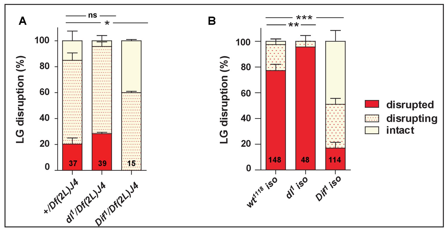
Dif but not Dorsal is required for proper lymph gland disruption in response to wasp parasitism.
Quantification (%) of lymph gland disruption in (A): +/Df(2L)J4 (control), dl1/Df(2L)J4 and Dif1/Df(2L)J4 mutant larvae and (B) in the isogenic strains wt1118 (control), dl1 and Dif1. Error bars correspond to SEM, *p<0.1, **p<0.01, ***p<0.001, ns (not significant) (Pearson’s Chi-squared test).
-
Figure 1—figure supplement 1—source data 1
Lymph gland disruption quantification.
- https://doi.org/10.7554/eLife.25496.004
Figure 2 with 2 supplements

Dif, lymph gland lamellocyte differentiation and lymph gland disruption are required for wasp egg neutralization.
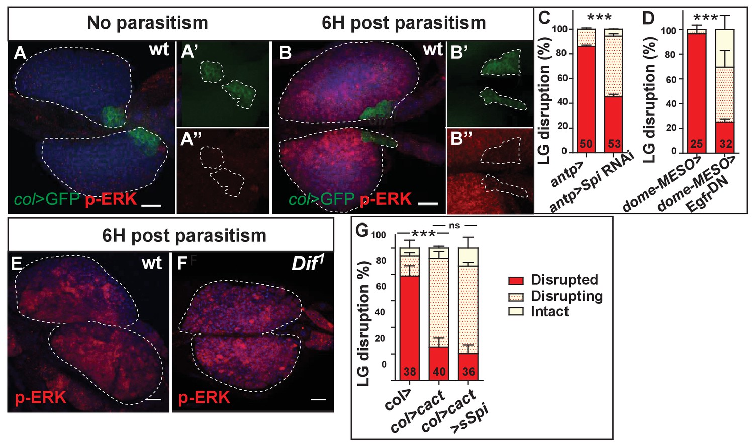
(A, B) Brightfield images of control (col>, A) and Dif1 mutant larvae 48 hr post parasitism (B). The arrow indicates a melanotic capsule. (C) Quantification (%) of wasp egg hatching in control (col>) and Dif1 mutant. In this and subsequent similar analyses, numbers of (infected) larvae analyzed are indicated on histograms. (D–I) Phalloidin (red) staining of circulating hemocytes in control (hml>, D, E) and Dif1 mutant (G, H) just prior (D, G) and following (E, H) lymph gland disruption. Arrows indicate discoidal-shaped lamellocytes that are larger than plasmatocytes. (F, I) Quantification (%) of circulating plasmatocytes and lamellocytes in control (F) and Dif1 mutant (I). Blue, TOPRO3. (J) Quantification of lymph gland disruption (%) in control larvae (dome-MESO>) and when latran is down-regulated in lymph gland progenitors (dome-MESO > lat RNAi). (K) Quantification of wasp egg hatching in control larvae (dome-MESO>) or (antp>) and when latran is down-regulated in lymph gland progenitors (dome-MESO > lat RNAi) or in PSC cells (antp > lat RNAi). Error bars correspond to SEM, ***p<0.001 and ns (not significant) (Pearson’s Chi-squared test).
-
Figure 2—source data 1
Wasp egg hatching quantification.
- https://doi.org/10.7554/eLife.25496.010
-
Figure 2—source data 2
Circulating hemocyte quantification.
- https://doi.org/10.7554/eLife.25496.011
-
Figure 2—source data 3
Lymph gland disruption quantification.
- https://doi.org/10.7554/eLife.25496.012
-
Figure 2—source data 4
Wasp egg hatching quantification.
- https://doi.org/10.7554/eLife.25496.013
Figure 2—figure supplement 1
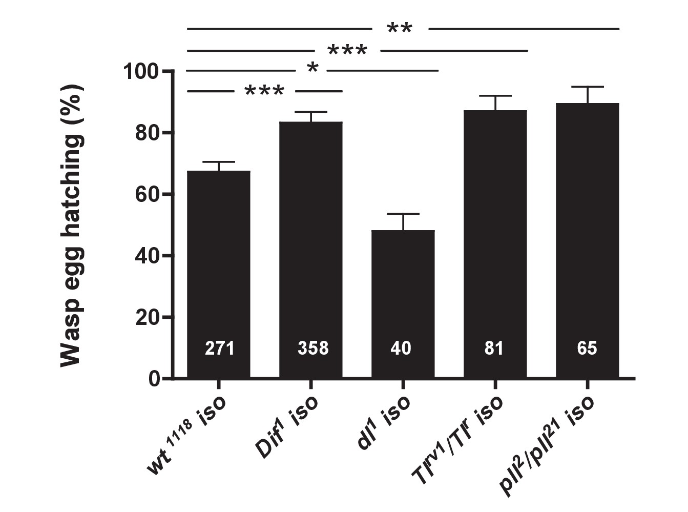
Toll/NF-κB signaling is required for wasp egg encapsulation.
Quantification (%) of wasp egg hatching in isogenic strains wt1118 (control), Dif1, dl1, Tlrv1/Tlr and pll2/pll21. Note high level of wasp egg hatching in the wt1118 iso background. Error bars correspond to SEM, *p<0.1, **p<0.01, ***p<0.001 (Pearson’s Chi-squared test).
-
Figure 2—figure supplement 1—source data 1
Wasp egg hatching quantification.
- https://doi.org/10.7554/eLife.25496.008
Figure 2—figure supplement 2
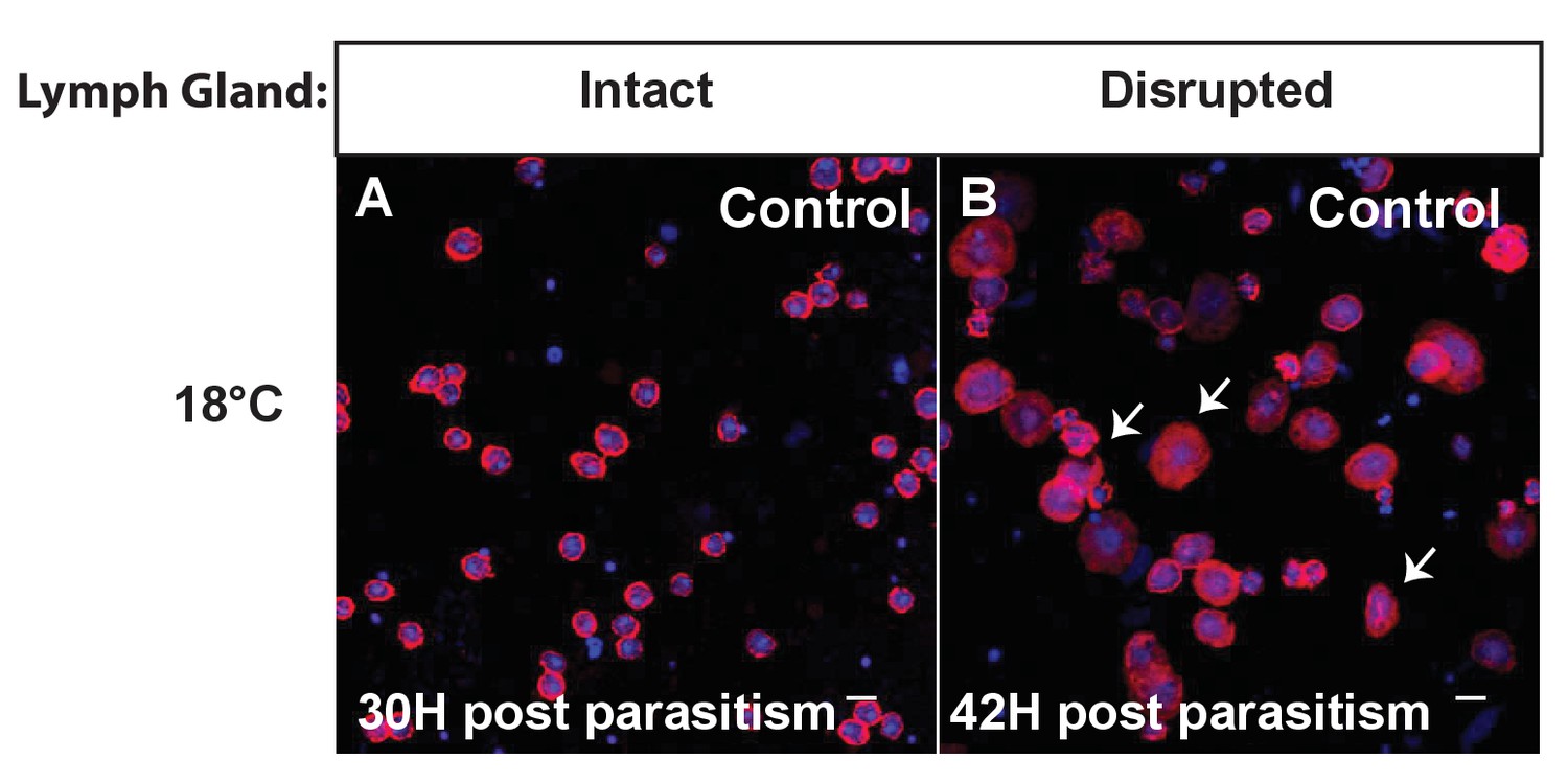
At 18°C, lymph gland disruption and the presence of lamellocytes in circulation are delayed.
Circulating hemocytes stained by Phalloidin (red) and TOPRO3 (blue) in control larvae (hml>) 30 hr post-parasitism, prior to lymph gland disruption (A) and 42 hr post-parasitism following lymph gland disruption (B). Arrows indicate lamellocytes. Scale bars represents 0.1 µm.
Figure 3 with 1 supplement
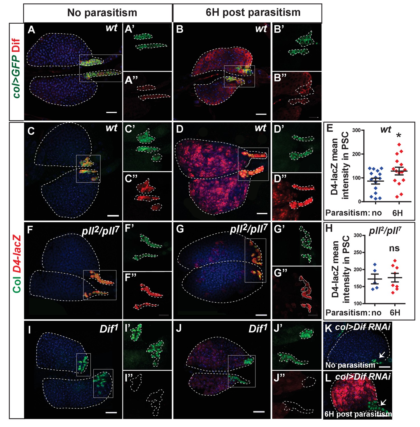
Dif-dependent Toll/NF-kB activation in PSC cells.
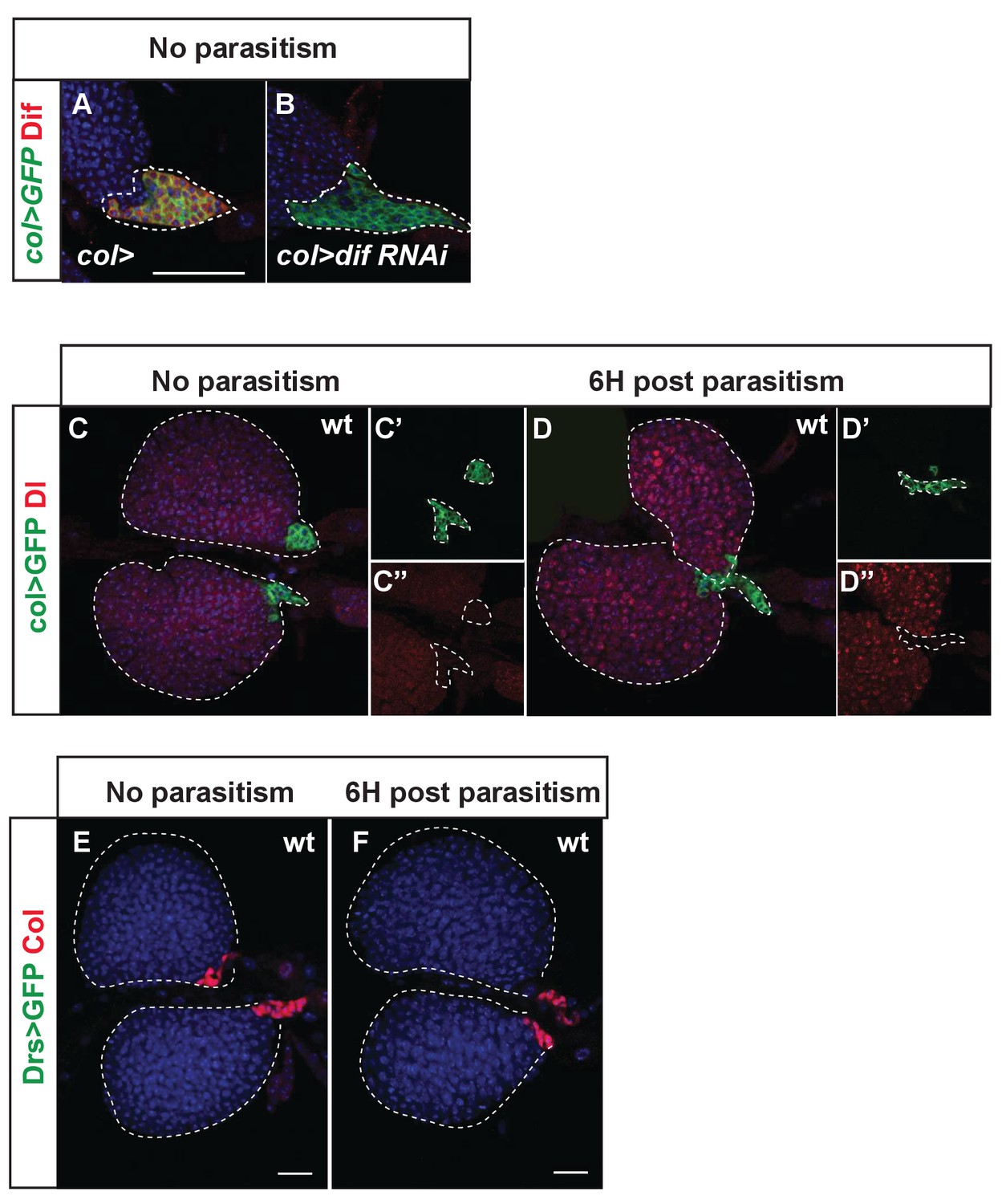
(A, B) Dif (red) immunostaining of col > mCD8 GFP (col > GFP, green) lymph glands without (A) and 6 hr post-parasitism (B). (A’, A’’, B’, B’’) enlarged views of the PSC. (C, D, F, G, I, J, K, L) LacZ (red) and Col (green) immunostaining of D4–LacZ of wt (C, D), pll2/pll7 (F, G), Dif1 (I, J), and col > Dif RNAi (K, L) lymph glands without (C, F, I, K), and 6 hr post-parasitism (D, G, J, L). (C’, C'’, D’, D'’, F’, F'’, G’, G”, I’, I”, J’ and J”) enlarged views of the PSC. Arrows (K, L) indicate Col-positive PSC cells that do not express D4–LacZ. (E, H) Quantifications of D4-LacZ mean intensity in PSC cells. Error bars represent SDs, *p<0.1 and ns (not significant) (Mann-Whitney nonparametric test).
-
Figure 3—source data 1
D4-lacZ staining quantification.
- https://doi.org/10.7554/eLife.25496.016
-
Figure 3—source data 2
D4-lacZ staining quantification.
- https://doi.org/10.7554/eLife.25496.017
Figure 3—figure supplement 1
Dif, Dorsal and Drosomycin-GFP expression in lymph gland cells.
(A and B) Efficient RNAi-mediated downregulation of Dif (red) in PSC cells (green, col > dif RNAi). (C and D) Immunodetection of Dorsal (red) in lymph gland cells under normal conditions (C) and 6 hr post-parasitism (D). Under both conditions, progenitors but not PSC cells express Dorsal. (E and F) Drosomycin-GFP (Drs-GFP, green) is not expressed in lymph gland cells under normal conditions (E) nor 6 hr post-parasitism (F). Nuclei are labeled with Draq5 (blue).
Figure 4 with 2 supplements
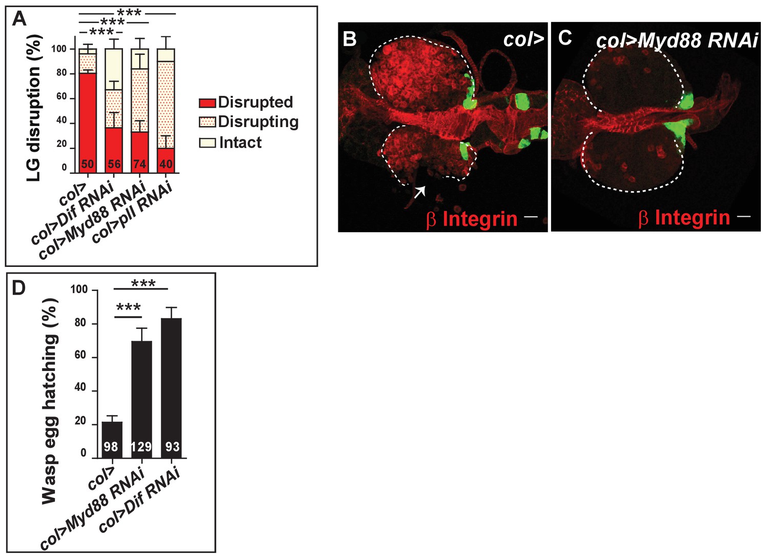
Toll/NF-kB activation in PSC cells controls lymph gland dispersal, lamellocyte differentiation and wasp egg encapsulation.
(A) Quantification (%) of lymph gland disruption post parasitism in col> (control), col > Dif RNAi, col > Myd88 RNAi and col > pll RNAi lymph glands. (B–C) Representative confocal images of lamellocyte differentiation 20 hr post-parasitism (β Integrin, red) in control lymph glands (col > GFP) (B) and when Toll/NF-kB is downregulated in PSC cells (col > GFP > Myd88 RNAi) (C). At least 60% (n = 44) of lymph glands displayed numerous lamellocytes in the control, whereas less than 15% (n = 41) are observed in col > Myd88 RNAi lymph glands. Note early signs of lymph gland disruption in (B, arrow). (D) Quantification (%) of wasp egg hatching 48 hr post-parasitism in col> (control), col > Myd88 RNAi and col > Dif RNAi lymph glands. Error bars correspond to SEM, ***p<0.001 (Pearson’s Chi-squared test).
-
Figure 4—source data 1
Lymph gland disruption quantification.
- https://doi.org/10.7554/eLife.25496.022
-
Figure 4—source data 2
Wasp egg hatching quantification.
- https://doi.org/10.7554/eLife.25496.023
Figure 4—figure supplement 1
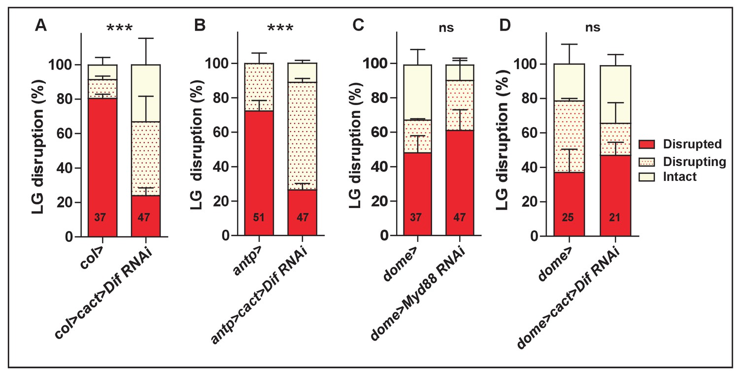
Toll/NF-kB activation in PSC cells but not in lymph gland progenitors is required for proper lymph gland disruption in response to wasp parasitism.
Quantifications (%) of lymph gland disruption post-parasitism upon downregulation of Toll/NF-kB signaling in PSC cells (col>, A), (antp>, B) and lymph gland progenitors (dome>, C and D). Error bars correspond to SEM, ***p<0.001, ns (not significant) (Pearson’s Chi-squared test).
-
Figure 4—figure supplement 1—source data 1
Lymph gland disruption quantification.
- https://doi.org/10.7554/eLife.25496.020
Figure 4—figure supplement 2
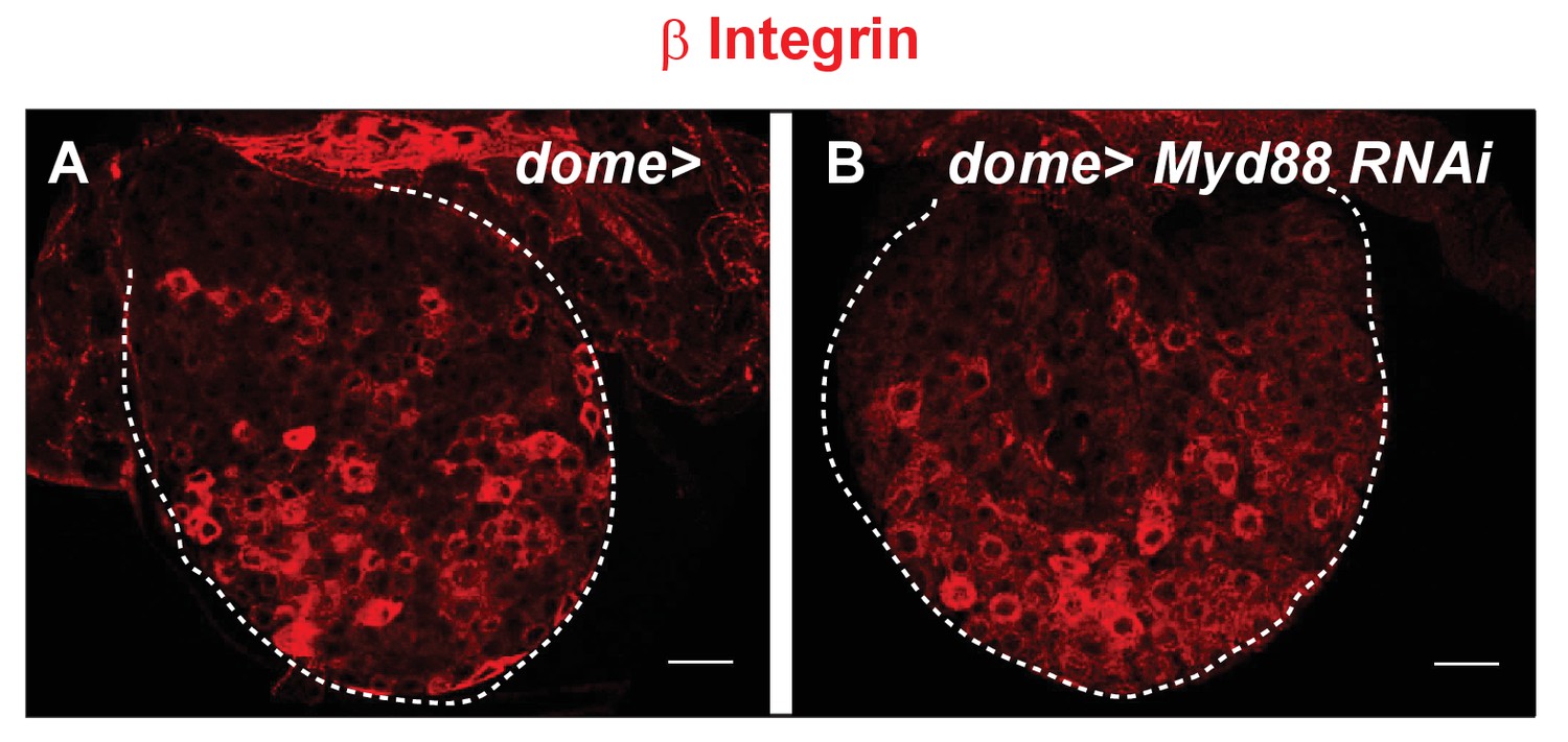
Toll/NF-kB signaling is not required in lymph gland progenitors for lamellocyte differentiation.
Representative confocal images of β integrin (red) immunostaining of lamellocytes 20 hr post-parasitism in control lymph gland (dome>) (A) (n = 20) and when Toll/NF-kB is impaired in progenitors (dome > Myd88 RNAi) (B) (n = 12).
Figure 5 with 1 supplement
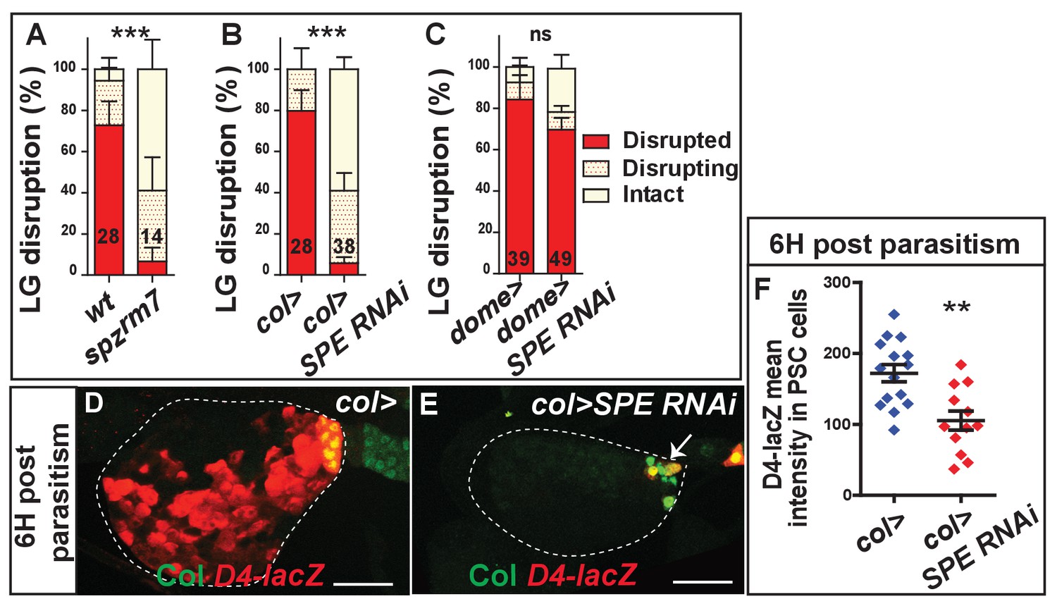
Toll/NF-kB activation depends on SPE expression in PSC cells.
(A–C) Quantification (%) of lymph gland disruption post parasitism in spzrm7 larvae (A) and when SPE is down-regulated in either PSC cells (col > SPE RNAi) (B) or lymph gland progenitors (dome > SPE RNAi) (C). Error bars correspond to SEM, ***p<0.001 and ns (not significant) (Pearson’s Chi-squared test). (D, E) LacZ (red) and Col immunostaining (green) of D4–LacZ expressing lymph glands 6 hr post-parasitism in wt (col>) (D) and when SPE is down-regulated in the PSC (white arrow) (col > SPE RNAi) (E). (F) Quantifications of D4-lacZ mean intensity in PSC cells 6 hr post-parasitism. Error bars represent SDs. **p<0.01 (Mann-Whitney nonparametric test).
-
Figure 5—source data 1
Lymph gland disruption quantification.
- https://doi.org/10.7554/eLife.25496.028
-
Figure 5—source data 2
Lymph gland disruption quantification.
- https://doi.org/10.7554/eLife.25496.029
-
Figure 5—source data 3
Lymph gland disruption quantification.
- https://doi.org/10.7554/eLife.25496.030
Figure 5—figure supplement 1
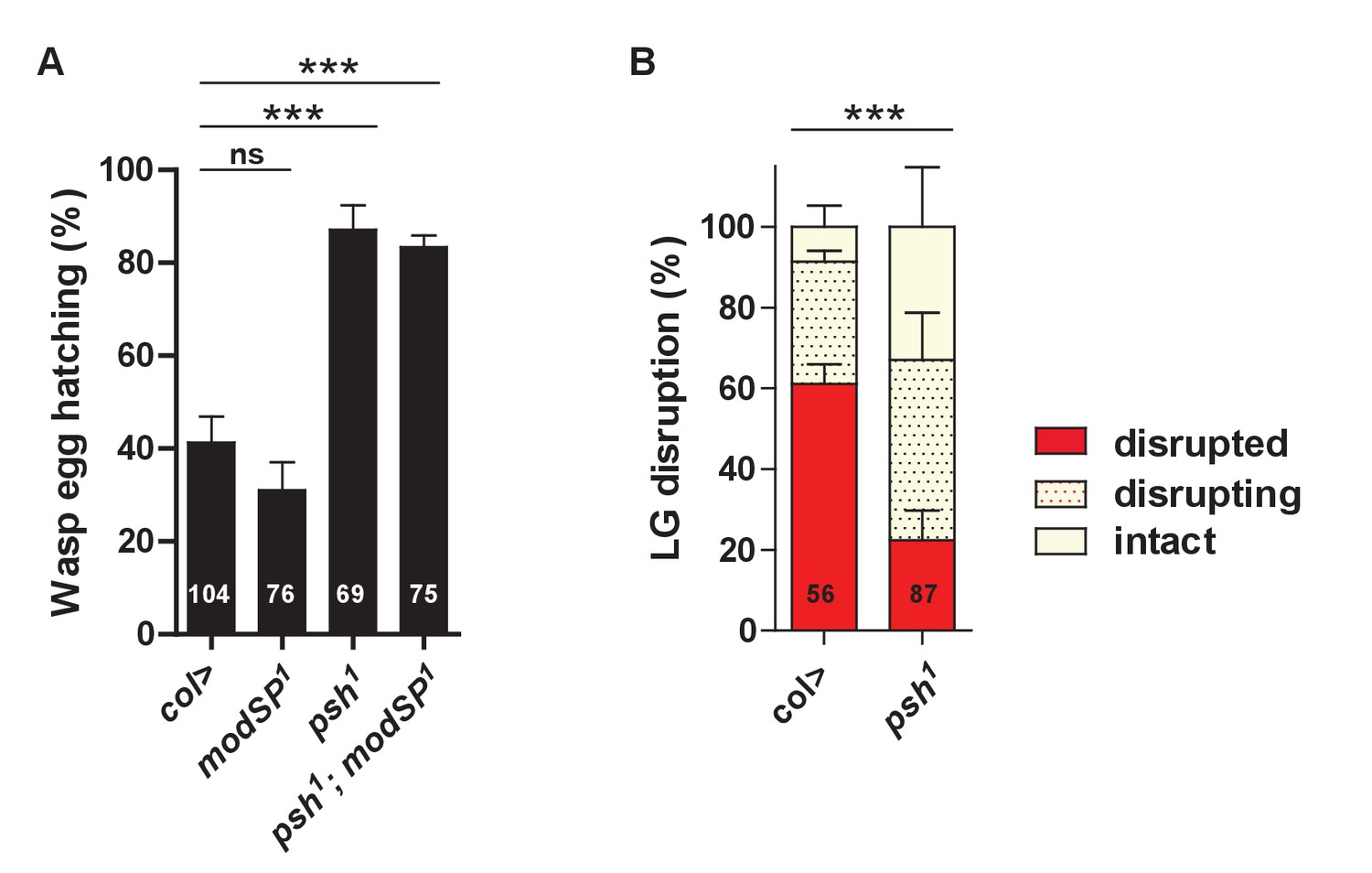
Psh is required for wasp egg neutralization and timely lymph gland disruption.
(A) Quantification (%) of wasp egg hatching in control (col>), modSP1, psh1 and double psh1; modSP1 mutants. (B) Quantification (%) of lymph gland disruption in control (col>) and psh1 mutants. Error bars correspond to SEM, ***p<0.001, ns (not significant) (Pearson’s Chi-squared test).
-
Figure 5—figure supplement 1—source data 1
Wasp egg hatching quantification.
- https://doi.org/10.7554/eLife.25496.026
-
Figure 5—figure supplement 1—source data 2
Lymph gland disruption quantification.
- https://doi.org/10.7554/eLife.25496.027
Figure 6 with 3 supplements
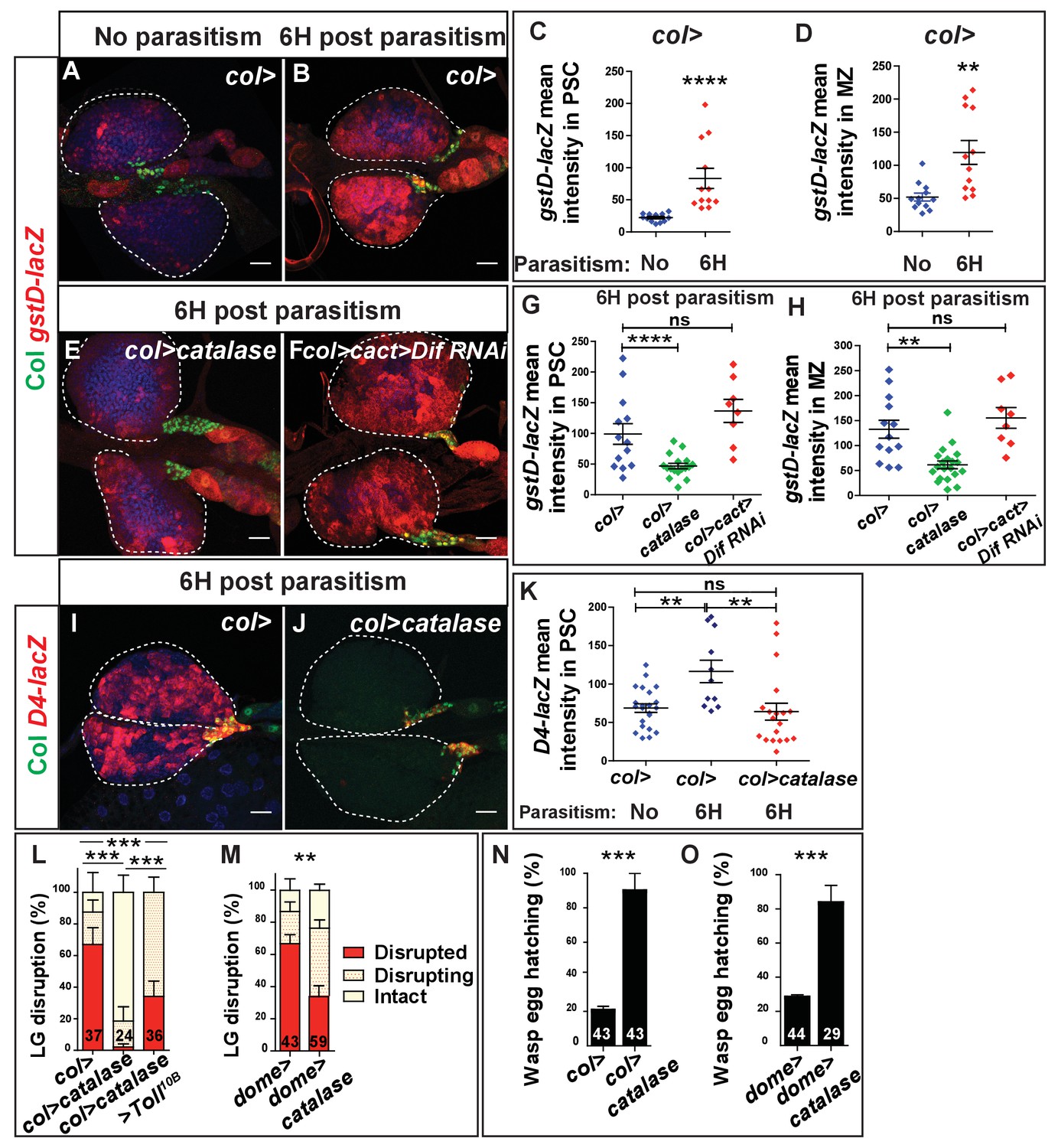
Wasp-mediated oxidative stress promotes Toll/NF-kB activation in the PSC and lymph gland disruption.
(A, B, E, F) LacZ (red) and Col (green) immunostaining in gstD-lacZ expressing lymph glands in wt context (col>; A, B), when Catalase is expressed in the PSC (col > catalase) (E) and when Toll/NF-kB is inactivated in PSC cells (col > cact > Dif RNAi)(F). (C, D, G, H) Quantifications of gstD-lacZ mean intensity in PSC and progenitor (MZ) cells. Error bars represent SDs. **p<0.01, ****p<0.0001 and ns (not significant) (Mann-Whitney nonparametric test). (I, J) LacZ (red) and Col (green) immunostaining in D4-LacZ expressing lymph glands in wt (col>) (I) and when an intracellular catalase is expressed in the PSC (col > catalase) (J). (K) Quantifications of D4-lacZ mean intensity in PSC cells. **p<0.01 and ns (not significant) (Mann-Whitney nonparametric test). (L–M) Quantifications of lymph gland disruption post parasitism. (N,O) Quantification (%) of wasp egg hatching. Error bars correspond to SEM, **p<0.01, ***p<0.001 (Pearson’s Chi-squared test).
-
Figure 6—source data 1
gstD-lacZ staining quantification.
- https://doi.org/10.7554/eLife.25496.035
-
Figure 6—source data 2
D4-lacZ staining quantification.
- https://doi.org/10.7554/eLife.25496.036
-
Figure 6—source data 3
Lymph gland disruption quantification.
- https://doi.org/10.7554/eLife.25496.037
-
Figure 6—source data 4
Lymph gland disruption quantification.
- https://doi.org/10.7554/eLife.25496.038
-
Figure 6—source data 5
Wasp egg hatching quantification.
- https://doi.org/10.7554/eLife.25496.039
Figure 6—figure supplement 1
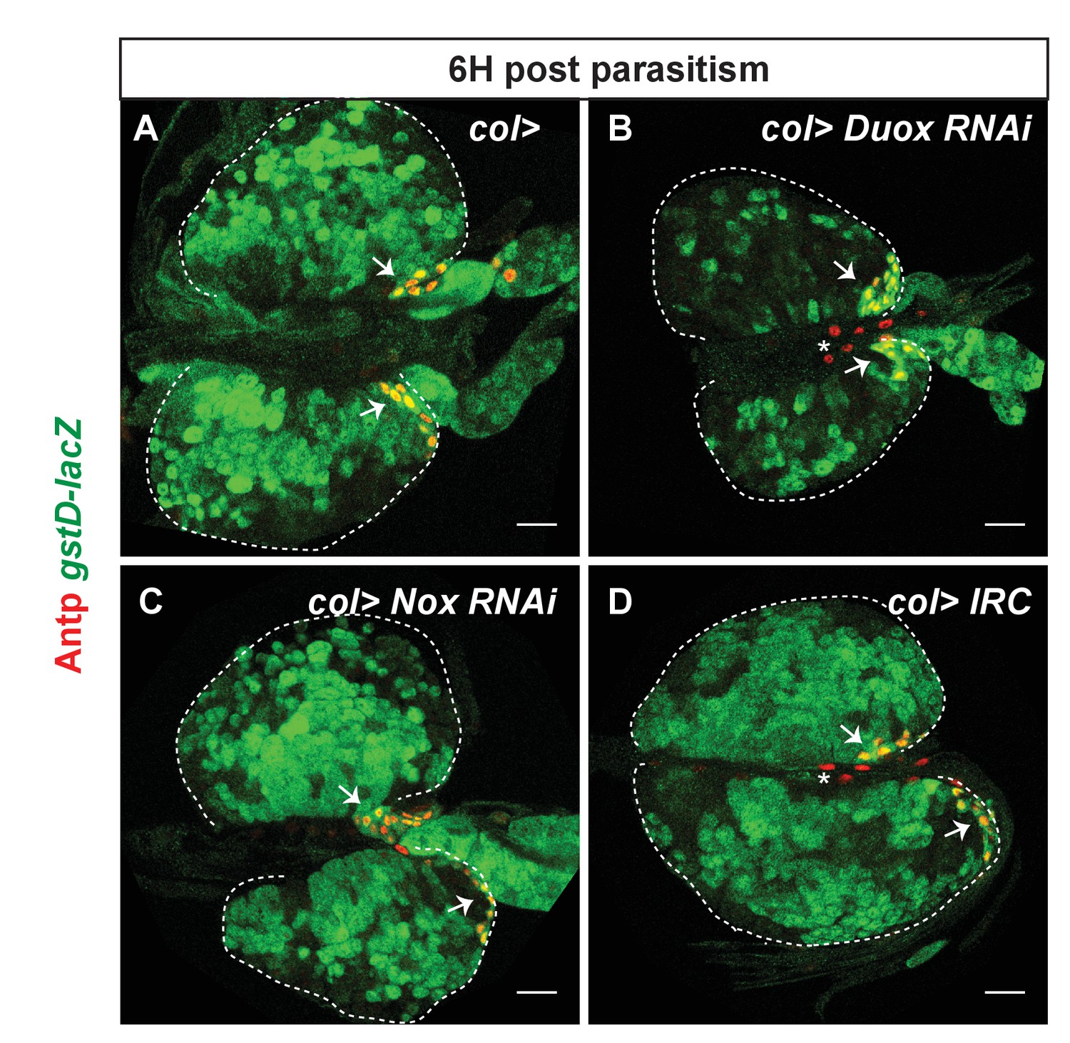
Duox- and Nox-independent generation of ROS in PSC cells 6 hr post-parasitism.
LacZ (green) and Antp (red) immunostaining in gstD-lacZ expressing lymph glands in control larvae (col>) (A), when Duox (col > Duox RNAi) (B) or Nox (col > Nox RNAi) (C) are downregulated in PSC cells, or when the intracellular catalase IRC is expressed in PSC cells (col > IRC) (D). In all contexts, similar levels of gstD-lacZ are observed in PSC cells (arrows). Reduced levels of gstD-lacZ are observed in progenitors of col > Duox RNAi lymph glands (B). Arrows and asterisks indicate the PSC and cardiac cells, respectively, marked with Antp.
Figure 6—figure supplement 2
ROS are required both in PSC cells and lymph gland progenitors for lamellocyte differentiation.
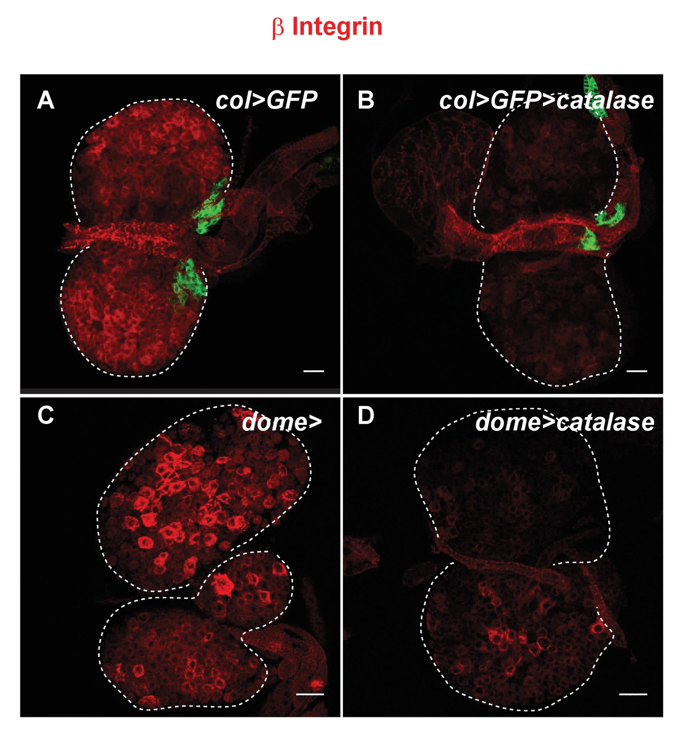
β integrin (red) immunostaining of control lymph glands (col > GFP, A) or (dome>, C) and when catalase is expressed in the PSC (col > GFP > catalase, B) or in progenitors (dome > catalase, D) 20 hr post-parasitism. Whereas at least 60% (n = 44) of col > GFP lymph glands have numerous lamellocytes less than 10% (n = 45) displayed lamellocytes in col > catalase. Likewise, more than 70% (n = 7) of dome> control lymph glands have lamellocytes, whereas only 13% (n = 15) contained lamellocytes in dome > catalase.
Figure 6—figure supplement 3
Scavenging ROS in PSC cells or silencing the EGFR pathway in MZ progenitors delays lymph gland disruption upon parasitism.
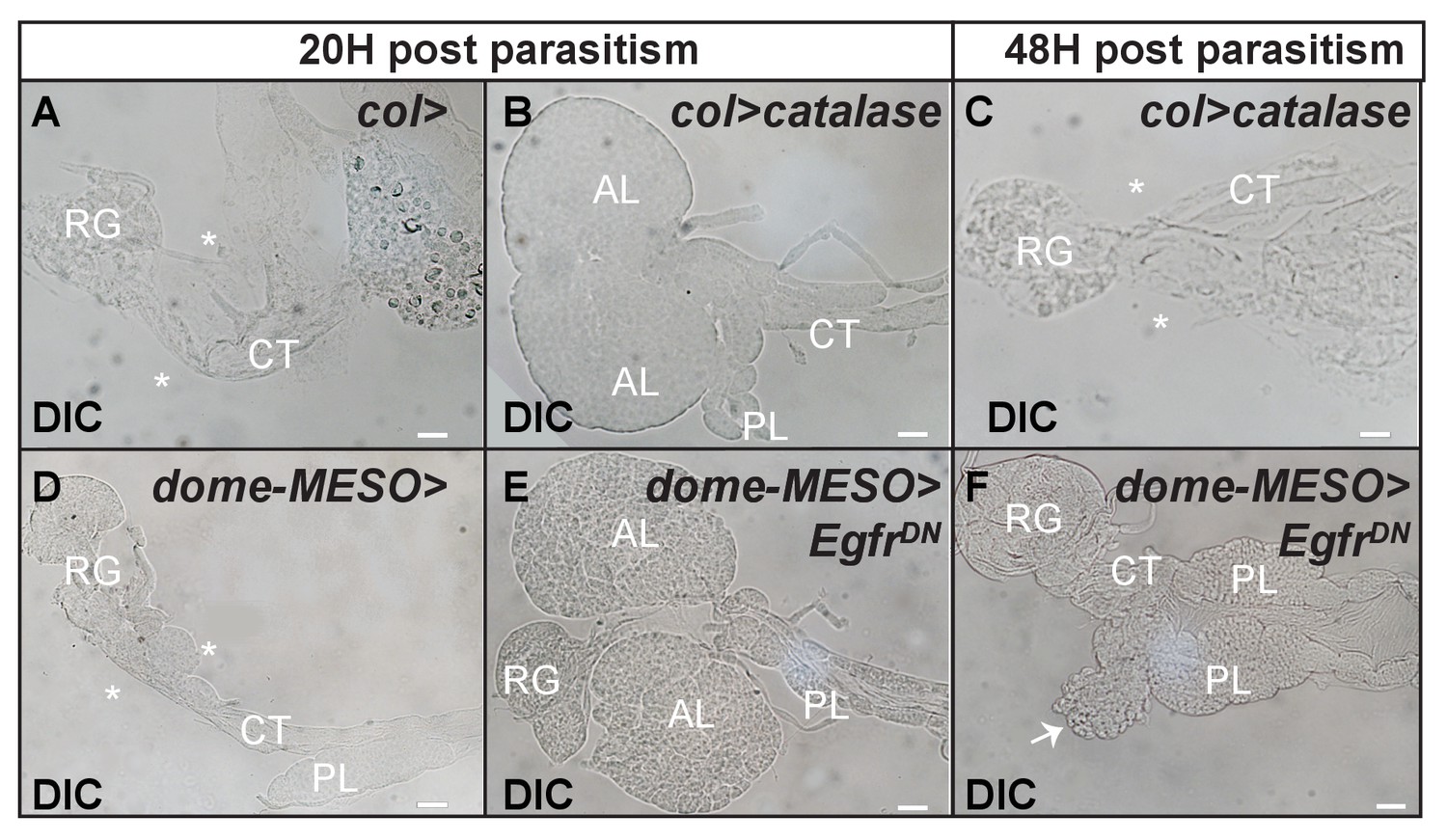
Differential interference contrast (DIC) microscopy of lymph glands 20 hr (A, B, D, E) and 48 hr (C, F) post parasitism in control (A, D) and when Catalase is expressed in the PSC (col > catalase) (B, C) or when the EGFR pathway is inactivated in lymph gland progenitors (dome-MESO > EgfrDN) (E, F). Ring gland, RG; cardiac tube, CT; anterior lobe, AL; posterior lobe, PL; disrupted (*) and disrupting (arrow in F) anterior lobes are shown. The scale bar represents 20 µm. All source data are supplied as Excel files.
Figure 7
Epistatic relationships between EGFR/Erk and Toll/NF-κB signaling.
(A, B) p-ERK (red) immunostaining in col > GFP (PSC, green) larvae. (A’, A”, B’, B”), enlarged views of PSC cells. (C, D) Quantifications of lymph gland disruption post parasitism. (E, F) p-ERK (red) immunostaining in wt (E) and Dif1 (F) mutant lymph glands. (G) Quantifications (%) of lymph gland disruption post parasitism. Error bars correspond to SEM, ***p<0.001, ns (not significant) (Pearson’s Chi-squared test).
-
Figure 7—source data 1
Lymph gland disruption quantification.
- https://doi.org/10.7554/eLife.25496.041
-
Figure 7—source data 2
Lymph gland disruption quantification.
- https://doi.org/10.7554/eLife.25496.042
-
Figure 7—source data 3
Lymph gland disruption quantification.
- https://doi.org/10.7554/eLife.25496.043
Figure 8
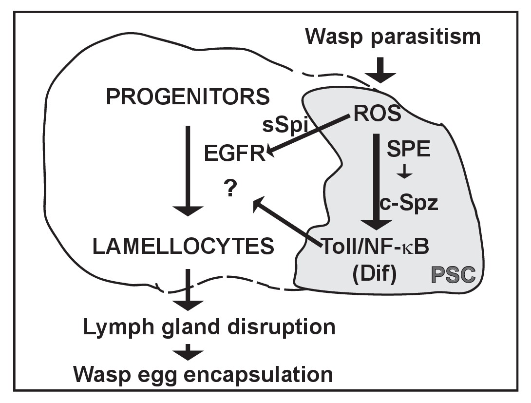
Proposed gene regulatory network that controls lymph gland rupture upon wasp parasitism.
The PSC is drawn in grey. Wasp parasitism increases ROS in PSC cells that activate Toll/NF-κB and Spitz secretion (sSpi). Toll/NF-kB activation in PSC cells requires SPE in the same cells for Spätzle processing (c-Spz). sSpi non cell-autonomously activates the EGFR pathway in lymph gland progenitors. Both EGFR and Toll/NF-κB activation are required for lymph gland lamellocyte differentiation, lymph gland disruption and wasp egg encapsulation.
Additional files
-
Transparent reporting form
- https://doi.org/10.7554/eLife.25496.045
Download links
A two-part list of links to download the article, or parts of the article, in various formats.
Downloads (link to download the article as PDF)
Open citations (links to open the citations from this article in various online reference manager services)
Cite this article (links to download the citations from this article in formats compatible with various reference manager tools)
Reactive oxygen species-dependent Toll/NF-κB activation in the Drosophila hematopoietic niche confers resistance to wasp parasitism
eLife 6:e25496.
https://doi.org/10.7554/eLife.25496


