Transmission genetics of drug-resistant hepatitis C virus
- Stanford University School of Medicine, United States
- The Scripps Research Institute, United States
- Dalhousie University, Canada
Figures
Figure 1 with 3 supplements

Construction of codon altered JFH1.
(A) Cell cultures coinfected with two strains of HCV result in four populations: uninfected, two types of singly infected, and coinfected cells. (B) Three segments of the JFH1 genome, that were roughly 1000 nucleotides in length and had altered codon usage, were designed using GeneArt algorithms and synthesized. These genome fragments were then cloned into the JFH1 strain of HCV. Huh7.5.1 cells were transfected with each construct using electroporation to create long-term HCVcc cultures and viability was followed over time by immunoblotting of cell lysates for the HCV core protein. (C) Viral passages shown in (B) were monitored by qRT-PCR analysis of viral RNA in culture supernatants. Only the CA-3 virus demonstrated growth kinetics comparable to wild-type (WT) JFH1.
Figure 1—figure supplement 1
Alignment of codon-altered and wild-type JFH1 viral RNAs.
Sequence alignments were generated using ClustalW to highlight the nucleotide changes incorporated into the CA-1 viral RNA. Sequences highlighted with orange bars identify regions containing covarying mutations indicating functional RNA secondary structures.
Figure 1—figure supplement 2
Alignment of codon-altered and wild-type JFH1 viral RNAs.
Sequence alignments were generated using ClustalW to highlight the nucleotide changes incorporated into the CA-2 viral RNA. Sequences highlighted with orange bars identify regions containing covarying mutations indicating functional RNA secondary structures.
Figure 1—figure supplement 3
Alignment of codon-altered and wild-type JFH1 viral RNAs.
Sequence alignments were generated using ClustalW to highlight the nucleotide changes incorporated into the CA-3 viral RNA. No regions of covarying mutations were observed within this region.
Figure 2
Differentiation of codon-altered and wild-type JFH1 using confocal microscopy and flow cytometry.
(A) RNA in situ hybridization probes were designed to differentiate between wild-type (WT) and codon-altered (CA) viral RNAs. These probes utilize branched DNA technology to amplify the contiguous DNA branches and not the RNA target itself. Roughly 8000 fluorophores labeled each target RNA. (B) Huh7.5.1 cells on coverslips were infected with WT or CA virus at MOIs of 0.01 FFU/cell. Cells were fixed after 72 hr, co-stained with WT and CA probe sets that recognized HCV positive strands and visualized by confocal microscopy. (C) Confocal microscopy of cells infected as in (B) but co-stained with probe sets to identify negative strands. (D) Huh7.5.1 cells were transfected with WT RNA, CA RNA, or both by electroporation. Cells were fixed at 72 hr post transfection and costained with WT and CA RNA probe sets. Flow cytometry was performed on (i) cells transfected with WT RNA, (ii) cells transfected with CA RNA, (iii) a mixture of cells in i and ii, and (iv) cells transfected with both WT and CA RNAs.
Figure 3 with 3 supplements
Flow cytometry to test dominance of viruses resistant to NS3 protease inhibitor.
(A) Structure of protease inhibitor BILN-2061. (B) Structure of NS3 protein. D168 (red) is located in the protease domain adjacent to the active site (lavender). D168A confers resistance to BILN-2061. (C) Diagram of CA virus with altered sequence (green hatches) and WT virus with location of D168A mutation identified. (D) The four types of cells present in the absence of inhibitors are uninfected (U), infected with drug-susceptible virus cells (S), infected with both drug-susceptible and drug-resistant virus (S + R) and drug-resistant virus (R). In the presence of a DAA, three outcomes are possible and are indicated by the changing density of the cell populations: (E) Drug-resistance is cis-dominant, (F) Drug resistance is dominant and (G) Drug susceptibility is dominant. Huh7.5.1 cells were coinfected with CA and WT-D168A for 72 hr followed by treatment with (H/I) DMSO or (J/K) 2 μM BILN-2061 for 36 hr. Cells were stained with CA and WT vRNA probes and analyzed by flow cytometry (H, J) and results from three replicates quantified (I, K). NS3 drug-resistance was found to be cis-dominant.
Figure 3—figure supplement 1
NS3-D168A confers resistance to BILN-2061 in both the wild-type and the codon-altered backgrounds.
D168A was cloned into both the WT (left) and CA (right) backgrounds and tested for resistance to BILN-2061. Huh7.5.1 cells were infected with WT, WT-D168A, CA or CA-D168A viruses at MOI = 0.1 FFU/cell in the absence or presence of 2 μM BILN-2061 as indicated. Viral RNA was harvested from culture supernatants collected at 72 hr post infection and analyzed by qRT-PCR.
Figure 3—figure supplement 2
Exogenously expressed drug-resistant NS3 and NS5A do not rescue drug-susceptible HCV in trans.
(A) Schematic of codon-altered HCV RNA, and non-structural protein expression plasmids (pTM-NS3-5B) that contain either no mutation, the D168A mutation that confers resistance to BILN-2061 or the Y93N mutation that confers resistance to SR2486. (B) Huh7-Lunet-T7 cells were infected with CA virus for 72 hr and then transfected with wild-type or D168A pTM constructs in the absence and presence of 2 μM BILN-2061 for 24 hr as indicated. Viral RNA was harvested from cell culture supernatants and quantified by qRT-PCR. Results from samples harvested in triplicate are shown. (C) Cells were infected with CA virus for 72 hr and then transfected with wild-type or Y93N pTM constructs in the absence and presence of SR2486 for 24 hr as indicated. Viral RNA was harvested from cell culture supernatants and quantified by qRT-PCR. Results from samples harvested in triplicate are shown.
Figure 3—figure supplement 3

Drug resistance to R1479 confers major fitness cost to JFH1.
(A) R1479 is a nucleotide analog inhibitor of the HCV NS5B polymerase. (B) WT virus was passaged every 72 hr ten times in the presence of 25 μM R1479. Extracted vRNA from each passage was quantified and sequenced to determine if selection of resistance associated variants occurred. Of the variants identified by sequencing only T481A and F427L demonstrated any replication capacity as measured by core expression in transfected cells or the presence of vRNA in culture supernatants at day (B) 7 or (C) 21 post transfection. (D) We attempted to select for compensatory mutations that increased the fitness of WT-T481A, WT-F427L or the double mutant WT-T481A/F427L by passaging these viruses in the presence of 25 μM R1479 ten times and sequencing total viral RNA from these passages. No compensatory mutations were identified.
Figure 4
NS5A-resistant HCV is cis-dominant.
(A) Structures of SR2486 or Daclatasvir. (B) Structure of NS5A dimer with Y93 identified (orange). NS5A variants, Y93N and Y93H have previously been shown to confer drug resistance to multiple NS5A inhibitors. (C) Huh7.5.1 cells were infected with WT, WT-Y93N or WT-Y93H at MOI of 0.1 FFU/cell in the absence or presence of 500nM SR2486 or 50 nM Daclatasvir to determine the drug resistance profiles. (D) Diagram of CA virus with altered sequence (green hatches) and WT virus with location of Y93N mutation identified. (E/F) Huh7.5.1 cells were coinfected with CA and WT-Y93N for 72 hr followed by treatment with (F) or without (E) 500nM SR2486 as indicated for 24 hr and analyzed by flow cytometry. Results from four replicates of the experiment shown are quantified. (G) Huh7.5.1 cells were infected with CA and WT-Y93N at a MOI of 1 for 24 hr followed by treatment with DMSO or 500nM SR2486. Resistance to SR2486 was found to be cis-dominant. (H) Results from three replicate experiments in which Huh7.5.1 cells were coinfected with CA and WT-Y93H for 72 hr followed by treatment without (left) or with 50 nM Daclatasvir (right) are shown. Resistance to Daclatasvir was found to be cis-dominant.
Figure 5

Complex formation between drug susceptible and drug-resistant NS5A in the absence of RNA replication.
(A) HCV NS3-5B constructs driven by T7 polymerase and encoding tandem tagged copies of NS5A (Berger et al., 2014) were created to co-express HA- and GFP-tagged NS5A alleles. Huh7-Lunet-T7 cells were transfected with pTM-Dual-NS5A constructs that contained two drug-resistant (R), two drug susceptible (S), or mixed alleles of NS5A. Proteins from transfected cell extracts that were incubated in the absence (B) or presence (C) of SR2486 were subjected to SDS-PAGE without further fractionation (Input) or after immunoprecipitation with anti-HA antibodies (IP αHA). The gel was subjected to immunoblotting with GFP or HA antibodies as indicated. (D) Cells were transfected with dual-NS5A constructs in the absence or presence of SR2486 for 24 hr. Cells were then fixed, stained with anti-HA antibodies and visualized by confocal microscopy. Representative images from over 25 cells are presented. (E) Cells transfected with dual-NS5A constructs that expressed drug-susceptible (S) and drug-resistant (R) alleles as shown were prepared for electron microscopy by high-pressure freezing and freeze-substitution and visualized by transmission electron microscopy. (F) Numbers of cytoplasmic lipid droplets per cell formed in the absence (left) or presence (right) of SR2486; at least 25 images per sample such as those shown in (E) were quantified.
Figure 6
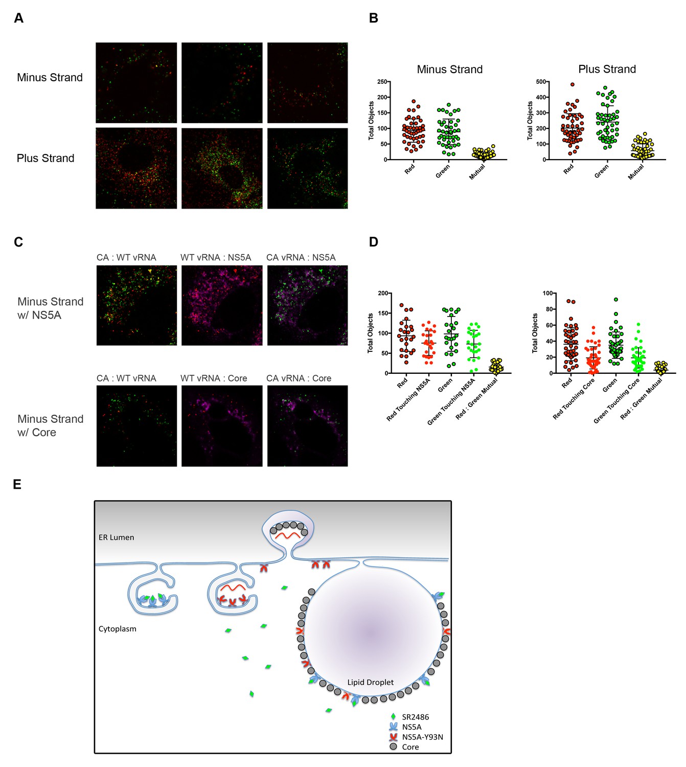
Drug-susceptible and drug-resistant RNA replication complexes segregate in coinfected cells.
(A) Huh7.5.1 cells were coinfected with CA and WT-Y93N at a MOI of 1 FFU/cell for 24 hr. Cells were fixed and co-stained with WT and CA negative-strand or positive strand viral RNA probe sets and visualized by confocal microscopy. Co-infected cells were identified and three representative images are displayed of more than 50 captured images. (B) Volocity was used to identify and quantify vRNA puncta within coinfected cells. These puncta were then assessed for colocalization and quantified. (C) Huh7.5.1 cells were coinfected with CA and WT-Y93N at a MOI of 1 FFU/cell for 24 hr. Cells were costained to visualize Core or NS5A together with CA and WT vRNAs. Representative cells are displayed demonstrating all pairwise comparisons analyzed for colocalization. (D) Volocity was used to quantify the number of vRNAs per cell, the number of colocalized vRNAs, as well as the number of vRNA puncta touching NS5A or Core. (E) Depiction of the clonal nature of individual RNA replication sites (Adapted from Figure 9 in Zayas et al., 2016]). Membrane invaginations house either drug-resistant (red) or drug-susceptible (blue) genomes. In the model, the RNA replication sites are segregated and therefore only the RNA from drug-resistant virus is amplified in the presence of inhibitors of RNA replication. It is visually suggested that NS5A molecules that bring core protein to lipid droplets for viral assembly mix on this surface and that this could lead to genetic dominance of drug susceptibility at a packaging step.
Additional files
-
Transparent reporting form
- https://doi.org/10.7554/eLife.32579.015
Download links
A two-part list of links to download the article, or parts of the article, in various formats.
Downloads (link to download the article as PDF)
Open citations (links to open the citations from this article in various online reference manager services)
Cite this article (links to download the citations from this article in formats compatible with various reference manager tools)
Transmission genetics of drug-resistant hepatitis C virus
eLife 7:e32579.
https://doi.org/10.7554/eLife.32579


