A SoxB gene acts as an anterior gap gene and regulates posterior segment addition in a spider
- Department of Biological and Medical Sciences, Oxford Brookes University, United Kingdom
- University of Cambridge, United Kingdom
Figures
Figure 1 with 1 supplement
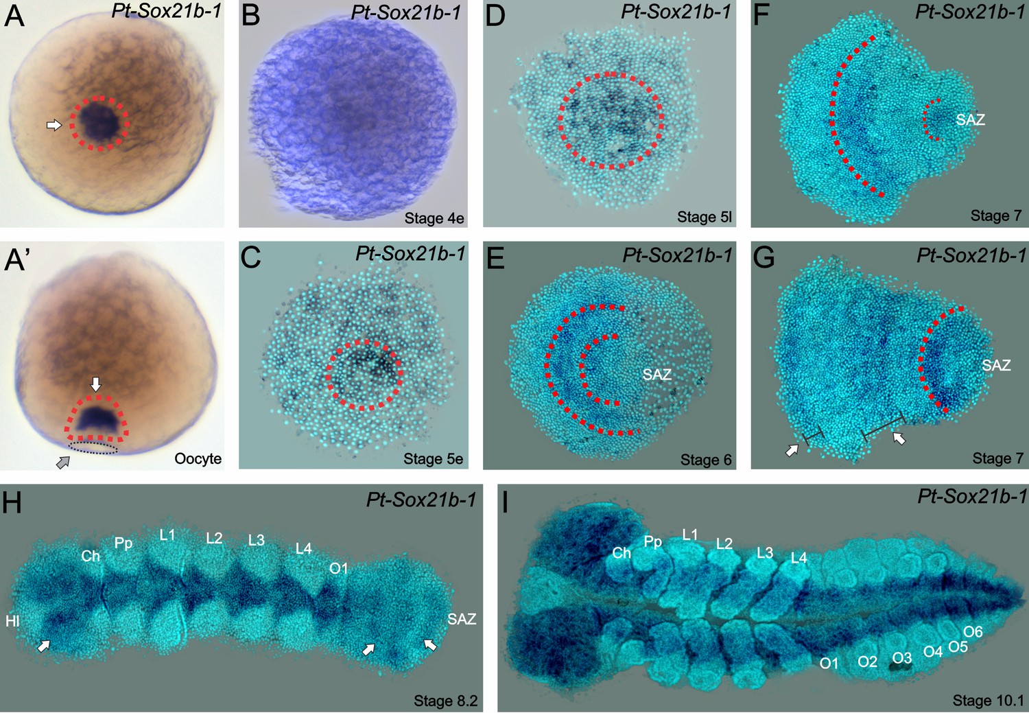
Expression of Sox21b-1 in P. tepidariorum oocytes and embryos.
(A) Dorsal (left) and lateral (right) views of pre-vitellogenic oocytes showing Sox21b-1 mRNA in the Balbiani’s body (red dashed circle and white arrows). The sperm implantation groove is indicated by a black dashed circle and grey arrow. (B) Overstained early stage 4 embryo evidencing the lack of expression of Sox21b-1 at this stage. (C) At early stage 5, the expression of Sox21b-1 appears in a salt and pepper pattern in the germ disc. (D) Expression in the cumulus becomes stronger at late stage 5, with less expression at the periphery of the germ disc (dashed red circle). (E) At stage 6 Sox21b-1 is expressed in a broad stripe in the anterior (between the red dashed lines). (F) At stage 7 there is expression in the region of the presumptive leg-bearing segments and in the SAZ (both indicated by red dashed lines). (G) At late stage 7 Sox21b-1 is still expressed in the SAZ (red dashed line) and the presumptive leg-bearing segments (indicated by a white arrow and wide black bracket), but nascent expression is observed at the anterior of the germ band (indicated by a white arrow and narrow black bracket). (H) At stage 8.2, when the limb buds are visible the expression of Sox21b-1 becomes restricted to the ventral nerve cord (anterior white arrow) and can be observed in the SAZ (posterior arrows). (I) At stage 10.1, Sox21b-1 expression is restricted to the ventral nerve cord and the head lobes. Ch: Chelicerae; Hl: Head lobes; L1 to L4: Prosomal leg- bearing segments 1 to 4; O1 to O6: Opisthosomal segments 1 to 6; SAZ: Segment Addition Zone. Ventral views are shown with anterior to the left, except as described for oocytes.
-
Figure 1—source data 1
ClustalW alignment of Sox protein HMG domains.
- https://doi.org/10.7554/eLife.37567.005
Figure 1—figure supplement 1
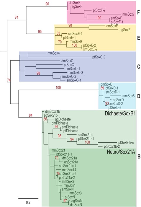
RAxML phylogeny of eumetazoan Sox genes.
Mid-point rooted phylogenetic tree made with RAxML algorithm showing the relationship between Anopheles gambiae (Ag), Mus musculus (Mm), D. melanogaster (Dm), P. tepidariorum (Pt) and S. mimosarum (Sm) Sox proteins based on HMG domain sequences. Bootstrap values over 70 shown in red and the scale bar represents the expected rate of substitution per site.
Figure 2 with 4 supplements
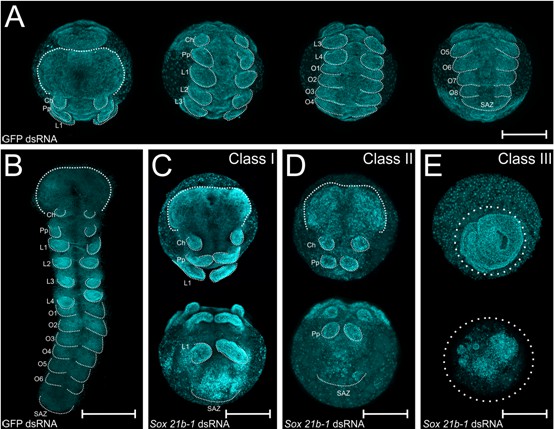
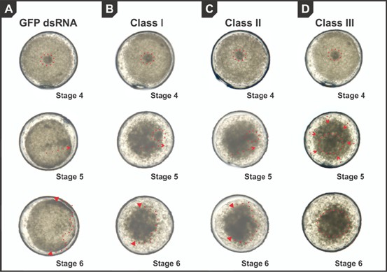
Embryo phenotypes after Sox21b-1 parental RNAi knockdown.
Whole mount (A) and flat mount (B) control embryos at stage 9 stained with DAPI. Stage 9, Class I (C), Class II (D) and Class III (E) phenotypes from Sox21b-1 knockdown. In the control embryos (A and B), the head, cheliceral, pedipalpal, prosomal walking limbs, opisthosomal segments and a posterior SAZ are all clearly visible as indicated. (C) Class I phenotype embryos show a morphologically normal head, and pairs of chelicerae, pedipalps and first walking limbs, but a disorganised cluster of cells in the posterior where L2-L4, opisthosomal segments and the SAZ should be. (D) Class II phenotype embryos consist of fewer cells, but still form a head, chelicerae, pedipalps and a structure resembling the SAZ in the posterior. (E) Class III embryos exhibit the most severe phenotype, where, after the germ disc stage, the embryo fails to form an organised germ band. Ch: Cheliceral segment; L1-L4: Prosomal segments 1 to 4; O1-O6: opisthosomal segments 1 to 6; Pp: Pedipalpal segment; SAZ: Segment Addition Zone. Anterior is to the top, scale bars: 150 µm.
Figure 2—figure supplement 1
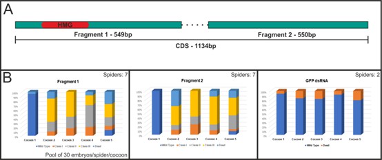
dsRNA design and phenotypical class frequencies for each fragment and GFP control injections.
(A) Two non-overlapping fragments were designed for the Sox21b-1 coding sequence. Fragment 1 contains the HMG conserved domain (549 bp) and fragment 2 has no conserved domains (550 bp). (B) Frequencies for each fragment, cocoon number and phenotype class. Seven spiders were injected for each Pt-Sox21b-1 fragment and two spiders for the GFP dsRNA controls. For the phenotypical class frequencies, 30 embryos per spider per cocoon were pooled, DAPI stained and analysed (total n = 210 for each).
Figure 2—figure supplement 2

Homeotic and mesodermal gene expression at stage 9 in Sox21b-1 pRNAi embryos.
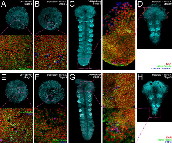
(A) Ventral view showing Pt-Dfd-A expression in the limb buds of L1 to L4 segments in the control embryos (white arrows). (B) Expression of Pt-Dfd-A is also observed in L1 in Sox21b-1 pRNAi embryos (n = 9) (white arrow in B). (C) Pt-lab is expressed in the pedipalpal segment (white arrow in D) and faintly in L1 segment in control embryos. In Sox21b-1 pRNAi embryos, Pt-lab expression can still be observed in the pedipalpal and L1 segments (n = 10) (white arrows in D). (E) The mesodermal marker Pt-twi is expressed in the anterior-most medial region of the head (white arrow), limb buds of L1 to L4, and with a stripped pattern in the O1 to O4 segments. (F) In Sox21b-1 knockdown embryos, only the head expression is maintained (n = 14) (white arrow in F). (G–J) show orthogonal projections of the cumulus (stage 5) and the head (stage 9) at 40x magnification of whole mount control embryos (left panels) and Sox21b-1 knockdown embryos (right panels), respectively. In control embryos, the formation of subectodermal layers is visible, but fewer internalisied cells (white arrows in I and J) are observed in the knockdown embryos. Note that nuclei also appeared to be larger in Sox21b-1 knockdown embryos (white arrow in H). DAPI stained nuclei are shown in cyan and the membrane marker alpha-Tubulin in red. Anterior is to the left in all panels.
Figure 2—figure supplement 3
Snapshots from live imaged videos in control and Sox21b-1 knockdown embryos.
Germ discs of (A) GFP dsRNA control embryos, showing the cumulus formation (red dotted lines), cumulus migration (red dotted arrow) and dorsal field opening (red dotted line and arrows). (B) Class I Sox21b-1 knockdown embryos showing cumulus formation, the partial migration of mesenchymal cells and limited dorsal field opening, which is also seen but more severely disrupted in Class II embryos (C), and absent in class III (D). .
Figure 2—figure supplement 4
Cell death and cell proliferation in Sox21b-1 knockdown embryos.
Ventral view of stage 5 control embryos stained for Cleaved-Caspase3 (A) and PHH3 (E). Cell death is not detectable in control embryos, but a high level of proliferation can be seen. In Sox21b-1 knockdown embryos, clusters of cells undergoing cell death can be found (B), as well as a decrease in proliferation in the knockdown embryos compared to controls (n = 15 for each staining) (F). Embryos at stage 9 stained for Cleaved-Caspase 3 (C) and PHH3 (G) show that only a small amount of cell death occurs in the SAZ, and that there is proliferation detectable throughout the entire embryo. Cell death is visible in the head extraembryonic layer in Sox21b-1 pRNAi embryos (D), and less proliferation is detected in stage 9 knockdown embryos (n = 15 for each staining). Anterior is to the top in panels C, D, G and H. Magnifications are 100X and 400x, respectively.
Figure 3
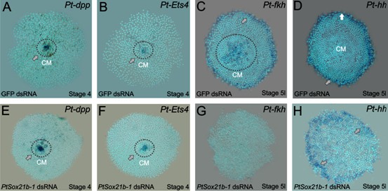
Gene expression in control and Sox21b-1 knockdowns at the germ disc stage.
Pt-dpp (A) and Pt-Ets4 (B) are expressed in the forming cumulus (CM) in the centre of the germ disc at stage 4 (grey arrow and dotted circle). This expression is unaffected by knockdown of Sox21b-1 (E and F) (n = 30 for each gene). (C) Pt-fkh is expressed at the rim and centre of the germ disc at late stage 5 (grey arrow and dotted circle in C), but expression is lost in Sox21b-1 knockdown embryos (n = 30) (G). Pt-hh expression at the rim of the germ disc (D) is normal in Sox21b-1 knockdown embryos (H) (grey arrows).
Figure 4
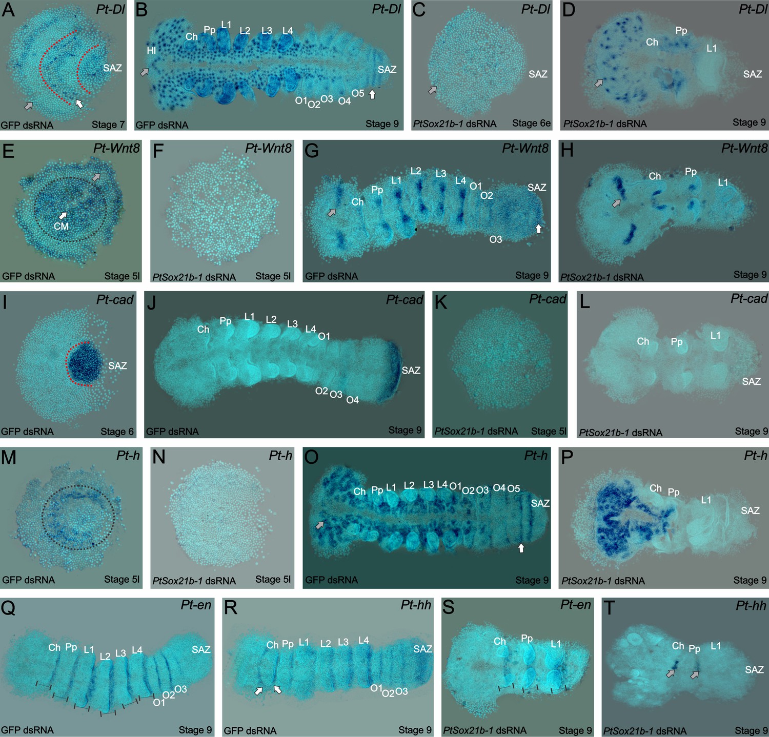
Expression of segmentation genes in Sox21b-1 pRNAi embryos.
(A) Pt-Dl expression at late stage 6/early stage 7 is dynamic in the SAZ and is also observed in the presumptive head region (grey arrow) and prosoma of the embryo (red dotted lines and white arrows). (B) At stage 9, Pt-Dl expression is seen in the SAZ (white arrow) but is restricted to the clusters of proneural differentiation in the anterior region of the embryo (anterior indicated by a grey arrow). (C) In Sox21b-1 knockdown embryos, Pt-Dl expression is not detectable in late stage 5/early stage 6 embryos (anterior indicated by a grey arrow) but can still be observed in the anterior ventral neuroectoderm at stage 9 (D) up to the pedipalpal segment (n = 17 and n = 14 for stage 5/6 and 9 respectively). Pt-Wnt8 expression is observed in the centre and at the rim of the germ disc in stage 5 control embryos (black dotted circle around the centre, grey arrow indicating the rim) but these expression domains are lost in Sox21b-1 knockdown embryos (n = 11) (E and F). Control embryos at stage 9 show the expression of Pt-Wnt8 in the medial region of the head (grey arrow in G), and in distal parts of each segment up to the SAZ (white arrow in G). In Sox21b-1 knockdown embryos at the same stage, the brain (grey arrow), cheliceral and pedipalpal expression is still present, but the posterior expression is lost (H) (n = 17). Pt-cad is expressed in the SAZ at late stage 5/early stage 6 (I), which persists to stage 9 in control embryos (J). However, Pt-cad expression is lost upon Sox21b-1 knockdown in embryos of both stages (n = 20 for each stage) (K and L). Pt-h expression at stage 5 in control embryos is seen at the rim and in the centre of the germ disc (black dotted circle in M), which is lost in Sox21b-1 knockdown embryos (N). At stage 9, Pt-h expression resembles Pt-Dl, both in the control (anterior is indicated by a grey arrow; the SAZ is indicated by a white arrow) and Sox21b-1 knockdown embryos (O and P) (n = 15 for both stages). Pt-en expression is present in the posterior of each segment (black lines in Q), and in cheliceral, pedipalpal and L1 segments in Sox21b-1 knockdown embryos at stage 9 (S) (n = 10). Pt-hh expression in control embryos at stage 9 is seen in the posterior of each segment, in the SAZ and also in a splitting wave between the cheliceral and pedipalpal segments (indicated by the white arrows in R). When Sox21b-1 is knocked-down, Pt-hh embryos show expression in the middle-posterior region of the cheliceral and pedipalpal segments (T) (n = 8). Ch: Chelicerae; Hl: Head Lobes; L1 to L4: Prosomal leg-bearing segments; O1 to O5: Opisthosomal segments; SAZ: Segment Addition Zone. Anterior is to the left in stage 9 embryos.
Figure 5

Expression of Sox21b-1 in Dl and Wnt8 pRNAi embryos.
Ventral view of stage 7 and 9 knockdown embryos for Pt-Dl (A and B) and Pt-Wnt8 (C and D). In knockdown embryos for both Pt-Dl and Pt-Wnt8, Sox21b-1 is still expressed at stage 7 (A and C) in the remaining SAZ cells, and in the forming segments of the presumptive prosoma of the embryo (white arrows). In stage 9 Pt-Dl knockdown embryos, Sox21b-1 remains highly expressed in the ventral nerve cord (B). Pt-Dl knockdown embryos lack the posterior L4 segment (white arrow), but brain formation appears normal (grey arrow) (B). Pt-Wnt8 embryos show a fusion of the L4 limb buds (white arrow), and Sox21b-1 is still expressed in the remaining SAZ cells (D). Anterior is to the left in all panels.
Figure 6
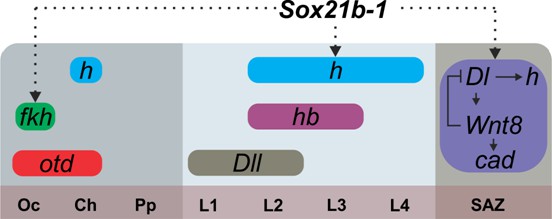
Summary of the regulation of spider segmentation.
The interaction of Sox21b-1 is presented in relation to genes involved in spider embryogenesis. We found that fkh expression requires Sox21b-1 in the most anterior part of the head . In the prosoma Sox21b-1 also has a gap gene like function and is required for the expression of hairy, while Distal-less (Pechmann et al., 2011), hunchback and orthodenticle (Pechmann et al., 2009) also act in a gap gene like manner during prosomal segmentation. The molecular control of segmentation in the SAZ involves a feedback loop between Dl and Wnt8, which acts upstream of cad and also controls the dynamic expression of hairy (Schönauer et al., 2016; McGregor et al., 2008b). We can infer from our results that Sox21b-1 acts upstream of these genes in the SAZ. Sox21b-1 could directly regulate these genes or the observed loss of expression could be indirect through incorrect specification of germ disc cells. Oc: Ocular segment; Ch: Cheliceral segment; Pp: Pedipalpal segment; L1-L4: leg bearing segments 1 to 4; SAZ: Segment Addition Zone.
Tables
Key resources table
| Reagent type (species) or resource | Designation | Source or reference | Identifiers | Additional information |
|---|---|---|---|---|
| Gene (Parasteatoda tepidariorum) | caudal | NA | AB096075 | |
| Gene (P. tepidariorum) | deformed-a | NA | AB433904 | |
| Gene (P. tepidariorum) | decapentaplegic | NA | AB096072 | |
| Gene (P. tepidariorum) | delta | NA | AB287420 | |
| Gene (P. tepidariorum) | engrailed | NA | AB125741 | |
| Gene (P. tepidariorum) | ets4 | NA | XP_015923392 | |
| Gene (P. tepidariorum) | forkhead | NA | AB096073 | |
| Gene (P. tepidariorum) | hairy | NA | AB125743 | |
| Gene (P. tepidariorum) | hedgehog | NA | AB125742 | |
| Gene (P. tepidariorum) | labial | NA | AB433903 | |
| Gene (P. tepidariorum) | sox21b-1 | NA | XP_015916301 | |
| Gene (P. tepidariorum) | twist | NA | AB167807 | |
| Gene (P. tepidariorum) | Wnt8 | NA | ACH88002 | |
| Antibody | Donkey anti-mouse IgG Alexa Fluor 555 | Invitrogen | A-31570 | (1:200) |
| Antibody | Goat anti-rabbit Alexa Fluor 647 | Invitrogen | A-21244 | (1:200) |
| Antibody | Mouse anti-α- Tubulin | Sigma | DM1a | (1:50) |
| Antibody | Rabbit Anti-phospho -Histone H3 (Ser10) | Merck Millipore | 06–570 | (1:200) |
| Antibody | Rabbit α cleaved caspase 3 | Cell Signaling | 9661 | (1:200) |
| Chemical compound, drug | 4′,6-Diamidino-2 -phenylindole dihydrochloride | Sigma -Aldrich | D8417 | |
| Chemical compound, drug | Halocarbon Oil 700 | Sigma -Aldrich | H8898 | |
| Chemical compound, drug | Poly-L-lysine | Sigma -Aldrich | P9155 | |
| Chemical compound, drug | TWEEN 20 | Sigma -Aldrich | P9416 | |
| Commercial assay or kit | TOPO-TA Cloning Kit | Invitrogen | K457502 | |
| Software, algorithm | Corel Graphics Suite | RRID:SCR_013674 | ||
| Software, algorithm | Code used for genomics data analysis - PROTGAMMALG | https://github.com/stamatak/standard-RAxML/blob/master/usefulScripts/ProteinModelSelection.pl | ||
| Software, algorithm | ImageJ | RRID:SCR_003070 | http://imagej.nih.gov/ij/ | |
| Software, algorithm | Helicon Focus | RRID:SCR_014462 |
Additional files
-
Supplementary file 1
Phenotypic frequencies for each fragment (1 and 2) and GFP (control) dsRNA – 30 embryos per cocoon for each spider were pooled and the characteristics were divided into three phenotypical classes.
- https://doi.org/10.7554/eLife.37567.015
-
Supplementary file 2
Sample sizes for the PtSox21b-1 phenotype analysis with in situ hybridization, and phenotypic classes used in each experiment.
- https://doi.org/10.7554/eLife.37567.016
-
Supplementary file 3
List of primers used in this paper.
- https://doi.org/10.7554/eLife.37567.017
-
Transparent reporting form
- https://doi.org/10.7554/eLife.37567.018
Download links
A two-part list of links to download the article, or parts of the article, in various formats.
Downloads (link to download the article as PDF)
Open citations (links to open the citations from this article in various online reference manager services)
Cite this article (links to download the citations from this article in formats compatible with various reference manager tools)
A SoxB gene acts as an anterior gap gene and regulates posterior segment addition in a spider
eLife 7:e37567.
https://doi.org/10.7554/eLife.37567


