MAPK activity dynamics regulate non-cell autonomous effects of oncogene expression

  1. Timothy J Aikin
  2. Amy F Peterson
  3. Michael J Pokrass
  4. Helen R Clark
  5. Sergi Regot  Is a corresponding author
  1. Department of Molecular Biology and Genetics, The Johns Hopkins University School of Medicine, United States
  2. The Biochemistry, Cellular, and Molecular Biology Graduate Program, The Johns Hopkins Universtiy School of Medicine, United States
  3. Department of Oncology, The Johns Hopkins University School of Medicine, United States

Abstract

A large fraction of human cancers contain genetic alterations within the Mitogen Activated Protein Kinase (MAPK) signaling network that promote unpredictable phenotypes. Previous studies have shown that the temporal patterns of MAPK activity (i.e. signaling dynamics) differentially regulate cell behavior. However, the role of signaling dynamics in mediating the effects of cancer driving mutations has not been systematically explored. Here, we show that oncogene expression leads to either pulsatile or sustained ERK activity that correlate with opposing cellular behaviors (i.e. proliferation vs. cell cycle arrest, respectively). Moreover, sustained–but not pulsatile–ERK activity triggers ERK activity waves in unperturbed neighboring cells that depend on the membrane metalloprotease ADAM17 and EGFR activity. Interestingly, the ADAM17-EGFR signaling axis coordinates neighboring cell migration toward oncogenic cells and is required for oncogenic cell extrusion. Overall, our data suggests that the temporal patterns of MAPK activity differentially regulate cell autonomous and non-cell autonomous effects of oncogene expression.

eLife digest

In animals, the MAPK pathway is a network of genes that helps a cell to detect and then respond to an external signal by switching on or off a specific genetic program. In particular, cells use this pathway to communicate with each other. In an individual cell, the MAPK pathway shows fluctuations in activity over time.

Mutations in the genes belonging to the MAPK pathway are often one of the first events that lead to the emergence of cancers. However, different mutations in the genes of the pathway can have diverse effects on a cell’s behavior: some mutations cause the cell to divide while others make it migrate. Recent research has suggested that these effects may be caused by changes in the pattern of MAPK signaling activity over time.

Here, Aikin et al. used fluorescent markers to document how different MAPK mutations influence the behavior of a human breast cell and its healthy neighbors. The experiments showed that cells with different MAPK mutations behaved in one of two ways: the signaling quickly pulsed between high and low levels of activity, or it remained at a sustained high level. In turn, these two signaling patterns altered cell behavior in different ways. Pulsed signaling led to more cell division, while sustained signaling stopped division and increased migration.

Aikin et al. then examined the effect of the MAPK mutations on neighboring healthy cells. Sustained signaling from the cancerous cell caused a wave of signaling activity in the surrounding cells. This led the healthy cells to divide and migrate toward the cancerous cell, pushing it out of the tissue layer. It is not clear if these changes protect against or promote cancer progression in living tissue. However, these results explain why specific cancer mutations cause different behaviors in cells.

Introduction

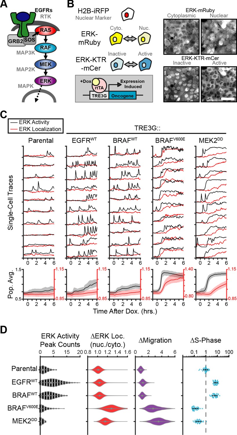
The Receptor-Tyrosine Kinase (RTK)/RAS/ERK signaling axis (Figure 1A) is mutated in most human cancers (Sanchez-Vega et al., 2018). In normal conditions, the ERK pathway promotes proliferation, differentiation, survival and cell migration (Johnson and Lapadat, 2002). During oncogenesis, mutations or amplification of ERK pathway components can also promote oncogene-induced senescence (Hahn and Weinberg, 2002) (OIS) or oncogenic cell extrusion from epithelial monolayers in the so-called Epithelial Defense Against Cancer response (EDAC) (Hogan et al., 2009; Kajita et al., 2010). The mechanisms underlying dose-dependent effects of ERK signaling have been intensely studied using bulk cell population assays. However, the advent of single-cell analysis has shown that single cells often behave qualitatively different than bulk populations. In fact, in vivo and in vitro studies have now shown that pulsatile or sustained ERK activity have different effects on cell behavior (Albeck et al., 2013; Aoki et al., 2013; de la Cova et al., 2017; Johnson and Toettcher, 2019; Santos et al., 2007; Bugaj et al., 2018; Aoki et al., 2017). Whether different oncogenic perturbations also have different functional outcomes depending on downstream signaling dynamics remains unknown. To address this question, an isogenic single-cell approach with temporal control of oncogene expression is needed.

Figure 1 with 4 supplements see all
Oncogenic ERK signaling dynamics promote qualitatively different cell behaviors.

(A) Schematic representation of the RTK/RAS/ERK signaling pathway. (B) MCF10A cells were transduced with lentiviral vectors expressing ERK KTR-mCerulean3 and ERK-mRuby2. The doxycycline inducible system (rTtA and TRE3G) was used to drive the expression of oncogenes during live imaging. Representative images of cytoplasmic and nuclear ERK-mRuby2 (top) and inactive or active ERK as reported by ERK KTR-mCerulean3 (bottom). Scale bar = 50 µm. (C) Cells described in B with indicated inducible oncogenes were imaged every 5 min for 6 hr upon doxycycline induction (2 µg/ml) at t = 0. Single cells were analyzed as described in methods. Population averages represent more than 1000 cells per condition. Shaded regions indicate the 25th-75th percentiles. (D) Quantification of data obtained in C. Single-cell counts of ERK activity peaks after induction (6–12 hr), ERK kinase localization fold change (final N/C ratio over basal N/C ratio per cell), and cell migration (final over basal distance traveled per cell) were extracted as described in methods. For proliferation analysis the fraction of S phase cells was measured using Edu incorporation and the change over the no dox control was calculated and normalized to the mean of parental cells (dashed line) (see Materials and methods). Data represents 36 independent observations.

Recent in vivo studies revealed that oncogene expression can trigger tissue level responses involving normal neighboring cells (Brown et al., 2017; Ellis et al., 2019; Clavería et al., 2013; Sancho et al., 2013). In specific cases, mosaic oncogene expression leads to either basal extrusion or apical extrusion (Hogan et al., 2009; Kajita et al., 2010); however, the signaling mechanism responsible for recognition between normal and diseased cells is poorly understood (Kajita and Fujita, 2015; Clavería and Torres, 2016; Maruyama and Fujita, 2017). Coincidentally, propagating ERK signaling waves requiring the sheddase ADAM17 have been observed in mouse epidermis and intestinal organoids, but the physiological role of these signaling events remains unclear (Hiratsuka et al., 2015; Muta et al., 2018). Observation of interactions between oncogenic and neighboring epithelium with live-cell biosensors could provide insights into the collective signaling preceding oncogenic extrusion. In fact, a recent study using live imaging of calcium biosensors during EDAC of HRASG12V cells showed a calcium signaling wave which propagated through neighboring epithelium to coordinate actin rearrangements and polarized movements during apical extrusion (Takeuchi et al., 2020). The mechanistic basis underlying EDAC calcium waves remains unknown.

Here, we combine live cell imaging of MAPK activity biosensors with inducible expression of oncogenes to study the effects of oncogene expression on signaling dynamics and how altered MAPK dynamics impact both cell autonomous and non-cell autonomous behaviors in epithelial tissues. Our data shows that pulsatile or sustained ERK signaling resulting from oncogenic perturbations triggers different dynamics-dependent cell behaviors including oncogene-induced paracrine signaling via the ADAM17-AREG-EGFR signaling axis. The resulting signaling gradients are required to coordinate neighboring cell migration and active oncogenic cell extrusion (EDAC). Our study highlights the role of MAPK signaling dynamics in coordinating individual and collective cell behaviors.

Results

To study the effects of oncogene expression on the temporal patterns of MAPK signaling we generated a reporter cell line derived from the chromosomally-normal human breast epithelial line, MCF10A, expressing the ERK Kinase Translocation Reporter (Regot et al., 2014) (ERK KTR) and a fluorescently tagged ERK kinase (ERK-mRuby2). This combination of biosensors allowed independent measurement of ERK activity and ERK localization in live single cells at high temporal resolution. Then, we introduced 12 different doxycycline-inducible oncogenic perturbations via lentiviral infection and measured ERK signaling dynamics during overexpression (Figure 1B). Our results revealed two qualitatively different responses to oncogene induction: (i) increased frequency of ERK activity pulses with no change in ERK kinase localization (i.e. EGFR, B-RafWT), and (ii) sustained ERK activity with subsequent nuclear translocation of ERK kinase (i.e. B-RafV600E, MEK2DD) (Figure 1, and Figure 1—Video 1). We refer to these distinct dynamics as pulsatile or sustained ERK, respectively. Of note, MEK1/2WT expression is capable of exporting ERK into the cytoplasm without changing kinase activity (Adachi et al., 2000; Figure 1—figure supplement 1) and ERK nuclear accumulation occurs only when activity is sustained, suggesting that ERK activity and ERK localization are not always correlated. Interestingly, expression of B-RafWT or B-RafV600E elicit qualitatively different downstream dynamics even though they differ in a single amino acid and show similar expression levels by immunoblotting (Figure 1—figure supplement 2). Given that the B-RafV600E is insensitive to negative feedback regulation by ERK (Yao et al., 2015), this result suggests that ERK inhibition to B-RafWT is mechanistically involved in the characteristic pulsatile dynamics.

Next, we assessed how ERK dynamics affect cell behaviors by measuring cell migration and proliferation. While pulsatile ERK dynamics (i.e. EGFR or B-RafWT) consistently correlated with increased cell cycle progression, sustained ERK activity (i.e. B-RafV600E or MEK2DD) caused cell cycle arrest and increased migration (Figure 1D). Importantly, observed differences in cell behavior correlated with dynamics independently of the point in the cascade that perturbations were introduced (EGFR, Raf or MEK), suggesting that ERK is responsible for differences in cell behaviors rather than alternate downstream pathways. Moreover, expression of B-RafWT or B-RafV600E, which activate the cascade at the same point, caused different ERK activity dynamics (i.e. pulsatile or sustained respectively) and triggered opposing cellular behaviors (Figure 1D). Taken together, these data suggest that ERK activity dynamics can either promote or inhibit proliferation cell autonomously.

The sudden increase in migration and the loss of cell-cell contacts observed in cases where ERK activity is sustained (B-RafV600E and MEK2DD, Figure 1—Video 1) are reminiscent of phenotypes described for cells undergoing Epithelial-to-Mesenchymal Transition, or EMT (Hao et al., 2019). We sought out to address the role of EMT in oncogene-dependent cell behaviors by immunofluorescent staining of an epithelial marker E-Cadherin (E-Cad) and the mesenchymal marker N-Cadherin (N-Cad). While cell migration was clearly increased at 24 hr post-oncogene expression, cells retain E-Cad expression with no clear increase in N-Cad, as was observed in TGFβ-induced EMT (Figure 1—figure supplement 3). These results indicate that at the time points studied here, altered cell behaviors are either distinct from or precede those resulting from EMT.

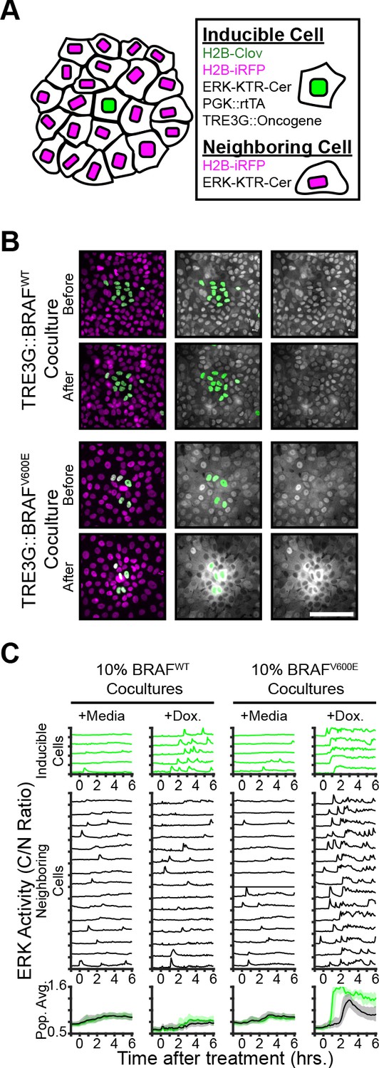
To examine the non-cell autonomous effects of oncogene expression in epithelial monolayers, we cocultured ‘inducible’ cells (expressing doxycycline-inducible oncogenes, a constitutively expressed H2B-mClover, and the ERK biosensors) with ‘neighboring’ reporter cells (expressing ERK biosensors without inducible oncogenes) and monitored signaling dynamics upon induction (Figure 2A). Interestingly, expression of B-RafV600E, but not B-RafWT, resulted in waves of ERK activation of neighboring cells (Figure 2B–C and Figure 2—Video 1). This comparison suggests that oncogenic perturbations that elicit sustained ERK activity propagate ERK activity pulses to neighboring cells. In agreement, other oncogenes that triggered sustained, but not pulsatile, ERK activity also promoted ERK activity waves in the neighboring cells (Figure 2—figure supplement 1). By using KTRs for p38 and JNK, we observed that neighboring epithelia did not activate other the MAPK pathways (Figure 2—figure supplement 1). Notably, spontaneous cell death events were also followed by similar ERK signaling waves (Figure 2—Video 2), indicating that oncogene expression and cell death may be similarly perceived by neighboring cells.

Figure 2 with 3 supplements see all
Oncogene induction results in dynamics-dependent paracrine ERK signaling.

( A) Schematic representation of coculture assay. H2B-iRFP (magenta) and ERK KTR are expressed in all cells for segmentation and quantification. H2B-mClover (green) was used to label inducible cells. (B) BRAFWT or BRAFV600E inducible cells were cocultured at 10% with ERK KTR cells and treated with doxycycline (2 µg/ml). Representative images are shown. Scale bar = 100 µm. (C) BRAFWT or BRAFV600E cocultures, as in B, were treated with vehicle (+Media) or with doxycycline (+Dox, 2 µg/ml). Single cells were quantified as described in methods. ERK activity traces in inducible (top, green) and neighboring cells (bottom, black) are shown. Population averages and 25th-75th percentiles (shaded) are shown for n > 450 cells per coculture condition.

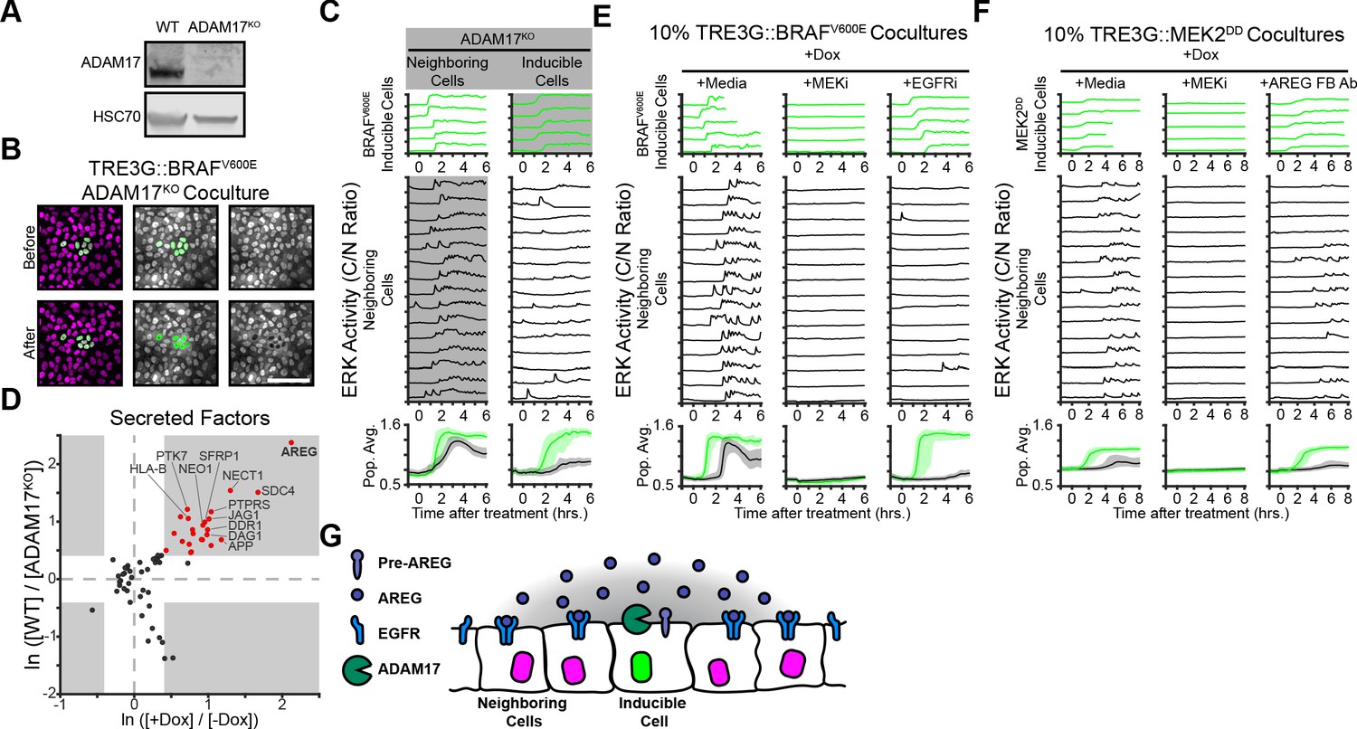
We then addressed the mechanistic basis of oncogene-dependent paracrine signaling. Previous studies demonstrated that ERK waves in epithelial monolayers depend on the membrane-tethered sheddase ADAM17, which releases membrane-bound growth factors that activate EGFR signaling in adjacent cells (Aoki et al., 2013; Aoki et al., 2017; Hiratsuka et al., 2015). Thus, we hypothesized that oncogenic cell ADAM17 may be decoding ERK signaling dynamics to trigger growth factor release. To test this hypothesis, we generated an ADAM17 knockout (ADAM17KO) cell line (Figure 3A) and used it as either ‘inducible’ or ‘neighboring’ cells in our coculture assay. Live imaging of WT and ADAM17KO cocultures indicated that ADAM17 is necessary in inducible, but not neighboring cells, to trigger ERK waves in the monolayer (Figure 3B–C and Figure 2—Video 1). Therefore, ADAM17 decodes ERK activity dynamics in inducible cells to transmit ERK signaling to neighboring cells. Previous work has shown that ADAM17 is weakly phosphorylated compared to other ERK substrates (Díaz-Rodríguez et al., 2002), thus the phosphorylation-dephosphorylation kinetics of ADAM17 and the temporal patterns of ERK activity may explain dynamics-specific ADAM17 activation.

ERK activity waves require ADAM17 release of AREG and neighboring cell EGFRs.

(A) Immunoblot against ADAM17 and HSC70 in WT and ADAM17KO cells generated by CRISPR-Cas9 editing (see Materials and methods for details). (B) Representative images of ADAM17KO BRAFV600E inducible cells cocultured and treated as in Figure 2B. (C) ADAM17KO cells (gray boxed traces) were used as inducible cells (right) or neighboring cells (left) in cocultures. Data for n > 1100 cells is presented as in Figure 2C. (D) ADAM17 substrates profiled by TMT mass spectrometry. Supernatants from ADAM17KO or WT cells expressing (+Dox) or not expressing (-Dox) BRAFV600E were collected and analyzed by Tandem-Mass-Tag (TMT) mass spectrometry as described in methods. Scatter plots show the natural log of fold change values of all statistically significant (p<0.05) proteins in both WT vs. ADAM17KO and +Dox vs. -Dox comparisons. Grey boxes indicate >1.5 fold change. (E) BRAFV600E co-cultured monolayers were plated as in Figure 2C and pretreated with indicated inhibitors (MEKi, 5 µM PD0325901; EGFRi, 5 µM Gefitinib) for one hour before induction with doxycycline (2 µg/ml). Representative single cell traces and population averages for n > 1000 cells are shown as in 2C. (F) MEK2DD co-cultured monolayers were plated as in Figure 2C and pretreated with indicated inhibitors (MEKi, 5 µM PD0325901; AREG FB Ab, 50 µg/ml function-blocking antibody) for one hour before induction with doxycycline (2 µg/ml). Representative single cell traces and population averages for n > 1000 cells are shown as in Figure 2C. (G) Schematic representation of ADAM17-AREG-EGFR paracrine signaling.

ADAM17-released growth factors include HB-EGF, TGF-α, Epiregulin, and Amphiregulin (Zunke and Rose-John, 2017; Rios-Doria et al., 2015). In order to identify the factors mediating oncogene-induced paracrine signaling we used Tandem-Mass-Tag Mass Spectrometry of supernatant proteins following induction of sustained ERK activity in WT and ADAM17KO cells. A variety of known and unknown ADAM17 substrates were present in the induced cell supernatants, including immune surveillance (HLA-A/B/C), Delta-Notch (JAG1), and Wnt (SFRP) signaling proteins (Figure 3D and Supplementary file 1). Of note, the EGFR ligand Amphiregulin (AREG) was the most upregulated, ADAM17-dependent protein in the supernatant, suggesting that AREG released from inducible cells could act as an oncogene-dependent paracrine signaling molecule. Accordingly, cocultures pre-incubated with AREG function-blocking antibodies or EGFR inhibitors prevented neighboring cell ERK activation without affecting ERK signaling in inducible cells (Figure 3E–F). These results indicate that oncogene-dependent ERK waves are mediated by ADAM17 (in inducible cells), AREG release, and EGFR signaling (Figure 3G).

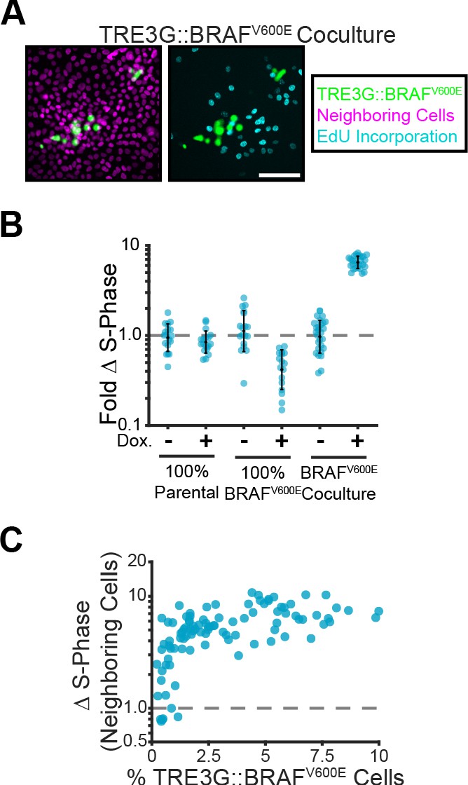
Given that cells surrounding B-RafV600E expressing cells showed pulsatile ERK activity (Figure 2C), we hypothesized that oncogene expression may promote cell proliferation in a non-cell autonomous manner. Accordingly, sustained ERK signaling in inducible cells increased proliferation of neighboring cells up to 10-fold (Figure 4A–C). Together, these data indicate that, depending on ERK dynamics, oncogenic cells can have either cell autonomous or non-cell autonomous contributions to tissue growth.

Paracrine ERK signaling leads to non-cell autonomous proliferation.

(A) Representative images of BRAFV600E cocultures treated with doxycycline and EdU as described in methods. Inducible cell nuclei (H2B-mClover, green), all nuclei (H2B-iRFP, magenta) and EdU staining (cyan) are shown. Scale bar = 100 µm. (B) Indicated monolayers were cultured and incubated with or without doxycycline for 24 hr. The change in S-phase cell fractions was determined by EdU incorporation as described in methods and normalized to parental mean (dashed line). Bar represents mean and standard deviation for n ≥ 16 observations. (C) Inducible BRAFV600E cocultures were plated at different proportions and labelled with EdU as in A. The fold-change in S-phase cell fractions is plotted against the percent of BRAFV600E-expressing cells for each position. 98 total observations shown.

In addition to proliferation, ERK waves have been shown to orient collective cell migration during wound healing (Aoki et al., 2017). In cocultures, sustained ERK activity in B-RafV600E-inducible cells correlated with neighboring cell migration towards inducible cells in an ADAM17 and EGFR-dependent manner (Figure 5A). We hypothesized that coordinated migration of neighboring cells could physically contribute to oncogenic cell extrusion (Hogan et al., 2009; Leung and Brugge, 2012; Slattum et al., 2014). To address this hypothesis, we used confocal Z stacks to quantify extrusion of oncogene-expressing cells from monolayers (Figure 5B and Figure 5—Video 1). Interestingly, while pulsatile ERK activity (i.e. EGFR and B-Raf) was not sufficient to extrude cells, sustained ERK activity (i.e. B-RafV600E and MEK2DD) led to efficient epithelial cell extrusion apically (Figure 5C). KRASG12V induction did not result in apical extrusion to the extent observed for HRASG12V (Hogan et al., 2009Figure 5—figure supplement 1). However, since sustained ERK activation in KRASG12V occurs later than B-RafV600E (Figure 1—figure supplement 1) further apical extrusion may also occur at a later time. Taken together, our data suggests that apical extrusion occurs when oncogenic perturbations trigger sustained ERK activity.

Figure 5 with 4 supplements see all
Paracrine ERK activation coordinates extrusion of aberrantly signaling cells through directed migration of the neighboring epithelium.

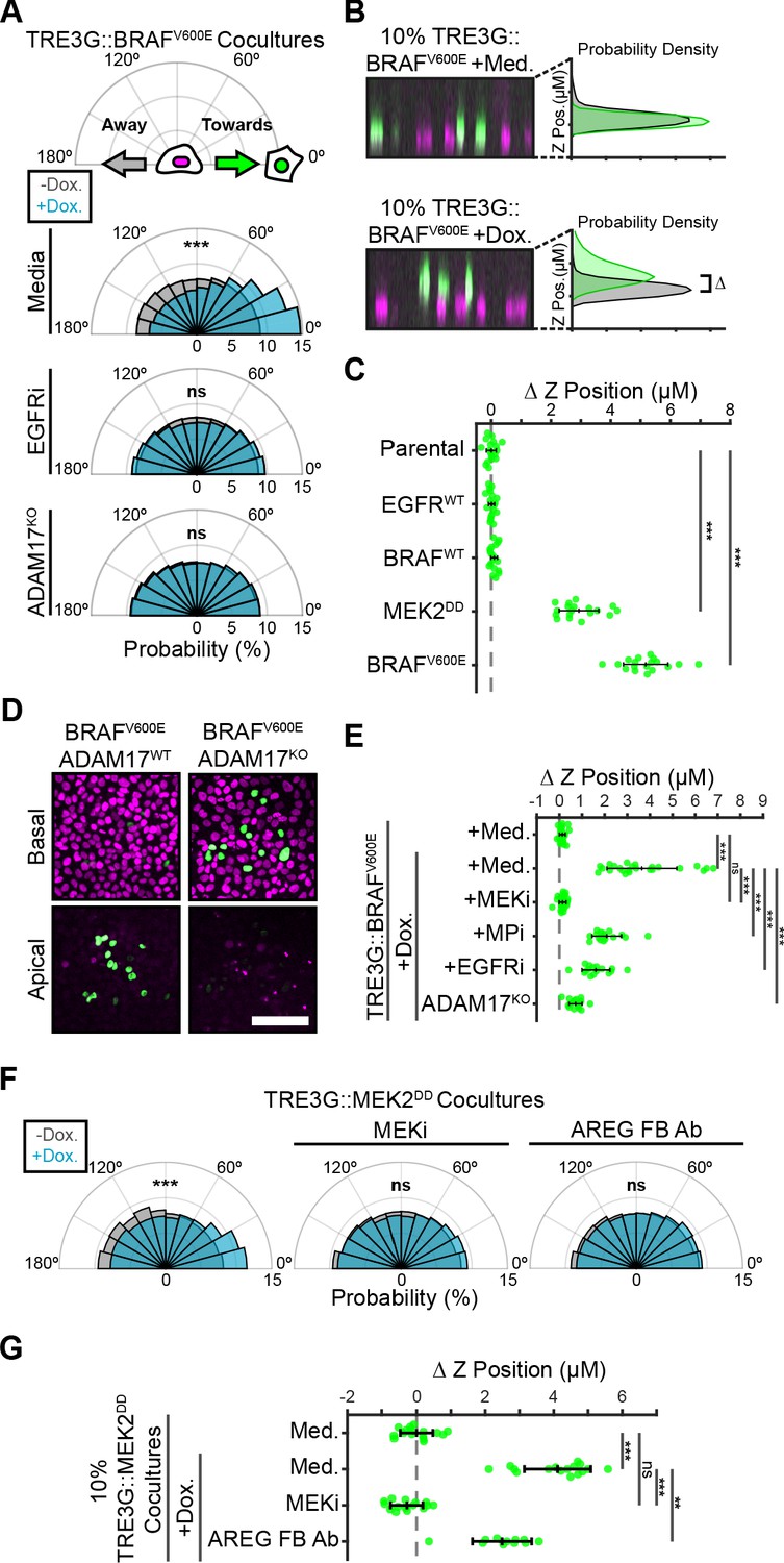
(A) Inducible BRAFV600E cells (WT or ADAM17KO) were plated in 1% cocultures and treated with doxycycline (2 µg/ml) in the presence or absence of EGFR inhibitor gefitinib (5 µM) as indicated. Radial histograms represent migration angle distributions of neighboring cells before (grey) and 2–6 hr after (cyan) induction (see Materials and methods). Data represents angles from n > 1000 cells from 10 independent observations per condition. Data was assessed using subsampling and a two-sample KS test with ‘ns’ not significant, ***p<0.001 (see Materials and methods). (B) 10% BRAFV600E cocultured monolayers were seeded as described in methods. After 24 hr with doxycycline (2 µg/ml), monolayers were imaged by spinning disk confocal. Representative orthogonal Z projections and probability densities for nuclear height of inducible (green) and neighboring (grey) cells are shown (see methods). Extrusion (ΔZ) is calculated as the height difference between gaussian-fitted maxima of the green and black distributions. (C) 10% cocultures of indicated parental or inducible cells were treated with 24 hr doxycycline (2 μg/ml), imaged, and analyzed as in B. Data represents difference in nuclear height (ΔZ) for n = 18 observations normalized to the mean height of parental cells (dashed line), with mean and +/- standard deviation (black bars). Significance was calculated by two-sample t-test with ‘ns’ indicating no significance, ***p<0.001. (D) Representative basal and apical images (+6 µm) of WT or ADAM17KO, BRAFV600E inducible cells (green) in WT monolayers (red) after 24 hr of doxycycline treatment. (E) 10% BRAFV600E cocultures were pretreated with inhibitors (MEKi, 5 μM PD0325901, MPi, 5 μM Batimastat, EGFRi, 5 μM Gefitinib) and 24 hr doxycycline (2 μg/ml) or media, imaged and analyzed as in B. Data represents difference in nuclear height (ΔZ) for n ≥ 16 independent observations presented as in C. (F) Inducible MEK2DD cells were plated in 1% cocultures and treated with doxycycline (2 µg/ml) in the presence of MEK inhibitor (MEKi, 5 µM PD0325901) or amphiregulin function-blocking antibody (AREG FB Ab, 50 ng/ml) as indicated. Radial histograms are presented as in A for angles of n > 100 cells from two to three independent observations per condition. Data was assessed using subsampling and a two-sample KS test with ‘ns’ not significant, ***p<0.001 (see methods). (G) 10% MEK2DD cocultures were pretreated with MEK inhibitor (MEKi, 5 µM PD0325901) or Amphiregulin function-blocking antibody (AREG FB Ab, 50 ng/ml) and 24 hr doxycycline (2 µg/ml) or media, as indicated, then imaged and analyzed as in B-C. Data represents difference in nuclear height (ΔZ) for n ≥ 11 independent observations normalized to the mean height of media-treated MEK2DD cells (dashed line), with mean and +/- standard deviation (black bars). Significance was calculated by two-sample t-test with ‘ns’ indicating no significance, **p<0.01, and ***p<0.001.

In mammalian epithelia, apical extrusion eliminates apoptotic cells or crowded cells to maintain homeostasis (Rosenblatt et al., 2001; Eisenhoffer et al., 2012). Similar, but mechanistically different, apical extrusion has been observed for some oncogenic cells during EDAC (Kajita and Fujita, 2015). We wanted to know whether the extrusion of inducible cells with sustained ERK activity resembled EDAC. To differentiate between pseudostratified or de-laminated (Grieve and Rabouille, 2014) epithelium and extrusion we analyzed confocal images of E-cad membrane staining in induced cocultures. Cells with sustained ERK activity were fully extruded, sitting above WT cells in the plane of the monolayer (Figure 5—figure supplement 2). These images also demonstrate maintenance of E-Cad at the junctions between WT cells below extruded cells. Quantification of fully-extruded cells at several timepoints showed the majority of oncogenic cells being extruded from 4 to 8 hr after induction, but continuing until 24 hr, when 91% of inducible cells are fully extruded.

Both the oncogenic and apoptotic extrusion models involve cytoskeletal rearrangements at the site of extrusion (Rosenblatt et al., 2001Kajita and Fujita, 2015). To observe live actin dynamics in cocultures, we made cell lines stably expressing Utrophin-261-EGFP (Belin et al., 2014). Using this tool, we observed transient accumulation of actin at the basal interface of B-RafV600E expressing and neighboring cells that first closed off the basal attachments of inducible cells before they were pushed apically out of the monolayer (Figure 5—figure supplement 2 and Figure 5—Video 2). These polarized, actin-containing basal protrusions were dependent on EGFR activity as they could be inhibited by EGFR inhibitor. Apoptotic extrusion relies on Sphingosine-1-phosphate (S1P) signaling through intrinsic S1P production and juxtracrine activation of the GPCR S1PR2 (Gu et al., 2011), yet inhibition of S1P production had only moderate effects on apical extrusion of MEK2DD cells (Figure 5—figure supplement 2). Together, our results suggest that the apical extrusion of oncogenic cells observed in our experiments are similar to the EDAC mechanism previously described for HRASG12V, V-Src, and other cells (Hogan et al., 2009; Kajita et al., 2010; Kajita and Fujita, 2015).

The requirement for paracrine signals in collective migration led to the question of whether paracrine signals were also required for extrusion. To test the role of ADAM17-mediated AREG-EGFR paracrine signals in promoting extrusion, we performed extrusion assays using ADAM17KO cells or in the presence of EGFR inhibitor or AREG function-blocking antibodies. Extrusion of inducible cells was abolished in these conditions (Figure 5D–G), suggesting that ERK signaling waves are required for extrusion. Of note, since ADAM17KO and EGFR inhibition affect ERK activation of neighboring cells without altering ERK dynamics in inducible cells, we hypothesized that that activation of inducible cells alone is not sufficient for extrusion, but that neighboring cell ERK activation may be required. To address this question, ERK-independent ADAM17 activation is needed.

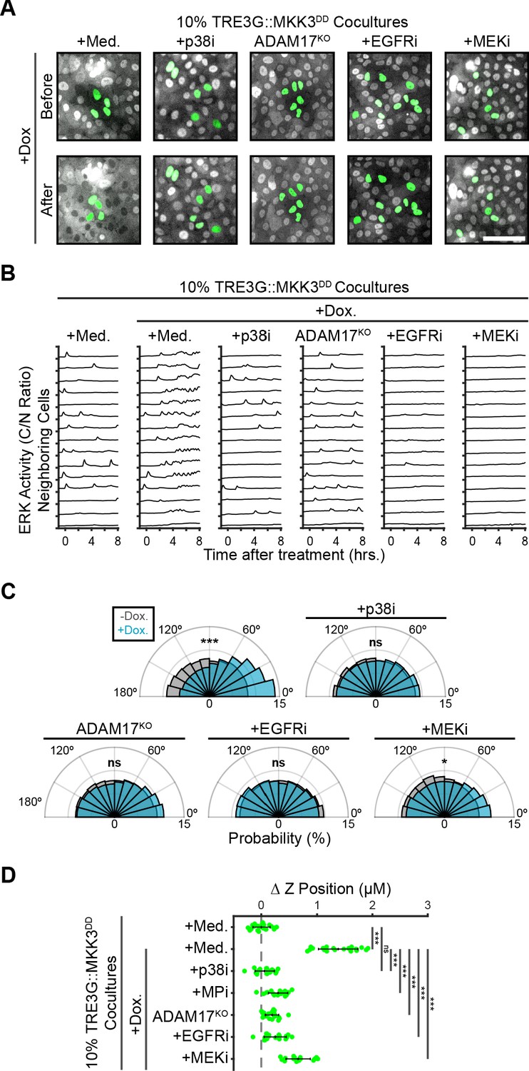
Previous studies have shown that the stress MAPK p38 phosphorylates and activates ADAM17 (Xu and Derynck, 2010). Thus, we used our doxycycline-inducible system to drive the expression of MKK3DD, a constitutively-active MAP2K specific for p38 (Figure 6—figure supplement 1), to activate ADAM17 independently of ERK. As expected, we found that p38 activation leads to ERK signaling waves (Figure 6A–B), proliferation (Xu and Derynck, 2010; Figure 6—figure supplement 2), directed migration (Figure 6C) and extrusion in an ADAM17 and EGFR dependent manner (Figure 6D and Figure 6—Video 1). However, B-RafV600E oncogenic signaling, extrusion and proliferation were unaffected by p38 inhibition (Figure 6—figure supplement 3), suggesting that sustained ERK or p38 activity are each capable of activating ADAM17 paracrine signaling. Using this ERK-independent MKK3DD extrusion system, we found that MEK inhibition decreased directed migration and prevented extrusion, confirming that extrusion requires ERK activity in the neighboring cells (Figure 6C–D). Thus, ERK activity is required for extrusion in both oncogenic and neighboring cells with qualitatively different temporal dynamics. Notably, inhibition of S1P signaling in MKK3DD cocultures also reduced extrusion efficiency despite having unaffected signaling (Figure 6—figure supplement 4). This result suggests that cell-autonomous ERK or p38 activation in extruded cells may underlie the fundamental differences reported between extrusion of oncogenic and stressed cells.

Figure 6 with 5 supplements see all
ERK activity in neighboring cells is required for coordinating extrusion.

(A) Representative images showing WT or ADAM17KO cells with inducible MKK3DD (green), cocultured at 10% with neighboring ERK-KTR cells (grey). Cocultures were treated with doxycycline (2 µg/m) in the presence of media, p38 inhibitor (5 µM BIRB-796), EGFR inhibitor (5 µM Gefitinib), or MEK inhibitor (5 µM PD 0325901). Scale bar = 100 µm. (B) ERK activity traces of neighboring cells in coculture with MKK3DD-inducible cells (WT or ADAM17KO) plated at 10%, pretreated with inhibitors (p38i, 5 µM BIRB 796; EGFRi, 5 µM Gefitinib; MEKi, 5 µM PD 0325901) and doxycycline (2 µg/ml) or media, and imaged as in Figure 2C. 15 representative neighboring cell ERK activity traces are shown for each condition. (C) Inducible MKK3DD cells (WT or ADAM17KO) were plated in 1% cocultures and treated with doxycycline (2 µg/ml) in the presence or absence of inhibitors. Radial histograms of migration angles before (grey) and 6–9 hr after (cyan) induction presented as in Figure 5A. Data represents angles from n > 900 cells from ≥6 observations per condition assessed using subsampling and a two-sample KS test with ‘ns’ not significant, *p<0.05, ***p<0.001 (see Materials and methods). (D) 10% MKK3DD cocultures were pretreated with inhibitors (p38i, 5 µM BIRB 796; EGFRi, 5 µM Gefitinib; MEKi, 5 µM PD 0325901) and 24 hr doxycycline (2 µg/ml) or media, imaged and analyzed as in Figure 5B–D. Data represents difference in nuclear height (ΔZ) for n ≥ 16 observations normalized to the mean height of parental cells (dashed line), with mean and +/- standard deviation (black bars). Significance was calculated by two-sample t-test with ‘ns’ indicating no significance, ***p<0.001.

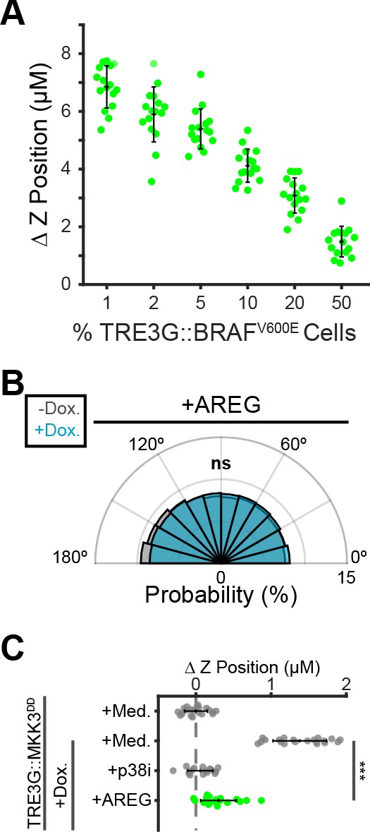
Finally, we asked whether the spatiotemporal properties of paracrine ERK signaling waves are important to coordinate extrusion. We first tested the efficiency of extrusion with altered proportions of B-RafV600E cells in the coculture, as higher proportions will have de-centralized and overlapping signaling events. The proportion of inducible cells was inversely correlated with extrusion efficiency (Figure 7A). Moreover, exogenous addition of AREG, which triggers widespread ERK activation preventing any spatially defined waves, eliminated directed migration of neighboring cells and extrusion (Figure 7B–C). The observation that in cocultures, polarized actin enrichment in neighboring cell basal protrusions is absent with EGFR inhibition, also indicates that growth factor signaling provides directional information during extrusion (Figure 5—figure supplement 2). Together this data suggests that locally generated paracrine signaling coordinates directed migration of neighboring epithelia to promote extrusion of oncogenic cells (Figure 8).

Localized paracrine signals coordinate directed migration and extrusion.

(A) Inverse relationship between fraction of oncogenic cells in coculture and extrusion efficiency. Inducible BRAFV600E cells were cocultured at indicated proportions, treated 24 hr with doxycycline (2 µg/ml), imaged, and analyzed as in Figure 5B. Data represents difference in nuclear height (ΔZ) for n ≥ 15 observations presented as in 5C. (B) Inducible MKK3DD cells were plated in 1% cocultures and treated with doxycycline (2 µg/ml) in the presence or absence of Amphiregulin (20 ng/ml). Radial histograms of migration angles before (grey) and after (cyan) induction presented as in Figure 6C. Data represents angles of n > 900 cells from ≥6 observations assessed using subsampling and a two-sample KS test with ‘ns’ not significant, ***p<0.001 (see Materials and methods). (C) 10% MKK3DD cocultures were pretreated with Amphiregulin (20 ng/ml, green) and 24 hr doxycycline (2 µg/ml) or media, imaged and analyzed as in Figure 5B–D, and compared to selected conditions reproduced from Figure 6D (grey). Data represents difference in nuclear height (ΔZ) for n ≥ 16 observations normalized to the mean height of parental cells (dashed line), with mean and +/- standard deviation (black bars). Significance was calculated by two-sample t-test with ‘ns’ indicating no significance, ***p<0.001.

Graphical summary.

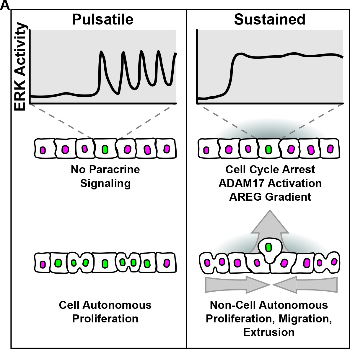
(A) Model summarizing cell autonomous and non-cell autonomous effects resulting from pulsatile and sustained ERK signaling dynamics.

Discussion

A wide variety of ERK pathway alterations occur across human tumors, often resulting in different cancer phenotypes (Sanchez-Vega et al., 2018; Bailey et al., 2018; Hoadley et al., 2018). To better understand the signaling effects of oncogenic mutations, we used live cell imaging of signaling biosensors upon induction of oncogenes and found that different ERK pathway oncogenes trigger two distinct temporal patterns: pulsatile or sustained ERK activity. While our approach is admittedly different than acquisition of point mutations in vivo, ERK dynamics resulting from oncogene overexpression robustly correlated with the same cellular phenotypes: (i) pulsatile ERK activity correlates with increased proliferation and, (ii) sustained ERK activity leads to cell cycle arrest similar to OIS (Hahn and Weinberg, 2002; Courtois-Cox et al., 2006; Kuilman et al., 2008). Moreover, we showed that sustained ERK activity in oncogenic cells triggers ERK signaling waves through unperturbed neighboring cells. These signaling waves depend on the ADAM17-EGFR paracrine signaling axis and lead to different non cell-autonomous behaviors such as (i) proliferation, (ii) directed migration of neighboring cells toward oncogenic cells, and (iii) oncogenic cell extrusion (Figure 8).

Our data indicates that cancer mutations can have non-cell autonomous contributions to tissue growth (Figure 4). Interestingly, studies in mouse epidermis have shown that mosaic oncogene expression promotes proliferation of wild type surrounding cells, which is required to expel mutant outgrowths from the tissue (Brown et al., 2017). While the role of ADAM17 in this phenomenon and during early tumorigenesis in vivo is yet unknown, it is tempting to speculate that polypous outgrowths may occur in the presence of non-proliferative oncogenic cells that release growth factors via ADAM17. The mechanisms that mediate tissue expelling in vivo remain unknown.

Previous work in described the process of oncogenic cell extrusion as part of the so called Epithelial Defense Against Cancer (i.e. EDAC) (Hogan et al., 2009). However, the signals involved in recognition of oncogenic cells, and why only specific oncogenes trigger oncogenic cell extrusion was unclear (Kajita and Fujita, 2015; Clavería and Torres, 2016; Maruyama and Fujita, 2017). Our data suggests that perturbations that elicit sustained ERK activity (eg. B-RafV600E, MEK2DD), activate ADAM17, which in turn releases EGFR ligands (Figure 3). This paracrine signal is critical for oncogenic extrusion (Figure 5). However, we acknowledge that different cellular states such as apoptosis or overcrowding lead to extrusion by different mechanisms. Of note, our data showed that ERK activation drives extrusion to a higher extent than p38 activation (Figure 6), which may result from a difference in overall cell autonomous migration in these two cases (Figure 6—figure supplement 1). Moreover, sphingosine kinase inhibition caused greater defects in extrusion of p38-active cells than ERK-active cells (Figure 5—figure supplement 2 and Figure 6—figure supplement 4). This finding agrees with work showing that EDAC of transformed HRASG12V cells is less dependent on sphingosine-1-phosphate production than extrusion of crowded or apoptotic cells (Yamamoto et al., 2016), where stress signaling may be involved.

We and others have identified AREG as one of the key EGFR ligands in mammary epithelial cells (Sternlicht et al., 2005; Figure 3); however, different ligands may be required in other tissues. These ligands, released by ADAM17, coordinate the migration of neighboring cells by mechanisms that remain unclear. Cultured monolayers are fundamentally different than in vivo tissues; however, the chemo-attractive properties of growth factors for directed migration have been modeled and studied in cell culture (Devreotes et al., 2017; Tranquillo et al., 1988). We propose that local signaling gradients are created by oncogenic cells to coordinate directed migration of neighbors. To support this idea, we show that addition of exogenous AREG or increased fractions of oncogenic cells both prevent directed migration of neighboring cells (Figure 7), and that during extrusion, polarized actin-containing basal protrusions require growth factor signaling (Figure 5—figure supplement 2). Localized ERK signaling gradients have also been observed during morphogenesis of Drosophila, avian, and mammalian embryos (Yang et al., 2002; Ogura et al., 2018; Corson et al., 2003), and in preserving homeostasis of mammalian epidermis and intestinal organoids (Hiratsuka et al., 2015; Muta et al., 2018; Liang et al., 2017). Thus, in addition to roles in oncogenesis, the ADAM17-EGFR paracrine signaling axis may direct collective behaviors during development.

Overall, our results highlight the importance of quantitative live-cell approaches to understand the effects of genetic perturbations and cell-cell communication in tissues. We propose a critical role for ERK signaling dynamics and the ADAM17-EGFR signaling axis in coordinating cell behaviors at the tissue level.

Materials and methods

Cell lines & reagents

Request a detailed protocol

 MCF10A human mammary epithelial cells (ATCC) were grown at 37° and 5% CO2 in DMEM/F12 (Gibco) with 5% horse serum (HS) (Sigma), 10 µg/ml Insulin (Sigma), 20 ng/ml EGF (Peprotech), 1x Penicillin-Streptomycin (P/S) (Gibco), 0.5 mg/ml Hydrocortisone (Sigma), 100 ng/ml Cholera Toxin (Sigma). Cells were passaged every 3 days with 0.25% Trypsin-EDTA (Gibco), are mycoplasma free, and were verified by STR-profiling (ATCC).

Cell lines were generated with lentivirus produced in HEK293-FTs (Thermo) with third-generation packaging plasmids and Lipofectamine 2000 (Thermo). Viral supernatants were collected 48 hr after transfection and incubated in MCF10As with polybrene (10 µg/ml, EMD Millipore). To create dual-sensor cells, MCF10As were infected with a lentiviral H2B-iRFP vector (Addgene) and sorted. We used gateway cloning (Campeau et al., 2009) to introduce ERK-KTR-mCer3 and ERK1-mRuby2 into PGK pLenti DEST vectors (Addgene), infected and selected the H2B-iRFP MCF10As (Blasticidin 3 µg/ml and Hygromycin 10 µg/ml Corning). We isolated moderately expressing clones using cloning cylinders (EMD Milipore). For inducible cells, a gateway-ready reverse TET trans-activator (rtTA) plasmid was created by adding the rtTA with a 2A peptide to the Puromycin resistance gene in a CMV Puro DEST plasmid (Addgene) by gibson cloning (Gibson et al., 2009). Human coding sequences were acquired from either Addgene or the Thermo Ultimate ORF Collection, sequence verified, and introduced in the rtTA CMV Puro DEST plasmid by gateway cloning (Campeau et al., 2009). These plasmids were used for lentivirus, and infected cells were selected with Puromycin (1 µg/ml, Sigma). Utrophin-261-EGFP cell lines were made by cloning the coding region from pEGFP-C1 Utr261-EGFP (Addgene) into a pENTR backbone by Gibson cloning, and then introduced into the pLenti PGK Puro DEST plasmid by gateway cloning. These plasmids were used to generate lentivirus, and infected cells were selected with Puromycin.

For inhibitor experiments, small molecules or antibodies and doxycycline were dissolved to a 10X working concentration in imaging media before addition. Final DMSO concentration did not exceed 0.15%. Inhibitors used include the MEK inhibitor PD-0325901, the MMP/ADAM inhibitor Batimastat, the EGFR inhibitor Gefitinib, the p38 inhibitor BIRB-796, the Sphingosine Kinase inhibitor SKII, and the S1PR2, inhibitor JTE-013 all from Selleck Chemicals. The p38 inhibitor SB-203580 was obtained from Sigma. Amphiregulin was ordered from Peprotech. Amphiregulin function-blocking antibody is from R and D systems.

The ADAM17KO cell lines were created using the CRISPR V2 Neo system (a gift from Dr. Andrew Holland) and gRNA oligos targeting R241 of exon 6. Dual sensor cells were infected with lentivirus carrying this plasmid, selected with Neomycin (500 µg/ml, Sigma) and clonally expanded before western blot validation (Figure 2B).

Live imaging

Request a detailed protocol

Cells were plated at 3*105 cells/well in fibronectin-treated (EMD Millipore) 96-well glass-bottom plates (Thermo Scientific) 48 hr before imaging. The following day, monolayers were serum-starved with 0.5% HS, phenol-red-free DMEM/F12 containing P/S with 1 mM Na Pyruvate and 10 mM HEPES. For signaling experiments in Figure 1 and Figure 1—figure supplement 1, media was switched to 0% HS several hours before imaging to limit basal signaling. Monolayers were imaged using a Metamorph-controlled Nikon Eclipse Ti-E epifluorescence microscope with a 20x air objective and a Hamamatsu sCMOS camera. The multi LED light source SpectraX (Lumencor) and the multiband dichroic mirrors DAPI/FITC/Cy3/Cy5 and CFP/YPF/mCherry (Chroma) where used for illumination and imaging without any spectral overlap. For extrusion and live-actin experiments, a Metamorph-controlled Nikon Eclipse Ti-E spinning-disc confocal (Yokogawa W1) with a 20x or 40X objective, Prime 95-B sCMOS camera (Photometrics) and a Multiline laser launch (Cairn Research) was used to capture H2B-iRFP and H2B-mClover or Utrophin-261-EGFP images every 1 µm of a 25–30 µm range through monolayers. Temperature (37°C), humidity and CO2 (5%) were maintained throughout all imaging using OKO Labs control units. Sample sizes were selected by attempting to capture at least 100 cells from each population, with several hundred cells preferred. Key conditions from imaging experiments were performed at least twice, with one replicate presented in figures.

Image analysis and quantification

Request a detailed protocol

Primary time-lapse images were subjected to flat-fielding and registration (custom software Aikin et al., 2020) before object segmentation and measurements in Cell Profiler. Nuclear positions were used to track individual cells through time-series (custom software Aikin et al., 2020) and intensity ratios were calculated as previously described (Regot et al., 2014). Minimal cleaning of traces excluded cells where tracks switched between two objects, where the KTR ratios were affected by segmentation errors, or where traces represent less than two thirds of the entire time-course. In conditions where cells move rapidly, such as B-RafV600E and MEK2DD, and traces are shorter due tracking errors, track-length restraints were relaxed to include more cells for analysis. Single-cell traces were chosen by random plotting of distinct cells and selection of those that were tracked throughout the whole experiment. Peak counting was performed with software based on findPeaks (O'Haver, 2014; Mathworks.com) and modified to detect peaks based on the rate of change between gaussian-fitted minima and maxima from single-cell traces.

For directed migration, positions were selected where distinct groups of inducible cells were present in the center of the field of view. Migration was quantified by positional changes over 20 min intervals for specified time windows, from all WT neighboring cells within a 200 µM X 200 µM area centered on the group of inducible cells. The migration angles of neighboring cells are plotted as radial histograms where 0° indicates migration directly towards, and 180° directly away from the center of isolated inducible cell groups. Migration datasets contain many sampled angles from large populations of cells. To overcome issues with high power, we applied subsampling techniques using 1000 iterations of 1000 randomly-selected migration angles each, and presented the median Two-Sample Kolmogorov-Smirnov (KS) Test P-values from these iterations (‘ns’, not significant, *p<0.05, **p<0.01, ***p<0.001).

For extrusion experiments, histograms of mClover and mRuby pixel intensities across each z-stack were fit to gaussian curves using Matlab. The difference in gaussian fitted maxima of inducible cells and neighboring cells for each observation are plotted. Extrusion experiment sample size represents all non-overlapping positions from 2 to 3 independent wells excluding outliers resulting from imaging artifacts. Two-sample T-test significance values compare indicated conditions (‘ns’, not significant, *p<0.05, **p<0.01, ***p<0.001).

For live-actin imaging experiments, ERK-KTR, H2B-iRFP were infected with the TRE3G::BRAFV600E. These cells were plated in 1% coculture with neighboring ERK-KTR, H2B-iRFP cells containing the Utrophin-261-EGFP construct. Oncogenic cells were identified by lack of green fluorescence and confirmed by images of the KTR, showing activation after induction with doxycycline (2 µg/ml). Actin enrichment was quantified by manually tracing the border of adjacent Utrophin-261-EGFP cells at the leading edge in contact with oncogenic cells on Fiji. All cells from a single extrusion event are plotted along with their mean.

Immunoblotting

Request a detailed protocol

For assessment of protein expression in Figure 1—figure supplement 2 , parental, TRE3G::BRAFWT and TRE3G::BRAFV600E cells were plated in 6-well plastic culture plates, and starved with 0.5% HS, DMEM/F12 containing P/S with 1 mM Na Pyruvate and 10 mM HEPES overnight before treatment with media or doxycycline (2 µg/ml) for 24 hr. Samples were lysed with RIPA buffer (CST) containing HALT protease and phosphatase inhibitors (Thermo), and reduced in Laemelli SDS buffer (BioRad) with BME (Sigma). Samples underwent electrophoresis on 4–15% gradient polyacrylamide gels (BioRad) and were immunoblotted with Rabbit anti-BRAF (CST) and mouse anti-HSC70 (Santa Cruz Biotechnology), and IRDye donkey anti-rabbit 800 and goat anti-mouse 680 secondary antibodies (LiCor) before imaging. For validation of ADAM17 CRISPR-KOs in Figure 3 , suspected clones were grown, lysed, and run on a gel as described above, before immunoblotting with Rabbit anti-ADAM17 (CST) and mouse anti-HSC70 (Santa Cruz Biotechnology) primary and IRDye donkey anti-rabbit 800 and goat anti-mouse 680 secondary antibodies (LiCor). All images were acquired on an Odyssey Infrared Scanner (LiCor).

Proteomics

Request a detailed protocol

For mass spectrometry, cells were grown to 90% confluency in T175 flasks and serum starved 24 hr (see live imaging) before switching to 15 mL growth factor/serum-free DMEM/F12 +/- Dox for 4 hr. The supernatant was collected and concentrated using 3 kDa cut-off centrifugal filters (Millipore-Sigma). Triplicate samples were quantified by the Pierce Assay (Thermo Scientific), reduced, alkylated, and trypsin digested before labeling with Tandem Mass Tag labels. Peptide fractions were analyzed by LC/MSMS using an Easy-LC 1200 HPLC system interfaced with an Orbitrap Fusion Lumos Tribrid Mass Spectrometer (Thermo Fisher Scientific). Isotopically resolved masses in precursor and fragmentation spectra were processed in Proteome Discoverer software (v2.3, Thermo Scientific). Data were searched using Mascot (2.6.2, Matrix Science) against the 2017_Refseq 83 Human database and filtered at a 1% FDR confidence threshold.

Cell proliferation assay

Request a detailed protocol

Monolayers were plated and starved as described above and treated with doxycycline (Dox, 2 µg/ml) in the presence of indicated inhibitors for 24 hr. During the final 4 hr, EdU (10 µM, Thermo Fischer Scientific) was added into cultures to label S phase cells then fixed with methanol and washed before Alexa-Fluor Azide 488 click labelling (Thermo Fischer Scientific) and DAPI staining (Thermo Scientific). Monolayers were imaged by epifluorescence. Because methanol fixation eliminates fluorescence from fluorescent proteins, cocultures were imaged just before fixation and registered with DAPI and EdU images to determine positions of inducible and neighboring cells. Sample size for population EdU experiments represents all non-overlapping positions from 2 to 3 independent wells, excluding outliers resulting from imaging artifacts. Key conditions were replicated at least twice.

Immunofluorescence

Request a detailed protocol

Monolayers were plated and starved as described above, and treated with media or doxycycline (Dox, 2 µg/ml) in the presence of any indicated inhibitors for 24 hr or timepoints as marked. To induce EMT, parental cells were maintained in full serum supplemented with TGFβ (5 ng/ml or 50 ng/ml, R and D Systems) through splittings over 8 days to induce EMT (Hao et al., 2019), then cells were plated and starved as described with consistent TGFβ. Cells were fixed 15 min with 4% PFA in PBS, washed with PBS before incubating 1.5 hr in blocking buffer (PBS + 0.3% Triton X-100 + 5% BSA), followed by PBS washing and incubation overnight in blocking buffer with added primary antibodies (Rabbit anti-E-Cadherin, or Rabbit anti-N-Cadherin, both CST). The following day, cells were washed in PBS before incubating 2 hr in blocking buffer with secondary antibody (Donkey anti-Rabbit IgG Alexa Fluor 405, Abcam). Cells were then washed with PBS and stored at 4°C until imaging via spinning disk confocal as described above. All incubations occurred at room temp in the dark, except the overnight primary, which was incubated at 4°C.

Appendix 1

Appendix 1—key resources table
Reagent type
(species) or
resource
DesignationSource or
reference
IdentifiersAdditional
information
Cell line (Human)MCF10AATCC
Cell line (Human)HEK293FTThermo-Fisher
Recombinant DNA reagentpLenti H2B-iRFPPMID:24949979H2B-iRFP; 'Nuclear marker'
pSR1881
Recombinant
DNA reagent
pLenti PGK-ERK1-mRuby2This paperERK Localization SensorpSR1214, Regot Lab
Recombinant DNA reagentpLenti PGK-ERK-KTR-mCerulean3Addgene #90229 PMID:24949979ERK-KTR; ERK Kinase Translocation ReporterpTA30, Regot Lab
Recombinant DNA reagentpLenti H2B-mCloverThis paperH2B-CloverpTA54, Regot Lab
Recombinant DNA reagentpLenti PGK::rtTA,
TRE3G::FGFR1WT
This paperTRE3G::FGFR1WTpTA46, Regot Lab
Recombinant DNA reagentpLenti PGK::rtTA,
TRE3G::FGFR2WT
This paperTRE3G::FGFR2WTpHC127, Regot Lab
Recombinant DNA reagentpLenti PGK::rtTA,
TRE3G::EGFR1WT
This paperTRE3G::EGFR1WTpHC132, Regot Lab
Recombinant DNA reagentpLenti PGK::rtTA,
TRE3G::HER2WT
This paperTRE3G::HER2WTpHC123, Regot Lab
Recombinant DNA reagentpLenti PGK::rtTA,
TRE3G::KRASWT
This paperTRE3G::KRASWTpHC131, Regot Lab
Recombinant DNA reagentpLenti PGK::rtTA,
TRE3G::KRASG12V
This paperTRE3G::KRASG12VpHC136, Regot Lab
Recombinant DNA reagentpLenti PGK::rtTA,
TRE3G::BRAFWT
This paperTRE3G::BRAFWTpHC142, Regot Lab
Recombinant DNA reagentpLenti PGK::rtTA,
TRE3G::BRAFV600E
This paperTRE3G::BRAFV600EpHC125, Regot Lab
Recombinant DNA reagentpLenti PGK::rtTA,
TRE3G::MEK1WT
This paperTRE3G::MEK1WTpHC134, Regot Lab
Recombinant DNA reagentpLenti PGK::rtTA,
TRE3G:: MEK1DD
This paperTRE3G:: MEK1DDpAP53, Regot Lab
Recombinant DNA reagentpLenti PGK::rtTA,
TRE3G:: MEK2WT
This paperTRE3G:: MEK2WTpHC126, Regot Lab
Recombinant DNA reagentpLenti PGK::rtTA,
TRE3G:: MEK2DD
This paperTRE3G:: MEK2DDpHC141, Regot Lab
Recombinant DNA reagentpLenti PGK::rtTA,
TRE3G:: MKK3DD
This paperTRE3G:: MKK3DDpAP55, Regot Lab
Recombinant DNA reagentCMV Puro DESTAddgene #17452 PMID:19657394
Recombinant DNA reagentPGK Puro DESTAddgene #19068
PMID:19657394
Recombinant DNA reagentpEGFP-C1 Utr261-EGFPAddgene #58471
PMID:26317264
Recombinant DNA reagentpLenti PGK-Utr261-EGFP puroThis paperpTA152, Regot Lab
Recombinant DNA reagentpLenti PGK-P38-KTR-mCloverThis paperpAP50, Regot Lab
Recombinant DNA reagentpLenti PGK-JNK-KTR-mRuby2Addgene #59154
PMID:24949979
pSR1846
recombinant DNA reagentlentiCRISPR_V2_NeoGift from Dr. Andrew HollandlentiCRISPR_V2_Puro on addgene as #52961
sequence-based reagentADAM17KO guideThis paper, from IDT5’-CTACAGATACATGGGCAGAG-3’ (targets R241 of exon 6)
Recombinant DNA reagentpLenti CRISPR ADAM17KO NeoThis paperADAM17KOpTA70, Regot Lab
Chemical compound, drugPD-0325901Selleck Chemicals #S1036MEKi; MEK inhibitor5µM
Chemical compound, drugBatimastatSelleck Chemicals
#S7155
MPi; MMP/ADAM inhibitor5µM
Chemical compound, drugGefitinibSelleck Chemicals #S1025EGFRi; EGFR inhibitor5µM
Chemical compound, drugBIRB-796Selleck Chemicals
# S1574
P38i; p38 inhibitor;
BIRB
5µM
Chemical compound, drugSB-203580Sigma # S8307P38i; p38 inhibitor;
SB
25µM
Chemical compound, drugSKIISelleck Chemicals #S7176SKi; Sphingosine Kinase inhibitor10µM
Chemical compound, drugJTE-013Selleck Chemicals # S128S1PR2i; S1PR2 inhibitor10µM
Peptide, recombinant proteinDoxycyclineSigma #D9891Dox2μg/ml
Peptide, recombinant proteinAmphiregulinPeprotech #100-55BAREG20ng/ml
Peptide, recombinant proteinTGFβR&D Systems
#7754-BH
TGFβ5ng/ml or 50ng/ml
Peptide, recombinant proteinEGFPeprotech
#AF-100-15
EGFMCF10A culture
Peptide, recombinant proteinInsulinSigma #I0516MCF10A culture
Peptide, recombinant proteinPenicillin/
Streptomycin
Sigma #P0781MCF10A culture
Peptide, recombinant proteinCholera ToxinSigma # C-8052MCF10A culture
Peptide, recombinant proteinHydrocortisoneSigma #H-0888MCF10A culture
OtherHorse SerumGibco #16050-122MCF10A culture
OtherDMEM/F12Gibco #11030-032MCF10A culture
Other0.25% Trypsin-EDTAGibco #25300-054MCF10A culture
OtherPuromycinSigma #P88331 μg/ml
OtherBlasticidinCorning #30-100-RB3μg/ml
OtherHygromycinGibco #1068701010 μg/ml
OtherNeomycinSigma #N6386500μg/ml
OtherLipofectamine 2000Thermo Fisher #11668-027For lentiviral production
OtherPolybreneEMD/Millipore
#TR-1000-G
10 μg/ml, for lentiviral infection
OtherHuman Plasma FibronectinEMD/Millipore #FC010
Chemical compound, drugEdUThermo Fisher # A10044EdU10μM
Chemical compound, drugAlexa-Fluor Azide 488 click labellingThermo Fisher # A10266
Chemical compound, drugDAPIThermo Fisher # D3571
AntibodyAnti-Amphiregulin Antibody (mouse monoclonal)R & D Systems #MAB262AREG FB-Ab50ng/ml
AntibodyAnti-ADAM17 Antibody (rabbit polyclonal)CST #3976Sα-ADAM171:1,000
AntibodyAnti-BRAF Antibody (rabbit monoclonal)CST #14814Sα-BRAF1:1,000
AntibodyAnti-E-Cadherin Antibody (rabbit monoclonal)CST #3195Sα-ECad1:500
AntibodyAnti-N-Cadherin Antibody
(rabbit monoclonal)
CST #13116Sα-NCad1:200
AntibodyAnti-HSC70 Antibody (mouse monoclonal)Santa Cruz Biotechnologyα-HSC701:1,000
AntibodyIRDye anti-rabbit 800 (donkey polyclonal)Licor #925-322121:10,000
AntibodyIRDye anti-mouse 680 (goat polyclonal)Licor #925-680701:10,000
Antibodyanti-Rabbit IgG Alexa Fluor 405 (donkey polyclonal)Abcam #1756491:400
Software, algorithmCellProfilerhttps://cellprofiler.org/
Software, algorithmfindPeaks matlab scriptT. C. O’Haver, 2014; Mathworks.commodified to detect rate of change between gaussian-fitted minima and maxima from single cell traces
Software, algorithmProteome DiscovererThermo Fisher, v2.3
Software, algorithmMascotMatrix Science, v2.6.2
Software, algorithmpreprocessImagesCaller.pyAikin T., Peterson A., Pokrass M., Clark H., Regot S., PreprocessImagesCaller. GitHub.
https://github.com/tjaikin/Regot-Lab/blob/Aikin_2020/preprocessImagesCaller.py. dc08aeb.
Software, algorithmpreprocessImages.pyAikin T., Peterson A., Pokrass M., Clark H., Regot S., PreprocessImages. GitHub.
https://github.com/tjaikin/Regot-Lab/blob/Aikin_2020/preprocessImages.py. dc08aeb.
Software, algorithmflatfielding.pyAikin T., Peterson A., Pokrass M., Clark H., Regot S., Flatfielding. GitHub.
https://github.com/tjaikin/Regot-Lab/blob/Aikin_2020/flatfielding.py.dc08aeb.
Software, algorithmregisterAndCrop.pyAikin T., Peterson A., Pokrass M., Clark H., Regot S., registerAndCrop. GitHub.
https://github.com/tjaikin/Regot-Lab/blob/Aikin_2020/registerAndCrop.py.dc08aeb.
Software, algorithmtrackOrganizeCpDataCaller.pyAikin T., Peterson A., Pokrass M., Clark H., Regot S., trackOrganizeCpDataCaller. GitHub.
https://github.com/tjaikin/Regot-Lab/blob/Aikin_2020/trackOrganizeCpDataCaller.py.dc08aeb.
Software, algorithmtrackOrganizeCpData.pyAikin T., Peterson A., Pokrass M., Clark H., Regot S., PreprocessImages. GitHub.
https://github.com/tjaikin/Regot-Lab/blob/Aikin_2020/trackOrganizeCpData.py.dc08aeb.

Data availability

All data generated or analysed during this study are included in the manuscript and supporting files.

References

    1. Bailey MH
    2. Tokheim C
    3. Porta-Pardo E
    4. Sengupta S
    5. Bertrand D
    6. Weerasinghe A
    7. Colaprico A
    8. Wendl MC
    9. Kim J
    10. Reardon B
    11. Ng PK-S
    12. Jeong KJ
    13. Cao S
    14. Wang Z
    15. Gao J
    16. Gao Q
    17. Wang F
    18. Liu EM
    19. Mularoni L
    20. Rubio-Perez C
    21. Nagarajan N
    22. Cortés-Ciriano I
    23. Zhou DC
    24. Liang W-W
    25. Hess JM
    26. Yellapantula VD
    27. Tamborero D
    28. Gonzalez-Perez A
    29. Suphavilai C
    30. Ko JY
    31. Khurana E
    32. Park PJ
    33. Van Allen EM
    34. Liang H
    35. Lawrence MS
    36. Godzik A
    37. Lopez-Bigas N
    38. Stuart J
    39. Wheeler D
    40. Getz G
    41. Chen K
    42. Lazar AJ
    43. Mills GB
    44. Karchin R
    45. Ding L
    46. Caesar-Johnson SJ
    47. Demchok JA
    48. Felau I
    49. Kasapi M
    50. Ferguson ML
    51. Hutter CM
    52. Sofia HJ
    53. Tarnuzzer R
    54. Wang Z
    55. Yang L
    56. Zenklusen JC
    57. Zhang J
    58. Chudamani S
    59. Liu J
    60. Lolla L
    61. Naresh R
    62. Pihl T
    63. Sun Q
    64. Wan Y
    65. Wu Y
    66. Cho J
    67. DeFreitas T
    68. Frazer S
    69. Gehlenborg N
    70. Getz G
    71. Heiman DI
    72. Kim J
    73. Lawrence MS
    74. Lin P
    75. Meier S
    76. Noble MS
    77. Saksena G
    78. Voet D
    79. Zhang H
    80. Bernard B
    81. Chambwe N
    82. Dhankani V
    83. Knijnenburg T
    84. Kramer R
    85. Leinonen K
    86. Liu Y
    87. Miller M
    88. Reynolds S
    89. Shmulevich I
    90. Thorsson V
    91. Zhang W
    92. Akbani R
    93. Broom BM
    94. Hegde AM
    95. Ju Z
    96. Kanchi RS
    97. Korkut A
    98. Li J
    99. Liang H
    100. Ling S
    101. Liu W
    102. Lu Y
    103. Mills GB
    104. Ng K-S
    105. Rao A
    106. Ryan M
    107. Wang J
    108. Weinstein JN
    109. Zhang J
    110. Abeshouse A
    111. Armenia J
    112. Chakravarty D
    113. Chatila WK
    114. de Bruijn I
    115. Gao J
    116. Gross BE
    117. Heins ZJ
    118. Kundra R
    119. La K
    120. Ladanyi M
    121. Luna A
    122. Nissan MG
    123. Ochoa A
    124. Phillips SM
    125. Reznik E
    126. Sanchez-Vega F
    127. Sander C
    128. Schultz N
    129. Sheridan R
    130. Sumer SO
    131. Sun Y
    132. Taylor BS
    133. Wang J
    134. Zhang H
    135. Anur P
    136. Peto M
    137. Spellman P
    138. Benz C
    139. Stuart JM
    140. Wong CK
    141. Yau C
    142. Hayes DN
    143. Parker JS
    144. Wilkerson MD
    145. Ally A
    146. Balasundaram M
    147. Bowlby R
    148. Brooks D
    149. Carlsen R
    150. Chuah E
    151. Dhalla N
    152. Holt R
    153. Jones SJM
    154. Kasaian K
    155. Lee D
    156. Ma Y
    157. Marra MA
    158. Mayo M
    159. Moore RA
    160. Mungall AJ
    161. Mungall K
    162. Robertson AG
    163. Sadeghi S
    164. Schein JE
    165. Sipahimalani P
    166. Tam A
    167. Thiessen N
    168. Tse K
    169. Wong T
    170. Berger AC
    171. Beroukhim R
    172. Cherniack AD
    173. Cibulskis C
    174. Gabriel SB
    175. Gao GF
    176. Ha G
    177. Meyerson M
    178. Schumacher SE
    179. Shih J
    180. Kucherlapati MH
    181. Kucherlapati RS
    182. Baylin S
    183. Cope L
    184. Danilova L
    185. Bootwalla MS
    186. Lai PH
    187. Maglinte DT
    188. Van Den Berg DJ
    189. Weisenberger DJ
    190. Auman JT
    191. Balu S
    192. Bodenheimer T
    193. Fan C
    194. Hoadley KA
    195. Hoyle AP
    196. Jefferys SR
    197. Jones CD
    198. Meng S
    199. Mieczkowski PA
    200. Mose LE
    201. Perou AH
    202. Perou CM
    203. Roach J
    204. Shi Y
    205. Simons JV
    206. Skelly T
    207. Soloway MG
    208. Tan D
    209. Veluvolu U
    210. Fan H
    211. Hinoue T
    212. Laird PW
    213. Shen H
    214. Zhou W
    215. Bellair M
    216. Chang K
    217. Covington K
    218. Creighton CJ
    219. Dinh H
    220. Doddapaneni H
    221. Donehower LA
    222. Drummond J
    223. Gibbs RA
    224. Glenn R
    225. Hale W
    226. Han Y
    227. Hu J
    228. Korchina V
    229. Lee S
    230. Lewis L
    231. Li W
    232. Liu X
    233. Morgan M
    234. Morton D
    235. Muzny D
    236. Santibanez J
    237. Sheth M
    238. Shinbrot E
    239. Wang L
    240. Wang M
    241. Wheeler DA
    242. Xi L
    243. Zhao F
    244. Hess J
    245. Appelbaum EL
    246. Bailey M
    247. Cordes MG
    248. Ding L
    249. Fronick CC
    250. Fulton LA
    251. Fulton RS
    252. Kandoth C
    253. Mardis ER
    254. McLellan MD
    255. Miller CA
    256. Schmidt HK
    257. Wilson RK
    258. Crain D
    259. Curley E
    260. Gardner J
    261. Lau K
    262. Mallery D
    263. Morris S
    264. Paulauskis J
    265. Penny R
    266. Shelton C
    267. Shelton T
    268. Sherman M
    269. Thompson E
    270. Yena P
    271. Bowen J
    272. Gastier-Foster JM
    273. Gerken M
    274. Leraas KM
    275. Lichtenberg TM
    276. Ramirez NC
    277. Wise L
    278. Zmuda E
    279. Corcoran N
    280. Costello T
    281. Hovens C
    282. Carvalho AL
    283. de Carvalho AC
    284. Fregnani JH
    285. Longatto-Filho A
    286. Reis RM
    287. Scapulatempo-Neto C
    288. Silveira HCS
    289. Vidal DO
    290. Burnette A
    291. Eschbacher J
    292. Hermes B
    293. Noss A
    294. Singh R
    295. Anderson ML
    296. Castro PD
    297. Ittmann M
    298. Huntsman D
    299. Kohl B
    300. Le X
    301. Thorp R
    302. Andry C
    303. Duffy ER
    304. Lyadov V
    305. Paklina O
    306. Setdikova G
    307. Shabunin A
    308. Tavobilov M
    309. McPherson C
    310. Warnick R
    311. Berkowitz R
    312. Cramer D
    313. Feltmate C
    314. Horowitz N
    315. Kibel A
    316. Muto M
    317. Raut CP
    318. Malykh A
    319. Barnholtz-Sloan JS
    320. Barrett W
    321. Devine K
    322. Fulop J
    323. Ostrom QT
    324. Shimmel K
    325. Wolinsky Y
    326. Sloan AE
    327. De Rose A
    328. Giuliante F
    329. Goodman M
    330. Karlan BY
    331. Hagedorn CH
    332. Eckman J
    333. Harr J
    334. Myers J
    335. Tucker K
    336. Zach LA
    337. Deyarmin B
    338. Hu H
    339. Kvecher L
    340. Larson C
    341. Mural RJ
    342. Somiari S
    343. Vicha A
    344. Zelinka T
    345. Bennett J
    346. Iacocca M
    347. Rabeno B
    348. Swanson P
    349. Latour M
    350. Lacombe L
    351. Têtu B
    352. Bergeron A
    353. McGraw M
    354. Staugaitis SM
    355. Chabot J
    356. Hibshoosh H
    357. Sepulveda A
    358. Su T
    359. Wang T
    360. Potapova O
    361. Voronina O
    362. Desjardins L
    363. Mariani O
    364. Roman-Roman S
    365. Sastre X
    366. Stern M-H
    367. Cheng F
    368. Signoretti S
    369. Berchuck A
    370. Bigner D
    371. Lipp E
    372. Marks J
    373. McCall S
    374. McLendon R
    375. Secord A
    376. Sharp A
    377. Behera M
    378. Brat DJ
    379. Chen A
    380. Delman K
    381. Force S
    382. Khuri F
    383. Magliocca K
    384. Maithel S
    385. Olson JJ
    386. Owonikoko T
    387. Pickens A
    388. Ramalingam S
    389. Shin DM
    390. Sica G
    391. Van Meir EG
    392. Zhang H
    393. Eijckenboom W
    394. Gillis A
    395. Korpershoek E
    396. Looijenga L
    397. Oosterhuis W
    398. Stoop H
    399. van Kessel KE
    400. Zwarthoff EC
    401. Calatozzolo C
    402. Cuppini L
    403. Cuzzubbo S
    404. DiMeco F
    405. Finocchiaro G
    406. Mattei L
    407. Perin A
    408. Pollo B
    409. Chen C
    410. Houck J
    411. Lohavanichbutr P
    412. Hartmann A
    413. Stoehr C
    414. Stoehr R
    415. Taubert H
    416. Wach S
    417. Wullich B
    418. Kycler W
    419. Murawa D
    420. Wiznerowicz M
    421. Chung K
    422. Edenfield WJ
    423. Martin J
    424. Baudin E
    425. Bubley G
    426. Bueno R
    427. De Rienzo A
    428. Richards WG
    429. Kalkanis S
    430. Mikkelsen T
    431. Noushmehr H
    432. Scarpace L
    433. Girard N
    434. Aymerich M
    435. Campo E
    436. Giné E
    437. Guillermo AL
    438. Van Bang N
    439. Hanh PT
    440. Phu BD
    441. Tang Y
    442. Colman H
    443. Evason K
    444. Dottino PR
    445. Martignetti JA
    446. Gabra H
    447. Juhl H
    448. Akeredolu T
    449. Stepa S
    450. Hoon D
    451. Ahn K
    452. Kang KJ
    453. Beuschlein F
    454. Breggia A
    455. Birrer M
    456. Bell D
    457. Borad M
    458. Bryce AH
    459. Castle E
    460. Chandan V
    461. Cheville J
    462. Copland JA
    463. Farnell M
    464. Flotte T
    465. Giama N
    466. Ho T
    467. Kendrick M
    468. Kocher J-P
    469. Kopp K
    470. Moser C
    471. Nagorney D
    472. O’Brien D
    473. O’Neill BP
    474. Patel T
    475. Petersen G
    476. Que F
    477. Rivera M
    478. Roberts L
    479. Smallridge R
    480. Smyrk T
    481. Stanton M
    482. Thompson RH
    483. Torbenson M
    484. Yang JD
    485. Zhang L
    486. Brimo F
    487. Ajani JA
    488. Gonzalez AMA
    489. Behrens C
    490. Bondaruk J
    491. Broaddus R
    492. Czerniak B
    493. Esmaeli B
    494. Fujimoto J
    495. Gershenwald J
    496. Guo C
    497. Lazar AJ
    498. Logothetis C
    499. Meric-Bernstam F
    500. Moran C
    501. Ramondetta L
    502. Rice D
    503. Sood A
    504. Tamboli P
    505. Thompson T
    506. Troncoso P
    507. Tsao A
    508. Wistuba I
    509. Carter C
    510. Haydu L
    511. Hersey P
    512. Jakrot V
    513. Kakavand H
    514. Kefford R
    515. Lee K
    516. Long G
    517. Mann G
    518. Quinn M
    519. Saw R
    520. Scolyer R
    521. Shannon K
    522. Spillane A
    523. Stretch J
    524. Synott M
    525. Thompson J
    526. Wilmott J
    527. Al-Ahmadie H
    528. Chan TA
    529. Ghossein R
    530. Gopalan A
    531. Levine DA
    532. Reuter V
    533. Singer S
    534. Singh B
    535. Tien NV
    536. Broudy T
    537. Mirsaidi C
    538. Nair P
    539. Drwiega P
    540. Miller J
    541. Smith J
    542. Zaren H
    543. Park J-W
    544. Hung NP
    545. Kebebew E
    546. Linehan WM
    547. Metwalli AR
    548. Pacak K
    549. Pinto PA
    550. Schiffman M
    551. Schmidt LS
    552. Vocke CD
    553. Wentzensen N
    554. Worrell R
    555. Yang H
    556. Moncrieff M
    557. Goparaju C
    558. Melamed J
    559. Pass H
    560. Botnariuc N
    561. Caraman I
    562. Cernat M
    563. Chemencedji I
    564. Clipca A
    565. Doruc S
    566. Gorincioi G
    567. Mura S
    568. Pirtac M
    569. Stancul I
    570. Tcaciuc D
    571. Albert M
    572. Alexopoulou I
    573. Arnaout A
    574. Bartlett J
    575. Engel J
    576. Gilbert S
    577. Parfitt J
    578. Sekhon H
    579. Thomas G
    580. Rassl DM
    581. Rintoul RC
    582. Bifulco C
    583. Tamakawa R
    584. Urba W
    585. Hayward N
    586. Timmers H
    587. Antenucci A
    588. Facciolo F
    589. Grazi G
    590. Marino M
    591. Merola R
    592. de Krijger R
    593. Gimenez-Roqueplo A-P
    594. Piché A
    595. Chevalier S
    596. McKercher G
    597. Birsoy K
    598. Barnett G
    599. Brewer C
    600. Farver C
    601. Naska T
    602. Pennell NA
    603. Raymond D
    604. Schilero C
    605. Smolenski K
    606. Williams F
    607. Morrison C
    608. Borgia JA
    609. Liptay MJ
    610. Pool M
    611. Seder CW
    612. Junker K
    613. Omberg L
    614. Dinkin M
    615. Manikhas G
    616. Alvaro D
    617. Bragazzi MC
    618. Cardinale V
    619. Carpino G
    620. Gaudio E
    621. Chesla D
    622. Cottingham S
    623. Dubina M
    624. Moiseenko F
    625. Dhanasekaran R
    626. Becker K-F
    627. Janssen K-P
    628. Slotta-Huspenina J
    629. Abdel-Rahman MH
    630. Aziz D
    631. Bell S
    632. Cebulla CM
    633. Davis A
    634. Duell R
    635. Elder JB
    636. Hilty J
    637. Kumar B
    638. Lang J
    639. Lehman NL
    640. Mandt R
    641. Nguyen P
    642. Pilarski R
    643. Rai K
    644. Schoenfield L
    645. Senecal K
    646. Wakely P
    647. Hansen P
    648. Lechan R
    649. Powers J
    650. Tischler A
    651. Grizzle WE
    652. Sexton KC
    653. Kastl A
    654. Henderson J
    655. Porten S
    656. Waldmann J
    657. Fassnacht M
    658. Asa SL
    659. Schadendorf D
    660. Couce M
    661. Graefen M
    662. Huland H
    663. Sauter G
    664. Schlomm T
    665. Simon R
    666. Tennstedt P
    667. Olabode O
    668. Nelson M
    669. Bathe O
    670. Carroll PR
    671. Chan JM
    672. Disaia P
    673. Glenn P
    674. Kelley RK
    675. Landen CN
    676. Phillips J
    677. Prados M
    678. Simko J
    679. Smith-McCune K
    680. VandenBerg S
    681. Roggin K
    682. Fehrenbach A
    683. Kendler A
    684. Sifri S
    685. Steele R
    686. Jimeno A
    687. Carey F
    688. Forgie I
    689. Mannelli M
    690. Carney M
    691. Hernandez B
    692. Campos B
    693. Herold-Mende C
    694. Jungk C
    695. Unterberg A
    696. von Deimling A
    697. Bossler A
    698. Galbraith J
    699. Jacobus L
    700. Knudson M
    701. Knutson T
    702. Ma D
    703. Milhem M
    704. Sigmund R
    705. Godwin AK
    706. Madan R
    707. Rosenthal HG
    708. Adebamowo C
    709. Adebamowo SN
    710. Boussioutas A
    711. Beer D
    712. Giordano T
    713. Mes-Masson A-M
    714. Saad F
    715. Bocklage T
    716. Landrum L
    717. Mannel R
    718. Moore K
    719. Moxley K
    720. Postier R
    721. Walker J
    722. Zuna R
    723. Feldman M
    724. Valdivieso F
    725. Dhir R
    726. Luketich J
    727. Pinero EMM
    728. Quintero-Aguilo M
    729. Carlotti CG
    730. Dos Santos JS
    731. Kemp R
    732. Sankarankuty A
    733. Tirapelli D
    734. Catto J
    735. Agnew K
    736. Swisher E
    737. Creaney J
    738. Robinson B
    739. Shelley CS
    740. Godwin EM
    741. Kendall S
    742. Shipman C
    743. Bradford C
    744. Carey T
    745. Haddad A
    746. Moyer J
    747. Peterson L
    748. Prince M
    749. Rozek L
    750. Wolf G
    751. Bowman R
    752. Fong KM
    753. Yang I
    754. Korst R
    755. Rathmell WK
    756. Fantacone-Campbell JL
    757. Hooke JA
    758. Kovatich AJ
    759. Shriver CD
    760. DiPersio J
    761. Drake B
    762. Govindan R
    763. Heath S
    764. Ley T
    765. Van Tine B
    766. Westervelt P
    767. Rubin MA
    768. Lee JI
    769. Aredes ND
    770. Mariamidze A
    (2018) Comprehensive characterization of Cancer driver genes and mutations
    Cell 173:371–385.
    https://doi.org/10.1016/j.cell.2018.02.060
    1. Hoadley KA
    2. Yau C
    3. Hinoue T
    4. Wolf DM
    5. Lazar AJ
    6. Drill E
    7. Shen R
    8. Taylor AM
    9. Cherniack AD
    10. Thorsson V
    11. Akbani R
    12. Bowlby R
    13. Wong CK
    14. Wiznerowicz M
    15. Sanchez-Vega F
    16. Robertson AG
    17. Schneider BG
    18. Lawrence MS
    19. Noushmehr H
    20. Malta TM
    21. Stuart JM
    22. Benz CC
    23. Laird PW
    24. Caesar-Johnson SJ
    25. Demchok JA
    26. Felau I
    27. Kasapi M
    28. Ferguson ML
    29. Hutter CM
    30. Sofia HJ
    31. Tarnuzzer R
    32. Wang Z
    33. Yang L
    34. Zenklusen JC
    35. Zhang J
    36. Chudamani S
    37. Liu J
    38. Lolla L
    39. Naresh R
    40. Pihl T
    41. Sun Q
    42. Wan Y
    43. Wu Y
    44. Cho J
    45. DeFreitas T
    46. Frazer S
    47. Gehlenborg N
    48. Getz G
    49. Heiman DI
    50. Kim J
    51. Lawrence MS
    52. Lin P
    53. Meier S
    54. Noble MS
    55. Saksena G
    56. Voet D
    57. Zhang H
    58. Bernard B
    59. Chambwe N
    60. Dhankani V
    61. Knijnenburg T
    62. Kramer R
    63. Leinonen K
    64. Liu Y
    65. Miller M
    66. Reynolds S
    67. Shmulevich I
    68. Thorsson V
    69. Zhang W
    70. Akbani R
    71. Broom BM
    72. Hegde AM
    73. Ju Z
    74. Kanchi RS
    75. Korkut A
    76. Li J
    77. Liang H
    78. Ling S
    79. Liu W
    80. Lu Y
    81. Mills GB
    82. Ng K-S
    83. Rao A
    84. Ryan M
    85. Wang J
    86. Weinstein JN
    87. Zhang J
    88. Abeshouse A
    89. Armenia J
    90. Chakravarty D
    91. Chatila WK
    92. de Bruijn I
    93. Gao J
    94. Gross BE
    95. Heins ZJ
    96. Kundra R
    97. La K
    98. Ladanyi M
    99. Luna A
    100. Nissan MG
    101. Ochoa A
    102. Phillips SM
    103. Reznik E
    104. Sanchez-Vega F
    105. Sander C
    106. Schultz N
    107. Sheridan R
    108. Sumer SO
    109. Sun Y
    110. Taylor BS
    111. Wang J
    112. Zhang H
    113. Anur P
    114. Peto M
    115. Spellman P
    116. Benz C
    117. Stuart JM
    118. Wong CK
    119. Yau C
    120. Hayes DN
    121. Parker JS
    122. Wilkerson MD
    123. Ally A
    124. Balasundaram M
    125. Bowlby R
    126. Brooks D
    127. Carlsen R
    128. Chuah E
    129. Dhalla N
    130. Holt R
    131. Jones SJM
    132. Kasaian K
    133. Lee D
    134. Ma Y
    135. Marra MA
    136. Mayo M
    137. Moore RA
    138. Mungall AJ
    139. Mungall K
    140. Robertson AG
    141. Sadeghi S
    142. Schein JE
    143. Sipahimalani P
    144. Tam A
    145. Thiessen N
    146. Tse K
    147. Wong T
    148. Berger AC
    149. Beroukhim R
    150. Cherniack AD
    151. Cibulskis C
    152. Gabriel SB
    153. Gao GF
    154. Ha G
    155. Meyerson M
    156. Schumacher SE
    157. Shih J
    158. Kucherlapati MH
    159. Kucherlapati RS
    160. Baylin S
    161. Cope L
    162. Danilova L
    163. Bootwalla MS
    164. Lai PH
    165. Maglinte DT
    166. Van Den Berg DJ
    167. Weisenberger DJ
    168. Auman JT
    169. Balu S
    170. Bodenheimer T
    171. Fan C
    172. Hoadley KA
    173. Hoyle AP
    174. Jefferys SR
    175. Jones CD
    176. Meng S
    177. Mieczkowski PA
    178. Mose LE
    179. Perou AH
    180. Perou CM
    181. Roach J
    182. Shi Y
    183. Simons JV
    184. Skelly T
    185. Soloway MG
    186. Tan D
    187. Veluvolu U
    188. Fan H
    189. Hinoue T
    190. Laird PW
    191. Shen H
    192. Zhou W
    193. Bellair M
    194. Chang K
    195. Covington K
    196. Creighton CJ
    197. Dinh H
    198. Doddapaneni H
    199. Donehower LA
    200. Drummond J
    201. Gibbs RA
    202. Glenn R
    203. Hale W
    204. Han Y
    205. Hu J
    206. Korchina V
    207. Lee S
    208. Lewis L
    209. Li W
    210. Liu X
    211. Morgan M
    212. Morton D
    213. Muzny D
    214. Santibanez J
    215. Sheth M
    216. Shinbrot E
    217. Wang L
    218. Wang M
    219. Wheeler DA
    220. Xi L
    221. Zhao F
    222. Hess J
    223. Appelbaum EL
    224. Bailey M
    225. Cordes MG
    226. Ding L
    227. Fronick CC
    228. Fulton LA
    229. Fulton RS
    230. Kandoth C
    231. Mardis ER
    232. McLellan MD
    233. Miller CA
    234. Schmidt HK
    235. Wilson RK
    236. Crain D
    237. Curley E
    238. Gardner J
    239. Lau K
    240. Mallery D
    241. Morris S
    242. Paulauskis J
    243. Penny R
    244. Shelton C
    245. Shelton T
    246. Sherman M
    247. Thompson E
    248. Yena P
    249. Bowen J
    250. Gastier-Foster JM
    251. Gerken M
    252. Leraas KM
    253. Lichtenberg TM
    254. Ramirez NC
    255. Wise L
    256. Zmuda E
    257. Corcoran N
    258. Costello T
    259. Hovens C
    260. Carvalho AL
    261. de Carvalho AC
    262. Fregnani JH
    263. Longatto-Filho A
    264. Reis RM
    265. Scapulatempo-Neto C
    266. Silveira HCS
    267. Vidal DO
    268. Burnette A
    269. Eschbacher J
    270. Hermes B
    271. Noss A
    272. Singh R
    273. Anderson ML
    274. Castro PD
    275. Ittmann M
    276. Huntsman D
    277. Kohl B
    278. Le X
    279. Thorp R
    280. Andry C
    281. Duffy ER
    282. Lyadov V
    283. Paklina O
    284. Setdikova G
    285. Shabunin A
    286. Tavobilov M
    287. McPherson C
    288. Warnick R
    289. Berkowitz R
    290. Cramer D
    291. Feltmate C
    292. Horowitz N
    293. Kibel A
    294. Muto M
    295. Raut CP
    296. Malykh A
    297. Barnholtz-Sloan JS
    298. Barrett W
    299. Devine K
    300. Fulop J
    301. Ostrom QT
    302. Shimmel K
    303. Wolinsky Y
    304. Sloan AE
    305. De Rose A
    306. Giuliante F
    307. Goodman M
    308. Karlan BY
    309. Hagedorn CH
    310. Eckman J
    311. Harr J
    312. Myers J
    313. Tucker K
    314. Zach LA
    315. Deyarmin B
    316. Hu H
    317. Kvecher L
    318. Larson C
    319. Mural RJ
    320. Somiari S
    321. Vicha A
    322. Zelinka T
    323. Bennett J
    324. Iacocca M
    325. Rabeno B
    326. Swanson P
    327. Latour M
    328. Lacombe L
    329. Têtu B
    330. Bergeron A
    331. McGraw M
    332. Staugaitis SM
    333. Chabot J
    334. Hibshoosh H
    335. Sepulveda A
    336. Su T
    337. Wang T
    338. Potapova O
    339. Voronina O
    340. Desjardins L
    341. Mariani O
    342. Roman-Roman S
    343. Sastre X
    344. Stern M-H
    345. Cheng F
    346. Signoretti S
    347. Berchuck A
    348. Bigner D
    349. Lipp E
    350. Marks J
    351. McCall S
    352. McLendon R
    353. Secord A
    354. Sharp A
    355. Behera M
    356. Brat DJ
    357. Chen A
    358. Delman K
    359. Force S
    360. Khuri F
    361. Magliocca K
    362. Maithel S
    363. Olson JJ
    364. Owonikoko T
    365. Pickens A
    366. Ramalingam S
    367. Shin DM
    368. Sica G
    369. Van Meir EG
    370. Zhang H
    371. Eijckenboom W
    372. Gillis A
    373. Korpershoek E
    374. Looijenga L
    375. Oosterhuis W
    376. Stoop H
    377. van Kessel KE
    378. Zwarthoff EC
    379. Calatozzolo C
    380. Cuppini L
    381. Cuzzubbo S
    382. DiMeco F
    383. Finocchiaro G
    384. Mattei L
    385. Perin A
    386. Pollo B
    387. Chen C
    388. Houck J
    389. Lohavanichbutr P
    390. Hartmann A
    391. Stoehr C
    392. Stoehr R
    393. Taubert H
    394. Wach S
    395. Wullich B
    396. Kycler W
    397. Murawa D
    398. Wiznerowicz M
    399. Chung K
    400. Edenfield WJ
    401. Martin J
    402. Baudin E
    403. Bubley G
    404. Bueno R
    405. De Rienzo A
    406. Richards WG
    407. Kalkanis S
    408. Mikkelsen T
    409. Noushmehr H
    410. Scarpace L
    411. Girard N
    412. Aymerich M
    413. Campo E
    414. Giné E
    415. Guillermo AL
    416. Van Bang N
    417. Hanh PT
    418. Phu BD
    419. Tang Y
    420. Colman H
    421. Evason K
    422. Dottino PR
    423. Martignetti JA
    424. Gabra H
    425. Juhl H
    426. Akeredolu T
    427. Stepa S
    428. Hoon D
    429. Ahn K
    430. Kang KJ
    431. Beuschlein F
    432. Breggia A
    433. Birrer M
    434. Bell D
    435. Borad M
    436. Bryce AH
    437. Castle E
    438. Chandan V
    439. Cheville J
    440. Copland JA
    441. Farnell M
    442. Flotte T
    443. Giama N
    444. Ho T
    445. Kendrick M
    446. Kocher J-P
    447. Kopp K
    448. Moser C
    449. Nagorney D
    450. O’Brien D
    451. O’Neill BP
    452. Patel T
    453. Petersen G
    454. Que F
    455. Rivera M
    456. Roberts L
    457. Smallridge R
    458. Smyrk T
    459. Stanton M
    460. Thompson RH
    461. Torbenson M
    462. Yang JD
    463. Zhang L
    464. Brimo F
    465. Ajani JA
    466. Gonzalez AMA
    467. Behrens C
    468. Bondaruk O
    469. Broaddus R
    470. Czerniak B
    471. Esmaeli B
    472. Fujimoto J
    473. Gershenwald J
    474. Guo C
    475. Lazar AJ
    476. Logothetis C
    477. Meric-Bernstam F
    478. Moran C
    479. Ramondetta L
    480. Rice D
    481. Sood A
    482. Tamboli P
    483. Thompson T
    484. Troncoso P
    485. Tsao A
    486. Wistuba I
    487. Carter C
    488. Haydu L
    489. Hersey P
    490. Jakrot V
    491. Kakavand H
    492. Kefford R
    493. Lee K
    494. Long G
    495. Mann G
    496. Quinn M
    497. Saw R
    498. Scolyer R
    499. Shannon K
    500. Spillane A
    501. Stretch J
    502. Synott M
    503. Thompson J
    504. Wilmott J
    505. Al-Ahmadie H
    506. Chan TA
    507. Ghossein R
    508. Gopalan A
    509. Levine DA
    510. Reuter V
    511. Singer S
    512. Singh B
    513. Tien NV
    514. Broudy T
    515. Mirsaidi C
    516. Nair P
    517. Drwiega P
    518. Miller J
    519. Smith J
    520. Zaren H
    521. Park J-W
    522. Hung NP
    523. Kebebew E
    524. Linehan WM
    525. Metwalli AR
    526. Pacak K
    527. Pinto PA
    528. Schiffman M
    529. Schmidt LS
    530. Vocke CD
    531. Wentzensen N
    532. Worrell R
    533. Yang H
    534. Moncrieff M
    535. Goparaju C
    536. Melamed J
    537. Pass H
    538. Botnariuc N
    539. Caraman I
    540. Cernat M
    541. Chemencedji I
    542. Clipca A
    543. Doruc S
    544. Gorincioi G
    545. Mura S
    546. Pirtac M
    547. Stancul I
    548. Tcaciuc D
    549. Albert M
    550. Alexopoulou I
    551. Arnaout A
    552. Bartlett J
    553. Engel J
    554. Gilbert S
    555. Parfitt J
    556. Sekhon H
    557. Thomas G
    558. Rassl DM
    559. Rintoul RC
    560. Bifulco C
    561. Tamakawa R
    562. Urba W
    563. Hayward N
    564. Timmers H
    565. Antenucci A
    566. Facciolo F
    567. Grazi G
    568. Marino M
    569. Merola R
    570. de Krijger R
    571. Gimenez-Roqueplo A-P
    572. Piché A
    573. Chevalier S
    574. McKercher G
    575. Birsoy K
    576. Barnett G
    577. Brewer C
    578. Farver C
    579. Naska T
    580. Pennell NA
    581. Raymond D
    582. Schilero C
    583. Smolenski K
    584. Williams F
    585. Morrison C
    586. Borgia JA
    587. Liptay MJ
    588. Pool M
    589. Seder CW
    590. Junker K
    591. Omberg L
    592. Dinkin M
    593. Manikhas G
    594. Alvaro D
    595. Bragazzi MC
    596. Cardinale V
    597. Carpino G
    598. Gaudio E
    599. Chesla D
    600. Cottingham S
    601. Dubina M
    602. Moiseenko F
    603. Dhanasekaran R
    604. Becker K-F
    605. Janssen K-P
    606. Slotta-Huspenina J
    607. Abdel-Rahman MH
    608. Aziz D
    609. Bell S
    610. Cebulla CM
    611. Davis A
    612. Duell R
    613. Elder JB
    614. Hilty J
    615. Kumar B
    616. Lang J
    617. Lehman NL
    618. Mandt R
    619. Nguyen P
    620. Pilarski R
    621. Rai K
    622. Schoenfield L
    623. Senecal K
    624. Wakely P
    625. Hansen P
    626. Lechan R
    627. Powers J
    628. Tischler A
    629. Grizzle WE
    630. Sexton KC
    631. Kastl A
    632. Henderson J
    633. Porten S
    634. Waldmann J
    635. Fassnacht M
    636. Asa SL
    637. Schadendorf D
    638. Couce M
    639. Graefen M
    640. Huland H
    641. Sauter G
    642. Schlomm T
    643. Simon R
    644. Tennstedt P
    645. Olabode O
    646. Nelson M
    647. Bathe O
    648. Carroll PR
    649. Chan JM
    650. Disaia P
    651. Glenn P
    652. Kelley RK
    653. Landen CN
    654. Phillips J
    655. Prados M
    656. Simko J
    657. Smith-McCune K
    658. VandenBerg S
    659. Roggin K
    660. Fehrenbach A
    661. Kendler A
    662. Sifri S
    663. Steele R
    664. Jimeno A
    665. Carey F
    666. Forgie I
    667. Mannelli M
    668. Carney M
    669. Hernandez B
    670. Campos B
    671. Herold-Mende C
    672. Jungk C
    673. Unterberg A
    674. von Deimling A
    675. Bossler A
    676. Galbraith J
    677. Jacobus L
    678. Knudson M
    679. Knutson T
    680. Ma D
    681. Milhem M
    682. Sigmund R
    683. Godwin AK
    684. Madan R
    685. Rosenthal HG
    686. Adebamowo C
    687. Adebamowo SN
    688. Boussioutas A
    689. Beer D
    690. Giordano T
    691. Mes-Masson A-M
    692. Saad F
    693. Bocklage T
    694. Landrum L
    695. Mannel R
    696. Moore K
    697. Moxley K
    698. Postier R
    699. Walker J
    700. Zuna R
    701. Feldman M
    702. Valdivieso F
    703. Dhir R
    704. Luketich J
    705. Pinero EMM
    706. Quintero-Aguilo M
    707. Carlotti CG
    708. Dos Santos JS
    709. Kemp R
    710. Sankarankuty A
    711. Tirapelli D
    712. Catto J
    713. Agnew K
    714. Swisher E
    715. Creaney J
    716. Robinson B
    717. Shelley CS
    718. Godwin EM
    719. Kendall S
    720. Shipman C
    721. Bradford C
    722. Carey T
    723. Haddad A
    724. Moyer J
    725. Peterson L
    726. Prince M
    727. Rozek L
    728. Wolf G
    729. Bowman R
    730. Fong KM
    731. Yang I
    732. Korst R
    733. Rathmell WK
    734. Fantacone-Campbell JL
    735. Hooke JA
    736. Kovatich AJ
    737. Shriver CD
    738. DiPersio J
    739. Drake B
    740. Govindan R
    741. Heath S
    742. Ley T
    743. Van Tine B
    744. Westervelt P
    745. Rubin MA
    746. Lee JI
    747. Aredes ND
    748. Mariamidze A
    (2018) Cell-of-Origin patterns dominate the molecular classification of 10,000 tumors from 33 types of Cancer
    Cell 173:291–304.
    https://doi.org/10.1016/j.cell.2018.03.022
  1. Software
    1. O'Haver TC
    (2014)
    findPeaks
    MathWorks.
    1. Sanchez-Vega F
    2. Mina M
    3. Armenia J
    4. Chatila WK
    5. Luna A
    6. La KC
    7. Dimitriadoy S
    8. Liu DL
    9. Kantheti HS
    10. Saghafinia S
    11. Chakravarty D
    12. Daian F
    13. Gao Q
    14. Bailey MH
    15. Liang W-W
    16. Foltz SM
    17. Shmulevich I
    18. Ding L
    19. Heins Z
    20. Ochoa A
    21. Gross B
    22. Gao J
    23. Zhang H
    24. Kundra R
    25. Kandoth C
    26. Bahceci I
    27. Dervishi L
    28. Dogrusoz U
    29. Zhou W
    30. Shen H
    31. Laird PW
    32. Way GP
    33. Greene CS
    34. Liang H
    35. Xiao Y
    36. Wang C
    37. Iavarone A
    38. Berger AH
    39. Bivona TG
    40. Lazar AJ
    41. Hammer GD
    42. Giordano T
    43. Kwong LN
    44. McArthur G
    45. Huang C
    46. Tward AD
    47. Frederick MJ
    48. McCormick F
    49. Meyerson M
    50. Van Allen EM
    51. Cherniack AD
    52. Ciriello G
    53. Sander C
    54. Schultz N
    55. Caesar-Johnson SJ
    56. Demchok JA
    57. Felau I
    58. Kasapi M
    59. Ferguson ML
    60. Hutter CM
    61. Sofia HJ
    62. Tarnuzzer R
    63. Wang Z
    64. Yang L
    65. Zenklusen JC
    66. Zhang J
    67. Chudamani S
    68. Liu J
    69. Lolla L
    70. Naresh R
    71. Pihl T
    72. Sun Q
    73. Wan Y
    74. Wu Y
    75. Cho J
    76. DeFreitas T
    77. Frazer S
    78. Gehlenborg N
    79. Getz G
    80. Heiman DI
    81. Kim J
    82. Lawrence MS
    83. Lin P
    84. Meier S
    85. Noble MS
    86. Saksena G
    87. Voet D
    88. Zhang H
    89. Bernard B
    90. Chambwe N
    91. Dhankani V
    92. Knijnenburg T
    93. Kramer R
    94. Leinonen K
    95. Liu Y
    96. Miller M
    97. Reynolds S
    98. Shmulevich I
    99. Thorsson V
    100. Zhang W
    101. Akbani R
    102. Broom BM
    103. Hegde AM
    104. Ju Z
    105. Kanchi RS
    106. Korkut A
    107. Li J
    108. Liang H
    109. Ling S
    110. Liu W
    111. Lu Y
    112. Mills GB
    113. Ng K-S
    114. Rao A
    115. Ryan M
    116. Wang J
    117. Weinstein JN
    118. Zhang J
    119. Abeshouse A
    120. Armenia J
    121. Chakravarty D
    122. Chatila WK
    123. de Bruijn I
    124. Gao J
    125. Gross BE
    126. Heins ZJ
    127. Kundra R
    128. La K
    129. Ladanyi M
    130. Luna A
    131. Nissan MG
    132. Ochoa A
    133. Phillips SM
    134. Reznik E
    135. Sanchez-Vega F
    136. Sander C
    137. Schultz N
    138. Sheridan R
    139. Sumer SO
    140. Sun Y
    141. Taylor BS
    142. Wang J
    143. Zhang H
    144. Anur P
    145. Peto M
    146. Spellman P
    147. Benz C
    148. Stuart JM
    149. Wong CK
    150. Yau C
    151. Hayes DN
    152. Parker JS
    153. Wilkerson MD
    154. Ally A
    155. Balasundaram M
    156. Bowlby R
    157. Brooks D
    158. Carlsen R
    159. Chuah E
    160. Dhalla N
    161. Holt R
    162. Jones SJM
    163. Kasaian K
    164. Lee D
    165. Ma Y
    166. Marra MA
    167. Mayo M
    168. Moore RA
    169. Mungall AJ
    170. Mungall K
    171. Robertson AG
    172. Sadeghi S
    173. Schein JE
    174. Sipahimalani P
    175. Tam A
    176. Thiessen N
    177. Tse K
    178. Wong T
    179. Berger AC
    180. Beroukhim R
    181. Cherniack AD
    182. Cibulskis C
    183. Gabriel SB
    184. Gao GF
    185. Ha G
    186. Meyerson M
    187. Schumacher SE
    188. Shih J
    189. Kucherlapati MH
    190. Kucherlapati RS
    191. Baylin S
    192. Cope L
    193. Danilova L
    194. Bootwalla MS
    195. Lai PH
    196. Maglinte DT
    197. Van Den Berg DJ
    198. Weisenberger DJ
    199. Auman JT
    200. Balu S
    201. Bodenheimer T
    202. Fan C
    203. Hoadley KA
    204. Hoyle AP
    205. Jefferys SR
    206. Jones CD
    207. Meng S
    208. Mieczkowski PA
    209. Mose LE
    210. Perou AH
    211. Perou CM
    212. Roach J
    213. Shi Y
    214. Simons JV
    215. Skelly T
    216. Soloway MG
    217. Tan D
    218. Veluvolu U
    219. Fan H
    220. Hinoue T
    221. Laird PW
    222. Shen H
    223. Zhou W
    224. Bellair M
    225. Chang K
    226. Covington K
    227. Creighton CJ
    228. Dinh H
    229. Doddapaneni H
    230. Donehower LA
    231. Drummond J
    232. Gibbs RA
    233. Glenn R
    234. Hale W
    235. Han Y
    236. Hu J
    237. Korchina V
    238. Lee S
    239. Lewis L
    240. Li W
    241. Liu X
    242. Morgan M
    243. Morton D
    244. Muzny D
    245. Santibanez J
    246. Sheth M
    247. Shinbrot E
    248. Wang L
    249. Wang M
    250. Wheeler DA
    251. Xi L
    252. Zhao F
    253. Hess J
    254. Appelbaum EL
    255. Bailey M
    256. Cordes MG
    257. Ding L
    258. Fronick CC
    259. Fulton LA
    260. Fulton RS
    261. Kandoth C
    262. Mardis ER
    263. McLellan MD
    264. Miller CA
    265. Schmidt HK
    266. Wilson RK
    267. Crain D
    268. Curley E
    269. Gardner J
    270. Lau K
    271. Mallery D
    272. Morris S
    273. Paulauskis J
    274. Penny R
    275. Shelton C
    276. Shelton T
    277. Sherman M
    278. Thompson E
    279. Yena P
    280. Bowen J
    281. Gastier-Foster JM
    282. Gerken M
    283. Leraas KM
    284. Lichtenberg TM
    285. Ramirez NC
    286. Wise L
    287. Zmuda E
    288. Corcoran N
    289. Costello T
    290. Hovens C
    291. Carvalho AL
    292. de Carvalho AC
    293. Fregnani JH
    294. Longatto-Filho A
    295. Reis RM
    296. Scapulatempo-Neto C
    297. Silveira HCS
    298. Vidal DO
    299. Burnette A
    300. Eschbacher J
    301. Hermes B
    302. Noss A
    303. Singh R
    304. Anderson ML
    305. Castro PD
    306. Ittmann M
    307. Huntsman D
    308. Kohl B
    309. Le X
    310. Thorp R
    311. Andry C
    312. Duffy ER
    313. Lyadov V
    314. Paklina O
    315. Setdikova G
    316. Shabunin A
    317. Tavobilov M
    318. McPherson C
    319. Warnick R
    320. Berkowitz R
    321. Cramer D
    322. Feltmate C
    323. Horowitz N
    324. Kibel A
    325. Muto M
    326. Raut CP
    327. Malykh A
    328. Barnholtz-Sloan JS
    329. Barrett W
    330. Devine K
    331. Fulop J
    332. Ostrom QT
    333. Shimmel K
    334. Wolinsky Y
    335. Sloan AE
    336. De Rose A
    337. Giuliante F
    338. Goodman M
    339. Karlan BY
    340. Hagedorn CH
    341. Eckman J
    342. Harr J
    343. Myers J
    344. Tucker K
    345. Zach LA
    346. Deyarmin B
    347. Hu H
    348. Kvecher L
    349. Larson C
    350. Mural RJ
    351. Somiari S
    352. Vicha A
    353. Zelinka T
    354. Bennett J
    355. Iacocca M
    356. Rabeno B
    357. Swanson P
    358. Latour M
    359. Lacombe L
    360. Têtu B
    361. Bergeron A
    362. McGraw M
    363. Staugaitis SM
    364. Chabot J
    365. Hibshoosh H
    366. Sepulveda A
    367. Su T
    368. Wang T
    369. Potapova O
    370. Voronina O
    371. Desjardins L
    372. Mariani O
    373. Roman-Roman S
    374. Sastre X
    375. Stern M-H
    376. Cheng F
    377. Signoretti S
    378. Berchuck A
    379. Bigner D
    380. Lipp E
    381. Marks J
    382. McCall S
    383. McLendon R
    384. Secord A
    385. Sharp A
    386. Behera M
    387. Brat DJ
    388. Chen A
    389. Delman K
    390. Force S
    391. Khuri F
    392. Magliocca K
    393. Maithel S
    394. Olson JJ
    395. Owonikoko T
    396. Pickens A
    397. Ramalingam S
    398. Shin DM
    399. Sica G
    400. Van Meir EG
    401. Zhang H
    402. Eijckenboom W
    403. Gillis A
    404. Korpershoek E
    405. Looijenga L
    406. Oosterhuis W
    407. Stoop H
    408. van Kessel KE
    409. Zwarthoff EC
    410. Calatozzolo C
    411. Cuppini L
    412. Cuzzubbo S
    413. DiMeco F
    414. Finocchiaro G
    415. Mattei L
    416. Perin A
    417. Pollo B
    418. Chen C
    419. Houck J
    420. Lohavanichbutr P
    421. Hartmann A
    422. Stoehr C
    423. Stoehr R
    424. Taubert H
    425. Wach S
    426. Wullich B
    427. Kycler W
    428. Murawa D
    429. Wiznerowicz M
    430. Chung K
    431. Edenfield WJ
    432. Martin J
    433. Baudin E
    434. Bubley G
    435. Bueno R
    436. De Rienzo A
    437. Richards WG
    438. Kalkanis S
    439. Mikkelsen T
    440. Noushmehr H
    441. Scarpace L
    442. Girard N
    443. Aymerich M
    444. Campo E
    445. Giné E
    446. Guillermo AL
    447. Van Bang N
    448. Hanh PT
    449. Phu BD
    450. Tang Y
    451. Colman H
    452. Evason K
    453. Dottino PR
    454. Martignetti JA
    455. Gabra H
    456. Juhl H
    457. Akeredolu T
    458. Stepa S
    459. Hoon D
    460. Ahn K
    461. Kang KJ
    462. Beuschlein F
    463. Breggia A
    464. Birrer M
    465. Bell D
    466. Borad M
    467. Bryce AH
    468. Castle E
    469. Chandan V
    470. Cheville J
    471. Copland JA
    472. Farnell M
    473. Flotte T
    474. Giama N
    475. Ho T
    476. Kendrick M
    477. Kocher J-P
    478. Kopp K
    479. Moser C
    480. Nagorney D
    481. O’Brien D
    482. O’Neill BP
    483. Patel T
    484. Petersen G
    485. Que F
    486. Rivera M
    487. Roberts L
    488. Smallridge R
    489. Smyrk T
    490. Stanton M
    491. Thompson RH
    492. Torbenson M
    493. Yang JD
    494. Zhang L
    495. Brimo F
    496. Ajani JA
    497. Gonzalez AMA
    498. Behrens C
    499. Bondaruk J
    500. Broaddus R
    501. Czerniak B
    502. Esmaeli B
    503. Fujimoto J
    504. Gershenwald J
    505. Guo C
    506. Lazar AJ
    507. Logothetis C
    508. Meric-Bernstam F
    509. Moran C
    510. Ramondetta L
    511. Rice D
    512. Sood A
    513. Tamboli P
    514. Thompson T
    515. Troncoso P
    516. Tsao A
    517. Wistuba I
    518. Carter C
    519. Haydu L
    520. Hersey P
    521. Jakrot V
    522. Kakavand H
    523. Kefford R
    524. Lee K
    525. Long G
    526. Mann G
    527. Quinn M
    528. Saw R
    529. Scolyer R
    530. Shannon K
    531. Spillane A
    532. Stretch J
    533. Synott M
    534. Thompson J
    535. Wilmott J
    536. Al-Ahmadie H
    537. Chan TA
    538. Ghossein R
    539. Gopalan A
    540. Levine DA
    541. Reuter V
    542. Singer S
    543. Singh B
    544. Tien NV
    545. Broudy T
    546. Mirsaidi C
    547. Nair P
    548. Drwiega P
    549. Miller J
    550. Smith J
    551. Zaren H
    552. Park J-W
    553. Hung NP
    554. Kebebew E
    555. Linehan WM
    556. Metwalli AR
    557. Pacak K
    558. Pinto PA
    559. Schiffman M
    560. Schmidt LS
    561. Vocke CD
    562. Wentzensen N
    563. Worrell R
    564. Yang H
    565. Moncrieff M
    566. Goparaju C
    567. Melamed J
    568. Pass H
    569. Botnariuc N
    570. Caraman I
    571. Cernat M
    572. Chemencedji I
    573. Clipca A
    574. Doruc S
    575. Gorincioi G
    576. Mura S
    577. Pirtac M
    578. Stancul I
    579. Tcaciuc D
    580. Albert M
    581. Alexopoulou I
    582. Arnaout A
    583. Bartlett J
    584. Engel J
    585. Gilbert S
    586. Parfitt J
    587. Sekhon H
    588. Thomas G
    589. Rassl DM
    590. Rintoul RC
    591. Bifulco C
    592. Tamakawa R
    593. Urba W
    594. Hayward N
    595. Timmers H
    596. Antenucci A
    597. Facciolo F
    598. Grazi G
    599. Marino M
    600. Merola R
    601. de Krijger R
    602. Gimenez-Roqueplo A-P
    603. Piché A
    604. Chevalier S
    605. McKercher G
    606. Birsoy K
    607. Barnett G
    608. Brewer C
    609. Farver C
    610. Naska T
    611. Pennell NA
    612. Raymond D
    613. Schilero C
    614. Smolenski K
    615. Williams F
    616. Morrison C
    617. Borgia JA
    618. Liptay MJ
    619. Pool M
    620. Seder CW
    621. Junker K
    622. Omberg L
    623. Dinkin M
    624. Manikhas G
    625. Alvaro D
    626. Bragazzi MC
    627. Cardinale V
    628. Carpino G
    629. Gaudio E
    630. Chesla D
    631. Cottingham S
    632. Dubina M
    633. Moiseenko F
    634. Dhanasekaran R
    635. Becker K-F
    636. Janssen K-P
    637. Slotta-Huspenina J
    638. Abdel-Rahman MH
    639. Aziz D
    640. Bell S
    641. Cebulla CM
    642. Davis A
    643. Duell R
    644. Elder JB
    645. Hilty J
    646. Kumar B
    647. Lang J
    648. Lehman NL
    649. Mandt R
    650. Nguyen P
    651. Pilarski R
    652. Rai K
    653. Schoenfield L
    654. Senecal K
    655. Wakely P
    656. Hansen P
    657. Lechan R
    658. Powers J
    659. Tischler A
    660. Grizzle WE
    661. Sexton KC
    662. Kastl A
    663. Henderson J
    664. Porten S
    665. Waldmann J
    666. Fassnacht M
    667. Asa SL
    668. Schadendorf D
    669. Couce M
    670. Graefen M
    671. Huland H
    672. Sauter G
    673. Schlomm T
    674. Simon R
    675. Tennstedt P
    676. Olabode O
    677. Nelson M
    678. Bathe O
    679. Carroll PR
    680. Chan JM
    681. Disaia P
    682. Glenn P
    683. Kelley RK
    684. Landen CN
    685. Phillips J
    686. Prados M
    687. Simko J
    688. Smith-McCune K
    689. VandenBerg S
    690. Roggin K
    691. Fehrenbach A
    692. Kendler A
    693. Sifri S
    694. Steele R
    695. Jimeno A
    696. Carey F
    697. Forgie I
    698. Mannelli M
    699. Carney M
    700. Hernandez B
    701. Campos B
    702. Herold-Mende C
    703. Jungk C
    704. Unterberg A
    705. von Deimling A
    706. Bossler A
    707. Galbraith J
    708. Jacobus L
    709. Knudson M
    710. Knutson T
    711. Ma D
    712. Milhem M
    713. Sigmund R
    714. Godwin AK
    715. Madan R
    716. Rosenthal HG
    717. Adebamowo C
    718. Adebamowo SN
    719. Boussioutas A
    720. Beer D
    721. Giordano T
    722. Mes-Masson A-M
    723. Saad F
    724. Bocklage T
    725. Landrum L
    726. Mannel R
    727. Moore K
    728. Moxley K
    729. Postier R
    730. Walker J
    731. Zuna R
    732. Feldman M
    733. Valdivieso F
    734. Dhir R
    735. Luketich J
    736. Pinero EMM
    737. Quintero-Aguilo M
    738. Carlotti CG
    739. Dos Santos JS
    740. Kemp R
    741. Sankarankuty A
    742. Tirapelli D
    743. Catto J
    744. Agnew K
    745. Swisher E
    746. Creaney J
    747. Robinson B
    748. Shelley CS
    749. Godwin EM
    750. Kendall S
    751. Shipman C
    752. Bradford C
    753. Carey T
    754. Haddad A
    755. Moyer J
    756. Peterson L
    757. Prince M
    758. Rozek L
    759. Wolf G
    760. Bowman R
    761. Fong KM
    762. Yang I
    763. Korst R
    764. Rathmell WK
    765. Fantacone-Campbell JL
    766. Hooke JA
    767. Kovatich AJ
    768. Shriver CD
    769. DiPersio J
    770. Drake B
    771. Govindan R
    772. Heath S
    773. Ley T
    774. Van Tine B
    775. Westervelt P
    776. Rubin MA
    777. Lee JI
    778. Aredes ND
    779. Mariamidze A
    (2018) Oncogenic signaling pathways in the Cancer genome atlas
    Cell 173:321–337.
    https://doi.org/10.1016/j.cell.2018.03.035

Decision letter

  1. Jody Rosenblatt
    Reviewing Editor; King's College London, United Kingdom
  2. Jonathan A Cooper
    Senior Editor; Fred Hutchinson Cancer Research Center, United States
  3. Jody Rosenblatt
    Reviewer; King's College London, United Kingdom

In the interests of transparency, eLife publishes the most substantive revision requests and the accompanying author responses.

Acceptance summary:

This work highlights how pulsed vs. sustained ERK signalling differentially effect cell migration, proliferation, and elimination by imaging single cell signaling within epithelial monolayers. While pulsatile ERK signalling causes cell proliferation, sustained ERK signals lead to cycle arrest and elimination by Epithelial Defense Against Cancer, where oncogenic cells are extruded. This new ability to visualize single cell signals shows how ERK causes differential effects on both oncogenic and nearby cells to coordinate their removal and replacement.

Decision letter after peer review:

[Editors’ note: the authors submitted for reconsideration following the decision after peer review. What follows is the decision letter after the first round of review.]

Thank you for submitting your work entitled "Collective MAPK signaling dynamics coordinates epithelial homeostasis" for consideration by eLife. Your article has been reviewed by three peer reviewers, one of whom is a member of our Board of Reviewing Editors, and the evaluation has been overseen by a Senior Editor. The reviewers have opted to remain anonymous.

Our decision has been reached after consultation between the reviewers. Based on these discussions and the individual reviews below, we regret to inform you that your work will not be considered for publication in eLife in its current state. While you are welcome to send it elsewhere, we would like to give you an option to resubmit it as a new submission and I will try to get the same reviewers to address your changes.

While you will see that all the reviewers felt that the level of data was high, we all felt the main problem with the manuscript in its current form is that it overstates what the data says. I, myself, felt that the manuscript lacked a simple overarching finding. Instead, it was many findings, culminating with a title that is so general that we don't actually understand a clear take-home message. Indeed, we even felt you could publish in its current form (possibly) if rewritten entirely to only make claims that are actually supported by the data. While we do not want to prescribe how you might rewrite it, I think it would be helpful if you to state the questions and then answer each with solid data (without overreaching). Additionally, we felt that some data needs to actually be shown, especially if it is mentioned in the Abstract, on “extrusion”. There are different ways to delaminate cells and the signaling that does so is different, therefore, we need to know if what you are describing as extrusion is the (Piezo1)-S1P-Rho-actomyosin one or the Epithelial Defense against Cancer one or simply elongation of cells in a pseudostratification, each of which have different cell biological hallmarks. We hope that the following complete reviews will help you with your decision on how you might revise this or submit elsewhere.

Reviewer #1:

Aikin et al. combine biosensors, inducible oncogene expression systems and an impressive live-imaging and analytical capacity to study the early effects of oncogenic signalling on ERK activity (pulses/sustained) and location (nuclear/cytoplasmic), and how these differently affect migration, proliferation, and extrusion in epithelial monolayers. Oncogene-driven sustained ERK activation promotes mutant cell cycle arrest and ADAM17-mediated growth factor shedding. Through paracrine signalling, this increases ERK waves in oncogene-free neighbouring cells to promote their proliferation. Moreover, these waves orient wild type cell migration towards the oncogene-expressing cell, resulting in its extrusion. This is an interesting study that highlights the relevance of cell signalling and the resulting collective dynamics in tissue homeostasis.

1) While this is an interesting foray into how cells differentially expressing different oncogenes interact within a monolayer, the story as a whole, lacks a coherent, unifying message. This could be addressed by addition of a schematic summarizing the findings, with colour coding representing different types of ERK waves. It could also use some rationale for why different signals might behave in such polarizing ways. At the moment the work is interesting but feels like a bunch of separate stories that do not fit a thematic story.

2) While the authors refer to extrusion throughout the text, there is no data within the paper supporting that the cells extrude through the classical mechanisms published, actomyosin ring assembly. Indeed, it seems that extrusion here does not require S1P, which it does in canonical extrusion studies. Might this be like the HRas and Src studies that are eliminated by a similar mechanism, EDAC? They are using MCF10A, which do not form proper junctions unless grown in cysts, so perhaps this is not extrusion that is driven by epithelia that make tight junctions to each other. It is important to learn if the signalling they have identified is really controlling bonified extrusion or something like it.

Reviewer #2:

In this article, Aikin and colleagues describe how epithelial cells propagate ERK activity waves in pathological conditions: i.e. in presence of a tumor subpopulation or in acute UV-mediated injury. Methodologically, they combined fluorescent reporters of Erk activity and Erk localization, an oncogene inducible system, time-lapse microscopy and computer-vision image analysis. Using these tools, the authors found that oncogene induction in an epithelial subpopulation induces a change in Erk signalling dynamics and Erk localization in the oncogene-induced cells but also in the neighbors. They identified an AREG-EGFR paracrine cell-cell communication system responsible for the activation of Erk dynamics into the neighbors. Such aberrant communication between oncogene-induced and neighboring cells in responsible for the extrusion of oncogene-induced cells and increased proliferation in the neighbors. In addition, they identified that the same Erk activity propagation happens from UV-stressed epithelium into the neighboring healthy cells.

The data is of high quality, and the findings are clearly important for the cancer signaling community. However, a lot of the data is strongly overinterpreted, and I think that the authors have to make a large effort to rethink what they can conclude and not conclude from their data. The authors did not focus particularly on one of the many mechanisms they document to provide clear mechanistic details. In the Discussion, the results are interpreted in light of a mix of cell competition, epithelial homeostasis, signaling dynamics conceptual frameworks but no clear mechanistical explanations emerges for some of the (very) strong claims the authors make. Also, very often, the authors make some very strong statements without going into the details of what they want to say (just providing some references). This caused quite some confusion for me while reading the paper. In my opinion the paper should be rewritten completely to be appropriate for eLife (I do not have problems with the data). Also, the authors have to make additional experiments if they want to stick to the strong claims they make (or simply tone down the interpretation of the results).

1) The authors propose that the different cell cycle entry versus motility are solely induced by the different patterns of ERK activity (pulsatile versus sustained)! This is not compatible with the view that different oncogenic mutations can trigger a large number of pathway: (1) EGFR controls MAPK, PI3K and PLC; (2) B-RAF crosstalks with ROCK (see the work of the Baccarini lab).

2) The authors make a strong case that the temporal patterns of ERK activity in the cancer cells are decoded by a paracrine growth factor signaling mechanism leading to ERK waves in the healthy cells. Despite this strong claim, the authors do not provide a mechanistic explanation about how this happens. It is not clear to me how healthy cells can decode a sustained ERK signaling state in the cancer cells, and if ERK dynamics is the feature that switches ON paracrine signaling/ERK waves in the surrounding healthy cells?

3) Rather, the BRAFV600E cells and MEK2DD cells have clearly undergone an EMT (clearly visible in the Video 1) and are thus might be much less adhesive. I think that this can provide a simple explanation how they can be detected/extruded by healthy cells in the monolayer. If the authors want a causal link between mutation state and paracrine signaling, they should rather work towards that EMT concept (e.g. checking adhesive state of the cells – E-cadherin versus N-Cadherin expression, restoring adhesion, understanding how sustained ERK activity leads to EMT/increased motility – I am sure there must be abundant literature about ERK and motility).

4) Overexpression of oncogenes has been shown to lead to different phenotypes than knockin of these oncogenes that better mimic the (especially evident for the Ras pathway). This should be strongly emphasized in the Discussion.

5) The title is misleading and overreaching. There isn't any single experiment to investigate the role of ERK signalling dynamics in epithelial homeostasis conditions (e.g. in absence of cancer mutations). Rather, the authors investigated the propagation of MAPK activation from two pathological conditions (i.e. oncogene-induced subpopulation and acute UV-induced injury).

6) EGFR-ligands/EGFR communicates the Erk signalling pulses to the neighboring cells. However, it doesn't prove that AREG is the only EGFR ligand involved in such communication. I suggest the author to specifically block this ligand with an AREG neutralizing antibody or to knock-down AREG to prove the specificity of the pathway.

7) The expression levels of BRAF seems to have a strong effect on signalling. For instance, in Figure 1C BRAFWT overexpression induces a significant increase of the number of Erk pulses. It is not clear to me how the authors can distinguish the effects of the V600E mutation from the simple BRAFoverexpression. How could the author be sure that the expression of BRAFWT and BRAFV600E are comparable in Figure 2C and other experiments? The use of a V600E-specific inhibitor in the experiments shown if Figures 2 and 3 would help to distinguish between the effects of overexpression and mutation.

8) The extrusion assay based on nuclear position shown throughout the paper is practical and informative, however doesn't distinguish between elongated or actually extruded cells. For instance, it could be that cells acquire an elongated morphology, while maintaining contact with the substrate. I recommend the authors to use a cytoplasm or membrane staining to prove that cells are really extruded and not just elongated or pseudostratified. E-Cadherin should be visualized to test if the cancer mutations induce EMT, and if this leads to lower adhesion of the cancer cells to the healthy monolayer.

Reviewer #3:

The manuscript by Aikin et al. demonstrates that two different patterns of ERK activity lead to distinct cellular outcomes. They have used the ERK signaling biosensor to detect ERK activity patterns in single cells and found that sustained ERK activity induced by the oncogene expression or UV exposure results in EGFR ligand secretions shed by ADAM17. The ADAM17-EGFR signaling triggers ERK waves in the neighboring cells, thereby promoting their polarized movement and extrusion of oncogenic or stressed cells in a non-cell autonomous manner. Overall, most of the experiments are properly performed, and the data are of high quality. I feel that this study is thus potentially suitable for eLife. However, some of the conclusions are not sufficiently supported by the presented data, thus the authors should address the following concerns before publication.

ERK waves have been demonstrated and analyzed in previous studies. The novel finding shown in this study is that ERK waves around transformed or apoptotic cells induce the polarized movement of the surrounding cells and promote cell extrusion. To consolidate this finding, the following concern should be addressed. In its current form, some of the authors' conclusions fall into overstatement.

1) The authors describe that “activation of inducible cells alone is not sufficient for extrusion, and neighboring cell EGFR-ERK activation is required”. However, they have only tested the effect of inhibitors. The authors should establish EGFR-knockdown or knockout cells and examine the effect of EGFR-depletion in the surrounding cells on ERK activation, cell movement and cell extrusion.

2) How ERK waves induce the polarized movement of the surrounding cells? The link between the two processes is not studied or described in this study at all. If the authors' conclusion is right, secreted EGFR ligands bind to EGFR on the apical surface of the surrounding epithelial cells. It is hard to imagine (believe) whether and how epithelial cells sense the direction of extruding cells from this input and generate the planar polarity. The authors should make some efforts to link the two processes. Any planar polarized molecules or structures after ERK waves (e.g. centrosomes or Golgi)?

[Editors’ note: further revisions were suggested prior to acceptance, as described below.]

Thank you for submitting your article "MAPK Activity Dynamics Regulate Non-Cell Autonomous Effects of Oncogene Expression" for consideration by eLife. Your article has been reviewed by three peer reviewers, including Jody Rosenblatt as the Reviewing Editor and Reviewer #1, and the evaluation has been overseen by Jonathan Cooper as the Senior Editor.

The reviewers have discussed the reviews with one another and the Reviewing Editor has drafted this decision to help you prepare a revised submission.

We would like to draw your attention to changes in our revision policy that we have made in response to COVID-19 (https://elifesciences.org/articles/57162). Specifically, we are asking editors to accept without delay manuscripts, like yours, that they judge can stand as eLife papers without additional data, even if they feel that they would make the manuscript stronger. Thus the revisions requested below only address clarity and presentation.

Summary:

The manuscript by Aikin et al. highlight how pulsed vs sustained ERK signalling differentially effect cell migration, proliferation, and elimination in mammalian epithelial monolayers. Using live imaging of biosensors and inducible oncogenes, they reveal that mutations resulting in pulsatile ERK signalling lead to mutant cell proliferation, while mutations resulting in sustained ERK signals lead to their cycle arrest and elimination by Epithelial Defense Against Cancer. Sustained ERK causes ADAM17-mediated AREG shedding, inactivating EGFR, but increasing ERK waves in oncogene-free neighbouring cells, promoting their cycle progression and oriented cell migration towards the oncogene-expressing cell.

Revisions:

We have found the revised manuscript much improved and, while all reviewers are in favor of publication now, we would like you to edit it for clarity and to highlight a few points and secure some statistical analysis. I will leave below the specific comments below for your reference. In general, two of us felt that the discussion on “extrusion” was not all that clear and that your data are more supportive of EDAC so that this should be clarified throughout, not supported by disrupting actin dynamics or showing a clear ring. While we do not request more experiments, we think it is important to not confuse the audience, as EDAC and Extrusion are controlled by different signaling and mechanisms and we need to be clear on what signaling drives each. Additionally, I felt the manuscript would benefit a bit by more clear and concise language, especially in sections that are most frequently read, such as the Abstract, last paragraph of Introduction, and the first paragraph of the Discussion. Therefore, I offered a few suggestions to tighten up the text. I liked the schematic and felt that it serves as a great talking point for the Abstract and Discussion.

Reviewer #1

Several articles have recently highlighted differential outcomes of pulsatile vs sustained ERK signalling and their relevance to homeostasis versus transformation, making this addition to the field timely. We feel that the authors have greatly improved their data since the last submission and are in favour of its acceptance, once they address the following concerns.

1) The Abstract describes the work in a very diffuse way, not concisely stating what the results are. One should be able to understand what the take-home message of the paper is saying from the Abstract. I think that the Nature website gives a good guide on how to write a useful Abstract. It is important to note that most people will only ever read this. Relay exactly what your paper's results are, i.e. exactly what you find in Figure 8.

2) The same is true in your Discussion. The first paragraph should be a nice recap of your findings that points to Figure 8. Instead that first paragraph is vague and difficult to follow. The last paragraph should be more of a zoom out to talk about the impact of this work in general, instead of just repeating what you have said several times.

3) The authors have addressed the previous concerns by adding a substantial amount of data, but some quantifications (e.g. radial histograms in Figures 5, 6, 7) still lack statistical analysis.

Reviewer #2:

In the revised manuscript, the authors have responded to most of my comments properly, and the paper has been substantially improved. This study is, in principle, acceptable for eLife.

Reviewer #3:

In this revision of the paper by Aikin et al. about non-autonomous ERK signalling dynamics and implications in cell-extrusion and non-autonomous proliferation, the authors revised the manuscript according to my and other reviewer's suggestions. The manuscript is now more linear and better supported by additional experiments. In particular, the authors provided a better characterization of the extrusion process induced by oncogene expression, about EMT, and the role of S1P and AREG during extrusion. Moreover, they provided additional controls about the expression levels of WT and mutated BRAF, as well as a dose-response curve for Gefitinib. In my opinion the manuscript is much improved, and could be published without the need of any additional experiments.

https://doi.org/10.7554/eLife.60541.sa1

Author response

[Editors’ note: the authors resubmitted a revised version of the paper for consideration. What follows is the authors’ response to the first round of review.]

Reviewer #1:

Aikin et al. combine biosensors, inducible oncogene expression systems and an impressive live-imaging and analytical capacity to study the early effects of oncogenic signalling on ERK activity (pulses/sustained) and location (nuclear/cytoplasmic), and how these differently affect migration, proliferation, and extrusion in epithelial monolayers. Oncogene-driven sustained ERK activation promotes mutant cell cycle arrest and ADAM17-mediated growth factor shedding. Through paracrine signalling, this increases ERK waves in oncogene-free neighbouring cells to promote their proliferation. Moreover, these waves orient wild type cell migration towards the oncogene-expressing cell, resulting in its extrusion. This is an interesting study that highlights the relevance of cell signalling and the resulting collective dynamics in tissue homeostasis.

1) While this is an interesting foray into how cells differentially expressing different oncogenes interact within a monolayer, the story as a whole, lacks a coherent, unifying message. This could be addressed by addition of a schematic summarizing the findings, with colour coding representing different types of ERK waves. It could also use some rationale for why different signals might behave in such polarizing ways. At the moment the work is interesting but feels like a bunch of separate stories that do not fit a thematic story.

We agree with the reviewer and have now implemented the following changes:

1) We have sharpened our message by specifically addressing the role of ERK pathway oncogene expression on signaling dynamics and resulting cell behaviors by rewriting the text and rearranging the main figures.

2) We have removed separate findings about UV-induced apoptosis that were distracting of the central message.

3) We have added a summary schematic (Figure 8) to illustrate how single cell ERK dynamics lead to specific autonomous and non-cell autonomous behaviors.

4) We speculate on the mechanistic basis of dynamics-specific paracrine signaling. Previous studies showed that ADAM17 is a weaker ERK substrate than ELK, thus sustained activity may be required to accumulate active ADAM17 (Results paragraph five).

2) While the authors refer to extrusion throughout the text, there is no data within the paper supporting that the cells extrude through the classical mechanisms published, actomyosin ring assembly. Indeed, it seems that extrusion here does not require S1P, which it does in canonical extrusion studies. Might this be like the HRas and Src studies that are eliminated by a similar mechanism, EDAC? They are using MCF10A, which do not form proper junctions unless grown in cysts, so perhaps this is not extrusion that is driven by epithelia that make tight junctions to each other. It is important to learn if the signalling they have identified is really controlling bonified extrusion or something like it.

We performed several experiments to relate the extrusion observed here to the processes of delamination, cell elongation, and apoptotic or EDAC extrusion (Figure 5 and Figure 5—figure supplement 2):

1) We show that oncogenic cells are apically extruded (not elongated) and maintain E-Cad expression (not delaminated).

2) We used live imaging of actin dynamics using Utrophin-261-EGFP and showed polarized actin enrichment in neighboring cells.

3) Since our system seems to be only partially affected by S1P inhibitors we agree with the reviewer that the process observed resembles EDAC more than extrusion. Thus, we have changed the text accordingly.

In studying the effects of S1P in these processes we made an interesting observation: blocking S1P production had no effect on the signaling, migration, or extrusion of MEK2DD cells (Figure 5—figure supplement 2E-G). However it did have an effect on extrusion in MKK3DD cells (Figure 6—figure supplement 4). We attribute the different requirement for S1P production to a difference in whether extruded cells have either active ERK (with increased migration) or active p38 (Figure 6—figure supplement 1C). These observations are summarized in the Results and Discussion.

Reviewer #2:

[…]

1) The authors propose that the different cell cycle entry versus motility are solely induced by the different patterns of ERK activity (pulsatile versus sustained)! This is not compatible with the view that different oncogenic mutations can trigger a large number of pathway: (1) EGFR controls MAPK, PI3K and PLC; (2) BRAF crosstalks with ROCK (see the work of the Baccarini lab).

We acknowledge that oncogenes at different levels will activate different downstream pathways likely having different phenotypic consequences. However, our results show that independently of what signaling node is perturbed in the cascade the temporal patterns of ERK activity (sustained or pulsatile) were always correlated to the same phenotypes (cell cycle arrest with paracrine signaling or cellautonomous proliferation respectively). These data provide correlative evidence linking ERK dynamics to cell behavior. The text has been modified to indicate the correlative nature of these observations.

In particular, if BRAFV600E effects were a result of BRAF-ROCK crosstalk through RAF1 (as demonstrated by the Baccarini lab), ROCK activation would be absent in the case of sustained ERK activity after MEK2DD induction. Yet BRAFV600E and MEK2DD both lead to increased migration and decreased proliferation. Similarly, if EGFR acted through the alternate pathways mentioned (PI3K, PLC) to elicit increased proliferation, that crosstalk would be absent during BRAFWT induction. Thus cell behavior correlated with ERK dynamics even though the different perturbations are likely to activate different downstream pathways.

We highlight the comparison of BRAFWT and BRAFV600E because they are nearly identical proteins that show qualitatively different dynamics yet opposed cellular behaviors. The potential mechanisms behind these different downstream ERK dynamics are now discussed.

2) The authors make a strong case that the temporal patterns of ERK activity in the cancer cells are decoded by a paracrine growth factor signaling mechanism leading to ERK waves in the healthy cells. Despite this strong claim, the authors do not provide a mechanistic explanation about how this happens. It is not clear to me how healthy cells can decode a sustained ERK signaling state in the cancer cells, and if ERK dynamics is the feature that switches ON paracrine signaling/ERK waves in the surrounding healthy cells?

We apologize for the confusion. We have tried to better explain the proposed mechanism for paracrine signaling in the text and figures, and have provided a graphical summary (see Figure 7).

Briefly, we show that inducing sustained ERK activity in a subset of cells results in ERK activity waves through the surrounding monolayer. This process is ADAM17, AREG and EGFR dependent. Thus, we propose that sustained ERK activity is decoded by activation of ADAM17 within the oncogenic cells. ADAM17 then cleaves AREG from the membrane to diffuse and engage EGF receptors on neighboring cells. ADAM17 paracrine signaling is dynamics-dependent since it only occurs upon sustained ERK activity (See Figure 2, Figure 3G and Figure 7).

We also propose a mechanism for how ADAM17 activation may depend on the temporal patterns of ERK activity. Previous studies showed that phosphorylation of ADAM17 by ERK is weaker than ELK, thus if dephosphorylation occurs at the same rate, sustained activity may be required to accumulate active ADAM17 at meaningful levels.

3) Rather, the BRAFV600E cells and MEK2DD cells have clearly undergone an EMT (clearly visible in the Video 1) and are thus might be much less adhesive. I think that this can provide a simple explanation how they can be detected/extruded by healthy cells in the monolayer. If the authors want a causal link between mutation state and paracrine signaling, they should rather work towards that EMT concept (e.g. checking adhesive state of the cells – E-cadherin versus N-Cadherin expression, restoring adhesion, understanding how sustained ERK activity leads to EMT/increased motility – I am sure there must be abundant literature about ERK and motility).

We thank the reviewer for these insightful comments. We agree that the rapid migration and loss of contact observed following induction of BRAFV600E and MEK2DD in Video 1 are reminiscent of EMT.

1) To directly address the role of EMT in these contexts, we performed immunofluorescent staining for E-Cadherin (E-Cad) and N-Cadherin (N-Cad) and compared induced cells to cells treated with TGF-b to induce EMT (Figure 1—figure supplement 3). We did not find loss of E-Cad expression in the cases of BRAFV600E and MEK2DD induction, and N-Cad expression did not compare to the levels observed in EMT’d cells. We conclude that “results indicate that at the time points studied here, altered cell behaviors are either distinct from or precede those resulting from EMT”.

2) E-Cad immunofluorescence helped us address the reviewer’s question about loss of adhesion. Our experiments show that single oncogenic cells maintain E-Cad expression in coculture, and E-Cad is enriched at the boundaries with neighboring cells. We have presented this finding in the text and figures (Figure 5—figure supplement 2A).

4) Overexpression of oncogenes has been shown to lead to different phenotypes than knockin of these oncogenes that better mimic the (especially evident for the Ras pathway). This should be strongly emphasized in the Discussion.

The reviewer is correct about this distinction, and our results are now discussed alongside mention of this caveat, reading:

“While our approach is admittedly different than acquisition of point mutations in vivo, ERK dynamics resulting from oncogene overexpression robustly correlated with cellular phenotypes.”

In addition, we have included a western blot showing expression of BRAF inducible constructs compared to WT cells (Figure 1—figure supplement 2).

5) The title is misleading and overreaching. There isn't any single experiment to investigate the role of ERK signalling dynamics in epithelial homeostasis conditions (e.g. in absence of cancer mutations). Rather, the authors investigated the propagation of MAPK activation from two pathological conditions (i.e. oncogene-induced subpopulation and acute UV-induced injury).

We modified the title in order to reflect the restructured focusing of the text on ERK signaling dynamics specifically in the oncogenic context.

To support this new focus, we eliminated data and text related to the response to UV-light. We kept a single video documenting similar ERK waves following spontaneous cell death (Figure 2—video 2 ), because we hypothesize that the waves occur via similar mechanisms to facilitate a similar protective effect, as has been suggested by two recent preprints since the submission of our work (see Gagliardi et al., 2020; Valon et al., 2020; Results and Discussion).

6) EGFR-ligands/EGFR communicates the Erk signalling pulses to the neighboring cells. However, it doesn't prove that AREG is the only EGFR ligand involved in such communication. I suggest the author to specifically block this ligand with an AREG neutralizing antibody or to knock-down AREG to prove the specificity of the pathway.

We thank the reviewer for suggesting the use of function blocking antibodies (FB Ab’s) against AREG. We treated 10% MEK2DD cocultures with AREG FB Ab’s and they reduced paracrine signaling to the neighboring cells. This result is now presented in Figure 3F.

7) The expression levels of BRAF seems to have a strong effect on signalling. For instance, in Figure 1C BRAFWT overexpression induces a significant increase of the number of Erk pulses. It is not clear to me how the authors can distinguish the effects of the V600E mutation from the simple BRAF overexpression. How could the author be sure that the expression of BRAFWT and BRAFV600E are comparable in Figure 2C and other experiments? The use of a V600E-specific inhibitor in the experiments shown if Figures 2 and 3 would help to distinguish between the effects of overexpression and mutation.

We thank the reviewer for this insightful comment. We have analyzed the expression level of BRAFWT to BRAFV600E by immunoblotting and found them to be similarly overexpressed when compared to the uninduced controls (Figure 1—figure supplement 2). We believe that since these we use these expression systems only to alter downstream ERK signaling dynamics in a temporally controlled manner, differentiation between the effects of overexpression alone or activity+overexpression is beyond the scope of the current study.

8) The extrusion assay based on nuclear position shown throughout the paper is practical and informative, however doesn't distinguish between elongated or actually extruded cells. For instance, it could be that cells acquire an elongated morphology, while maintaining contact with the substrate. I recommend the authors to use a cytoplasm or membrane staining to prove that cells are really extruded and not just elongated or pseudostratified. E-Cadherin should be visualized to test if the cancer mutations induce EMT, and if this leads to lower adhesion of the cancer cells to the healthy monolayer.

We thank the reviewer for this insightful comment. As discussed in the response to major comment 3, ECad and N-Cad staining did not indicate that oncogenic cells underwent EMT by the time of extrusion.

The reviewer accurately assesses the disadvantages of using nuclear position to measure extrusion. To rigorously test whether cells were elongated, delaminated, or fully extruded, we looked at E-Cad immunostaining of oncogenic cells in coculture. This assay revealed that (i) BRAFV600E cells maintain ECad expression, (ii) that E-Cad is enriched at sites of contact with neighboring cells, and (iii) that the majority (91%) of oncogenic cells are extruded by 24 hours, sitting on top of other cells without any basal attachment. These results are presented in the text and figures (Figure 5—figure supplement 2A-B).

Finally, we used Utrophin-261-EGFP to observe actin dynamics during extrusion (see reviewer 1 major comment 2). These images show complete eclipsing of any basal attachment of an oncogenic cell by its neighbors (Figure 5—figure supplement 2C and Video 4).

Reviewer #3:

[…]

1) The authors describe that “activation of inducible cells alone is not sufficient for extrusion, and neighboring cell EGFR-ERK activation is required”. However, they have only tested the effect of inhibitors. The authors should establish EGFR-knockdown or knockout cells and examine the effect of EGFR-depletion in the surrounding cells on ERK activation, cell movement and cell extrusion.

We thank the reviewer for this thoughtful comment. Throughout the manuscript we cite previous work characterizing the mechanisms of spontaneous ERK waves in the skin, and during collective migration following scratch depending on ADAM17 and EGFR signaling. To our knowledge, ERK activity waves have not been observed in the context of oncogenesis, and we discovered that the waves are MAPK dynamics-dependent (depend on sustained ERK activity or p38 activity in single cells).

Knockout of EGFRs is technically challenging because MCF10A cells rely on EGFR signals for growth and survival, and because a family of potentially redundant EGFR receptors would have to be knocked down simultaneously. We now acknowledge in the text that experiments with EGFR inhibitor cannot discriminate between inducible or neighboring cells.

Regarding the requirement of ERK activation specifically in neighboring cells, we use p38-active cells to provide an ERK-independent stimulation of ADAM17-mediated shedding. In this context we can inhibit ERK without interrupting the generation of paracrine signals. By blocking the activation of neighboring cells (in MKK3DD cocultures pretreated with MEK or EGFR inhibitor), directed migration and extrusion are both significantly reduced. These results are presented in Figure 6 and discussed in the text.

2) How ERK waves induce the polarized movement of the surrounding cells? The link between the two processes is not studied or described in this study at all. If the authors' conclusion is right, secreted EGFR ligands bind to EGFR on the apical surface of the surrounding epithelial cells. It is hard to imagine (believe) whether and how epithelial cells sense the direction of extruding cells from this input and generate the planar polarity. The authors should make some efforts to link the two processes. Any planar polarized molecules or structures after ERK waves (e.g. centrosomes or Golgi)?

We thank the reviewer for this question, which led to an interesting set of experiments.

We have now grouped evidence supporting the directional information properties (or “coordination” role) of growth factor paracrine signaling into a new figure (Figure 7), and emphasized these results in the Discussion. To disrupt spatially-organized signals and potential gradients, we did the following experiments:

1) We increased the proportion of inducible cells in coculture, thus decentralizing paracrine signaling and coordination. The fraction of inducible cells in cocultures was inversely proportional to extrusion efficiency (Figure 7A),

2) We disrupted spatially organized signaling by flooding cocultures with saturating amounts of AREG. Exogenous AREG abolished directed migration and extrusion (Figure 7B-C).

Regarding the question about polarized structures forming as a result of signaling gradients, live-actin imaging experiments performed for a separate reviewer request were illuminating (see reviewer 1, point 2). In oncogenic cocultures, the Utrophin-261-EGFP reporter revealed polarized actin enrichment specifically at the leading edge of WT cells adjacent to oncogenic cells (Figure 5—figure supplement 2C-D). However, in the presence of EGFR inhibitor, polarized actin-containing basal protrusions were absent. This is a straightforward example of a polarized structure dependent on growth-factor signaling.

[Editors’ note: what follows is the authors’ response to the second round of review.]

Reviewer #1

Several articles have recently highlighted differential outcomes of pulsatile vs sustained ERK signalling and their relevance to homeostasis versus transformation, making this addition to the field timely. We feel that the authors have greatly improved their data since the last submission and are in favour of its acceptance, once they address the following concerns.

1) The Abstract describes the work in a very diffuse way, not concisely stating what the results are. One should be able to understand what the take-home message of the paper is saying from the Abstract. I think that the Nature website gives a good guide on how to write a useful Abstract. It is important to note that most people will only ever read this. Relay exactly what your paper's results are, i.e. exactly what you find in Figure 8.

We have now rewritten the Abstract to describe the findings in a more clear and direct way.

2) The same is true in your Discussion. The first paragraph should be a nice recap of your findings that points to Figure 8. Instead that first paragraph is vague and difficult to follow. The last paragraph should be more of a zoom out to talk about the impact of this work in general, instead of just repeating what you have said several times.

We have now restructured the Discussion as requested.

3) The authors have addressed the previous concerns by adding a substantial amount of data, but some quantifications (e.g. radial histograms in Figures 5, 6, 7) still lack statistical analysis.

Thanks. We have now added the missing statistics to all panels.

https://doi.org/10.7554/eLife.60541.sa2

Article and author information

Author details

  1. Timothy J Aikin

    1. Department of Molecular Biology and Genetics, The Johns Hopkins University School of Medicine, Baltimore, United States
    2. The Biochemistry, Cellular, and Molecular Biology Graduate Program, The Johns Hopkins Universtiy School of Medicine, Baltimore, United States
    3. Department of Oncology, The Johns Hopkins University School of Medicine, Baltimore, United States
    Contribution
    Conceptualization, Data curation, Formal analysis, Investigation, Methodology, Writing - original draft
    Competing interests
    No competing interests declared
    ORCID icon "This ORCID iD identifies the author of this article:" 0000-0003-4594-1691
  2. Amy F Peterson

    1. Department of Molecular Biology and Genetics, The Johns Hopkins University School of Medicine, Baltimore, United States
    2. The Biochemistry, Cellular, and Molecular Biology Graduate Program, The Johns Hopkins Universtiy School of Medicine, Baltimore, United States
    3. Department of Oncology, The Johns Hopkins University School of Medicine, Baltimore, United States
    Contribution
    Resources, Methodology, Writing - review and editing
    Competing interests
    No competing interests declared
    ORCID icon "This ORCID iD identifies the author of this article:" 0000-0002-0848-8181
  3. Michael J Pokrass

    1. Department of Molecular Biology and Genetics, The Johns Hopkins University School of Medicine, Baltimore, United States
    2. The Biochemistry, Cellular, and Molecular Biology Graduate Program, The Johns Hopkins Universtiy School of Medicine, Baltimore, United States
    3. Department of Oncology, The Johns Hopkins University School of Medicine, Baltimore, United States
    Contribution
    Resources, Writing - review and editing
    Competing interests
    No competing interests declared
    ORCID icon "This ORCID iD identifies the author of this article:" 0000-0002-9603-4189
  4. Helen R Clark

    1. Department of Molecular Biology and Genetics, The Johns Hopkins University School of Medicine, Baltimore, United States
    2. The Biochemistry, Cellular, and Molecular Biology Graduate Program, The Johns Hopkins Universtiy School of Medicine, Baltimore, United States
    3. Department of Oncology, The Johns Hopkins University School of Medicine, Baltimore, United States
    Contribution
    Resources, Writing - review and editing
    Competing interests
    No competing interests declared
    ORCID icon "This ORCID iD identifies the author of this article:" 0000-0002-2035-9796
  5. Sergi Regot

    1. Department of Molecular Biology and Genetics, The Johns Hopkins University School of Medicine, Baltimore, United States
    2. The Biochemistry, Cellular, and Molecular Biology Graduate Program, The Johns Hopkins Universtiy School of Medicine, Baltimore, United States
    3. Department of Oncology, The Johns Hopkins University School of Medicine, Baltimore, United States
    Contribution
    Conceptualization, Formal analysis, Supervision, Funding acquisition, Project administration, Writing - review and editing
    For correspondence
    sregot@jhmi.edu
    Competing interests
    No competing interests declared
    ORCID icon "This ORCID iD identifies the author of this article:" 0000-0001-9786-3897

Funding

National Institute of General Medical Sciences (1R35GM133499)

  • Sergi Regot

National Science Foundation (MCB-1844994)

  • Sergi Regot

American Cancer Society (133537-RSG-19-005-01-CCG)

  • Sergi Regot

National Institute of General Medical Sciences (T32-GM007445)

  • Timothy J Aikin
  • Amy F Peterson
  • Michael J Pokrass
  • Helen R Clark

National Science Foundation (DGE-1746891)

  • Timothy J Aikin
  • Michael J Pokrass
  • Helen R Clark

The Jerome L Greene Foundation (Jerome L Greene Foundation Discovery Award)

  • Sergi Regot

The funders had no role in study design, data collection and interpretation, or the decision to submit the work for publication.

Acknowledgements

We thank all members of the Regot lab, and members of the Holland lab (A Holland, B Lambrus, and C Gleitch) and Wantanabe lab (T Ogunmowo) for helpful discussions and technical advice, and the Mass Spectrometry and Proteomics Core at JHSOM for assistance processing samples. We acknowledge our funding sources: NIH T32 pre-doctoral training grants to TA, AP, MP, and HC (GM007445). NSF Graduate Research Fellowships to TA, MP and HC (DGE-1746891). An NSF CAREER award (MCB-1844994), NIGMS R35 (1R35GM133499), American Cancer Society Research Scholar Grant (133537-RSG-19-005-01-CCG), and Jerome L Greene Foundation Discovery Award to SR.

Senior Editor

  1. Jonathan A Cooper, Fred Hutchinson Cancer Research Center, United States

Reviewing Editor

  1. Jody Rosenblatt, King's College London, United Kingdom

Reviewer

  1. Jody Rosenblatt, King's College London, United Kingdom

Version history

  1. Received: June 30, 2020
  2. Accepted: August 19, 2020
  3. Version of Record published: September 17, 2020 (version 1)

Copyright

© 2020, Aikin et al.

This article is distributed under the terms of the Creative Commons Attribution License, which permits unrestricted use and redistribution provided that the original author and source are credited.

Metrics

  • 4,220
    Page views
  • 457
    Downloads
  • 33
    Citations

Article citation count generated by polling the highest count across the following sources: Scopus, Crossref, PubMed Central.

Download links

A two-part list of links to download the article, or parts of the article, in various formats.

Downloads (link to download the article as PDF)

Open citations (links to open the citations from this article in various online reference manager services)

Cite this article (links to download the citations from this article in formats compatible with various reference manager tools)

  1. Timothy J Aikin
  2. Amy F Peterson
  3. Michael J Pokrass
  4. Helen R Clark
  5. Sergi Regot
(2020)
MAPK activity dynamics regulate non-cell autonomous effects of oncogene expression
eLife 9:e60541.
https://doi.org/10.7554/eLife.60541

Further reading

    1. Biochemistry and Chemical Biology
    2. Cancer Biology
    Xiaoquan Zhu, Chao Chen ... Yanyang Zhao
    Research Article Updated

    Identification oncogenes is fundamental to revealing the molecular basis of cancer. Here, we found that FOXP2 is overexpressed in human prostate cancer cells and prostate tumors, but its expression is absent in normal prostate epithelial cells and low in benign prostatic hyperplasia. FOXP2 is a FOX transcription factor family member and tightly associated with vocal development. To date, little is known regarding the link of FOXP2 to prostate cancer. We observed that high FOXP2 expression and frequent amplification are significantly associated with high Gleason score. Ectopic expression of FOXP2 induces malignant transformation of mouse NIH3T3 fibroblasts and human prostate epithelial cell RWPE-1. Conversely, FOXP2 knockdown suppresses the proliferation of prostate cancer cells. Transgenic overexpression of FOXP2 in the mouse prostate causes prostatic intraepithelial neoplasia. Overexpression of FOXP2 aberrantly activates oncogenic MET signaling and inhibition of MET signaling effectively reverts the FOXP2-induced oncogenic phenotype. CUT&Tag assay identified FOXP2-binding sites located in MET and its associated gene HGF. Additionally, the novel recurrent FOXP2-CPED1 fusion identified in prostate tumors results in high expression of truncated FOXP2, which exhibit a similar capacity for malignant transformation. Together, our data indicate that FOXP2 is involved in tumorigenicity of prostate.

    1. Cancer Biology
    2. Genetics and Genomics
    Ann-Kathrin Herzfeldt, Marta Puig Gamez ... Lee Kim Swee
    Research Article

    Cytotoxic CD8+ T lymphocytes (CTLs) are key players of adaptive anti-tumor immunity based on their ability to specifically recognize and destroy tumor cells. Many cancer immunotherapies rely on unleashing CTL function. However, tumors can evade killing through strategies which are not yet fully elucidated. To provide deeper insight into tumor evasion mechanisms in an antigen-dependent manner, we established a human co-culture system composed of tumor and primary immune cells. Using this system, we systematically investigated intrinsic regulators of tumor resistance by conducting a complementary CRISPR screen approach. By harnessing CRISPR activation (CRISPRa) and CRISPR knockout (KO) technology in parallel, we investigated gene gain-of-function as well as loss-of-function across genes with annotated function in a colon carcinoma cell line. CRISPRa and CRISPR KO screens uncovered 187 and 704 hits respectively, with 60 gene hits overlapping between both. These data confirmed the role of interferon‑γ (IFN-γ), tumor necrosis factor α (TNF-α) and autophagy pathways and uncovered novel genes implicated in tumor resistance to killing. Notably, we discovered that ILKAP encoding the integrin-linked kinase-associated serine/threonine phosphatase 2C, a gene previously unknown to play a role in antigen specific CTL-mediated killing, mediate tumor resistance independently from regulating antigen presentation, IFN-γ or TNF-α responsiveness. Moreover, our work describes the contrasting role of soluble and membrane-bound ICAM-1 in regulating tumor cell killing. The deficiency of membrane-bound ICAM-1 (mICAM-1) or the overexpression of soluble ICAM-1 (sICAM-1) induced resistance to CTL killing, whereas PD-L1 overexpression had no impact. These results highlight the essential role of ICAM-1 at the immunological synapse between tumor and CTL and the antagonist function of sICAM-1.