Left-right side-specific endocrine signaling complements neural pathways to mediate acute asymmetric effects of brain injury

  1. Nikolay Lukoyanov
  2. Hiroyuki Watanabe
  3. Liliana S Carvalho
  4. Olga Kononenko
  5. Daniil Sarkisyan
  6. Mengliang Zhang
  7. Marlene Storm Andersen
  8. Elena A Lukoyanova
  9. Vladimir Galatenko
  10. Alex Tonevitsky
  11. Igor Bazov
  12. Tatiana Iakovleva
  13. Jens Schouenborg
  14. Georgy Bakalkin  Is a corresponding author
  1. Departamento de Biomedicina da Faculdade de Medicina da Universidade do Porto, Instituto de Investigação e Inovação em Saúde, Instituto de Biologia Molecular e Celular, Portugal
  2. Department of Pharmaceutical Biosciences, Uppsala University, Sweden
  3. Neuronano Research Center, Department of Experimental Medical Science, Lund University, Sweden
  4. Department of Molecular Medicine, University of Southern Denmark, Denmark
  5. Faculty of Mechanics and Mathematics, Lomonosov Moscow State University, Russian Federation
  6. Faculty of Biology and Biotechnology, National Research University Higher School of Economics, Russian Federation
  7. Shemyakin–Ovchinnikov Institute of Bioorganic Chemistry RAS, Russian Federation

Abstract

Brain injuries can interrupt descending neural pathways that convey motor commands from the cortex to spinal motoneurons. Here, we demonstrate that a unilateral injury of the hindlimb sensorimotor cortex of rats with completely transected thoracic spinal cord produces hindlimb postural asymmetry with contralateral flexion and asymmetric hindlimb withdrawal reflexes within 3 hr, as well as asymmetry in gene expression patterns in the lumbar spinal cord. The injury-induced postural effects were abolished by hypophysectomy and were mimicked by transfusion of serum from animals with brain injury. Administration of the pituitary neurohormones β-endorphin or Arg-vasopressin-induced side-specific hindlimb responses in naive animals, while antagonists of the opioid and vasopressin receptors blocked hindlimb postural asymmetry in rats with brain injury. Thus, in addition to the well-established involvement of motor pathways descending from the brain to spinal circuits, the side-specific humoral signaling may also add to postural and reflex asymmetries seen after brain injury.

eLife digest

Brain trauma or a stroke often lead to severe problems in posture and movement. These injuries frequently occur only on one side, causing asymmetrical motor changes: damage to the left brain hemisphere triggers abnormal contractions of the right limbs, and vice-versa.

The injuries can disrupt neural tracts between the brain and the spinal cord, the structure that conveys electric messages to muscles. However, research has also shed light on new actors: the hormones released into the bloodstream by the pituitary gland. Similar to the effects of brain lesions, several of these molecules cause asymmetric posture in healthy rats. In fact, a group of hormones can trigger muscle contraction of the left back leg, and another of the right one. Could pituitary hormones mediate the asymmetric effects of brain injuries?

To investigate this question, Lukoyanov, Watanabe, Carvalho, Kononenko, Sarkisyan et al. focused on rats in which the connection between the brain and the spinal cord segments that control the hindlimbs had been surgically removed. This stopped transmission of electric messages from the brain to muscles in the back legs.

Strikingly, lesions on one side of the brain in these animals still led to asymmetric posture, with contraction of the leg on the opposite side of the body. These effects were abolished when the pituitary gland was excised. Postural asymmetry also emerged when blood serum from injured rats was injected into healthy animals. The findings suggest that hormones play an essential role in signalling from the brain to the spinal cord.

Further experiments identified that two pituitary hormones, β-endorphin and Arg-vasopressin, induced contraction of the right but not the left hindlimb of healthy animals. In addition, small molecules that inhibit these hormones could block the deficits seen on the right side after an injury on the left hemisphere of the brain. Taken together, these results show that neurons in the spinal cord are not just controlled by the neural tracts that descend from the brain, but also by hormones which have left-right side-specific actions. This unique signalling could be a part of a previously unknown hormonal mechanism that selectively targets either the left or the right side of the body. This knowledge could help to design side-specific treatments for stroke and brain trauma.

Introduction

Brain lesions can interrupt descending neural pathways that convey motor commands from the cerebral cortex to motoneurons located in the brain stem and anterior horn of the spinal grey matter (Cai et al., 2019; Kuypers, 1981; Lemon, 2008; Purves et al., 2001; Smith et al., 2017; Tan et al., 2012; Zörner et al., 2014). Brain injury-induced motor deficits typically develop on the contralateral side of the body and include motor weakness, loss of voluntary movements, spasticity, asymmetric limb reflexes, and abnormal posture. Evidence suggests that these deficits are related to the impaired signaling through the descending motor tracts and to deafferentation-induced spinal neuroplasticity (Cai et al., 2019; Deliagina et al., 2014; Kanagal and Muir, 2009; Küchler et al., 2002; Li and Francisco, 2015; Morris and Whishaw, 2016; Tan et al., 2012; Whishaw et al., 1998; Zelenin et al., 2016; Zörner et al., 2014; Zörner et al., 2010). Mechanisms of these impairments are not well understood.

In animal experiments, a unilateral brain injury (UBI) induces hindlimb postural asymmetry (HL-PA) with contralesional limb flexion and asymmetry of the hindlimb withdrawal reflexes (Watanabe et al., 2021; Watanabe et al., 2020; Zhang et al., 2020). Consistently, lateral hemisection of the spinal cord impairs postural functions (Zelenin et al., 2016) and enhances monosynaptic and polysynaptic hindlimb reflexes on the ipsilesional side (Hultborn and Malmsten, 1983a; Hultborn and Malmsten, 1983b; Malmsten, 1983; Rossignol and Frigon, 2011). Asymmetry in posture and reflexes persists after complete transection of the spinal cord (Rossignol and Frigon, 2011; Watanabe et al., 2020; Zhang et al., 2020). This may be due to neuroplastic changes in the lumbar spinal cord induced by brain or spinal cord injury through the descending neural tracts. In addition, the side-specific signals from a lesion site to the lumbar domains may be conveyed by endocrine messengers. Signaling that is not mediated by the descending neural tracts has been proposed but generally has been disregarded (Bakalkin et al., 1986; Cope et al., 1980; Wolpaw and Lee, 1989).

Analysis of neurotransmitter mechanisms demonstrates that opioid peptides and Arg-vasopressin may induce the formation of HL-PA in rats with intact brains (Bakalkin et al., 1981; Bakalkin et al., 1986; Chazov et al., 1981; Klement'ev et al., 1986; Watanabe et al., 2020). Either the left or the right hindlimb can be flexed, depending on the compound injected. The κ-opioid agonists dynorphin, bremazocine and U-50,488, as well as the preferential endogenous µ-opioid agonist Met-enkephalin, induce flexion of the left hindlimb, whereas the δ-opioid agonist Leu-enkephalin and Arg-vasopressin cause the right limb to flex. These effects mimic the UBI-induced formation of the HL-PA, and suggest that these neurohormones may be involved in the left-right side specific postural and sensorimotor effects of the UBI. The left-right side-specific neurohormonal effects may be induced through lateralized receptors. Indeed, in the rat spinal cord, the expression of the opioid receptors and their co-expression patterns are different between the left and right sides (Kononenko et al., 2017; Watanabe et al., 2021).

In this study, we tested the hypothesis that the effects of UBI on the hindlimb posture and sensorimotor functions are mediated by a side-specific neuroendocrine pathway that operates in parallel with the descending neural tracts. Our strategy was to disable the descending neural influences in order to reveal the endocrine signaling. For this purpose, the spinal cord was completely transected before the brain injury was performed. The HL-PA and asymmetry of withdrawal reflexes that are regulated by neurohormones were studied as the readouts of UBI effects. Hypophysectomy and pharmacological antagonists of opioid and vasopressin receptors were used to disable hormonal signaling. We also tested whether the administration of serum collected from animals with UBI, as well as the administration of pituitary neurohormones β-endorphin and Arg-vasopressin, may replicate the effects of brain injury by inducing HL-PA in rats with intact brain.

Results

Brain injury induces postural asymmetry in rats with transected spinal cord

The hypothesis that a unilateral brain lesion may induce HL-PA through a pathway that bypasses the descending neural tracts was tested in rats that had complete transection of the spinal cord before the UBI was performed (Figure 1A–E; Figure 1—figure supplements 15). The spinal cord was transected at the T2-T3 level and then the hindlimb representation area of the sensorimotor cortex was ablated (Figure 1A; Figure 1—figure supplement 1A). HL-PA was analyzed within 3 hr after the UBI by both the hands-on and hands-off methods of hindlimb stretching followed by photographic and / or visual recording of the asymmetry in animals under pentobarbital anesthesia (for details, see ‘Materials and methods’ and Figure 1—figure supplement 2). HL-PA data are presented as the median values of HL-PA in mm (HL-PA size), and the probability to develop HL-PA (denoted as PA on the figures) that depicts the proportion of rats with HL-PA above the 1 mm threshold. The analysis was generally blind to the observer (for details, see ‘Materials and methods’). Control experiments demonstrated that this injury produced HL-PA with contralesional hindlimb flexion within 3 hr after the UBI in rats with intact spinal cord (Figure 1F–H; Figure 1—figure supplement 1B–D), and contralesional hindlimb motor deficits in the beam-walking and ladder rung tests (Figure 1—figure supplement 1E,F).

Figure 1 with 6 supplements see all
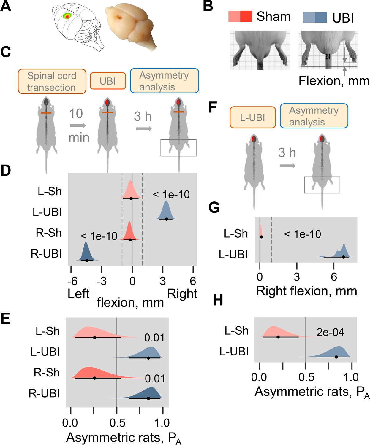
Postural asymmetry of hindlimbs induced by the unilateral ablation of the hindlimb representation area of sensorimotor cortex in rats with completely transected and intact spinal cord analyzed for comparison.

(A) Location of the right hindlimb representation area on the rat brain surface (adapted from Frost et al., 2013) and a representative UBI. (B) HL-PA analysis. (C–E) HL-PA 3 hr after left UBI (n = 9) or right UBI (n = 9), and left (n = 4) or right (n = 4) sham surgery, all performed after complete spinal cord transection. (F–H) HL-PA 3 hr after left UBI (n = 8) or left sham surgery (n = 7) in animals with intact spinal cord. (C,F) Experimental design. (D,G) The HL-PA in millimeters (mm), and (E,H) the probability to develop HL-PA (PA) above 1 mm threshold. Threshold is shown by vertical dotted lines in (D,G). The HL-PA and the probability are plotted as median (black circles), 95% HPDC intervals (black lines), and posterior density (colored distribution) from Bayesian regression. Negative and positive HL-PA values are assigned to rats with the left and right hindlimb flexion, respectively. Effects on asymmetry and differences between the groups: 95% HPDC intervals did not include zero, and adjusted p-values were ≤ 0.05. Adjusted p is shown for differences identified by Bayesian regression.

Strikingly, in the rats with transected spinal cords the UBI also induced HL-PA (Figure 1C–E). The HL-PA developed within 3 hr after the brain injury. Its size and probability were much greater than in rats with sham surgery (Left UBI, n = 31; Right UBI, n = 15; sham surgery, n = 29). An unanticipated observation was that in rats with HL-PA, the hindlimb was flexed on the contralesional side. The left or right hindlimb flexion was induced by the right and left UBI, respectively (Figure 1D; Figure 1—figure supplement 3B,C; Figure 1—figure supplement 4B,C,F,G). Both Wistar rats (Figure 1D–E; Figure 1—figure supplement 3, 4F-I) and Sprague Dawley rats (Figure 1—figure supplements 4B–E and 5) that were used in further molecular and electrophysiological experiments, respectively, developed HL-PA with hindlimb flexion on the contralesional side. To ensure the completeness of the transection, a 3–4 mm spinal segment was excised at the T2-T3 level in a subset of rats (Figure 1—figure supplement 5). After the excision, the left-side UBI-induced hindlimb postural asymmetry with the right limb flexion that replicated the other findings. The HL-PA size and probability, the time course of HL-PA development and formation of contralesional hindlimb flexion in rats with transected spinal cords that received UBI (Figure 1D,E; Figure 1—figure supplement 3, Figure 1—figure supplement 4) were similar to those of the UBI animals with intact spinal cords (Figure 1G,H; Figure 1—figure supplement 1C,D). We conclude that HL-PA formation in animals with transected spinal cord is mediated through a pathway that operates in parallel with the descending neural tracts and assures the development of contralesional flexion.

Brain injury induces asymmetry in withdrawal reflexes in rats with transected spinal cord

The withdrawal reflexes are instrumental in the investigation of brain injury-induced functional changes in hindlimb neural circuits activated by afferent input (Dewald et al., 1999; Schouenborg, 2002; Serrao et al., 2012; Spaich et al., 2006; Zhang et al., 2020). We next sought to determine whether UBI in rats with transected spinal cords produces changes in the hindlimb withdrawal reflexes, and whether these changes are asymmetric. Special care was taken to ensure that EMG recordings obtained from the left and right hindlimbs were quantitatively comparable. To achieve this, a number of strict technical criteria, such as maximally symmetrical positioning of the stimulation and recording electrodes, were applied. The criteria used in this study are described in details in ‘Materials and methods’, and are similar to those proposed by Hultborn and Malmsten (Hultborn and Malmsten, 1983a; Hultborn and Malmsten, 1983b; Malmsten, 1983). Furthermore, to minimize inter-individual variations, the asymmetry indices were used instead of the absolute values of the reflex size. This allowed double assessment: first, within both the UBI and control groups that identified asymmetric reflexes in each group, and, second, between these groups that revealed the effects of UBI vs. sham surgery. Because multiple responses were measured for the same animal, including two of its limbs, four muscles, and the varying stimulation conditions, and because they were analyzed within an animal group and between the groups, we applied mixed-effects models using Bayesian inference. Only strong and significant UBI effects were considered as biologically relevant.

Electromyographic responses were recorded from the extensor digitorum longus, interosseous, peroneus longus, and semitendinosus muscles of the contra- and ipsilesional hindlimbs in the rats with UBI (n = 18) or sham surgery (n = 11) performed after complete spinal transection and analyzed as the asymmetry index (AI = log2[Contra / Ipsi], where Contra and Ipsi were values for muscles of the contralesional and ipsilesional limbs) (Figure 2; Figure 2—figure supplement 1; Figure 2—figure supplement 2). When reflexes on both sides are equal (i.e. the Contra / Ipsi ratio equals 1), the asymmetry index is zero; if reflexes are doubled in size on the Contra or Ipsi side (i.e. the Contra / Ipsi ratio equals 2.0 or 0.5) the asymmetry index is +1 or –1, respectively.

Figure 2 with 3 supplements see all
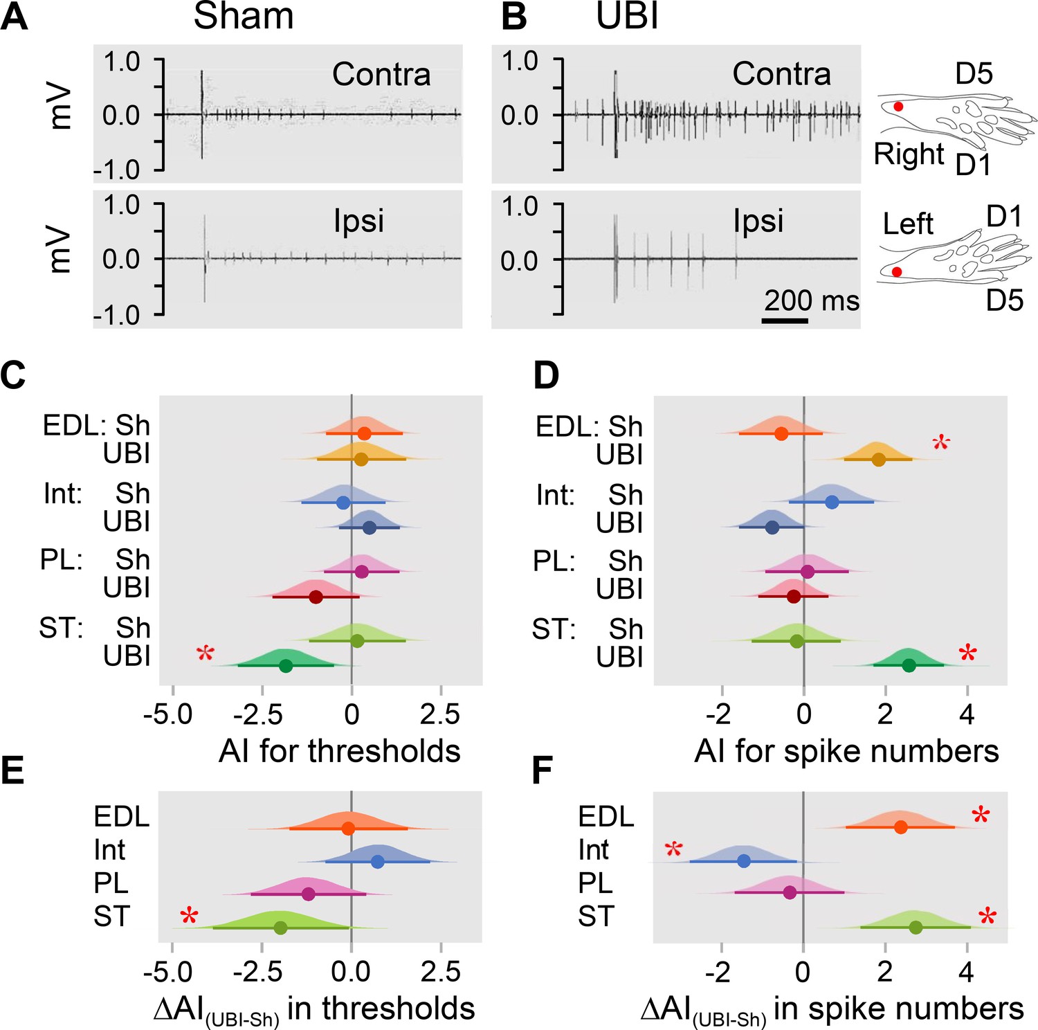
Hindlimb withdrawal reflexes in rats with UBI performed after complete spinal cord transection.

EMG activity of left and right extensor digitorum longus (EDL), interosseous (Int), peroneus longus (PL) and semitendinosus (ST) muscles were evoked by electrical stimulation of symmetric paw sites. The number of observations for both UBI and sham group is shown in Figure 2—figure supplement 2. The rats were subjected to left (n = 6) or right (n = 5) sham surgery, or to the left (n = 9) or right (n = 9) UBI. (A,B) Representative semitendinosus responses. (C,D) Asymmetry index (AI=log2[Contra/Ipsi]) for threshold and spike number. Differences in the asymmetry index from zero (AI = 0 when the ratio [Contra/Ipsi] = 1, that corresponds to a symmetric pattern) in UBI rats (C) in the current threshold for the semitendinosus muscle {median of the posterior distribution (median) = −1.840, 95% highest posterior density continuous interval (HPDCI) = [−3.169,–0.477], adjusted p-value (p) = 0.015, fold difference = 3.6}; and (D) in the number of spikes for the extensor digitorum longus (median = 1.818, HPDCI = [0.990, 2.655], p = 4×10⁻⁵, fold difference = 3.5) and semitendinosus (median = 2.560, HPDCI = [1.691, 3.415], p = 1×10⁻⁸, fold difference = 5.9) muscles. (E,F) Differences in the asymmetry index between the UBI and sham surgery (Sh) groups [ΔAI(UBI – Sh)]. Differences in the asymmetry index between the UBI and sham surgery groups for (E) the current threshold of the semitendinosus (median = −1.992, HPDCI = [−3.911,–0.106], p = 0.040, fold difference = 4.0); and (F) the number of spikes of the interosseous (median = −1.463, HPDCI = [−2.782,–0.159], p = 0.028, fold difference = 2.8), extensor digitorum longus (median = 2.379, HPDCI = [1.080, 3.743], p = 4×10⁻⁴, fold difference = 5.2), and semitendinosus (median = 2.745, HPDCI = [1.419, 4.128], p = 6×10⁻⁵, fold difference = 6.7). Medians, 95% HPDC intervals and densities from Bayesian sampler are plotted. * Asymmetry and differences between the groups: 95% HPDC intervals did not include zero, and adjusted p-values were ≤ 0.05.

Analysis of the electrically evoked electromyographic responses revealed that the asymmetry index was different from zero in the current threshold for the semitendinosus muscle (3.6-fold lower on the contra- vs. ipsilesional side), and in the number of spikes for the extensor digitorum longus (3.5-fold higher on the contra- vs. ipsilesional side) and semitendinosus (5.9-fold higher on the contra- vs. ipsilesional side) muscles in UBI rats (Figure 2C,D). No contra- vs. ipsilesional asymmetry was evident in the sham surgery group. Representative UBI-induced asymmetry in the number of spikes for the semitendinosus muscle is shown in Figure 2A,B (for those of extensor digitorum longus, interosseous and peroneus longus muscles, see Figure 2—figure supplement 1).

When compared to sham surgery, UBI substantially decreased the asymmetry index for the current threshold of the semitendinosus (4.0-fold), and the asymmetry index for the number of spikes of the interosseous (2.8-fold) that may be due to the decline in the responses on the contralesional side and/or their elevation on the ipsilesional side. Concomitantly, UBI elevated the asymmetry index for the number of spikes of the extensor digitorum longus (5.2-fold) and semitendinosus (6.7-fold) suggesting activation of the responses on the contralesional side and/or their inhibition on the ipsilesional side (Figure 2E,F). No changes in peroneus longus were revealed. Each group consisted of rats with left and right sided surgeries (Figure 2—figure supplement 2). Analysis of the asymmetry index for the four groups (i.e. the left UBI, left sham surgery, right UBI and right sham surgery groups) revealed virtually the same asymmetries in the UBI group, and the same UBI vs. sham differences in the asymmetry index, but not for all comparisons (Figure 2—figure supplement 3). Thus, the right UBI produced higher responses of the left (contralesional) extensor digitorum longus (4.1-fold) and the left (contralesional) semitendinosus (53.8-fold) compared to those on the right side, while responses of the left vs. right interosseous were decreased (5.5-fold) (Figure 2—figure supplement 3D). Additionally, the interosseous muscle was found to be asymmetric after the right side UBI. No effects on thresholds were identified.

Thus, in rats with transected spinal cord, UBI, but not sham surgery, induced asymmetry in withdrawal reflexes. The number of spikes of both flexor muscles, the extensor digitorum longus and semitendinosus was higher on the contra vs. ipsilateral side in the UBI rats. Consistently, the threshold was lower for the contra vs. ipsilesional semitendinosus. These effects may be due to (i) higher sensitivity of the afferent system reflected in a lower threshold on the contra vs. ipsilesional side for the semitendinosus; and (ii) an increased excitability of efferent systems for both muscles reflected in the increased number of spikes on the contra vs. ipsilesional side. At the cellular level, the increased excitatory drive may develop due to changes in local spinal circuits including those in presynaptic afferent inhibition, and/or changes in intrinsic membrane properties of motoneurons. Regardless of mechanism, robust differences in the asymmetry index between the UBI and sham groups suggested that the UBI markedly elevated both the sensitivity of semitendinosus afferents and the excitability of the extensor digitorum longus and semitendinosus efferents, all on the contralesional vs. ipsilesional side. The UBI also inhibited the contralesional interosseous. The UBI effects on the hindlimb withdrawal reflexes in rats with the transected spinal cords were similar in their range and contra-ipsilesional patterns to those of the UBI animals with intact spinal cords (Watanabe et al., 2021; Zhang et al., 2020). These effects corroborate clinical findings showing contralateral facilitation of withdrawal reflexes in stroke patients (Dewald et al., 1999; Serrao et al., 2012; Spaich et al., 2006).

Brain injury produces molecular changes in the lumbar spinal cord

We examined whether the UBI performed after complete spinal transection produced molecular changes in the lumbar spinal segments. Expression of 20 neuroplasticity-related, opioid and vasopressin genes, and the levels of three opioid peptides were analyzed in the ipsilesional and contralesional lumbar spinal cord of the rats with transected spinal cord that also had the left UBI (n = 12) or left sham surgery (n = 11). Genes coding for regulators of axonal sprouting, synapse formation, neuronal survival and neuroinflammation (Arc, Bdnf, Dlg4, Homer-1, Gap43, Syt4, and Tgfb1), transcriptional regulators of synaptic plasticity (cFos, Egr1, and Nfkbia), and essential components of the glutamate system critical for neuroplastic responses and regulation of spinal reflexes (GluR1, Grin2a, and Grin2b) were selected as neuroplasticity genes (for detailed description, see ‘Materials and methods, Neuroplasticity-related genes’). Genes of the opioid and vasopressin systems were included because of their involvement in asymmetric spinal responses to brain injury (see next section).

First, the mRNA levels and their median asymmetry index (AI = log2[Contra/Ipsi], where Contra and Ipsi were the levels in the contralesional and ipsilesional lumbar spinal cord) were compared between the UBI and sham surgery groups. Gene expression was either elevated on the ipsilesional side (Syt4, Grin2a, Grin2b, and Oprk1; Figure 3—figure supplement 1A–D) or decreased on the contralesional side (Gap43 and Penk; Figure 3—figure supplement 1E,F) (for all six genes, Punadjusted < 0.05). Consistently, the gene expression asymmetry index was decreased for Syt4 (Padjusted = 0.004), and for Oprk1, Oprm1, Dlg4, and Homer1 (for all four genes, Punadjusted < 0.05) (Figure 3—figure supplement 1G–K). These differences were subtle, between 0.13 and 0.38-fold, and therefore were discarded as evidence of the UBI effects.

These differences, however, pointed to the different direction in responses of the left and right spinal cord to the injury. Therefore, in the second step, we assessed whether the proportion of genes with lower expression on the contralesional vs. ipsilesional side was different between the UBI and sham surgery groups. The median gene expression asymmetry index of 19 out of 20 genes at the pairwise comparison was lower in the UBI rats compared to sham surgery group (sign-test: p = 4×10⁻⁵) (Figure 3A,B). Changes in the gene expression asymmetry index were consistent with decreased expression of 17 genes (sign test: p = 0.003) in the contralesional half (Figure 3—figure supplement 1E,F; Figure 3—figure supplement 2A–D) concomitantly with elevated expression of 15 genes (sign test: p = 0.041) in the ipsilesional half (Figure 3—figure supplement 1A–D; Figure 3—figure supplement 2A–D).

Figure 3 with 3 supplements see all
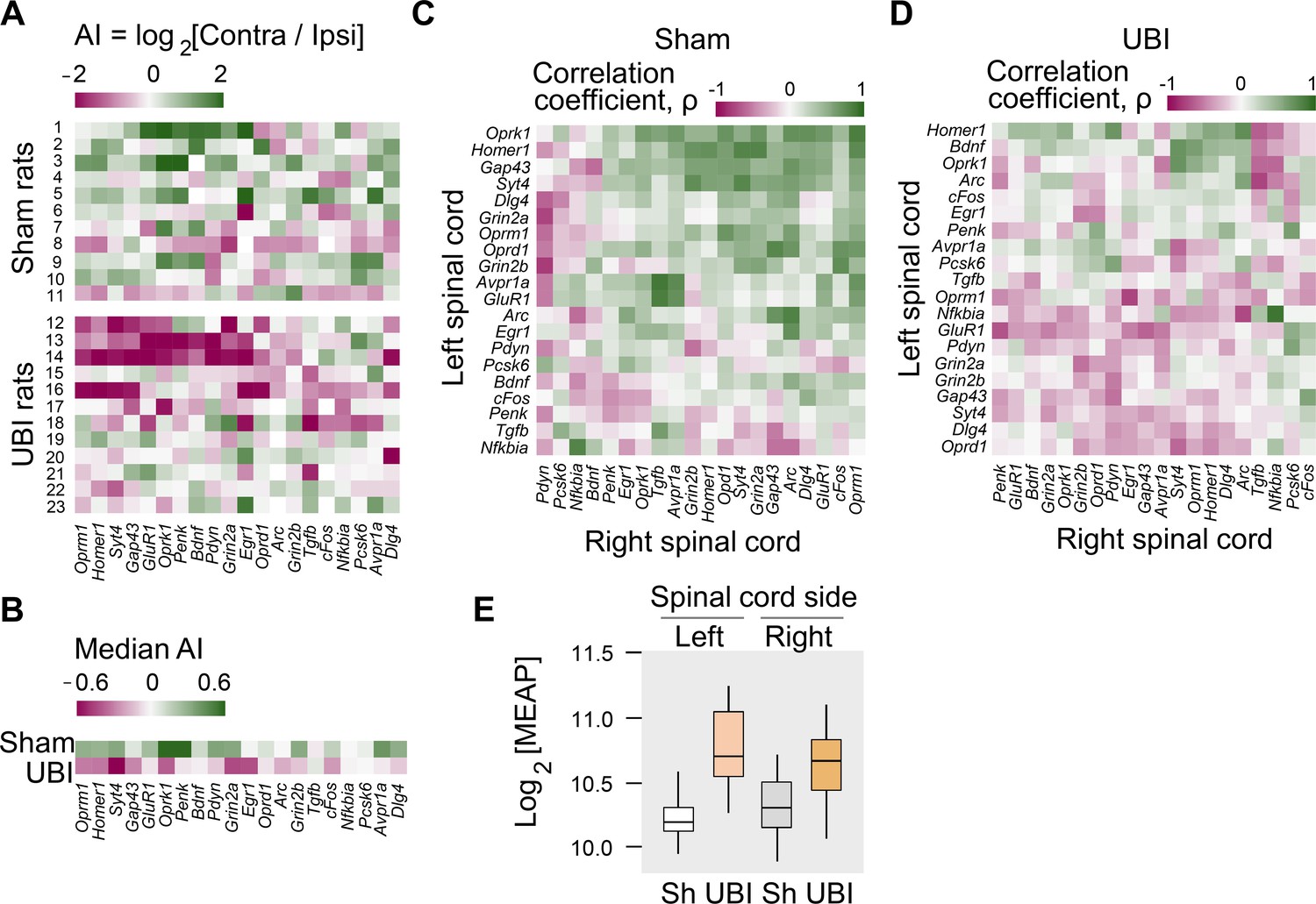
Expression of neuroplasticity-related and neuropeptide genes in the lumbar spinal cord of rats with left UBI performed after complete spinal cord transection.

The mRNA and peptide levels were analyzed in the ipsi- and contralesional halves of lumbar spinal cord isolated from rats 3 hr after the left UBI (n = 12) or left sham surgery (Sh; n = 11). (A,B) Heatmap for the (0,1)-standardized expression asymmetry index (AI=log2[Contra/Ipsi]) for each gene denoted for each rat individually, and as medians for rat groups. (C,D) Heatmap for Spearman’s rank correlation coefficients of expression levels between the left- and right lumbar halves for all gene pairs (inter-area correlations) in rats with transected spinal cord that were subjected to sham surgery or UBI. (E) UBI effects on the Met-enkephalin-Arg-Phe (MEAP) levels in the left (Padjusted = 9×10⁻⁴; fold change: 1.4×) and right (Pundjusted = 0.020; fold change: 1.3×) halves. Data are presented in fmol/mg tissue in the log2 scale as boxplots with median and hinges representing the first and third quartiles, and whiskers extending from the hinge to the highest/lowest value that lies within the 1.5 interquartile range of the hinge.

Figure 3—source data 1

The EXCEL source data file contains data for panels A,B of Figure 3.

https://cdn.elifesciences.org/articles/65247/elife-65247-fig3-data1-v1.xlsx
Figure 3—source data 2

The EXCEL source data file contains data for panel E of Figure 3.

https://cdn.elifesciences.org/articles/65247/elife-65247-fig3-data2-v1.xlsx

Gene co-expression patterns characterize regulatory interactions within and across tissues (Dobrin et al., 2009; Erola et al., 2020; Gerring et al., 2019; Zhang et al., 2020). Third, we examined whether the UBI induced changes in mRNA–mRNA correlations within the left and right half of the lumbar spinal cord (intra-area correlations), and between these halves (inter-area correlations). The proportion of intra-area positive correlations, which dominated in rats with sham surgery, was reduced after the UBI (Fisher's Exact Test: all correlations in the right half, p = 3×10⁻⁵; significant correlations in the left and right areas, p = 0.008 and 0.009, respectively) (Figure 3—figure supplement 2E–H). The inter-area gene-gene coordination strength was decreased after the UBI (Wilcoxon signed-rank test; all and significant correlations: p = 4×10⁻⁷ and 3×10⁻⁴, respectively) (Figure 3C,D). Positive inter-area correlations were predominant in rats with sham surgery (68%) in contrast to the UBI rats (42%) (Fisher's Exact Test: all and significant correlations, p = 6×10⁻¹⁴ and 0.004, respectively). Thus, the UBI robustly impairs coordination of expression of neuroplasticity-related and neuropeptide genes within and between the left and right halves of the lumbar spinal cord.

Fourth, analysis of opioid peptides demonstrated that the UBI substantially elevated the levels of the proenkephalin marker Met-enkephalin-Arg-Phe in the ipsilesional (Padjusted = 9×10⁻⁴) and contralesional (Punadjusted = 0.020) spinal halves (Figure 3E), and the prodynorphin-derived Dynorphin B and Leu-enkephalin-Arg in the ipsilesional spinal cord (for both, Punadjusted < 0.05) (Figure 3—figure supplement 3).

Altogether, the analysis of gene expression and of opioid peptides adds strong molecular evidence for the lateralized signaling from the injured brain to the lumbar neural circuits in rats with transected spinal cord.

UBI effects are mediated by neuroendocrine pathway

The left-right side specific mechanism that does not engage the descending neural tracts may operate through the neuroendocrine system by a release of pituitary hormones into the blood. Consistent with this hypothesis, no HL-PA developed in hypophysectomized animals that received left UBI after spinal transection (n = 8); the HL-PA median values and PA were nearly identical to those in sham operated rats (n = 8) (Figure 4A; Figure 4—figure supplement 1A–E). We next examined whether left UBI stimulates the release of chemical factors that may induce the development of HL-PA, into the blood. Serum that was collected 3 hr after performing a left UBI in rats with transected spinal cord was administered either centrally (into the cisterna magna; UBI serum, n = 13; sham serum, n = 7; Figure 4—figure supplement 1F–J) or intravenously (UBI serum, n = 13; sham serum, n = 7; Figure 4B; Figure 4—figure supplement 1K–O) to rats after their spinalization. Serum administration by either route resulted in formation of HL-PA with its values and its probability similar to those induced by the UBI in rats with intact and transected spinal cords. Remarkably, animals injected with serum from rats with left UBI displayed hindlimb flexion on the right side, which was the same as the flexion side in the donor rats (Figure 4B; Figure 4—figure supplement 1F–O). No HL-PA developed after administration of serum collected from rats with the left sham surgery. We conclude that the left UBI stimulates a release of chemical factors from the pituitary gland into the blood that induce HL-PA with contralesional flexion.

Figure 4 with 2 supplements see all
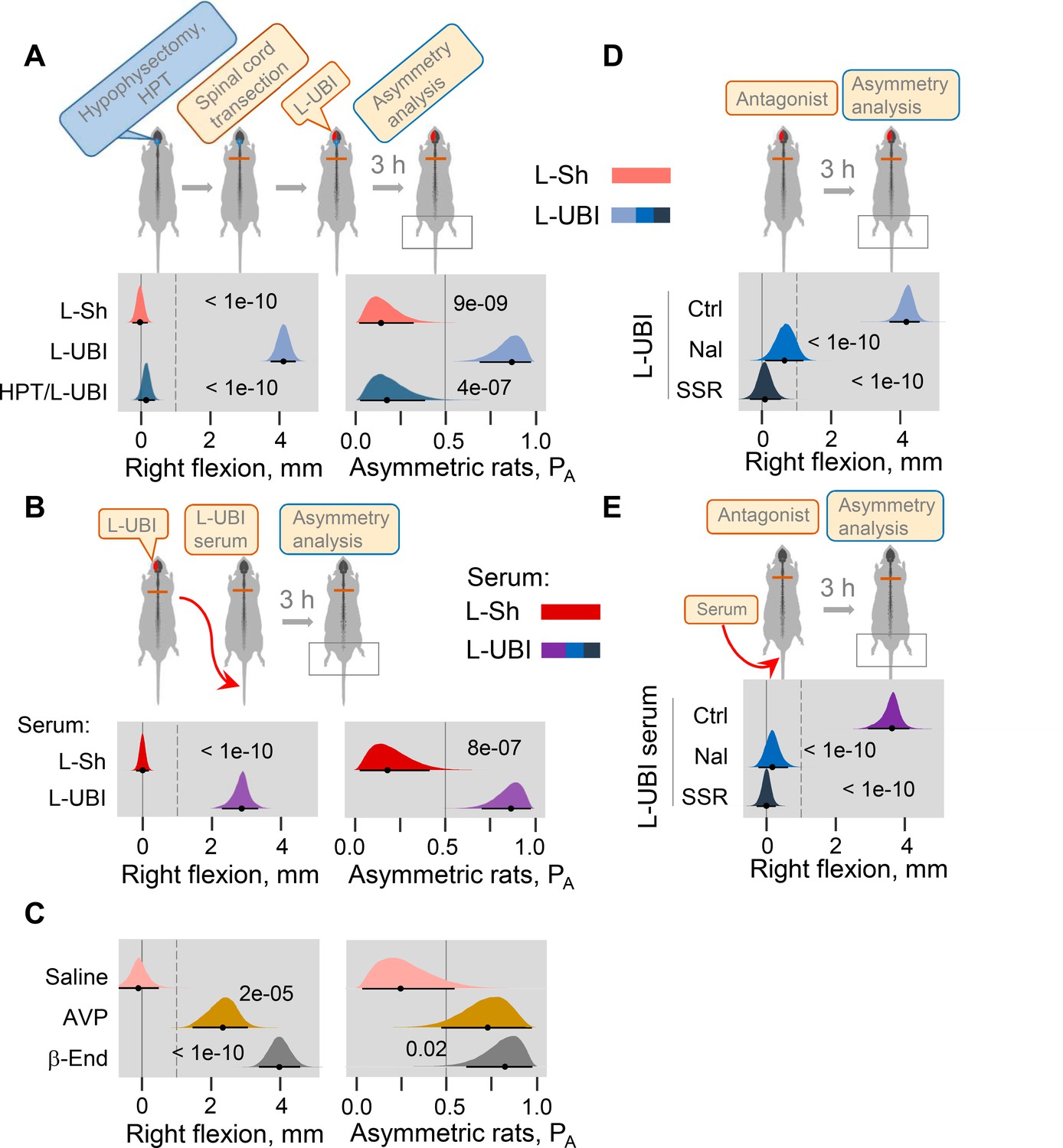
Neuroendocrine pathway mediating postural asymmetry formation in rats with transected spinal cord.

(A) HL-PA in hypophysectomized (HPT; n = 8) and control (n = 12) rats with transected spinal cord 3 hr after left UBI (L-UBI). Left sham surgery (L–Sh): n = 8. (B) HL-PA after intravenous administration of serum from rats with either left UBI (L-UBI serum) or left sham surgery (L-Sh serum) to rats with transected spinal cord (n = 13 and 7, respectively). (C) Induction of HL-PA by Arg-vasopressin (AVP) and β-endorphin (β-End) in rats with transected spinal cord. Synthetic β-endorphin or Arg-vasopressin (1 microgram and 10 nanogram / 0.3 ml saline / animal, respectively), or saline was administered intravenously to rats (n = 8, 7, and 4 rats, respectively) after spinal cord transection. The HL-PA was analyzed in rats 60 min after the injection and under pentobarbital anesthesia. (D) Effect of naloxone (Nal, n = 6) or saline (n = 6), and SSR-149415 (SSR, n = 6) or vehicle (n = 5) on HL-PA 3 h after left UBI in rats with transected spinal cord. Vehicle and saline groups were combined into a single control group (Ctrl; n = 11). (E) Effect of naloxone (n = 6) or saline (n = 3) and SSR-149415 (n = 6) or vehicle (n = 3) on HL-PA 3 hr after intravenous administration of the left UBI serum to rats with transected spinal cord. Ctrl = saline + vehicle; n = 6. In (D,E), naloxone (or saline) and SSR-149415 (or vehicle) were administered 0.5 and 3 hr before HL-PA analysis, respectively. HL-PA values in millimeters (mm) and probability (PA) to develop HL-PA above 1 mm threshold (denoted by vertical dotted lines) are plotted as median, 95% HPDC intervals, and posterior distribution from Bayesian regression. Negative and positive HL-PA values are assigned to rats with the left and right hindlimb flexion, respectively. Asymmetry and differences between the groups: 95% HPDC intervals did not include zero, and adjusted p-values were ≤ 0.05. Adjusted p is shown for differences identified by Bayesian regression.

Figure 4—source data 1

The EXCEL source data file contains data for panels A-E of Figure 4.

https://cdn.elifesciences.org/articles/65247/elife-65247-fig4-data1-v1.xlsx

Previous studies demonstrated that multiple peptide factors extracted from the brain, pituitary gland and serum may induce a side-specific hindlimb motor response. Several of them were identified as peptide neurohormones including opioid peptides (Bakalkin and Kobylyansky, 1989; Bakalkin et al., 1986; Chazov et al., 1981) and Arg-vasopressin (Klement'ev et al., 1986). It was found that Arg-vasopressin or Leu-enkephalin administered centrally induced HL-PA with right hindlimb flexion. The pituitary gland is the main source of the opioid neurohormone β-endorphin and the antidiuretic hormone Arg-vasopressin in the body. Here, we first replicated the effects of Arg-vasopressin that, consistent with a previous study (Klement'ev et al., 1986), produced flexion of the right hindlimb after its intracisternal administration (Figure 4—figure supplement 2; peptide, n = 22, and saline, n = 9 at the 180 min time point). We then tested if β-endorphin and Arg-vasopressin may evoke asymmetric motor response after intravenous administration. Injection of these neurohormones, but not saline, to rats with transected spinal cords resulted in development of HL-PA with right hindlimb flexion (Figure 4C; β-endorphin, n = 8; Arg-vasopressin, n = 7; saline, n = 4).

We next investigated whether opioid receptors and the vasopressin receptor V1B, that is expressed in the pituitary gland (Roper et al., 2011), mediate formation of HL-PA in UBI rats or in animals treated with serum from UBI rats. Naloxone and SSR-149415, the opioid and vasopressin V1B receptor antagonists, respectively, administered to animals with transected spinal cord that also received a left UBI (naloxone, n = 6; SSR-149415, n = 6; saline and vehicle, n = 11) inhibited HL-PA formation (Figure 4D). Similarly, the HL-PA induced by serum from animals with left UBI was abolished by administration of either naloxone (n = 6) or SSR-149415 (n = 6) (Figure 4E). Thus, the pituitary neurohormones β-endorphin and Arg-vasopressin released into the systemic circulation may serve as side-specific signals that mediate UBI effects on hindlimb motor circuits.

Discussion

The left and right hemispheres in top-down control of the endocrine system

During embryonic development, the left–right asymmetry of the body is generated by multiple paracrine signaling molecules that enable communications between the left and right halves of the embryo (Hamada et al., 2002). In the adult brain, functional lateralization is an organizing principle (Duboc et al., 2015; MacNeilage et al., 2009) and lateralized functions may be regulated by paracrine signaling molecules including peptide neurohormones (Deliagina et al., 2000; Hussain et al., 2012; Kononenko et al., 2017; Marlin et al., 2015; Nation et al., 2018; Phelps et al., 2019; Watanabe et al., 2015; Watanabe et al., 2021; Zink et al., 2011). Thus, oxytocin enables retrieval behavior by enhancing responses of the left, but not right, auditory cortex through its receptors expressed on the left side (Marlin et al., 2015). Arg-vasopressin targets the left but not right hemispheric areas to modulate social recognition-related activity (Zink et al., 2011). In the human brain, asymmetric distribution of the μ-opioid receptor along with opioid peptides that elicit euphoria and dysphoria may provide a basis for the lateralized processing of positive and negative emotions (Kantonen et al., 2020; Watanabe et al., 2015).

Top-down control of the hypothalamic-pituitary-adrenal and hypothalamic-pituitary-gonadal axes, as well as the immune system, is left-right hemisphere specific (Bakalkin et al., 1984; Inase and Machida, 1992; Kiss et al., 2020; Lueken et al., 2009; Madsen et al., 2012; Meador et al., 2004; Sullivan and Gratton, 2002; Xavier et al., 2013). The right-sided injuries to the brain or spinal nerves compared to those on the left side produce stronger effects on the neuroendocrine systems, including hormonal levels in the peripheral circulation (Hussain et al., 2012; Inase and Machida, 1992; Kononenko et al., 2017; Lueken et al., 2009). Cortisol (corticosterone) secretion is under excitatory control of the right hemisphere and, consistently, its phasic response is diminished in patients with right but not left sided stroke (Lueken et al., 2009). These features may be related to the asymmetry of the hypothalamic – pituitary axis. Thus, basal and corticotropin-releasing hormone–induced secretion of Arg-vasopressin and ACTH by the pituitary gland is lateralized to the right petrosal sinus (Kalogeras et al., 1996). Conversely, non-directional stimuli such as stress and stress-induced pain produce lateralized responses in the CNS (Bakalkin et al., 1982; Bakalkin et al., 1984; Nation et al., 2018; Sullivan and Gratton, 2002). Both left- and right-sided nerve or body injuries elicit functional and molecular responses that are often lateralized to the right, but not to the left, in the brain and spinal cord (Bakalkin et al., 1984; Hussain et al., 2012; Inase and Machida, 1992; Kononenko et al., 2017; Phelps et al., 2019). Neuropathic pain induced by either the left- or right-sided nerve injuries is controlled through κ-opioid receptors in the right amygdala (Phelps et al., 2019).

Taken together, these findings suggest that feedforward and feedback interactions between the lateralized CNS features and the endocrine system controlling peripheral processes are mediated either by neural circuits with unusual, asymmetric organization, or by left-right sided neuroendocrine pathways that may be similar to the left-right sided paracrine mechanism operating in the development. An alternative pathway that does not engage descending neural tracts may convey side-specific signals from the brain to the paired endocrine glands, the left and right spinal cord, and the left and right extremities had been suggested (Bakalkin et al., 1984; Cope et al., 1980; Wolpaw and Lee, 1989), and supported by preliminary evidence (Bakalkin et al., 1986) but not elaborated.

The left-right side-specific humoral signaling

This study provides evidence for left-right side-specific humoral signaling that mediates the effects of UBI on the formation of HL-PA, asymmetry in withdrawal reflexes, and asymmetric changes in gene expression patterns in the lumbar spinal cord (Figure 5). Encoding of information about the injury side in a hormonal message, humoral transmission of this message to its target sites on peripheral nerve endings or spinal neurons, and translation of this message into the left-right side-specific response, are the key stages of this phenomenon.

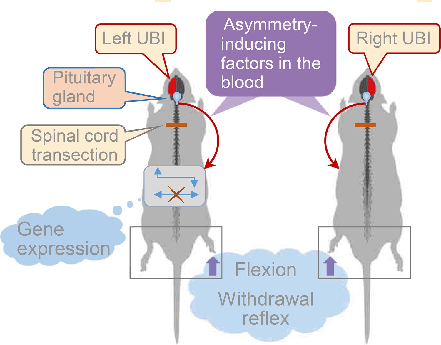
Model for the humoral neuroendocrine side-specific signaling from the unilaterally injured brain to the lumbar spinal cord.

In the rats with transected spinal cords, the UBI stimulates the release of the asymmetry inducing factors (neurohormones) from the pituitary gland into the circulation. They are transported to their target sites and induce flexion of the contralesional hindlimb and asymmetric, contra vs. ipsilesional side specific changes in withdrawal reflexes and spinal gene expression patterns.

The left- and right-side-specific responses evoked by hormonal molecules circulating in the blood is a core of the humoral signaling pathway. Together with previous reports (Bakalkin et al., 1981; Bakalkin and Kobylyansky, 1989; Bakalkin et al., 1986; Chazov et al., 1981; Watanabe et al., 2020), this study demonstrates that peptide neurohormones and opioids administered intravenously, intrathecally or intracisternally induce HL-PA in rats with intact brain. The critical finding is that the side of the flexed limb depends on the compound administered. Endogenous and synthetic κ-opioid agonists dynorphin, bremazocine, and U-50,488, along with the endogenous μ/δ-opioid agonist Met-enkephalin, induce flexion of the left hindlimb (Bakalkin et al., 1981; Bakalkin and Kobylyansky, 1989; Bakalkin et al., 1986; Chazov et al., 1981; Watanabe et al., 2020). In contrast, β-endorphin and Arg-vasopressin, and the δ-agonist Leu-enkephalin, cause the right limb to flex [the present study and Bakalkin et al., 1981; Chazov et al., 1981; Klement'ev et al., 1986]. Thus, topographical information conveyed by the ‘non-directional’ molecular messengers circulating in the blood is converted into side-specific motor responses. Hypophysectomy disables the endocrine pathway including its opioid and Arg-vasopressin components and abolishes the HL-PA. Consistent with this finding, serum from rats with left UBI induces HL-PA with contralesional hindlimb flexion in rats with intact brain. The pituitary gland is the main source of Arg-vasopressin and β-endorphin in the body that are secreted into the bloodstream (Autelitano et al., 1989; Day and Akil, 1989). Naloxone and SSR-149415 block the left UBI-induced formation of HL-PA. Altogether, these findings demonstrate that opioid and Arg-vasopressin neurohormones transmit side-specific signals from the injured brain to the spinal neural circuits.

Theoretically, the paravertebral chain of sympathetic ganglia, which is the remaining neural connection after complete spinal cord transection, may convey supraspinal signals to the muscle vasculature and through this mechanism may differentially affect ipsi- and contralesional muscles. However, the sympathetic ganglia likely do not mediate control of lumbar neural circuits by the supraspinal structures (Brodal, 1981; Wolpaw and Lee, 1989). Furthermore, the sympathetic system has a limited capacity to independently regulate blood flow to the left and right hindlimbs (Lee et al., 2007). Our present findings could not rule out a role of the sympathetic pathway. However, experiments with hypophysectomized rats, ‘pathological’ serum and neurohormones inducing the HL-PA strongly suggest the dominance of the humoral pathway in rats with transected spinal cord.

Functional and mechanistic implications

Clinical studies revealed robust functional changes induced by stroke or traumatic brain injury (TBI) in contralateral withdrawal reflexes that also control posture and locomotion (Dewald et al., 1999; Sandrini et al., 2005; Serrao et al., 2012). Patients with post-stroke motor deficits lose their ability to modulate the withdrawal reflexes that affects spatiotemporal interaction among joints and causes movement abnormalities during motor activities (Bohannon and Smith, 1987; Serrao et al., 2012).

Spinal withdrawal reflexes are regulated by the endogenous opioid peptides that may suppress the ipsilateral and contralateral segmental reflexes (Clarke et al., 1992; Duarte et al., 2019; Jankowska and Schomburg, 1998; Schmidt et al., 1991). Opioid receptors are expressed both in the dorsal and ventral horns of the spinal cord (Kononenko et al., 2017; Wang et al., 2018) and also in the periphery, in primary afferents including low-threshold mechanoreceptors that modulate cutaneous mechanosensation (Bardoni et al., 2014; Snyder et al., 2018). The left-right-specific endocrine mechanism may mediate the effects of UBI on the withdrawal reflexes through targeting peripherally or centrally located opioid receptors.

A large fraction of patients with stroke and cerebral palsy do not relax their muscles – they are tonically contracted without any voluntary command (Baude et al., 2019; Gracies, 2005; Lorentzen et al., 2018; Sheean and McGuire, 2009; Trompetto et al., 2019). This phenomenon is called ‘spastic dystonia’ and has a central mechanism that does not depend on afferent input (Gracies, 2005; Lorentzen et al., 2018; Sheean and McGuire, 2009). In the HL-PA analysis, no nociceptive stimulation is applied and tactile stimulation is negligible [this study and Zhang et al., 2020]. Stretch and postural limb reflexes are abolished immediately after complete spinal cord transection (Frigon et al., 2011; Miller et al., 1996; Musienko et al., 2010) and strongly inhibited by anesthesia (Fuchigami et al., 2011; Zhou et al., 1998). Therefore, in anesthetized rats with transected spinal cord, a role of nociceptive and stretch reflexes in the UBI-induced HL-PA formation may be limited. The finding that HL-PA is resistant to bilateral lumbar dorsal rhizotomy (Zhang et al., 2020) further supports this notion and suggests that the HL-PA and the clinical ‘spastic dystonia’ may be mechanistically similar.

Classical hyperreflexia develops during several weeks after the impact and is considered as pathology of the corticospinal tract (Williams et al., 2017). In contrast, the HL-PA is formed within 30 min after the UBI in rats with transected spinal cord. Whether the endocrine signaling has a role in exacerbation of stretch reflex after damage to the corticospinal tract would be interesting to study.

Single injection of either naloxone or SSR-149415 inhibited HL-PA asymmetry formation. The vasopressin receptor V1B is largely expressed in the anterior pituitary by corticotrophs producing proopiomelanocortin (Roper et al., 2011). A plausible scenario is that Arg-vasopressin released from neurohypophysis activates the V1B receptor and stimulates secretion of proopiomelanocortin-derived β-endorphin (Roper et al., 2011) that induces the HL-PA. Alternatively, Arg-vasopressin and β-endorphin may produce synergistic effects acting through the complex of the vasopressin receptor V1B and μ-opioid receptor that integrates two signaling pathways (Koshimizu et al., 2018).

The general opioid antagonists naloxone and naltrexone may normalize neurological functions that are impaired in animals and human patients after unilateral cerebral ischemia (Baskin and Hosobuchi, 1981; Baskin et al., 1984; Baskin et al., 1994; Hans et al., 1992; Hosobuchi et al., 1982; Jabaily and Davis, 1984; Namba et al., 1986; Skarphedinsson et al., 1989; Wang et al., 2019), and also reduce spasticity in patients with primary progressive multiple sclerosis (Gironi et al., 2008). Our findings suggest that the efficacy of pharmacological treatment may depend on topographical correspondence between the side of neuronal deficits and the side that is preferentially targeted by neurohormones and their antagonists.

Lateralized features of the spinal cord

A number of anatomical, functional, and molecular studies revealed left–right asymmetry in the spinal cord organization (de Kovel et al., 2017; Deliagina et al., 2000; Hultborn and Malmsten, 1983a; Hultborn and Malmsten, 1983b; Knebel et al., 2018; Kononenko et al., 2017; Malmsten, 1983; Nathan et al., 1990; Ocklenburg et al., 2017; Zhang et al., 2020). Three-quarters of cervical spinal cords are asymmetric with a larger right side (Nathan et al., 1990). Spinal-muscular systems are asymmetric in human fetuses, and the asymmetry correlates with lateralized gene transcription (de Kovel et al., 2017; Ocklenburg et al., 2017). Mono- and polysynaptic segmental reflexes evoked by stimulation of the dorsal roots and recorded in the ventral roots in intact rats and cats display higher activity on the right side (Hultborn and Malmsten, 1983a; Hultborn and Malmsten, 1983b; Malmsten, 1983). Similarly, EMG recordings of hindlimb withdrawal reflexes evoked by electrical stimulation in control rats display higher activity on the right side (Zhang et al., 2020). On this asymmetric background, neural circuits controlling the left and right limbs may be differently regulated by opioid peptides and Arg-vasopressin (Bakalkin et al., 1981; Bakalkin et al., 1986; Chazov et al., 1981; Klement'ev et al., 1986). The left-right side-specific neurohormonal effects may be mediated through lateralized receptors. In the rat spinal cord, the expression of three opioid receptors is lateralized to the left, and their proportions and co-expression patterns are different between the left and right sides (Kononenko et al., 2017; Watanabe et al., 2021). The asymmetry may be a critical feature of the spinal cord allowing translation of the ‘non-directional’ hormonal messages into the left-right side-specific response.

Limitations

The side-specific endocrine signaling was revealed in anaesthetized animals with transected spinal cords that were studied up to 180 min post UBI. Its biological and pathophysiological relevance has not been determined. Pathways from the injured brain area to the hypothalamic-pituitary system, neurohormones mediating effects of the right side injury, peripheral or central targets for the left- and right-side-specific endocrine messengers, and afferent, central or efferent mechanisms of the asymmetry formation have not been investigated. The study did not analyze forelimb postural asymmetry. It was not induced by lesion of the hindlimb sensorimotor cortex (Zhang et al., 2020) but it did develop after injury of the forelimb area (unpublished data).

The strategy was to selectively disable the neural and endocrine mechanisms by surgical means. In this approach, dissection of neural pathways does not allow us to assess a contribution of each pathway to asymmetric postural and motor deficits in awake animals. On the contrary, analysis of the UBI effects in the hypophysectomized rats with intact spinal cords may uncover a function of the left – right side-specific endocrine signaling. Rats with intact (Figure 1—figure supplement 1C,D) and transected (Figure 1—figure supplements 3 and 4) spinal cords developed HL-PA during the first 30 min following UBI. It would be worthwhile to analyze in more details whether the time course of development of asymmetry differs between these groups; and also to ascertain whether signals mediated by the neural and endocrine pathways are additive, synergistic, or even antagonistic with respect to each other during the initial impairment phase and the recovery period using naive animals and the hypophysectomized rats with intact spinal cords.

This study does not focus on clinical correlates and mechanisms of postural deficits. The withdrawal reflexes and hindlimb posture were studied as readouts of the UBI because they are regulated by neurohormones and may be analyzed after spinal cord transection. Furthermore, they are directed along the left-right axis, and, therefore, can reveal whether the endocrine system conveys the side-specific signals. At the same time, the HL-PA and withdrawal reflexes model several features of the brain injury-induced sensorimotor and postural deficits in humans. First, the changes induced by UBI have a contra-ipsilesional pattern. Second, the HL-PA is not dependent on the afferent input (Zhang et al., 2020) and in this regard it may be similar to ‘spastic dystonia’, a tonic muscle overactivity that contributes to ‘hemiplegic posture’ (Gracies, 2005; Lorentzen et al., 2018; Sheean and McGuire, 2009). Third, asymmetric exacerbated withdrawal reflexes that lead to flexor spasms in patients (Bussel et al., 1989; Dietz et al., 2009; Lavrov et al., 2006; Schouenborg, 2002) are similarly developed in rats.

TBI and stroke cause dysfunction of the hypothalamic–pituitary system and hypopituitarism manifested as changes in secretion of pituitary hormones (Bondanelli et al., 2005; Emelifeonwu et al., 2020; Klose and Feldt-Rasmussen, 2018; Lillicrap et al., 2018). Ischemic stroke activates the hypothalamus-pituitary-adrenal axis (Anne et al., 2007), while a unilateral ablation of sensorimotor cortex elevates the level of circulating ACTH and induces morphological changes in the pituitary corticotrophs that produce ACTH and β-endorphin (Lavrnja et al., 2014). Neurobiological mechanisms underlying these effects may be similar with those of the UBI-induced endocrine signaling described in this study. These mechanisms and anatomical pathways involved have not been identified. However, cortical projections to the hypothalamus that may potentially mediate effects of focal brain injury on secretion of pituitary hormones have been described (Jeong et al., 2016).

Conclusion

This study describes the left-right side-specific endocrine mechanism that, in addition to descending neural tracts, may mediate asymmetric effects of a unilateral brain injury on hindlimb postural asymmetry and spinal reflexes (Figure 5). Identification of features and the proportion of asymmetric sensorimotor deficits transmitted by neurohormonal signals vs. those mediated by neural pathways may be essential for understanding of stroke and TBI mechanisms.

Materials and methods

Key resources table
Reagent type
(species) or resource
DesignationSource or referenceIdentifiersAdditional information
Strain, strain background (Rattus norvegicus)Wistar Hannover rat, maleCharles River Laboratories, Spain
Strain, strain background (Rattus norvegicus)Sprague Dawley rat, maleTaconic, Denmark and Charles River Laboratories, France
Chemical compound, drugIsofluraneAbbott Laboratories, NorwayNDC 0044-5260-03Anesthesia agent
Chemical compound, drugLidocaine hydrochlorideMerck Group, GermanyPHR1257Anesthetic
Chemical compound, drugParaformaldehydeSigma-Aldridge, USACat#: 158127Perfusion
Chemical compound, drugSSR-149415Tocris Bioscience, United KingdomCat#: 6195vasopressin V1B antagonist
Chemical compound, drugNaloxoneTocris Bioscience, United KingdomCat#: 0599Opioid antagonist
Commercial assay or kitGiemsa StainSigma-Aldridge, USACat#: 32884Nissl staining
Commercial
assay or kit
RNasy Plus Mini kitQiagen, CA, USACat#: 74136Total RNA extraction
Commercial
assay or kit
iScript cDNA Synthesis KitBio-Rad Laboratories, USACat#: 1708891cDNA Synthesis
Commercial
assay or kit
iTaq Universal Probes supermixBio-Rad Laboratories, USACat# 1725131Real-Time PCR reagent
Peptide, recombinant proteinß-EndorphinBachem, SwitzerlandCat#
H-2814
Neurohormone
Peptide, recombinant proteinArg-vasopressinBachem, SwitzerlandCat#
H-1780
Neurohormone
AntibodyAnti-Dynorphin B
(rabbit, polyclonal)
Nguyen et al., 2005; Nylander et al., 1997; Yakovleva et al., 2006RIA
1: 350000
AntibodyAnti-Leu-enkephalin-Arg
(rabbit, polyclonal)
Nguyen et al., 2005; Nylander et al., 1997; Yakovleva et al., 2006RIA
1: 35000
AntibodyAnti-Met-enkephalin-Arg-Phe
(rabbit, polyclonal)
Nguyen et al., 2005; Nylander et al., 1997; Watanabe et al., 2015RIA
1: 15750
Sequence-based reagentActb (Rattus norvegicus)Bio-Rad Laboratories, USAqRnoCIP0050804PrimePCR Probe assay
Sequence-based reagentArc (Rattus norvegicus)Bio-Rad Laboratories, USAqRnoCEP0027389PrimePCR Probe assay
Sequence-based reagentAvpr1a (Rattus norvegicus)Bio-Rad Laboratories, USAqRnoCEP0023750PrimePCR Probe assay
Sequence-based reagentBdnf (Rattus norvegicus)Bio-Rad Laboratories, USAqRnoCEP0026843PrimePCR Probe assay
Sequence-based reagentcFos (Rattus norvegicus)Bio-Rad Laboratories, USAqRnoCEP0024078PrimePCR Probe assay
Sequence-based reagentDlg4 (Rattus norvegicus)Bio-Rad Laboratories, USAqRnoCIP0026242PrimePCR Probe assay
Sequence-based reagentEgr1 (Rattus norvegicus)Bio-Rad Laboratories, USAqRnoCEP0022872PrimePCR Probe assay
Sequence-based reagentGap43 (Rattus norvegicus)Bio-Rad Laboratories, USAqRnoCIP0027599PrimePCR Probe assay
Sequence-based reagentGapgh (Rattus norvegicus)Bio-Rad Laboratories, USAqRnoCIP0050838PrimePCR Probe assay
Sequence-based reagentGluR1 (Rattus norvegicus)Bio-Rad Laboratories, USAqRnoCIP0030725PrimePCR Probe assay
Sequence-based reagentGrin2a (Rattus norvegicus)Bio-Rad Laboratories, USAqRnoCIP0025244PrimePCR Probe assay
Sequence-based reagentGrin2b (Rattus norvegicus)Bio-Rad Laboratories, USAqRnoCIP0023973PrimePCR Probe assay
Sequence-based reagentHomer-1 (Rattus norvegicus)Bio-Rad Laboratories, USAqRnoCEP0023985PrimePCR Probe assay
Sequence-based reagentOprd1 (Rattus norvegicus)Bio-Rad Laboratories, USAqRnoCEP0029668PrimePCR Probe assay
Sequence-based reagentOprk1 (Rattus norvegicus)Bio-Rad Laboratories, USAqRnoCIP0029310PrimePCR Probe assay
Sequence-based reagentOprm1 (Rattus norvegicus)Bio-Rad Laboratories, USAqRnoCEP0024902PrimePCR Probe assay
Sequence-based reagentPdyn (Rattus norvegicus)Bio-Rad Laboratories, USAqRnoCEP0025357PrimePCR Probe assay
Sequence-based reagentPenk (Rattus norvegicus)Bio-Rad Laboratories, USAqRnoCEP0029455PrimePCR Probe assay
Sequence-based reagentPcsk6 (Rattus norvegicus)Bio-Rad Laboratories, USAqRnoCIP0045340PrimePCR Probe assay
Sequence-based reagentNfkbia (Rattus norvegicus)Bio-Rad Laboratories, USAqRnoCEP0026759PrimePCR Probe assay
Sequence-based reagentSyt4 (Rattus norvegicus)Bio-Rad Laboratories, USAqRnoCIP0029728PrimePCR Probe assay
Sequence-based reagentTgfb1 (Rattus norvegicus)Bio-Rad Laboratories, USAqRnoCIP0031022PrimePCR Probe assay
Software, algorithmSpike 2CED, UKEMG recording
Software, algorithmOffline SorterPlexon, USAversion 3
Software, algorithmNeuroExplorerNex Technologies, USA
Software, algorithmStanCarpenter et al., 2017v 2.19.2Scaling the data
Software, algorithmR programR Development Core Team, 2018v 3.6.1
Software, algorithmBrmsBurkner, 2017v 2.9.6Interface for R
Software, algorithmEmmeansSearle et al., 2012v 1.4R package
OtherTissuDuraBaxter, GermanyCat#: 0600096Covering material

Animals

Male Wistar Hannover rats (Charles River Laboratories, Spain), with body weight of 150–200 g were used in behavioral, HL-PA and molecular experiments (Figures 1, 3 and 4B–E; Figure 1—figure supplement 1, 3, 4F-I; Figure 3—figure supplement 1, 2, 3; Figure 4—figure supplement 1G–O, 2). Male Sprague Dawley rats were used for analysis of HL-PA (Figure 1—figure supplements 2, 4B–E and 5), in electrophysiological experiments (Figure 2, Figure 2—figure supplement 1, 2, 3) (Taconic, Denmark; 150–400 g body weight) and for hypophysectomy (Figure 4A and Figure 4—figure supplement 1B–E) (Charles River Laboratories, France; 115–125 g body weight). The animals received food and water ad libitum and were kept in a 12 hr day-night cycle at a constant environmental temperature of 21°C (humidity: 65%). Approval for animal experiments was obtained from the Malmö/Lund ethical committee on animal experiments (No.: M7-16), and the ethical committee of the Faculty of Medicine of Porto University and Portuguese Direção-Geral de Alimentação e Veterinária (No. 0421/000/000/2018).

Surgery

Request a detailed protocol

The animals were anesthetized with sodium pentobarbital (I.P.; 60 mg/kg body weight, as an initial dose and then 6 mg/kg every hour). If needed, the anesthesia was reinforced with ≈1.5% isoflurane (IsoFlo, Abbott Laboratories, Norway) in a mixture of 65% nitrous oxide-35% oxygen. Core temperature of the animals was controlled using a feedback-regulated heating system. In the experiments involving electrophysiological recordings, the rats were ventilated artificially via a tracheal cannula and the expiratory CO2 and mean arterial blood pressure (65–140 mmHg) was monitored continuously in the right carotid artery.

Aspiration brain injury and spinal cord transection

Request a detailed protocol

The experimental design included rats with either the UBI alone or the UBI which was preceded by a complete spinal cord transection. In the UBI-only experiments, anesthetized rats were placed on a surgery platform with stereotaxic head holder. The rat head was fixed in a position in which the bregma and lambda were located at the same horizontal level. After local injection of lidocaine (Xylocaine, 3.5 mg/ml) with adrenaline (2.2 μg/ml), the scalp was cut open and a piece of the parietal bone located 0.5–4.0 mm posterior to the bregma and 1.8–3.8 mm lateral to the midline (Paxinos and Watson, 2007) was removed. The part of the cerebral cortex located below the opening that includes the hind-limb representation area of the sensorimotor cortex (HL-SMC) was aspirated with a metallic pipette (tip diameter 0.5 mm) connected to an electrical suction machine (Craft Duo-Vec Suction unit, Rocket Medical Plc, UK). Care was taken to avoid damaging the white matter below the cortex. After the ablation, bleeding was stopped with a piece of Spongostone and the bone opening was covered with a piece of TissuDura (Baxter, Germany). For sham operations, animals underwent the same anesthesia and surgical procedures, but the cortex was not ablated.

In the experiments in which UBI was preceded by the spinal cord transection, the anaesthetized animals were first placed on a surgery platform and the skin of the back was incised along the midline at the level of the superior thoracic vertebrae. After the back muscles were retracted to the sides, a laminectomy was performed at the T2 and T3 vertebrae. The spinal cord between the two vertebrae then was completely transected using a pair of fine scissors. A piece of Spongostan (Medispon MDD sp. zo.o., Toruń, Poland) was placed between the rostral and caudal stumps of the spinal cord. The completeness of the transection was confirmed by (i) inspecting the cord during the operation to ensure that no spared fibers bridged the transection site and that the rostral and caudal stumps of the spinal cord were completely retracted; and (ii) examining the spinal cord in all animals after termination of the experiment. To further ensure the completeness of transection, a 3–4 mm spinal cord segment was dissected and removed after laminectomy at the T2-T4 level in a subset of rats. Inspection under the microscope demonstrated that the transection was complete. Following the surgery, the rats were mounted onto the stereotaxic frame and the UBI was performed as described above. After completion of all surgical procedures, the wounds were closed with the 3–0 suture (AgnTho’s, Sweden) and the rat was kept under an infrared radiation lamp to maintain body temperature during monitoring of postural asymmetry (up to 3 hr) and during EMG recordings.

To verify the UBI site the rats were perfused with 4% paraformaldehyde and the brains were removed from the skulls. Following post fixation overnight in the same fixative, the brains were soaked in phosphate-buffered saline for 2 days, dissected into blocks and the blocks containing the lesion area were cut into 50 µm sections using a freezing microtome. Every fourth section was mounted on slides and stained for Nissl with modified Giemsa solution (Sigma-Aldridge, USA; 1:5 dilution). The left drawing in Figure 1A shows the location of the right hindlimb representation area on the rat brain surface (adapted from Frost et al., 2013).

Hypophysectomy

Request a detailed protocol

The hypophysectomy was performed at the Charles River Laboratories (France) site and all the surgery-related procedures, including postoperative care and transportation of animals, were performed according to the ethical recommendations of that company. The procedure for transauricular hypophysectomy, performed under isoflurane anesthesia, was described elsewhere (Koyama, 1962). Briefly, a hypodermic needle fitted to a plastic syringe was introduced into the external acoustic meatus until its tip reached the medial wall of the tympanic cavity. The needle was then pushed slightly further, so that its tip pierced the bone and entered the pituitary capsule. The hypophysis was then sucked into the syringe. The success of hypophysectomy was assessed by visual inspection of the hypophyseal region of the skull under a microscope following animal sacrifice and removal of the brain. Only data obtained in rats in which complete hypophysectomy was confirmed were included in the analysis. Sham-operated rats underwent an identical procedure except that the needle was not introduced into the pituitary capsule. Following the hypophysectomy, the animals were given a 3-week recovery period before initiating the UBI experiments.

Behavioral tests

Experiments were performed between 10:00 and 15:00 hr over the course of the week preceding surgeries (pre-training) and one-day post-surgery (testing).

Beam-walking test (BWT)(Feeney et al., 1982)

Request a detailed protocol

The BWT apparatus consisted of a horizontally placed wooden beam, 130 cm long and 1.4 cm wide, which was elevated 55 cm above the surface of a table. One end of the beam was free, while another was connected to a square platform (10 x 10 cm, with the floor and two sidewalls painted in black) leading to the rat’s home cage. The rats were trained to walk along the beam from its free end to their home cages. Training continued during two consecutive days preceding the brain surgery, with three daily sessions and six trials per session. On the first, second, and third trials, the rat was placed on the beam close to the platform, at the midpoint, and at the starting point (free end of the beam), respectively. The rat was considered trained if it performed the task within 80 s on the second training day. On the day following the UBI/Sham surgery, rats were given one session consisting of three trials (runs). Each run was video-recorded for further offline analysis by an observer blind to the treatment groups. The number of times the left and the right hindlimbs slipped off the beam were recorded and averaged across all runs of a given session.

Ladder Rung Walking Test (LRWT) (Metz and Whishaw, 2002)

Request a detailed protocol

The horizontal LRWT apparatus, 100 cm long × 20 cm high, consisted of sidewalls made of clear Plexiglas and metal grid floor. The width of the apparatus was approximately 12 cm. The floor was composed of removable stainless steel bars (rungs), 3 mm in diameter, spaced 1 cm apart (center-to-center). The ladder was placed 30 cm above the surface of a table and was connected to the animal’s home cage at one of its ends. Its opposite end was open and served as a starting point. On each trial, the rat was placed on the starting point and allowed to cross the ladder to enter the home cage. The width of the apparatus was adjusted to the size of the animal in order to prevent the animal from running in the reverse direction. During training (one session consisting of five trials), every second bar was removed, so that the rungs were spaced regularly at 2 cm intervals. During post-surgery testing (one session of five trials), the rung spacing pattern was modified in order to increase the difficulty of the task. Five distinct irregular spacing patterns were implemented in the testing session: 001101, 011010100, 1010011100, 1000011010, and 10001011000, where one denotes a rung and 0 a missing rung. However, the rung spacing patterns and the order of their presentation were the same for all rats. Each ladder run was video-recorded for further offline analysis by an observer blind to the surgery type. A total number of steps made by the left and right hindlimbs during each run and the number of times the limbs slipped between the rungs were recorded. The limb slips/total steps ratio was averaged across the five testing trials and was used as an error score.

Analysis of postural asymmetry by the hands-on and hands-off methods

Request a detailed protocol

Analysis of the HL-PA by the hands-on method was described previously (Bakalkin and Kobylyansky, 1989; Zhang et al., 2020). Briefly, the postural asymmetry measurement was performed under pentobarbital (60 mg/kg, I.P.) anesthesia, or isoflurane anesthesia when rats with UBI were analyzed one or more days after the surgery. The level of anesthesia was characterized by a barely perceptible corneal reflex and a lack of overall muscle tone. The rat was placed in the prone position on 1 mm grid paper, and the hip and knee joints were straightened by gently pulling the hindlimbs back for 5–10 mm to reach the same level. Then, the hindlimbs were released and the magnitude of postural asymmetry was measured in millimeters as the length of the projection of the line connecting symmetric hindlimb distal points (digits 2–4) on the longitudinal axis of the rat. The procedure was repeated six times in immediate succession, and the mean HL-PA value for a given rat was calculated and used in statistical analyses. The measurements were performed 0.5, 1, and 3 hr after the brain injury, or at other time points as shown on figures. In a separate group of rats (Figure 1—figure supplement 1C,D), HL-PA was assessed 1, 4, 7, and 14 days after the UBI or sham surgery under isoflurane anesthesia (1.5% isoflurane in a mixture of 65% nitrous oxide and 35% oxygen). The rat was regarded as asymmetric if the magnitude of HL-PA exceeded the 1 mm threshold (see statistical section). The limb displacing a shorter projection was considered to be flexed.

In a subset of the rats with UBI or sham surgery (n = 11 and 10, respectively), the hindlimbs were stretched by gently pulling two threads glued to the nails of the middle three toes of both legs. In another subset (n = 6), the skin of the hindlimbs including and distal to the ankle joints was fully anesthetized by a topical application of 5% lidocaine cream 10 min before the assessments of HL-PA in rats with UBI. The absence of the pedal withdrawal reflexes following lidocaine application was confirmed in awake rats by pinching the skin between the toes with blunt forceps. None of these two procedures affected the resulting HL-PA suggesting that HL-PA formation does not dependent on tactile input from the hind paw.

For analysis in the supine position, the rat was placed in a V-shaped trough, a 90° - angled frame located on a leveled table surface with the 1 mm grid sheet; otherwise, the procedure was the same as for the prone position. The HL-PA values and the probability to develop asymmetry (PA) were essentially the same for both positions.

In the hands-off analysis (Figure 1—figure supplement 2), the anesthetized rat was placed on the bench in prone position. Silk threads were glued to the nails of the middle three toes of both hindlimbs, and their other ends were tied to hooks attached to the movable platform that was operated by a micromanipulator. To reduce potential friction between the hindlimbs and the surface with changes in their position during stretching and after releasing them, the bench under the rat was covered with plastic sheet and the movable platform was raised up to form a 10° angle between the threads and the bench surface. Stretching was initiated at the ‘natural’ hindlimb position that was either symmetric or asymmetric, and performed for the 2 cm distance at a rate of 2 cm/s (Variant 1, V1; Figure 1—video 1, episodes 1 and 2). Alternatively, the limbs were adjusted to an approximately symmetric position by gently pulling the thread on the flexed limb and then stretching it at a rate of 2 cm/sec for 1.5 cm (Variant 2, V2; Figure 1—video 1, episode 3). The threads then were relaxed, the limbs were released and the resulting HL-PA was photographed. The procedure was repeated six times in succession, and the mean value of postural asymmetry for a given rat was calculated and used in statistical analyses. Both variants 1 and 2 (V1 and V2) of the hands-off method, and the hands-on method produced virtually the same results; no differences (p > 0.40) in the magnitude and its direction were revealed between them (Figure 1—figure supplement 2).

The postural asymmetry analysis was blind to the observer excluding the analysis combined with the EMG. The ‘reverse design’ HL-PA results shown on Figure 1F were replicated by two groups in different laboratories (Figure 1—figure supplement 4B–E and F–L, respectively) and by both the hands-on and hands-off methods (Figure 1—figure supplement 5).

The HL-PA was measured in mm with negative and positive HL-PA values that are assigned to rats with the left and right hindlimb flexion, respectively. This measure shows the flexion side and HL-PA value. However, it does not show the proportion of the animals with asymmetry in each group; we could not see whether all or a small fraction of animals display the asymmetry. Furthermore, its interpretation may not be straightforward for groups with the similar number of left or right flexion; in this case, the HL-PA value would be about zero. Data are also presented as the probability of postural asymmetry (PA) that shows the proportion of animals exhibiting HL-PA at the imposed threshold (> 1 mm). The PA does not show flexion side and flexion size. These two measures are obviously dependent; however, they are not redundant and for this reason, both are required for data presentation and characterization of the HL-PA.

EMG experiments

Electromyography recordings

Request a detailed protocol

Core temperature was maintained between 36°C and 38°C using a thermostatically controlled, feedback-regulated heating system. The EMG activity of the extensor digitorum longus (EDL), interrossi (Int), peroneaus longus (PL), and semitendinosus (ST) muscles of both hindlimbs were recorded as described previously (Schouenborg et al., 1992; Weng and Schouenborg, 1996). EMG recordings were initiated approximately 3 hr after spinalization (i.e.; 2 hr and 20 min after UBI). Recordings were performed using gauge stainless steel disposable monopolar electrodes (DTM-1.00F, The Electrode Store, USA). The electrodes were insulated with Teflon except for ∼200 μm at the tip. The impedance of the electrodes was from 200 to 1000 kΩ. For EMG recordings, a small opening was made in the skin overlying the muscle, and the electrode was inserted into the mid-region of the muscle belly. A reference electrode was inserted subcutaneously in an adjacent skin region. The electrode position was checked by passing trains (100 Hz, 200 ms) of cathodal pulses (amplitude < 30 µA, duration 0.2 ms). The EMG signal was recorded with Spike two program (CED, UK) with a sampling rate of 5000 Hz. Low and high pass filter was set at 50,000 Hz and 500 respectively. Generally, the EMG activity of three or four pairs of hind limb muscles was recorded simultaneously in each experiment / rat.

Cutaneous stimulation

Request a detailed protocol

Stimulation sites were decided according to each muscle’s receptive field as described previously (Schouenborg et al., 1992; Weng and Schouenborg, 1996). After searching the muscle receptive field in responding to pinch stimulation, a pair of stimulation electrodes that were the same as the recording electrodes were inserted subcutaneously into the center of each muscle’s receptive field. The same pairs of digits (i.e. 2, 3, 4, and 5 of both limbs) were stimulated to induce ipsilateral reflex responses (Schouenborg et al., 1992). To detect the stimulation intensity that induce the maximal reflex in each muscle, graded current pulses (1 ms, 0.1 Hz) were used ranging mostly from 1 to 30 mA, and occasionally up to 50 mA. The reflex threshold was defined as the lowest stimulation current intensity evoking a response at least in 3 out of 5 stimulations. If a muscle response was induced by stimulation at more than one site, the lowest current was taken as a threshold value. For EMG data collection, the current level that induced submaximal EMG responses from both legs, usually ranging from 5 to 20 mA, was chosen. This was usually two to three times higher than the threshold currents. The same current level was used on symmetrical points from the most sensitive area on both paws. For each site, EMG responses from 18 to 20 stimulations at 0.1 Hz frequency were collected. No visible damage of the skin or marked changes in response properties at the stimulation sites were detectable at these intensities.

EMG data analysis

Request a detailed protocol

The spikes from Spike2 EMG data files were sorted with Offline Sorter (version 3, Plexon, USA). The EMG amplitude (spike number) from different muscles was calculated with NeuroExplorer (Nex Technologies, USA). To avoid stimulation artifacts, spikes from the first one or two stimulations were removed from further analysis. The number of spikes was calculated from 16 consecutive stimulations thereafter. The EMG thresholds and responses registered from 0.2 to 1.0 s were analyzed. In this animal preparation, characterized in our previous studies, the withdrawal reflexes evoked by innocuous stimulation were very weak compared to those evoked by noxious stimulation (Schouenborg, 2002; Schouenborg et al., 1992; Weng and Schouenborg, 1998; Zhang et al., 2020).

Criteria for comparison of two hindlimbs

Request a detailed protocol

Strict criteria were applied to ensure data comparability between two hindlimbs for (i) the experimental procedures including the symmetry of stimulation and recording conditions between the two sides; (ii) application of electrodes with similar resistance for analysis of symmetric muscles; (iii) selection of the reflex features for the analysis; and (iv) statistical analysis. The core criteria were similar to those described by Hultborn and Malmsten (Hultborn and Malmsten, 1983a; Hultborn and Malmsten, 1983b; Malmsten, 1983). Pairs of stimulation and recording electrodes were positioned as symmetrically as possible. Stimulation electrodes were inserted into the center of the receptive fields of the left and right muscles (Schouenborg et al., 1992; Weng and Schouenborg, 1996), and recording electrodes into the middle portion of the muscle belly. This was performed by an experienced investigator. The same stimulation patterns were used for stimulation of pairs of digits to induce reflexes in symmetric muscles. The same threshold level was used for the left and right muscles. In general, the current level that was applied exceeded the higher threshold recorded for each pair of muscles at a given stimulation site by 2–3-fold. Data recorded with stimulation of more than one site (digits 2, 3, 4, and 5) were processed as replicates to decrease experimental error. Only ipsilateral responses were recorded. Only data for pairs of left and right muscles of the same animal were included in the analysis. The sample size was sufficiently large (n = 11 rats in sham, and n = 18 rats in UBI groups) to ensure statistical power sufficient for analysis of responses typically recorded in this model (Schouenborg et al., 1992; Weng and Schouenborg, 1996; Zhang et al., 2020).

To minimize inter-individual variations that may be caused by differences in physiological and experimental conditions, including depth of anesthesia, and circulatory and respiratory states, the asymmetry index calculated for each animal but not the absolute values of the reflex size (i.e. reflex amplitude, thresholds and the number of spikes), was analyzed. Comparison between the two sides using the asymmetry index was based on the assumption that one side was a reference for the other side in each animal, and this approach largely diminished a contribution of the inter-individual variations (Hultborn and Malmsten, 1983a; Hultborn and Malmsten, 1983b; Malmsten, 1983).

Analysis of the asymmetry index allowed double assessment, first, within the UBI and control (sham) groups that identified the asymmetric vs. symmetric pattern, and, second, between the animal groups that revealed UBI-induced changes in the asymmetry. Analysis of the asymmetry index in control group established whether the observed distribution was close to the expected symmetric pattern (the size of variations around the symmetry point was assessed), and, therefore, demonstrated the validity of the approach.

Because multiple responses were measured for the same animal, including two limbs, four muscles, and differing stimulation conditions, and because they were analyzed within animal groups and between the groups, we applied mixed-effects models using Bayesian inference. To avoid bias in the acquisition of experimental observations that may be imposed by intermediate data analyses, the data processing and statistical analysis were performed after the completion of experiments. Only strong and significant UBI effects (in the study they were from 2.8- to 54-fold) were considered as biologically relevant. At the same time, the background reflex asymmetry in the control group that had a smaller effect size (1.7-fold difference from the symmetry), in its magnitude and direction was in an agreement with results that we published previously (Zhang et al., 2020) and that were published by other groups (Hultborn and Malmsten, 1983a; Hultborn and Malmsten, 1983b; Malmsten, 1983).

Effects of serum, neurohormones, and antagonists of opioid (naloxone) and vasopressin V1B (SSR-149415) receptors on HL-PA development

Request a detailed protocol

Serum was collected from three animals in each group of rats with transected spinal cord 3 hr after the UBI or sham surgery, pooled, kept at −80°C until use, and administered intravenously (0.3 mL / rat) to rats under pentobarbital anesthesia 10 min after complete spinal cord transection.

Serum and Arg-vasopressin were administered into the cisterna magna (intracisternal route; five microliters/rat) (Ramos et al., 2019; Xavier et al., 2018) of intact rats under pentobarbital anesthesia, which was followed by the spinal cord transection 10 min later. HL-PA was analyzed at the 0.5, 1 and 3 hr time points after injection while the animals remained under pentobarbital anesthesia.

SSR-149415 (5 mg/ml/kg, n=12) dissolved in a mixture of DMSO (10%) and saline (90%), or vehicle alone (n = 8) was administered I.P. 10 min before spinal cord transection. This was followed by either the left-side UBI (SSR-149415: n = 6; vehicle: n = 5) or intravenous administration of serum from UBI rats (SSR-149415: n = 6; vehicle: n = 3). In rodents, the effects of SSR149415 develop within 0.5–1 hr and last for 4–6 hr after administration (Ramos et al., 2006; Serradeil-Le Gal et al., 2002). Naloxone (5 mg/ml/kg in saline) or saline alone was injected I.P. 2 hr after delivering the UBI (naloxone: n = 6; saline: n = 6) or after injecting the UBI serum (naloxone: n = 6; saline: n = 3) to rats with transected spinal cord.

Analysis of mRNA levels by quantitative RT-PCR (qRT-PCR)

Total RNA was purified by using RNasy Plus Mini kit (Qiagen, Valencia, CA, USA). RNA concentrations were measured with Nanodrop (Nanodrop Technologies, Wilmington, DE, USA). RNA (1 μg) was reverse-transcribed to cDNA with the iScript cDNA Synthesis Kit (Bio-Rad Laboratories, CA, USA) according to manufacturer's protocol. cDNA samples were aliquoted and stored at –20°C. TagMan assay in 384-well format was applied. cDNAs were mixed with PrimePCR Probe assay and iTaq Universal Probes supermix (Bio-Rad) for qPCR with a CFX384 Touch Real-Time PCR Detection System (Bio-Rad Laboratories, CA, USA) according to manufacturer’s instructions. TagMan probes used are listed in Key resources table.

All procedures were conducted strictly in accordance with the established guidelines for the qRCR based analysis of gene expression, consistent with the minimum information for publication of quantitative real-time PCR experiments guidelines (MIQE) (Bustin et al., 2009; Taylor et al., 2019). The raw qRT-qPCR data were obtained by the CFX Maestro Software for CFX384 Touch Real-Time PCR Detection System (Bio-Rad Laboratories, CA, USA). mRNA levels of genes of interest were normalized to the geometric mean of expression levels of two reference genes Actb and Gapdh selected out of 10 genes (Actb, B2m, Gapdh, Gusb, Hprt, Pgk, Ppia, Rplpo13a, Tbp, and Tfrc) using the geNorm program [https://genorm.cmgg.be/ and Kononenko et al., 2017; Vandesompele et al., 2002]. The expression stability of candidate reference genes was computed for four sets of samples that were the left and right halves of the lumbar spinal cord obtained from the left-sided sham surgery group and the left-sided UBI group and was as follows (from high to low): Actb, Gapdh, Tbp, Rplpo13a, Hprt, Pgk, B2m, Tfrc, Ppia, and Gusb. In each experiment, the internal reference gene-stability measure M did not exceed 0.5 at the threshold value imposed by the MIQE equal to 1.5. The number of reference genes was optimized using the pairwise stability measure (V value) calculated by the geNorm program. The V value for Actb and Gapdh, the top reference genes was 0.12 that did not exceed the 0.15 threshold demonstrating that analysis of these two genes is sufficient for normalization.

Neuroplasticity-related and neurohormonal genes

Request a detailed protocol

Arc, activity-regulated cytoskeletal gene implicated in numerous plasticity paradigms; Bdnf, brain-derived neurotrophic factor regulating synaptogenesis; cFos, a neuronal activity dependent transcription factor; Dlg4 gene codes for PSD95 involved in AMPA receptor-mediated synaptic plasticity and post NMDA receptor activation events; Egr1 regulating transcription of growth factors, DNA damage, and ischemia genes; Gap-43 coding for growth-associated protein Gap-43 that regulates axonal growth and neural network formation; GluR1 and Grin2b coding for the glutamate ionotropic receptor AMPA Type Subunit one and NMDA receptor subunit, respectively, both involved in glutamate signaling and synaptic plasticity; Grin2a subunit of the glutamate receptors that regulates formation of neural circuits and their plasticity; Homer-1 giving rise to Homer Scaffold Protein 1, a component of glutamate signaling involved in nociceptive plasticity; Nfkbia (I-Kappa-B-Alpha) that inhibits NF-kappa-B/REL complexes regulating activity-dependent inhibitory and excitatory neuronal function; Syt4 (Synaptotagmin 4) playing a role in dendrite formation and synaptic growth and plasticity; and Tgfb1 that gives rise to Transforming Growth Factor β1 regulating inflammation, expression of neuropeptides and glutamate neurotoxicity, were selected as representatives of neuroplasticity-related genes (Adkins et al., 2006; Anderson and Winterson, 1995; Buisson et al., 2003; Dolan et al., 2011; Epstein and Finkbeiner, 2018; Grasselli and Strata, 2013; Harris et al., 2016; Hayashi et al., 2000; Joynes et al., 2004; Larsson and Broman, 2008; O'Mahony et al., 2006; Santibañez et al., 2011; Tappe et al., 2006; Vavrek et al., 2006; Won et al., 2016; You et al., 2004).

The prodynorphin (Pdyn) and proenkephalin (Penk) genes, and genes coding for opioid δ (Oprd1), κ (Oprk1) and µ (Oprm1) receptors, and arginine vasopressin receptor 1A (Avpr1a) were analyzed. Proopiomelanocortin (Pomc), arginine vasopressin receptor 1B (Avpr1B), arginine vasopressin receptor 2 (Avpr2), and arginine vasopressin (Avp) genes were found to be expressed at low levels in the spinal cord and were not included in the analysis.

Radioimmunoassay (RIA)

Request a detailed protocol

The procedure was described elsewhere (Christensson-Nylander et al., 1985; Merg et al., 2006). Briefly, 1 M hot acetic acid was added to finely powdered frozen tissues, and samples were boiled for 5 min, ultrasonicated, and centrifuged. Tissue extract was run through a SP-Sephadex ion exchange C-25 column, and peptides were eluted and analyzed by RIA. Anti-Dynorphin B antiserum showed 100% molar cross-reactivity with big dynorphin, 0.8% molar cross-reactivity with Leu-morphine (29 amino acid C-terminally extended Dynorphin B), and <0.1% molar cross-reactivity with Dynorphin A (1–17), Dynorphin A (1–8), α-neoendorphin, and Leu-enkephalin (Yakovleva et al., 2006). Cross-reactivity of Leu-enkephalin-Arg antiserum with Dynorphin B and Leu- and Met-enkephalin was <0.1% molar, with α-neoendorphin 0.5% molar, with Dynorphin A (1–8) 0.7% molar, with Met-enkephalin-Arg-Phe 1% molar and with Met-enkephalin-Arg 10% molar. Cross-reactivity of Met-enkephalin-Arg-Phe antiserum with Met-enkephalin, Met-enkephalin-Arg, Met-enkephalin-Arg-Gly-Leu, Leu-enkephalin and Leu-enkephalin-Arg was <0.1% molar (Nylander et al., 1997). Our variant of RIA readily detected Dynorphin B and Leu-enkephalin-Arg in wild-type mice (Nguyen et al., 2005) but not in Pdyn knockout mice; thus the assay was highly specific and not sensitive to the presence of contaminants. The peptide content is presented in fmol/mg tissue.

Statistical analysis

Processing and statistical analysis of the HL-PA, EMG and molecular data was performed after completion of the experiments by the statisticians (DS and VG), who were not involved in execution of experiments. Therefore, the results of intermediate statistical analyses could not affect acquisition of experimental data that otherwise might be biased.

Postural asymmetry and withdrawal reflexes

Request a detailed protocol

Predictors and outcomes were centered and scaled before Bayesian Regression Models were fitted by calling Stan 2.19.2 (Carpenter et al., 2017) from R 3.6.1 (R Development Core Team, 2018) using brms 2.9.6 (Burkner, 2017) interface. To reduce the influence of outliers, models used Student’s t response distribution with identity link function. Models had no intercepts with indexing approach to predictors (McElreath, 2019). Default priors were provided by brms according to Stan recommendations (Gelman, 2019). Residual SD and group-level SD were given weakly informative prior student_t(3, 0, 10). An additional parameter ν of Student’s distribution representing the degrees of freedom was given wide gamma prior gamma(2, 0.1). Group-level effects were given generic weakly informative prior normal(0, 1). Four Markov chain Monte Carlo (MCMC) chains of 40,000 iterations were simulated for each model, with a warm-up of 20,000 runs to ensure that effective sample size for each estimated parameter exceeded 10,000 (Kruschke, 2015), producing stable estimates of 95% highest posterior density continuous intervals (HPDCI). MCMC diagnostics were performed according to Stan manual.

The contrast in HL-PA between the groups [designated as ∆HL-PA(group 1 – group 2) on the figures] was a simple pairwise main effect (contrast), that is the difference between estimated marginal means of the groups computed by the R package emmeans (see https://cran.r-project.org/web/packages/emmeans/vignettes/comparisons.html#pairwise) given the fitted Bayesian model.

Median of the posterior distribution, 95% HPDCI and adjusted P-values were reported as computed by R package emmeans 1.4 (Searle et al., 2012). Adjusted P-values were produced by frequentist summary in emmeans using the multivariate t distribution with the same covariance structure as the estimates. The asymmetry and contrast between groups were defined as significant if the corresponding 95% HPDCI interval did not include zero and, simultaneously, the adjusted p-value was ≤ 0.05. R scripts are available upon request.

The 99th percentile of the HL-PA magnitude in rats after sham surgery (n = 36) combined at the 2 or 3 hr time points was 1.1 mm. Therefore, the rats with HL-PA magnitude > 1 mm threshold were defined as asymmetric. The probability PA to develop HL-PA above 1 mm in magnitude was modelled with Bernoulli response distribution and logit link function. The UBI effects remained significant for models with thresholds of 2 or 3 mm.

In the EMG analysis, asymmetry in stimulation threshold (Thr) and a spike number (SN) in EMG responses for each pair of hindlimb muscles analyzed was assessed using the Contra-/Ipsilesional asymmetry indexes AIThr (AIThr = log2 [Thrcontra / Thripsi]) and AISN (AISN = log2 [(1+SNcontra)/(1+SNipsi)]), where contra and ipsi designate the Contralateral and Ipsilesional sides relative to the brain injury side, or the Left-/Right asymmetry indexes. Operation type (UBI vs. sham) was the factor of interest analyzed for each muscle (EDL stimulated at D3, D4 and D5; Int at D2, D3, D4, and D5; PL at D4 and D5; and ST at the heel). Data recorded at stimulation of more than one site were processed as replicates for a given muscle. The number of rats for each pair of muscles in each UBI and sham group is shown in Figure 2—figure supplement 2.

The AISN was fitted using linear multilevel model with fixed effects of muscle (EDL, Int, PL and ST) interacting with operation type (UBI vs. sham) and log-transformed recording current. The sweep’s number was a fixed effect confounder with non-significant effect, showing that the AISN was not significantly affected by wind-up. The AIThr was fitted using the similar linear multilevel model without the recording current and sweep’s number factors. To get rid of No-U-Turn Sampler warnings, parameters adapt_delta=0.999 and max_treedepth=13 were used.

Gene expression and opioid peptide analyses

Request a detailed protocol

First, the mRNA levels of 14 neuroplasticity-related genes (Arc, Bdnf, cFos, Dlg4, Egr1, Homer-1, Gap43, GluR1, Grin2a, Grin2b, Nfkbia, Pcsk6, Syt4, and Tgfb1), and six opioid and vasopressin genes (Penk, Pdyn, Oprm1, Oprd1, Oprk1, and Avpr1a), along with the levels of opioid peptides Dynorphin B, Leu-enkephalin-Arg, and Met-enkephalin-Arg-Phe were compared separately for the left and right halves of the lumbar spinal cord between the left UBI (n = 12) and left sham (n = 11) rat groups. Only Avpr1a out of four genes of the vasopressin system (Avpr1a, Avpr1b, Avpr2, and Avp) was found to be expressed in the lumbar spinal cord and therefore included in the statistical analyses. The mRNA and peptide levels were compared between the animal groups for the left and right spinal halves separately using Mann-Whitney test followed by Bonferroni correction for a number of tests (correction factor for mRNAs was 40, for peptides 6). Results were considered significant if the p value corrected for multiple comparisons (Padjusted) did not exceed 0.05. Log fold change (logFC) was computed as a difference of median log2-scaled expression values.

Second, the expression asymmetry index (AI) defined as log2-scaled ratio of expression levels in Contra and Ipsilesional spinal halves (log2[Contra/Ipsi]), was compared between the rat groups. Comparison of AI was performed using a Mann-Whitney test followed by a Bonferroni correction for multiple tests (correction factor was 20).

Heatmaps of expression levels and AI were constructed using data (0,1)-standardized for each gene by subtraction of the median value and division by an inter-quartile range. In the analysis of expression levels, standardization was applied to log2-scaled expression levels pooled for the left and the right spinal halves.

Intra- and inter-regional gene coexpression patterns

Request a detailed protocol

Spearman's rank correlation coefficient was calculated for all gene pairs in each area (n = 190) and between the areas (n = 400). To circumvent the effects of differences in a number of animals between the groups (caused by differences in the number of rats or by missing values) on outcome of statistical analyses, the following procedure was applied. For a given pair of genes, animals with missing expression levels were excluded from calculations. For the group with smaller number of remaining animals (let N denote this number), the correlation coefficient was calculated in a standard way. For the other group, the correlation coefficient was calculated for all subsets consisting of N animals, and the median was taken. The procedure was applied separately for each pair of genes in each analysis. Significance of correlation coefficients was assessed using Spearman R package with precomputed null distribution (i.e. approximation parameter set to ‘exact’).

Statistical comparison of gene-gene coordination strength between the animal groups was performed by applying Wilcoxon signed-rank test to the set of absolute values of all correlation coefficients and, separately, to the set of absolute values of significant correlation coefficients (i.e. having associated p-value not exceeding 0.05 for at least one animal group). As the comparison of gene-gene coordination strength ignored correlation signs, a separate analysis was performed to assess differences in the proportion of positive and negative correlations between animal groups. This assessment was performed separately for the sets of (i) all and (ii) significant correlation coefficients using the Fisher's Exact test with two×two contingency table.

Data availability

All data generated or analysed during this study are included in the manuscript and supporting files. Source data files have been provided for Figures 1–4.

References

    1. Bakalkin GIa
    2. Iarygin KN
    3. Kobylianskiĭ AG
    4. Samovilova NN
    5. Klement'ev BI
    (1981)
    [Postural asymmetry induction by factors of the right and left hemispheres]
    Doklady Akademii Nauk SSSR 260:1271–1275.
    1. Klement'ev BI
    2. Molokoedov AS
    3. Bushuev VN
    4. Danilovskiĭ MA
    5. Sepetov NF
    (1986)
    [Isolation of the postural asymmetry factor following right hemisection of the spinal cord]
    Doklady Akademii Nauk SSSR 291:737–741.
  1. Book
    1. Kruschke J
    (2015)
    Doing Bayesian Data Analysis
    Academic Press.
  2. Book
    1. Kuypers H
    (1981)
    Section 1: The Nervous System
    In: Kuypers H, editors. Anatomy of the Descending Pathways. in Handbook of Physiology. Motor control. pp. 1–2.
  3. Book
    1. McElreath R
    (2019)
    Statistical Rethinking. a Bayesian Course with Examples in R and Stan ( & PyMC3 & Brms & Julia Too
    Chapman and Hall/CRC.
  4. Book
    1. Paxinos G
    2. Watson C
    (2007)
    The Rat Brain in Stereotaxic Coordinates
    Academic Press.
  5. Book
    1. Purves D
    2. Augustine GJ
    3. Fitzpatrick D
    (2001)
    Neuroscience
    Oxford University Press.
  6. Software
    1. R Development Core Team
    (2018) R: a language and environment for statistical computing
    R Foundation for Statistical Computing, Vienna, Austria.

Decision letter

  1. Peggy Mason
    Reviewing Editor; University of Chicago, United States
  2. Christian Büchel
    Senior Editor; University Medical Center Hamburg-Eppendorf, Germany
  3. Peggy Mason
    Reviewer; University of Chicago, United States
  4. Simon M Danner
    Reviewer; Drexel University College of Medicine, United States

In the interests of transparency, eLife publishes the most substantive revision requests and the accompanying author responses.

Acceptance summary:

This contribution is as important as it is unexpected. A wide variety of experiments makes a strong case for humoral signaling of forebrain damage to spinal circuits. The fundamental and clinical interest of these studies cannot be overestimated.

Decision letter after peer review:

[Editors’ note: the authors submitted for reconsideration following the decision after peer review. What follows is the decision letter after the first round of review.]

Thank you for submitting your work entitled "Endocrine signaling mediates asymmetric motor deficits after unilateral brain injury" for consideration by eLife. Your article has been reviewed by 4 peer reviewers, including X as the Reviewing Editor and Reviewer #1, and the evaluation has been overseen by a Senior Editor. The following individuals involved in review of your submission have agreed to reveal their identity: Simon M Danner (Reviewer #3); John H. Martin (Reviewer #4).

Our decision has been reached after consultation between the reviewers. Based on these discussions and the individual reviews below, we regret to inform you that your work will not be considered further for publication in eLife.

There was much about this manuscript that intrigued the reviewers. However, as the endpoint is an overthrow of more than a century old dogma, the bar of proof is raised. Thus, as editor, while I am appreciative of the opportunity to examine this interesting work, it is my opinion that this study does not meet the elevated bar needed.

The concerns are:

The lack of blinding.

Concerns about whether the transections were complete. Given the organization of the rodent spinal cord, even the sparing of a reticulospinal pathway is in the ventral column could account for cortical control of motor function in the distal spinal cord in a rodent.

Concern over papers from 30 years ago, from the same lab and never replicated, that form a critical piece of this story.

There is no information about the role of deafferentation. Is a background of deafferentation necessary?

There is no gold standard (UBI alone) for the reflex analysis.

Far more addressable but still worrisome is that the authors do not directly tackle the issue of the human correlate of postural asymmetry in the rodent.

Reviewer #1:

This is a study with the shocking message that several of the effects of a

somatomotor lesion on contralateral movement are mediated by humoral substances rather than by the loss of synaptic input. The experiments are thorough, showing that postural asymmetry results even if a thoracic transection is performed prior to the somatomotor cortical lesion. This does not happen for L brain injury after hypophysectomy but the effects of L brain injury can be recapitulated with vasopressin or b-endorphin. I have a few substantive comments.

The authors need to clearly state what they believe the postural asymmetry corresponds to in the human. Does this correspond to the hemiparetic stance, walk? Does it include the voluntary paralysis of the digits? I think it is easier to conceive of the former than the latter and when I first read the manuscript, I thought that was what the authors were postulating. However upon re-reading the introduction, it is less clear. The authors need to explicitly and clearly state their hypothesis in the introduction and support their interpretation on this point in the discussion.

I would like to see a comparison of the reflexes after L-UBI alone or L-UBI following thoracic transection. There is an effect in the dual manipulation but whether that mimics the L-UBI effect is not tested.

Secondly, as remarkable as the asymmetry in response to systemic peptides is the hindlimb preference (over the forelimb). One possibility for this is that there is a different set of molecules for the L hindlimb, R hl, L fl and R fl. Crazy-seeming but so is this result so at this point, the reader feels reluctant to rule anything out. A second possibility is that the deafferentation produced by the transection (or UBI in non-experimental conditions) is a necessary prerequisite for the humoral factors to work their magic. Related to this, do the authors have any data on whether VP, b-end do anything in the absence of a proceeding transection? These possibilities should be discussed.

The hyper- part of the reflexes is not analyzed, only the asymmetry. [actually there is mention of a decrease in threshold for the flexor reflexes but this is in the sum up and not in the figure or in the quantified text.] Why is this?

180-190 is written as though VP and b-endorphin are given together but the figure suggests they were given separately. Please make this clear. Did the authors try the two substances together? Same comments for the antagonists.

Reviewer #2:

This is an enormously interesting study. And, if its results are replicated, it is also an enormously important study. The belief that lateralized brain control of the somatic musculature is due entirely to lateralized neural pathways has been unquestioned truth for several thousand years. Other possibilities are seldom considered; when they are mentioned, they are quickly discounted (e.g., p. 568 in J Neurophysiol (1989) 61:563-576). This study indicates that this belief is not the whole story; that humoral factors also contribute to lateralized brain control. Given these fairly earth-shaking results (with really major scientific and clinical implications), the credibility of the study is critically important. In this regard, it is fortunate and impressive that the authors have addressed the issue in multiple ways – the basic postural study, the effects of specific agents, the effects of serum from other lesioned animals, the effects of hypophysectomy, the effects on gene expression. The Methods used for these experiments are not perfect, they raise a number of concerns of varying importance. Nevertheless, the multifaceted approach, and the fact that the results are largely consistent across experiments, is reassuring. In sum, the results as a whole are credible. They certainly need confirmation by other groups. Indeed, given their credibility and importance, they demand replication; they cannot be ignored.

It is good that the assessment of the postural asymmetry was confirmed by using alternative measurement techniques. However, it is unfortunate that the person doing the postural measurement was apparently not blinded to the rat type (i.e., Sham, RUBI, LUBI). Given the description of the measurement process (manually stretching the legs, etc.), it appears that there was room for unconscious R/L bias in the methods. I realize that blinding might have had practical problems (e.g., R or L post-surgical dressing or scarring on the head). Nevertheless, blinding could have been managed for at least a substantial subset of rats. (And it could presumably have been easily done for the serum-injection studies.) Furthermore, it would have been good to develop a hands-off method of stretching the legs, perhaps by attaching them to a bar perpendicular to the long axis of the rat and devising a simple mechanical method of ensuring bilaterally symmetrical stretch, with symmetrical grasp of the feet, symmetrical speed of stretch, etc. The lack of blinding and of hands-off stretch are major deficits. If the other experiments had not been conducted and yielded consistent results, these deficits would have made the results far less credible.

It would also have been preferable, in at least a subset of rats, to quantify EMG and force during the postural evaluation. The results show a marked postural asymmetry, but this is only in terms of limb displacement, not neural activity. While it is likely that the asymmetry reflects differences in neural drive to the limbs, it is conceivable that there was unconscious bias in the way the limbs were pulled back and released (or even to musculoskeletal differences caused by the rat lying or moving asymmetrically after the unilateral cerebral lesion). While the results do not include EMG during the asymmetry evaluation, this issue could be partially addressed by quantifying the number of spikes in the few seconds prior to the cutaneous stimulation test. This would evaluate the background EMG level, which presumably would be higher in the contralateral limb of the UBI rats (see Figures 2A and 2B) (and/or lower in the antagonist muscles). Lack of EMG asymmetry consistent with the postural asymmetry would be a very disturbing finding; it would suggest a non-neural origin for the postural asymmetry. It should be assessed.

I assume that the authors plan to continue experiments of the kind reported here. If that is the case, it is important, essentially imperative, that they markedly upgrade their methods for assessing the postural asymmetry. It is likely that many people will soon be looking at their studies, and groups with be trying to replicate them. The authors need to establish the best possible methods, both for their own credibility and for the future of the entire endeavor. Thus, the postural asymmetry should use an automated, hands-off device. The involvement of a mechanical engineer in its design and implementation is highly desirable. Also, the evaluation should be blinded, at least in substantial subsets of rats. And it should ideally include measurement of the right and left force and EMG associated with the stretch. In sum, relying simply on the amount of movement made by the limb after unblinded hand stretching is sloppy and inadequate. Blinded automated stretch of both legs together, with concurrent recording of right and left force and EMG at different lengths, is needed. The authors have shown that this research area is really important. They should now enable it to be done with the best possible, and thus most credible, methods.

In the methods (lines 600-604), they described how they verified that the spinal lesion was complete by: (1) visualizing the complete separation of the rostral and caudal sections of the spinal cord at the time that they made the incision; and (2) after termination of the experiment. The completeness of their spinal lesion is of the utmost importance for this manuscript to be believable. For (1), the lesion site tends to fill up with blood unless packed with Gelfoam or the like, and even if the bleeding is stopped, it can be difficult to be certain that the lesion is complete without some physical confirmation (e.g., passing a suture under the cord and then lifting it through the gap, or lifting one cut end and observing that the other cut end doesn't move). For (2), I was not sure if they just looked at it by eye or through a dissecting microscope, and/or did histology on sections and viewed it under a compound microscope. The latter approach is the only definitive way to say that there were no residual connections, but even this is not totally definitive--it is possible that there were descending connections present during the testing that were broken after the experiment has been completed; and thus the transection appeared to be complete in the post-experiment evaluation. Thus, the only way to guarantee that the transection is complete during the experiment is to show this before the experiment begins. It is likely that they completely transected the spinal cord, but they really haven't proved it. Given the potential importance of this paper, they should absolutely nail this issue down.

The analysis of the cutaneous stimulation-induced EMG responses appears to focus just on the number of post-stimulus spikes, without accounting for differences in the background activity. It would be nice to know if the increased response was due to a change in background activity (more motoneurons are closer to threshold), a change in afferent activation at the stimulation site (they would have needed to record the afferent volley in the dorsal root for this), or a change in processing of the afferent input. This is not a major issue.

The molecular biology raises several issues. First, heat maps are used to present the data rather than the CT values (or normalized expression values with associated error bars). This method tends to exaggerate very small differences in expression (i.e., the red to green transition is less that 2-fold and many genes show less than 1.5-fold change). It would be preferable to translate to differences in actual protein, at least for a few of the genes. This could allay the concern that the changes are not very big. The rationale for the gene selection is also not clear, at least to this reviewer. The reason these particular genes were chosen at all is a little confusing to me to begin with, but I might have missed something.

Another issue is the choice of control genes (actin and Gapdh). The authors do use a good tool for normalizing expression between samples (https://genorm.cmgg.be/). The approach is designed to minimize artifacts that crop up in QPCR when you use a limited pool of housekeeping genes to normalize sample to sample differences in expression. This tool would normally run more control genes (e.g., 6 or more for this many experimental genes) and then calculate a geometric mean of the group rather than a single gene normalization. However, this study used only two control genes, and compared them to over 20 experimental genes. Furthermore, it seems likely that the relative CT data would show that both control genes are expressed at far greater levels than many of the experimental genes. Thus, the signal used to normalize might be an order of magnitude greater than the results that are being normalized. Finally, the use of these particular control genes as house-keeping genes has been brought into question in recent years, since their expression has been shown to be altered by many neurophysiological challenges, including spinal cord injury ( https://www.ncbi.nlm.nih.gov/pmc/articles/PMC3614345/). The issue of appropriate QPCR controls for brain tissue is addressed in https://www.nature.com/articles/srep37116

At present, these two concerns limit the significance of the asymmetric gene expression results. They are probably fixable if the investigators add more controls to the QPCR study and provide the raw data with variance included for the RNA work.

Reviewer #3:

Lukoynov et al. show that unilateral brain injury (UBI) after spinal transection causes stereotypic asymmetrical posture and withdrawal reflexes. They further implicate that endocrine pathways play a vital role in this non-spinal lateralized response. This is a novel and very intriguing concept that is supported by ample evidence. The paper is a good fit for publication in eLife. My comments mainly concern the clarity of the presentation and several technical issues, mostly related to the electrophysiological data. None of my concerns invalidate the conclusions drawn by the authors.

1. My main concern is related to the clarity of the text. The article deals with unusual concepts and hence is difficult to follow. Some more guidance to the reader would be helpful. The concept that the left and right spinal cord are different (hormones, reflexes) should be stressed in the introduction and stressed in the parts of the results for which it is relevant (related to Figure 3 and 4). Often some results only become clear when looking up details in the methods. I highlighted more details in the minor comments.

2. The effect of UBI with spinal transection on the asymmetry has here only been shown up to 180 mins post UBI. Thus, no chronic effect has been shown. This should be explicitly stated. It is entirely possible that symmetry would reestablish in the chronic case. During acute spinalization many compensatory mechanisms will occur with which endochrine signaling could potentially interact with. Confirmation with chronic transection would be interesting and could be done in a followup (Research Advance) article.

3. All hormonal results seem to be only done with left UBI. Given the lateralization it would very interesting to see results from right UBI as well. I.e., confirm that and identify which neurohormones can induce the phenotype of right UBI. This is beyond the scope of the manuscript, but as a follow up would add evidence for the observed phenomena.

4. There are several potential issues with the withdrawal reflex data.

a. Stimulation thresholds and response amplitude strongly depend on the stimulation conditions (electrode positions, resistance, etc.) which are difficult to replicate on the contralateral side. Hultborn and Malmsten 1983 for example recorded the incoming afferent volley.

b. Stimulation intensities are not clearly reported (only in ranges) thus it is not clear if all responses are comparable. It seems to me that a certain stimulation condition was established on one side and then repeated on the other. This procedure has a high probability of error. Ideally, experimenters should have been blinded. This is a clear limitation and should be mentioned but, given the other results, it is likely that the conclusions drawn are valid.

c. There is no good reason to assume that C-fibers or nociception is involved in these responses. Stimulation intensities were around 2x the threshold and only one stimulation pulse was applied (5T and preceding subthreshold pulses are common) and responses were not validated to be similar to mechanical stimulation. Further, long lasting responses could also be explained with persistent inward currents. I recommend just referring to them as withdrawal reflexes.

d. Reflex results are reported as ipsi-/contralateral and were obtained from left and right UBI. HL-PA results were reported separately for left and right UBI and not together. This inconsistency is somewhat confusing for the reader. Furthermore, since asymmetry should be present in the sham condition (Hultborn and Malmsten 1983; Zhang et al. 2020), it would be preferable to also show the individual results of left and the right UBI.

Reviewer #4:

This study examines an intriguing phenomenon in which an aspect of the signs of unilateral lesion of the hind limb area of rat motor cortex (termed UBI, for unilateral brain injury) can be replicated by a presumed hypophysial factor in the serum. Importantly, serum taken from rats after receiving right UBI produces left (contralateral) hind leg flexion, whereas serum after left UBI produces right hind leg flexion. The effects of UBI are present in T3-4 full transection animals, implying descending motor pathways or intrinsic spinal circuits are not mediating the effect. The study seems to be motivated by the last author's earlier work that different systemically- or intrathecally-administered drugs can produce lateralized hind leg effects (e.g., left flexion produce by met-Enk; right flexion produced by Arg-vasopressin). Lesion of the hind limb area of motor cortex produces contralateral limb impairment, including hind limb flexion. As is often the case in motor impairment studies, the authors seem to be using the hindlimb flexion response as a proxy for the constellation of motor control impairments produced by the lesion.

This is a very carefully conducted study. I do not have any major criticisms about the procedures and analyses. The measurement of the extent of flexion is adequate, as is the asymmetry index. The EMG analysis provides some explanation for the unilateral flexion and, together with the supporting literature (i.e., early studies of Bakalkin lab; spinal asymmetry studies), provide strong evidence for a segmental/propriospinal locus for the effect triggered by a circulating molecule. I agree that currently there is no evidence to support a highly lateralized and muscle-specific projection from the hind leg area of motor cortex to the rostral/connected spinal sympathetic circuits. The laterality-specific sera experiment, a classical physiological demonstration, is clear. This leaves the endocrine-related explanation for the flexor phenomenon. My major concern rests in the functional and clinical significance of the contralateral hind limb flexor sign and whether, indeed, a "paradigm in neurology" has been successfully challenged.

1) Does hind limb asymmetry after UBI have a behavioral consequence other than hind limb asymmetry in the anesthetized state? The statement that this study questions "a paradigm in neurology" (Abstract) and that endocrine-based therapeutics might be helpful in ameliorating motor signs (Discussion) implies that the flexor asymmetry is part of the contralateral hemiplegia that is characteristic of a motor stroke. In my opinion, more experiments tackling whether they are studying a significant motor sign would need to be conducted. They show that there is an endocrine basis for the sign.

2) Is the asymmetry functional meaningful for limb control? There is no discussion of the effect of this asymmetry. Possibly the early pharmacology studies can address this; although the drugs do have complex behavioral effects, impacting more than the spinal cord. What is the manifestation of the flexor response in the awake animal?

3) What triggers the neuroendocrine response? I think it is necessary to identify this, at some level, to help calibrate the functional significance of the effect. Is the asymmetric hindlimb flexor response produced by any cortical lesion of similar size? Is there a neural basis or is that too, mediated by circulating molecules?

4) What is the cellular/biochemical target of the effect of hypophysectomy? The authors have clear neurochemical targets and demonstrate lateralized effects with drug action. Can this be leveraged histologically (in situ) to identify responding cell classes or biochemical changes in the pituitary?

5) What is the time course or persistence of the asymmetric flexor response after UBI. It seems that the focus was the very short-term. Although, fourteen days was shown in the supplementary material, but is this the same phenomenon as the short-term event (see next point)?

6) The asymmetric flexor response can occur at a very short time after the lesion. This is not like the classical hyperreflexia seen after cortical (or spinal) injuries, which can take weeks to develop. Is this short induction-time event a prodrome for hyperreflexia or a different process? The supplemental figure 1-1 shows effect strengthening over 3 hours and a persistence of 2 weeks. This makes me concerned that there may be changes at different joints or different processes. This needs to be clarified.

[Editors’ note: further revisions were suggested prior to acceptance, as described below.]

Thank you for choosing to send your work entitled "Endocrine signaling mediates asymmetric motor deficits after unilateral brain injury" for consideration at eLife. Your letter of appeal has been considered by a Senior Editor and Peggy Mason as Reviewing editor, and we are prepared to consider a revised submission with no guarantees of acceptance.

In addition to the changes and clarifications you have outlined in your appeal, we ask that you also address very specifically and thoroughly two questions:

Please expand very specifically and clearly on the possibility of connectivity and humoral signaling combining to explain the hindlimb assymetry as opposed to either mechanism alone.

What do the authors propose is the human correlate of postural asymmetry in the rodent? Is it hemiparalysis in toto or is it limited to gait and posture assymetries?

[Editors’ note: further revisions were suggested prior to acceptance, as described below.]

Thank you for choosing to send your work entitled "Endocrine signaling mediates asymmetric motor responses to unilateral brain injury" for consideration at eLife. Your article and your letter of appeal have been considered by Peggy Mason as Senior Editor, and we regret to inform you that we are upholding our original decision.

The title and first sentence of the abstract reveal a motivation for this study that is unwarranted. The title purports to demonstrate endocrine signaling in the asymmetric motor responses to unilateral brain injury. Then the first sentence makes clear that the asymmetric responses referred to are hemiplegia and hemiparesis. But in fact the authors go on to liken a one-sided flexion (we are not told which muscle acting at which joint is involved in this flexion) to the hemiparetic posture in humans that includes ankle inversion, plantar flexion, adduction at the hip and extension of the knee along with a decerebrate upper limb posture.

As stated in the initial review, the bar for upending a more than century-long neurological truth is high. This manuscript makes claims that surpass the evidence as nothing that the authors present contradicts the idea that the interruption of neural pathways is responsible for hemiplegia after motor cortex damage.

[Editors’ note: further revisions were suggested prior to acceptance, as described below.]

Thank you for submitting your article "Left-right side-specific endocrine signaling complements neural pathways to mediate asymmetric effects of brain injury" for consideration by eLife. Your article has been reviewed by 3 peer reviewers, including Peggy Mason as the Reviewing Editor and Reviewer #1, and the evaluation has been overseen by Christian Büchel as the Senior Editor. The following individual involved in review of your submission has agreed to reveal their identity: Simon M Danner (Reviewer #2).

The reviewers have discussed their reviews with one another, and the Reviewing Editor has drafted this to help you prepare a revised submission. We have also prepared public reviews of your work, which are designed to transform your unrefereed author manuscript into a publicly accessible refereed preprint (read more about this in the "Posting public reviews" section below).

Reviewer #1 (Recommendations for the authors (required)):

The authors look at a potential hormonal influence on postural symmetry of the hind limb and at flexion WD reflexes following lesion of the hindlimb somatomotor cortex on the left. They find that the flexion of the hindlimb contralateral to the cortical lesion persists even if the lesion is imposed after full spinal cord transection. This indirect evidence for a hormonal mechanism is supported by its dependence on an intact pituitary; its recapitulation with injection of serum from a lesioned animal into a transected animal; and the reversal of the effects by administration of antagonists of vasopressin or β-endorphin receptors.

Muscle relaxants are a broad pharmacological group and their antagonism of the flexion is weak evidence for any particular mechanism.

Either the authors are using language loosely or I am missing something. Eg on lines 98-99, "The HL-PA along with contralesional flexion…" What is the difference between HL-PA and the flexion? I understood them to be the same. Also see line 116.

No correlation is shown, no stats given although the authors state that a correlation exists between balance beam and gait on one hand and flexion on the other. Show the correlation or omit this statement.

What added info does Pa (probability of flexion) provide over the mm flexion metric shown? I can see none. And there are a large number of figures and supplemental figures; it would be lovely to cut down. Please either justify Pa's inclusion or omit it.

How is the difference – δ HL-PA – calculated?

In line 484, the authors state that animals were studied for up to 3 hours after brain injury. Yet Figure 1 Suppl 1 shows flexion measurements at 1, 7, and 14 days post lesion. Explain.

Two points in the response to reviewers are worth noting. First it is true that these results have never been replicated. It is also true that no one has published a failure to replicate. Given that 30 years have passed since this line of research began, the lack of replication may speak to a profound lack of interest or to an inability to replicate combined with the common tendency to not publish negative results. This observation is simply notable and more concerning than reassuring.

Second, there is no reflex analysis after UBI alone (without spinal transection) that I could find. The quoted lines and figure describe the hindlimb flexion analysis, NOT the withdrawal reflex. Thus the lack of a gold standard for the WD reflex changes remains a concern.

Reviewer #2 (Recommendations for the authors (required)):

The authors show that unilateral brain injury leads to an asymmetry in the spinal neural circuitry that is mediated by hormonal signaling. They show that asymmetric posture and reflex responses can be caused by unilateral brain injury in fully-spinalized animals. These effects could be replicated by transfusion of serum from animals with to animals without a brain injury. They further investigate and characterize the endochrine signaling involved. These are very surprising and exciting findings that (given independent replication) significantly add to our understanding of the mechanisms involved in the asymmetric effects of unilateral brain injury.

I have reviewed this paper before and it has since gone through several iterations with the editors. There were concerns about the methodology used for comparing the reflexes, blinding of the experimenters, relationship of postural asymmetry measures in rodents to the human, and how to best interpret the results. These concerns were of great importance because of the significance of the presented findings and their potential impact. The authors have addressed all my concerns (and I believe also those of raised by the other reviewers). I don't have any additional concerns and recommend the paper for publication.

Reviewer #3 (Recommendations for the authors (required)):

This manuscript reports remarkable results of very high scientific and possibly clinical importance. A fundamental and time-hallowed assumption in experimental and clinical neuroscience is that the lateralized deficits caused by a hemispheric lesion are due to pathological asymmetry in the activity of neural pathways that connect the brain and spinal cord. The results in this manuscript indicate that this is not the whole story; that vascular/humoral mechanisms also underlie the lateralized deficits. For basic and clinical neuroscientists, this is an essentially earthshaking finding, with huge implications.

The manuscript has been substantially improved by the revisions. We have no major problems with the current version. At the same time, we do think some additional revisions are desirable.

Lines 198-207: The authors seem to be saying that the afferent response to stimulation itself was different. Couldn't threshold be lower due to differences in presynaptic inhibition or intrinsic motoneuron properties?

Lines 238-242: While I don't think that the authors necessarily intended this, the statement could be interpreted as inferring causality from the results, which would not be appropriate. The results show that changes in gene expression correlate with ipsi/contra asymmetry. The text should be changed accordingly.

Lines 489-498: As indicated in the Discussion, further studies of the specific contributions of neural-pathway and vascular/humoral contributions to lateralized motor deficits after sensorimotor cortex lesions are certainly needed. In this context, Lines 489-498 are problematic. The statement "This strategy did not allow us to assess a role of these two mechanisms in the asymmetry formation in animals with intact spinal cord" is puzzling. In the future, why not examine UBI impact in hypophysectomized rats with intact spinal cords? Why not examine the effect of Arg-vasopressin, β-endorphin, opioid peptides on UBI impact in hypophysectomized rats with intact spinal cords (including differences for right vs. left UBI)? A future series of valuable studies would be possible with the same methods used very effectively in this paper. Such straightforward studies should be conducted first, and might reduce the need for genetic studies. Genetic manipulations introduce a host of potential complications in regard to interpretation (given their inevitable wider effects). In short, the Discussion should explicate the possible further extensions of the current well-established methods.

The present methods could also enable future explorations of the interactions of neural and vascular mechanisms underlying the laterality of the effects of unilateral cortical lesions. Their similarity in timing invite further questions about the extent of their mechanistic overlap, and about whether their effects are additive or even synergistic, or conversely, might saturate so that they add little to each other.

Finally, the Discussion might amplify consideration of the possible clinical implications of the findings in regard to inter-individual differences in stroke effects (particularly related to individual differences in functional lateralization), and in regard to possible novel therapeutic approaches.

This manuscript reports remarkable results of high scientific and possibly clinical importance. A fundamental and time-hallowed assumption in experimental and clinical neuroscience is that the lateralized deficits caused by a hemispheric lesion are due to pathological asymmetry in the activity of neural pathways that connect the brain and spinal cord. The results in this manuscript indicate that this is not the whole story; that vascular/humoral mechanisms also underlie the lateralized deficits. For basic and clinical neuroscientists, this is an essentially earthshaking finding, with huge implications.

The manuscript has been substantially improved by the revisions. We have no major problems with the current version. At the same time, we do think some additional revisions are desirable.

Lines 198-207: The authors seem to be saying that the afferent response to stimulation itself was different. Couldn't threshold be lower due to differences in presynaptic inhibition or intrinsic motoneuron properties?

Lines 238-242: While we don't think that the authors necessarily intended this, the statement could be interpreted as inferring causality from the results, which would not be appropriate. The results show that changes in gene expression correlate with ipsi/contra asymmetry. The text should be changed accordingly.

Lines 489-498: As indicated in the Discussion, further studies of the specific contributions of neural-pathway and vascular/humoral contributions to lateralized motor deficits after sensorimotor cortex lesions are certainly needed. In this context, Lines 489-498 are problematic. The statement "This strategy did not allow us to assess a role of these two mechanisms in the asymmetry formation in animals with intact spinal cord" is puzzling. In the future, why not examine UBI impact in hypophysectomized rats with intact spinal cords? Why not examine the effect of Arg-vasopressin, β-endorphin, opioid peptides on UBI impact in hypophysectomized rats with intact spinal cords (including differences for right vs. left UBI)? A future series of valuable studies would be possible with the same methods used very effectively in this paper. Such straightforward studies should be conducted first, and might reduce the need for genetic studies. Genetic manipulations introduce a host of potential complications in regard to interpretation (given their inevitable wider effects). In short, the Discussion should explicate the possible further extensions of the current well-established methods.

The present methods could also enable future explorations of the interactions of neural and vascular mechanisms underlying the laterality of the effects of unilateral cortical lesions. Their similarity in timing invite further questions about the extent of their mechanistic overlap, and about whether their effects are additive or even synergistic, or conversely, might saturate so that they add little to each other.

Finally, the Discussion might amplify consideration of the possible clinical implications of the findings in regard to inter-individual differences in stroke effects (particularly related to individual differences in functional lateralization), and in regard to possible novel therapeutic approaches.

[Editors’ note: further revisions were suggested prior to acceptance, as described below.]

Thank you for resubmitting your work entitled "Left-right side-specific endocrine signaling complements neural pathways to mediate asymmetric effects of brain injury" for further consideration by eLife. Your revised article has been evaluated by Christian Büchel (Senior Editor) and a Reviewing Editor.

The authors have responded to the exact critiques of the previous review. Here what we are looking for is a thorough re-examination of this manuscript using the spirit of the critiques and going above and beyond their specifics. We ask that the authors try this one more time:

Emblematic of the problems are the examples below. This is NOT an exhaustive list. Consequently the authors should comb through the entire manuscript with care and deliberate consideration.

– Intro sentence "Brain injury-induced sensorimotor deficits typically develop on

45 the contralateral side of the body. They include reduced voluntary control and muscle strength, 46 lack of dexterity, spasticity, asymmetric postural limb reflexes and abnormal posture."

"Reduced voluntary control" is euphemistic. A cut pyramidal tract yields voluntary paralysis. Reduced muscle strength is a poor way to put it. The muscle is only affected once atrophy occurs way down the time-line. Motor weakness would be a more accurate way to articulate the result.

– In the second paragraph of the Intro, there is talk of cerebellar lesions. Why? This manuscript is long (see more below on this) and complex enough already, without adding in completely ancillary points.

In the Intro it is stated that the strategy is to spinally transect and THEN damage somatomotor cortex, Figure 1 starts out with only damage to somatomotor cortex. Fine, the findings need to be anchored. But then "The extra-spinal mechanism of the HL-PA formation induced by brain lesion was tested in rats 131 that had complete transection of the spinal cord at the T2-3 level before the UBI was performed 132 (Figure 1F; Figure 1—figure supplement 3, Figure 1—figure supplement 4 and Figure 1—figure supplement 5 showing data of three replication experiments)" but Figure 1F is the wrong reference. It should be 1H.

This type of error should not be occurring on the nth submission of a manuscript.

There is even a typo in line 91 "effects of UBI on the on contra-ipsilesional …" This manuscript needs to be free of this sort of thing.

The manuscript is also extremely long, as in monograph-length. If this is necessary, fine. But this editor suspects that not all the supplementary figures are necessary. For example, you clearly performed calibration experiments on multiple methods for measuring hindlimb extension. There is no need to include such calibrations in the manuscript.

In sum, look beyond the problems stated in the review. Look at the manuscript afresh and tighten and clean it up. Use discipline: careful writing that uses words precisely and in deliberate and consistent ways.

[Editors’ note: further revisions were suggested prior to acceptance, as described below.]

Thank you for resubmitting your work entitled "Left-right side-specific endocrine signaling complements neural pathways to mediate asymmetric effects of brain injury" for further consideration by eLife. Your revised article has been evaluated by Christian Büchel (Senior Editor) and Peggy Mason as Reviewing Editor.

The manuscript has been improved but there are some remaining issues that need to be addressed, as outlined below:

I apologize for the delay in getting this decision back to you. This is a very interesting study and the authors have provided an excellent revision. Please address the following issues, send back and I will accept. That is, that I offer the bulk of my suggestions as strong suggestions but not requirements.

There are two exceptions to the suggestion over requirement and that is the first paragraph of the Results and Figure 1A-G. In these two areas you do two things: First you show that the time course of PA covers two weeks post UBI. Second you replicate the finding that PA persists after sc-tx is done on an animal with an existing UBI. Neither of these points is worthy of so much space (one paragraph, the bulk of one figure and 4 of 5 supplemental figures associated with that figure).

I ask that these results be telegraphed in one to two sentences because first, the rest of the manuscript deals with acute effects, 180 min and shorter; and second, the sc-tx after UBI experiments are not new. It would be fine to put a version of Figure 1 (without H-I) into the supplemental figures. To include time points up to 2 weeks gives the mistaken impression that is relevant to the paper. It is not.

Second, make clear in abstract and throughout results that effects are acute, 3 hr or less. Consider adding acute to the title as well.

https://doi.org/10.7554/eLife.65247.sa1

Author response

[Editors’ note: The authors appealed the original decision. What follows is the authors’ response to the first round of review.]

There was much about this manuscript that intrigued the reviewers. However, as the endpoint is an overthrow of more than a century old dogma, the bar of proof is raised. Thus, as editor, while I am appreciative of the opportunity to examine this interesting work, it is my opinion that this study does not meet the elevated bar needed.

The concerns are:

The lack of blinding.

This might be a misunderstanding.

i) Most of hindlimb asymmetry experiments were blinded as stated in the manuscript (see please lines 697-699, 643-645, and 661-662). Specifically, brain surgeries were performed by a researcher (N.L. or L.C.) in a surgery room, whereas the asymmetry was measured by another researcher in a behavioral room (L.C. or N.L., respectively, in a reverse order) who was blind to the surgery type. The hypophysectomy / sham surgeries were performed in another institution and weight-matched codified animals were used as control, that ruled out any bias. The same is true for the blood serum and agonist / antagonist experiments; the samples were codified and therefore it was not possible to know whether serum was derived from UBI or sham-operated animals. For molecular analyzes, the surgeries were performed by a researcher (usually L.C.) while tissue dissection by other persons (N.L. or Gisela Maia – who due to only technical support was not included in the list of authors).

ii) Furthermore, the most critical experiments were repeated by two independent groups of investigators, in Lund and Porto, with no prior knowledge of the results of another group (please, see Figure 1F; Figure 1—figure supplement 2; Figure 1—figure supplement 3. Replication experiments 1 and 2).

iii) Processing and statistical analysis of the EMG and molecular data was performed after completion of the experiments. Thereby the eventual results could not affect the row data acquisition that otherwise might be biased (this is added to the corrected manuscript).

Concerns about whether the transections were complete. Given the organization of the rodent spinal cord, even the sparing of a reticulospinal pathway is in the ventral column could account for cortical control of motor function in the distal spinal cord in a rodent.

The transection procedure was performed with all controlling procedures typically implemented in numerous studies. Thus controls for complete transection included (a) controls with Spongostan that was pressed against the rostral and caudal stumps to separate the two ends 2-3 mm apart; and (b) examination after the transection using a microsurgery microscope and performed before the testing that showed complete separation of the two parts with no dura mater connecting the two ends, and with bony structure of anterior surface

of the vertebral canal that was clearly seen at the transection site.

The revised manuscript will be supplemented with further assessment of the completeness of the transection requested by reviewer 2.

On the other hand, the demonstration of the left-right specific humoral brain-to-spinal cord signaling does not require spinal cord transection. The compelling evidence for the endocrine signaling is a) that the blockage of this pathway by hypophysectomy

Concern over papers from 30 years ago, from the same lab and never replicated, that form a critical piece of this story.

Leaving aside the ethical question, we consider this statement as incorrect in four issues. First, there is no documented failure to replicate these findings, and there are no published attempts to repeat the experiments. We would appreciate receiving a

reference on this issue.

Second, effects of Arg-vasopressin were discovered in another laboratory, the I.P. Pavlov Department of Physiology, Institute of Experimental Medicine, St. Petersburg Klement'ev et al., 1986).

Third, the conceptual core emerged from the early findings, the lateralization of the opioid system in the animal and human CNS has recently received substantial molecular evidence from publications of our group of collaborators (J. Neurotrauma

2012, 29: 1785; Cerebral Cortex 2015, 25: 97; FASEB J. 2017, 2017 31: 1953; Brain Res. 2018, 1695: 78). Furthermore, the opioid lateralization hypothesis has received a direct evidence in the recent PET study from another laboratory (NeuroImage 2020, 217: 116922).

Fourth, conclusions in this manuscript are solely based on the results generated in the past 6 years and presented in the manuscript; the early findings have only laid basis for the hypothesis.

There is no information about the role of deafferentation. Is a background of deafferentation necessary?

These data (effects of UBI alone) are presented in Figure 1E, and Figure 1—figure

supplement 1B-D, and described in lines 69 – 74.

We also attach our paper recently accepted for publication (Zhang et al., 2020, BRAIN Communications) that provides evidence for spinal cord asymmetry and methodological background for the present study.

There is no gold standard (UBI alone) for the reflex analysis.

These data (effects of UBI alone in animals with intact spinal cord) are presented in Figure 1E-G, and Figure 1—figure supplement 1B-D, and described in lines 120 – 128.

Furthermore, effects of UBI alone on the hindlimb postural asymmetry and asymmetry of withdrawal reflexes are described in details in our recent paper (Zhang et al., 2020, BRAIN Communications).

Far more addressable but still worrisome is that the authors do not directly tackle the issue of the human correlate of postural asymmetry in the rodent.

Reviewer #1:

This is a study with the shocking message that several of the effects of a

somatomotor lesion on contralateral movement are mediated by humoral substances rather than by the loss of synaptic input. The experiments are thorough, showing that postural asymmetry results even if a thoracic transection is performed prior to the somatomotor cortical lesion. This does not happen for L brain injury after hypophysectomy but the effects of L brain injury can be recapitulated with vasopressin or b-endorphin. I have a few substantive comments.

The authors need to clearly state what they believe the postural asymmetry corresponds to in the human. Does this correspond to the hemiparetic stance, walk? Does it include the voluntary paralysis of the digits? I think it is easier to conceive of the former than the latter and when I first read the manuscript, I thought that was what the authors were postulating. However upon re-reading the introduction, it is less clear. The authors need to explicitly and clearly state their hypothesis in the introduction and support their interpretation on this point in the discussion..

Please see the response to the general comment 2. The animal model and the hypothesis are presented in the introduction and discussed in details in the discussion. We would like to emphasize that the focus of this phenomenological study is not on clinical correlates but on evidence for or against the side-specific humoral signaling, which pathophysiological role could be addressed addressed in future studies. Please also see Response to comment 1 of the fourth reviewer.

I would like to see a comparison of the reflexes after L-UBI alone or L-UBI following thoracic transection. There is an effect in the dual manipulation but whether that mimics the L-UBI effect is not tested.

Please see the response to the general comment 6. Requested data (effects of UBI alone in rats with intact spinal cord) are presented in Figure 1E-G, and Figure 1—figure supplement 1B-D, and described in lines 120 – 128. Furthermore, UBI effects on the hindlimb postural asymmetry and asymmetry of withdrawal reflexes developed in animals with intact spinal cord are described in details in our recent paper (Zhang et al., 2020).

Secondly, as remarkable as the asymmetry in response to systemic peptides is the hindlimb preference (over the forelimb). One possibility for this is that there is a different set of molecules for the L hindlimb, R hl, L fl and R fl. Crazy-seeming but so is this result so at this point, the reader feels reluctant to rule anything out. A second possibility is that the deafferentation produced by the transection (or UBI in non-experimental conditions) is a necessary prerequisite for the humoral factors to work their magic. Related to this, do the authors have any data on whether VP, b-end do anything in the absence of a proceeding transection? These possibilities should be discussed.

This is exciting idea to examine if Arg-vasopressin and opioid peptides may induce side-specific effects in the forelimbs, and, if so, whether the effects would be contra- or ipsilateral relative to those in hindlimbs.

In this paper we do not analyze effects of neurohormones on the forelimb asymmetry however it may be induced by the unilateral injury to the forelimb area of the cortex (unpublished data). Furthermore, the previous molecular study revealed the lateralized expression of opioid genes to the left, and asymmetric organization of their co-expression pattern in the cervical spinal cord (Kononenko et al., 2017) suggesting that opioid peptides may also induce side-specific changes in the forelimb posture and reflexes, and mediate effects of the unilateral brain lesions on the forelimbs.

In this and previous studies, postural asymmetry was induced by neurohormones only in animals with transected spinal cord. In intact animals the descending neural pathways may interfere with effects of neurohormones or mediate the compensatory processes.

At the moment, we do not have data on postural changes induced by opioid peptides and vasopressin in intact animals. However, an asymmetric organization and function of the opioid and vasopressin / oxytocin systems in animal and human CNS is possibly a general phenomenon that was demonstrated in a number of molecular, pharmacological and human imaging studies (see please Introduction and Discussion in the revised manuscript). For example, µ-opioid receptor and opioid peptides that induce euphoria and dysphoria are lateralized in the human brain (Watanabe et al., 2015; Kantonen et al., 2020), while κ-receptor mediates pain processing in the right but not left amygdala in the rats (Nation et al., 2018; Phelps at al., 2019).References and discussion on the issues have been added to Introduction and Discussion sections.

The hyper- part of the reflexes is not analyzed, only the asymmetry. [actually there is mention of a decrease in threshold for the flexor reflexes but this is in the sum up and not in the figure or in the quantified text.] Why is this?

The reflexes (the current thresholds and the spike numbers) were analyzed but due to variations among animals only data on the asymmetry coefficients that are less affected by the variability are presented in the paper.

Strict criteria for evaluation of asymmetric EMG responses were imposed and considered in Limitations and Materials and methods. Please, see response to point 4 of the third reviewer.

Data for UBI effects on the asymmetry index in the current threshold are shown in the result section and on Figure 2C,E. Thus, the threshold for the semitendinosus muscle in rats exposed to brain injury was lower 3.6-fold on the contra- vs. ipsilesional side (P = 0.015), and the asymmetry index for the threshold of the semitendinosus was decreased 4.0-fold after the UBI when compared to sham surgery (P = 0.04).

180-190 is written as though VP and b-endorphin are given together but the figure suggests they were given separately. Please make this clear. Did the authors try the two substances together? Same comments for the antagonists.

Thank you, text has been corrected. We did not treat animals with two agonists or two antagonists. Indeed, two agonists may produce additive effects, and this is interesting to evaluate.

Reviewer #2:

This is an enormously interesting study. And, if its results are replicated, it is also an enormously important study. The belief that lateralized brain control of the somatic musculature is due entirely to lateralized neural pathways has been unquestioned truth for several thousand years. Other possibilities are seldom considered; when they are mentioned, they are quickly discounted (e.g., p. 568 in J Neurophysiol (1989) 61:563-576). This study indicates that this belief is not the whole story; that humoral factors also contribute to lateralized brain control. Given these fairly earthshaking results (with really major scientific and clinical implications), the credibility of the study is critically important. In this regard, it is fortunate and impressive that the authors have addressed the issue in multiple ways – the basic postural study, the effects of specific agents, the effects of serum from other lesioned animals, the effects of hypophysectomy, the effects on gene expression. The Methods used for these experiments are not perfect, they raise a number of concerns of varying importance. Nevertheless, the multifaceted approach, and the fact that the results are largely consistent across experiments, is reassuring. In sum, the results as a whole are credible. They certainly need confirmation by other groups. Indeed, given their credibility and importance, they demand replication; they cannot be ignored.

It is good that the assessment of the postural asymmetry was confirmed by using alternative measurement techniques. However, it is unfortunate that the person doing the postural measurement was apparently not blinded to the rat type (i.e., Sham, RUBI, LUBI). Given the description of the measurement process (manually stretching the legs, etc.), it appears that there was room for unconscious R/L bias in the methods. I realize that blinding might have had practical problems (e.g., R or L post-surgical dressing or scarring on the head). Nevertheless, blinding could have been managed for at least a substantial subset of rats. (And it could presumably have been easily done for the serum-injection studies.)

Please, see the response to the general comment 3. Briefly, most of hindlimb asymmetry experiments were blinded as stated in the manuscript.

Furthermore, it would have been good to develop a hands-off method of stretching the legs, perhaps by attaching them to a bar perpendicular to the long axis of the rat and devising a simple mechanical method of ensuring bilaterally symmetrical stretch, with symmetrical grasp of the feet, symmetrical speed of stretch, etc. The lack of blinding and of hands-off stretch are major deficits. If the other experiments had not been conducted and yielded consistent results, these deficits would have made the results far less credible.

The hands-off method of stretching the legs has been developed (Figure 1—figure supplement 2) and applied for analysis of a subset of rats with transected spinal cord that were exposed to the unilateral brain injury (Figure 1—figure supplement 5).

It would also have been preferable, in at least a subset of rats, to quantify EMG and force during the postural evaluation. The results show a marked postural asymmetry, but this is only in terms of limb displacement, not neural activity. While it is likely that the asymmetry reflects differences in neural drive to the limbs, it is conceivable that there was unconscious bias in the way the limbs were pulled back and released (or even to musculoskeletal differences caused by the rat lying or moving asymmetrically after the unilateral cerebral lesion). While the results do not include EMG during the asymmetry evaluation, this issue could be partially addressed by quantifying the number of spikes in the few seconds prior to the cutaneous stimulation test. This would evaluate the background EMG level, which presumably would be higher in the contralateral limb of the UBI rats (see Figures 2A and 2B) (and/or lower in the antagonist muscles). Lack of EMG asymmetry consistent with the postural asymmetry would be a very disturbing finding; it would suggest a non-neural origin for the postural asymmetry. It should be assessed.

I assume that the authors plan to continue experiments of the kind reported here. If that is the case, it is important, essentially imperative, that they markedly upgrade their methods for assessing the postural asymmetry. It is likely that many people will soon be looking at their studies, and groups with be trying to replicate them. The authors need to establish the best possible methods, both for their own credibility and for the future of the entire endeavor. Thus, the postural asymmetry should use an automated, hands-off device. The involvement of a mechanical engineer in its design and implementation is highly desirable. Also, the evaluation should be blinded, at least in substantial subsets of rats. And it should ideally include measurement of the right and left force and EMG associated with the stretch. In sum, relying simply on the amount of movement made by the limb after unblinded hand stretching is sloppy and inadequate. Blinded automated stretch of both legs together, with concurrent recording of right and left force and EMG at different lengths, is needed. The authors have shown that this research area is really important. They should now enable it to be done with the best possible, and thus most credible, methods.

In the methods (lines 600-604), they described how they verified that the spinal lesion was complete by: (1) visualizing the complete separation of the rostral and caudal sections of the spinal cord at the time that they made the incision; and (2) after termination of the experiment. The completeness of their spinal lesion is of the utmost importance for this manuscript to be believable. For (1), the lesion site tends to fill up with blood unless packed with Gelfoam or the like, and even if the bleeding is stopped, it can be difficult to be certain that the lesion is complete without some physical confirmation (e.g., passing a suture under the cord and then lifting it through the gap, or lifting one cut end and observing that the other cut end doesn't move). For (2), I was not sure if they just looked at it by eye or through a dissecting microscope, and/or did histology on sections and viewed it under a compound microscope. The latter approach is the only definitive way to say that there were no residual connections, but even this is not totally definitive--it is possible that there were descending connections present during the testing that were broken after the experiment has been completed; and thus the transection appeared to be complete in the post-experiment evaluation. Thus, the only way to guarantee that the transection is complete during the experiment is to show this before the experiment begins. It is likely that they completely transected the spinal cord, but they really haven't proved it. Given the potential importance of this paper, they should absolutely nail this issue down.

The analysis of the cutaneous stimulation-induced EMG responses appears to focus just on the number of post-stimulus spikes, without accounting for differences in the background activity. It would be nice to know if the increased response was due to a change in background activity (more motoneurons are closer to threshold), a change in afferent activation at the stimulation site (they would have needed to record the afferent volley in the dorsal root for this), or a change in processing of the afferent input. This is not a major issue.

We thank the reviewer for suggestions on how to address a mechanism of hindlimb postural asymmetry formation.

First, it should be emphasized that the asymmetry induced by the unilateral brain injury has a neurogenic origin because of pancuronium, a muscle relaxant abolished its formation (Zhang et al., BRAIN communications, 2020).

Second, examination of the background EMG that was recorded for 5 min before stimulation revealed virtually no spikes in sham rats and a few spike in rats with injury; no or only few of them were induced by stretching. This was because EMG was recorded when animals were under anesthesia, and within the “spinal chock” period after complete spinal cord transection that is characterized by the absence of most reflexes. EMG responses only may be evoked by strong sensory stimulation. At the same time, short periods of EMG activity, if any, at the initiation of limb stretching that trigger muscle contractions may contribute to formation of the hindlimb asymmetry observed in resting rats.

Third, the asymmetry with contralesional flexion may be caused by contraction of muscles that were not analyzed.

As described in Response to general comment 7, we have already analyzed i) postural asymmetry by the automated hands-off stretching device operated by a micromanipulator followed by photographic recording (Figure 1—figure supplement 2); as well as ii) the hindlimb stretching force that correlated with the postural asymmetry (Zhang et al., BRAIN communications, 2020).

In general terms, there are two phases in analysis of a phenomenon that are i) its discovery – the acquisition of primary evidence that is often accomplished with simple techniques as elegantly described by Hans Selye in his “in vivo” lectures; and ii) elaboration of the phenomenon, its mechanisms, biological role and clinical significance using advanced methods. Accordingly, this phenomenological manuscript presents the first evidence for the side-specific endocrine mechanism, and reserves the functional and mechanistic issues for further studies.

We agree that it is highly important to decipher afferent, central or efferent mechanisms of the asymmetry in electrophysiological experiments as proposed by the second reviewer; to identify a functional and pathophysiological role of the side-specific endocrine signaling in behavioral / functional experiments as proposed by the fourth reviewer; to reveal pathways from the injured brain area to the hypothalamic-pituitary system as suggested by the fourth reviewer; and to identify neurohormones selectively mediating effects of the left and right side injury by molecular techniques. However, all these are not trivial tasks and are beyond the scope of this first phenomenological study.

This is included in the Limitation section of Discussion.

The molecular biology raises several issues. First, heat maps are used to present the data rather than the CT values (or normalized expression values with associated error bars). This method tends to exaggerate very small differences in expression (i.e., the red to green transition is less that 2-fold and many genes show less than 1.5-fold change). It would be preferable to translate to differences in actual protein, at least for a few of the genes. This could allay the concern that the changes are not very big. The rationale for the gene selection is also not clear, at least to this reviewer. The reason these particular genes were chosen at all is a little confusing to me to begin with, but I might have missed something.

Another issue is the choice of control genes (actin and Gapdh). The authors do use a good tool for normalizing expression between samples (https://genorm.cmgg.be/). The approach is designed to minimize artifacts that crop up in QPCR when you use a limited pool of housekeeping genes to normalize sample to sample differences in expression. This tool would normally run more control genes (e.g., 6 or more for this many experimental genes) and then calculate a geometric mean of the group rather than a single gene normalization. However, this study used only two control genes, and compared them to over 20 experimental genes. Furthermore, it seems likely that the relative CT data would show that both control genes are expressed at far greater levels than many of the experimental genes. Thus, the signal used to normalize might be an order of magnitude greater than the results that are being normalized. Finally, the use of these particular control genes as house-keeping genes has been brought into question in recent years, since their expression has been shown to be altered by many neurophysiological challenges, including spinal cord injury ( https://www.ncbi.nlm.nih.gov/pmc/articles/PMC3614345/). The issue of appropriate QPCR controls for brain tissue is addressed in https://www.nature.com/articles/srep37116

At present, these two concerns limit the significance of the asymmetric gene expression results. They are probably fixable if the investigators add more controls to the QPCR study and provide the raw data with variance included for the RNA work.

Please see a response to the general comment 3.

Responses to specific points:

“First, heat maps are used to present the data ….”

We agree with the listed limitations of heatmap visualization and are sorry that the data were not described clearly that caused a misunderstanding. The manuscript has been corrected accordingly.

Heat maps were not used for presentation and statistical evaluation of differences in expression levels. Formal statistical analysis was applied to “heatmap-unscaled” expression levels and asymmetry indexes. The mRNA levels were shown as normalized values in log scale as boxplots with median and hinges (Figure 3—figure supplement 2A-F). The normalized data were also used for calculation of the asymmetry index presented as boxplots (Figure 3—figure supplement 2G-K). Due to subtle differences in both the mRNA levels and the asymmetry index between the animal groups (20-30%), that however were formally significant after adjustment for multiple comparisons, they were not considered as evidence for the UBI effects. At the same time, we note that “Subtle changes in gene expression can have important biological consequences in mammalian cells” (Michaels et al., Nature Communications: https://doi.org/10.1038/s41467-019-08777-y). We analyzed such differences to assess the direction of changes, either elevation or decrease in the expression levels (or in the asymmetry index) after brain injury. Our analysis was similar to the gene set enrichment analysis, where an importance of a pathway (or a gene set) is assessed using data for a sufficiently large number of genes with subtle individual changes in the expression. Heat maps were used to visualize the directions of changes in expression levels and the asymmetry index (Figure 3; Figure 3—figure supplement 3) for all analyzed genes in each animal, and for medians in groups of animals.

“It would be preferable to translate to differences in actual protein, at least for a few of the genes.”

We did not claim that differences in mRNA levels were significant and biologically relevant; therefore, there was no need to confirm these differences by analysis of respective proteins or peptides. Instead we identified robust changes in coordination of intra- and interregional gene expression patterns induced by the UBI (Figure 3; Figure 3—figure supplement 3). This transcriptional phenomenon could be hardly validated at the protein level due to nonlinear dependence between the mRNA and protein levels.

Nonetheless for characterization of molecular effects of the UBI on the opioid system that mediates UBI effects of postural asymmetry, we analyzed three opioid peptides using RIA, which is still a gold standard for quantitative analysis of peptides / proteins. The proenkephalin marker Met-enkephalin-Arg-Phe, and prodynorphin-derived dynorphin B and Leu-enkephalin-Arg were found to be substantially elevated in the left lumbar spinal cord of the rats with transected spinal cord exposed to the UBI (Figure 3E; Figure 3—figure supplement 4). In spite of recent technological advances in proteomics, there are substantial reservations for their application for quantification of subtle changes in the levels of proteins or peptides, and for analysis of low expressed proteins including numerous neuroplasticity-related proteins and neuropeptides. Western blotting is a semiquantitative method even in its best variants due to the problems with specificity and quantification (see e. g. our paper: Watanabe H et al., Addict Biol. 2009; 14: 294-297), while mass spectrometry should be further developed for quantitative protein analysis in multiple biological samples (lain Van Gool et al., Expert Review of Proteomics, 2020; 17, 257-273) including spinal cord tissue as described in our study (Sui et al. J Proteome Res. 2013; 12: 2245-2252).

“The rationale for the gene selection is also not clear, at least to this reviewer.”

There is no established view on how to categorize genes as neuroplasticity-related. Specifically, there are no lists of neuroplasticity-related genes consistent among the studies, and, consequently, there are no panels of neuroplasticity-related genes offered by biotech companies, providers of the platforms for mRNA analysis. Most companies (e. g. Thermofisher, Illumina, Nanostring) offer “Neurological” or “Neuropathology Research” panels that include neuroplasticity-related genes but are not designed for their targeted analysis.

Any selection is arbitrary and a set of selected genes could be different among the studies depending on the aims. At these circumstances, we selected genes as neuroplasticity related if they are considered as such in several major studies; the selection was justified for each gene by referring to these studies in Materials and methods, and was not biased. We do not claim that the gene set is comprehensive.

Importantly the main conclusion does not depend on a gene set analysed. The aim was not to identify neuroplasticity-related genes affected by the unilateral brain injury, but to analyse a gene transcription profile as a tool to assess whether the unilateral brain lesion produces asymmetric molecular changes in the lumbar spinal segments, that could provide a molecular evidence for the humoral pathway.

“Another issue is the choice of control genes.”

We are sorry that the geNorm procedure was not properly described. Additional information has been added and geNorm described in more details in Materials and methods in the re-submitted manuscript.

Thus, all procedures were conducted strictly in accordance with the established guidelines for the qRCR based analysis of gene expression, the minimum information for publication of quantitative real-time PCR experiments guidelines (MIQE) (Bustin et al. 2009; Taylor et al., 2019). Two reference genes Actb and Gapdh were selected out of ten candidate reference genes (Actb, B2m, Gapdh, Gusb, Hprt, Pgk, Ppia, Rplpo13a, Tbp, and Tfrc) using the geNorm program (https://genorm.cmgg.be/ and Vandesompele et al., 2002). The expression stability of candidate genes was computed for four sets of samples that were the left and right halves of the lumbar spinal cord obtained from the left-sided sham surgery group and the left-sided UBI group. All sample sets showed M value below 0.5 at the established limit with 1.5 as the maximum value. The V value was 0.12 that was lower than 0.15 imposed as the maximum established limit; thus two top reference genes (Actb and Gapdh) were sufficient for normalization.

The most stable genes are commonly highly expressed genes that have to be expressed in all or most cell types. According to the geNorm analysis, stability is prioritized over the expression level issue. However, this is not trivial question; housekeeping genes are characterized by high expression level whereas genes with low expression levels are generally cell type specific and adaptable, and their use as reference points may be compromised by differences in cell composition (the proportion of cells expressing and not expressing the genes) under transition between experimental conditions (for more details see our paper: Basov et al., 2017). Because the most stable reference genes were analyzed, and data were taken from the linear range of qPCR amplification, we could conclude that normalization was correct.

We agree with the reviewer that the expression of reference genes can differ among experimental conditions. For this reason, two reference genes with the stable expression levels were selected from ten candidate genes using geNorm before the main analysis (see, Materials and methods). Reference genes that demonstrated no difference in expression between the spinal sides and between the sham surgery and UBI groups were selected. The recommended study (https://www.nature.com/articles/srep37116) that compared a gene expression patterns among regions in the human brain, used the same reference genes, thus confirming that the geNorm was appropriate tool to evaluate the stability of reference genes.

“these two concerns limit the significance of the asymmetric gene expression results. They are probably fixable if the investigators add more controls to the QPCR study and provide the raw data with variance included for the RNA work”.

All requested controls have been performed and included in the revised manuscript, and normalized data with variance are provided as boxplots with medians and whiskers (Materials and methods; Figure 3E; Figure 3—figure supplement 2; and Figure 3—figure supplement 4).

Differences in the expression levels of individual genes and the asymmetry index between animal groups were significant but subtle and therefore these data were discarded as evidence for the UBI effects. However, subtle changes are biologically valid (Michaels et al., Nature Communications: https://doi.org/10.1038/s41467-019-08777-y) and may have a role in the revealed impressive changes in coordination of gene expression within and between the left and right spinal cord.

Reviewer #3:

Lukoynov et al. show that unilateral brain injury (UBI) after spinal transection causes stereotypic asymmetrical posture and withdrawal reflexes. They further implicate that endocrine pathways play a vital role in this non-spinal lateralized response. This is a novel and very intriguing concept that is supported by ample evidence. The paper is a good fit for publication in eLife. My comments mainly concern the clarity of the presentation and several technical issues, mostly related to the electrophysiological data. None of my concerns invalidate the conclusions drawn by the authors.

1. My main concern is related to the clarity of the text. The article deals with unusual concepts and hence is difficult to follow. Some more guidance to the reader would be helpful. The concept that the left and right spinal cord are different (hormones, reflexes) should be stressed in the introduction and stressed in the parts of the results for which it is relevant (related to Figure 3 and 4). Often some results only become clear when looking up details in the methods.

We are sorry about that; this was because the first variant of the manuscript was submitted as a brief report. The resubmitted manuscript has been supplemented with new background in Introduction, detailed presentation of the results and detailed discussion.

2. The effect of UBI with spinal transection on the asymmetry has here only been shown up to 180 mins post UBI. Thus, no chronic effect has been shown. This should be explicitly stated. It is entirely possible that symmetry would reestablish in the chronic case. During acute spinalization many compensatory mechanisms will occur with which endochrine signaling could potentially interact with. Confirmation with chronic transection would be interesting and could be done in a followup (Research Advance) article.

We thank the reviewer and have corrected the manuscript accordingly.

This is an excellent idea to examine i) whether the hindlimb postural asymmetry that is evident during first 3 h after the transection and brain lesion, could persist, or a symmetric patter could be reestablished in animals with chronically transected spinal cord; and ii) whether the side-specific neuroendocrine signaling would contribute to plastic changes in CPG and reflex transmission below the lesion, spasticity and the stepping recovery after the unilateral brain or spinal cord injury in the established models (Gossard et al., 2015; Tan et al., 2012).

3. All hormonal results seem to be only done with left UBI. Given the lateralization it would very interesting to see results from right UBI as well. I.e., confirm that and identify which neurohormones can induce the phenotype of right UBI. This is beyond the scope of the manuscript, but as a follow up would add evidence for the observed phenomena.

Thank you for this suggestion as well. We are working on it, and already have pharmacological evidence for a role of dynorphins in formation of the left hindlimb flexion after the right-side brain lesion in animals with intact spinal cord (manuscript in preparation).

4. There are several potential issues with the withdrawal reflex data.

a. Stimulation thresholds and response amplitude strongly depend on the stimulation conditions (electrode positions, resistance, etc.) which are difficult to replicate on the contralateral side. Hultborn and Malmsten 1983 for example recorded the incoming afferent volley.

b. Stimulation intensities are not clearly reported (only in ranges) thus it is not clear if all responses are comparable. It seems to me that a certain stimulation condition was established on one side and then repeated on the other. This procedure has a high probability of error. Ideally, experimenters should have been blinded. This is a clear limitation and should be mentioned but, given the other results, it is likely that the conclusions drawn are valid.

We are sorry that we did not clearly introduce the strict criteria that had been applied to ensure data comparability between two hindlimbs. They have been imposed for i) the experimental procedures including the symmetry of stimulation and recording conditions between the two sides; ii) application of electrodes with similar resistance for analysis of symmetric muscles; iii) selection of the reflex features for the analysis; and iv) statistical analysis. The core criteria were similar with those developed by Hultborn and Malmsten (Hultborn and Malmsten, 1983; Malmsten, 1983).

Specifically, in these experiments, stimulation and recording electrodes in pairs were positioned as symmetrically as possible. Stimulation electrodes were inserted into the center of receptive fields of the left and right muscles (Schouenborg et al., 1992; Weng and Schouenborg, 1996), and recording electrodes into the middle portion of the muscle belly. This was always done by an experienced investigator with knowledge in anatomy and physiology.

The same stimulation patterns were used for stimulation of pairs of digits to induce reflexes in symmetric muscles. The same threshold level was used for the left and right muscles. In general, the applied current was 2 to 3 times higher the recorded threshold for each pair of muscles at a given stimulation site. Data recorded with stimulation of more than one site (digits 2, 3, 4 and 5) were processed as replicates to decrease experimental error.

Only ipsilateral responses were recorded.

Only data for pairs of muscles of the same animal were included in the analysis. The sample size was sufficiently large (n = 11 in sham, and n = 18 in UBI groups) to ensure statistical power sufficient for analysis of responses typically registered in this model (Schouenborg et al., 1992; Weng and Schouenborg, 1996; Zhang et al., 2020).

To minimize inter-individual variations that may be caused by differences in physiological and experimental conditions including those in depth of anesthesia, and circulatory and respiratory state, the asymmetry index calculated for each animal but not absolute values of the reflex size including reflex amplitude, thresholds and the number of spikes, was analyzed. Comparison between the two sides using the asymmetry index was based on the assumption that one side was a reference for another side in each animal, and this approach largely diminished a contribution of the inter-individual variations (Hultborn and Malmsten, 1983; Malmsten, 1983).

Analysis of the asymmetry index allowed double assessment, first, within each the UBI and control (sham) groups that identified the asymmetric vs. symmetric pattern, and, second, between the animal groups that revealed UBI-induced changes in the asymmetry. Analysis of the asymmetry index in control group established whether the observed distribution was close to the expected symmetric pattern (the size of variations around the symmetry point was assessed), and, therefore, demonstrated the validity of the approach.

Because multiple responses were measured for the same animal, its two limbs, four muscles and stimulation conditions, and analyzed within the animal group and between the groups, we used mixed-effects models using Bayesian inference. To avoid bias in the acquisition of experiential observations that may be imposed by intermediate data analyses, the data processing and statistical analysis were performed after the completion of experiments. (The statistician (D.S.) who analyzed the data was not involved in execution of the experiments).

Only strong and significant UBI effects (they were from 2.8- to 54-fold) were considered as biologically relevant. At the same time, the background reflex asymmetry in the control group that had a smaller effect size (1.7-fold difference from the symmetry), in its magnitude and direction was in an agreement with previous results published by us (Zhang et al., 2020) and other groups (Hultborn and Malmsten, 1983; Malmsten, 1983).

In spite of strict criteria imposed and large size and high significance of UBI effects, the understanding of these effects due to complexity of the model requires more comprehensive mechanistic characterization of the UBI-induced reflex asymmetry e. g. by the complex assessment of system, cellular and synaptic potentials (for example see, Mahrous et al., 2019).

These issues have been added to Limitations and Materials and methods.

c. There is no good reason to assume that C-fibers or nociception is involved in these responses. Stimulation intensities were around 2x the threshold and only one stimulation pulse was applied (5T and preceding subtreshold pulses are common) and responses were not validated to be similar to mechanical stimulation. Further, long lasting responses could also be explained with persistent inward currents. I recommend just referring to them as withdrawal reflexes.

We thank the reviewer for the comment; the manuscript has been corrected accordingly. At the same time our previous studies demonstrated that while the withdrawal reflex could be evoked by innocuous stimulation in this experimental setting, these responses were weak compared to those evoked by noxious stimulation (Schouenborg et al., 1992; Weng and Schouenborg, 1998; Schouenborg, 2002; Zhang et al., 2020). This was added to section “EMG experiments” in Materials and methods.

d. Reflex results are reported as ipsi-/contralateral and were obtained from left and right UBI. HL-PA results were reported separately for left and right UBI and not together. This inconsistency is somewhat confusing for the reader. Furthermore, since asymmetry should be present in the sham condition (Hultborn and Malmsten 1983; Zhang et al. 2020), it would be preferable to also show the individual results of left and the right UBI.

Thank you for this comment. Effects of the left and right side brain injury were separately analyzed and the results were included in the manuscript (Figure 2—figure supplement 2; Figure 2—figure supplement 3). Virtually the same effects of brain injury on the spike number in rats with transected spinal cord were revealed, however not for all comparisons due to the lesser number of animals in the groups. In addition, the interosseous muscle was found to be asymmetric in the right UBI rats.

The withdrawal reflexes for all three muscles analyzed in the combined sham group consisting of the left and right sham animals displayed the left-right asymmetry in the number of spikes that was in the same direction (Left < Right) as described previously (Zhang et al., 2020; Hultborn and Malmsten, 1983). The asymmetry was substantial for the interosseous (median = -0.768, HPDCI = [-1.518, -0.013], fold difference = 1.7) and at the trend level for the extensor digitorum longus and semitendinosus due to the lesser number of observations compared to the preceding analysis (for each muscle, 7-10 vs. 14-15 in Zhang et al., 2020) (data are not included in the manuscript). The effect size for the background asymmetry (from 1.2- to 1.7-fold difference between the left and right hindlimbs) was much lower than the effects of the UBI (from 4- to 54-fold), and therefore the brain injury effects were readily identified on this background

Reviewer #4:

This study examines an intriguing phenomenon in which an aspect of the signs of unilateral lesion of the hind limb area of rat motor cortex (termed UBI, for unilateral brain injury) can be replicated by a presumed hypophysial factor in the serum. Importantly, serum taken from rats after receiving right UBI produces left (contralateral) hind leg flexion, whereas serum after left UBI produces right hind leg flexion. The effects of UBI are present in T3-4 full transection animals, implying descending motor pathways or intrinsic spinal circuits are not mediating the effect. The study seems to be motivated by the last author's earlier work that different systemically- or intrathecally-administered drugs can produce lateralized hind leg effects (e.g., left flexion produce by met-Enk; right flexion produced by Arg-vasopressin). Lesion of the hind limb area of motor cortex produces contralateral limb impairment, including hind limb flexion. As is often the case in motor impairment studies, the authors seem to be using the hindlimb flexion response as a proxy for the constellation of motor control impairments produced by the lesion.

This is a very carefully conducted study. I do not have any major criticisms about the procedures and analyses. The measurement of the extent of flexion is adequate, as is the asymmetry index. The EMG analysis provides some explanation for the unilateral flexion and, together with the supporting literature (i.e., early studies of Bakalkin lab; spinal asymmetry studies), provide strong evidence for a segmental/propriospinal locus for the effect triggered by a circulating molecule. I agree that currently there is no evidence to support a highly lateralized and muscle-specific projection from the hind leg area of motor cortex to the rostral/connected spinal sympathetic circuits. The laterality-specific sera experiment, a classical physiological demonstration, is clear. This leaves the endocrine-related explanation for the flexor phenomenon. My major concern rests in the functional and clinical significance of the contralateral hind limb flexor sign and whether, indeed, a "paradigm in neurology" has been successfully challenged.

1) Does hind limb asymmetry after UBI have a behavioral consequence other than hind limb asymmetry in the anesthetized state? The statement that this study questions "a paradigm in neurology" (Abstract) and that endocrine-based therapeutics might be helpful in ameliorating motor signs (Discussion) implies that the flexor asymmetry is part of the contralateral hemiplegia that is characteristic of a motor stroke. In my opinion, more experiments tackling whether they are studying a significant motor sign would need to be conducted. They show that there is an endocrine basis for the sign.

2) Is the asymmetry functional meaningful for limb control? There is no discussion of the effect of this asymmetry. Possibly the early pharmacology studies can address this; although the drugs do have complex behavioral effects, impacting more than the spinal cord. What is the manifestation of the flexor response in the awake animal?

We appreciate these comments; a functional role and clinical significance of the hindlimb postural asymmetry is also our concern.

The statement on “challenging” a “paradigm in neurology” has been omitted. We now hypothesize that the side-specific endocrine signaling may complement the descending neural pathways.

We should distinguish two consecutive phases in the analysis of a phenomenon: i) its discovery – the acquisition of primary evidence; and ii) its elaboration including identification of a functional role and pathophysiological significance that are very broad issues and generally require input from many studies and laboratories. Accordingly, this phenomenological manuscript presents the first evidence for the endocrine mechanism while leaving the second, functional and clinical issues for further studies.

The second phase requires development of novel pharmacological and genetic tools for selective inactivation of the endocrine mechanism in intact rats (e. g. antagonists that selectively block peripheral actions of the pituitary hormones when they are transported thought the blood; and transgenic animals in which this endocrine mechanism may be activated or inhibited). Development of both tool sets is not trivial.

Nevertheless, several findings discussed in the revised manuscript support the clinical relevance of the phenomenon. These findings suggest that the asymmetric changes in rats may recapitulate some clinical and pathophysiological features of the human upper motor neuron syndrome including the exaggerated asymmetric withdrawal reflexes and the asymmetric “hemiplegic posture”. Briefly, the asymmetrically exacerbated withdrawal reflexes, often leading to flexor spasms in patients, were developed in rats as shown in the study. Pathophysiological mechanisms of postural deficits are not well defined. Spastic dystonia, a tonic muscle overactivity of central origin that is developed without any trigger, may be a cause of “hemiplegic posture” with plantarflexion and inversion at the ankle, extension at the knee and associated flexion at the elbow in patients (Lorentzen et al., 2018; Gracies, 2005; Sheean and McGuire, 2009), and the hindlimb postural asymmetry in rats as it was demonstrated in the previous study (Zhang et a., 2020).

Formation of the contralesional limb flexion correlates with impaired performance of this limb in the in the beam-walking and ladder rung tests (Figure 1). However, analysis of a role of the endocrine signaling vs. that of neural pathway in such impairment requires development of a novel model in which spinal mechanisms may be analyzed in isolation (not surgical) from the abnormal influences of the descending neural tracts in animals with intact spinal cord. This is not a simple task. We are not aware of any report that describes such a model.

To illustrate a potential of pharmacological amelioration of asymmetric motor deficits, in the revised manuscript we discuss the studies in which opioid antagonists reversed asymmetric neurological deficits secondary to unilateral cerebral ischemia (Baskin and Hosobuchi, 1981; Baskin et al., 1984; Baskin et al., 1994; Hosobuchi et al., 1982; Hans et al., 1992; Namba et al., 1986; Jabaily and Davis. 1984; Skarphedinsson et al., 1989; Gunnarsson et al., 1994), and reduce spasticity in patients with primary progressive multiple sclerosis (Gironi et al., 2008).

In summary, we agree that identification of a functional and pathophysiological role of the side-specific endocrine signaling in behavioral experiments, as proposed by the fourth reviewer, are highly important. We believe that deciphering the afferent, central or efferent mechanisms of the asymmetry in electrophysiological experiments, as proposed by the second reviewer, also merit consideration. Likewise, the identification of pathways from the injured brain area to the hypothalamic-pituitary system, which was brought up by the fourth reviewer, and the identification of neurohormones that selectively mediate the effects of the left and right side injury by molecular techniques, are highly important as well. However, we must point out to the reviewers that these are not at all trivial tasks and are beyond the scope of this first phenomenological study. We assure the reviewers that will keep these important points under consideration in further studies arising from this work.

3) What triggers the neuroendocrine response? I think it is necessary to identify this, at some level, to help calibrate the functional significance of the effect. Is the asymmetric hindlimb flexor response produced by any cortical lesion of similar size? Is there a neural basis or is that too, mediated by circulating molecules?

Hindlimb postural asymmetry is produced by the unilateral ablation injury of the cerebellum, the hindlimb area of sensorimotor cortex, large ablation in one of the hemispheres, and by the unilateral controlled cortical impact, a TBI model. The asymmetric effects were retained after complete spinal cord transection but a role of the endocrine mechanism has not been examined.

We included discussion of mechanisms of signaling from the injured cortex to the hypothalamic-pituitary system in the revised manuscript. Cortical projections to the hypothalamus that may mediate effects of focal brain injury on the secretion of pituitary hormones have been described, as have dysfunctions of the hypothalamic–pituitary system that cause changes in secretion of pituitary hormones after each stroke and TBI. However, neurobiological mechanisms underlying these effects have not been identified and anatomical pathways and neurotransmitter systems affected have not been revealed.

4) What is the cellular/biochemical target of the effect of hypophysectomy? The authors have clear neurochemical targets and demonstrate lateralized effects with drug action. Can this be leveraged histologically (in situ) to identify responding cell classes or biochemical changes in the pituitary?

We thank the reviewer for this question. This is important for understanding the side-specific endocrine mechanism and is discussed in the revised manuscript.

Arg-vasopressin and β-endorphin may mediate effects of the brain injury. Expression of both the vasopressin receptor V1B, which is activated by Arg-vasopressin and is blocked by its selective antagonist SSR-149415, and of proopiomelanocortin cleaved to β-endorphin occurs mostly in the pituitary gland, specifically in corticotrophs. A plausible scenario is that Arg-vasopressin released from neurohypophysis activates the V1B receptor in corticotrophs and stimulates secretion of β-endorphin that, by acting on the peripheral or central opioid receptors, induces the postural asymmetry. Analysis of gene expression in the pituitary demonstrated that the unilateral cortical injury robustly and selectively upregulated expression of the Avpr1B gene coding for the V1B vasopressin receptor. More detailed characterization of this effect is needed before its publication.

5) What is the time course or persistence of the asymmetric flexor response after UBI. It seems that the focus was the very short-term. Although, fourteen days was shown in the supplementary material, but is this the same phenomenon as the short-term event (see next point)?

Effects of the unilateral brain lesion that are mediated by the endocrine mechanism were evident at least for several hours after complete spinal cord transection. The approach was based on surgical dissociation of the neural and endocrine signaling, and did not allow for the analyses of these pathways separately in animals with intact spinal cord (see, please, Table 1).

We could not conclude whether hindlimb postural asymmetry recorded fourteen days after the injury in animals with intact spinal cord is mechanistically the same phenomenon. This issue is discussed in the limitation section and the conclusion part of the discussion.

6) The asymmetric flexor response can occur at a very short time after the lesion. This is not like the classical hyperreflexia seen after cortical (or spinal) injuries, which can take weeks to develop. Is this short induction-time event a prodrome for hyperreflexia or a different process? The supplemental figure 1-1 shows effect strengthening over 3 hours and a persistence of 2 weeks. This makes me concerned that there may be changes at different joints or different processes. This needs to be clarified.

Animals that showed the asymmetry during two weeks had either an intact spinal cord, or a spinal cord that was transected on the day of analysis. We could not rule out that the side-specific endocrine signaling may have a role in asymmetric motor deficits including exacerbated stretch reflex at later stages after the injury.

These issues are discussed in the revised manuscript.

[Editors’ note: what follows is the authors’ response to the second round of review.]

In addition to the changes and clarifications you have outlined in your appeal, we ask that you also address very specifically and thoroughly two questions:

Please expand very specifically and clearly on the possibility of connectivity and humoral signaling combining to explain the hindlimb assymetry as opposed to either mechanism alone.

We have added such a discussion now to distinguish between the mechanisms. Briefly, the strategies applied to understand the contribution of neural vs. endocrine signaling to HL-PA development were, first, to selectively disable the former or the latter by surgical means; and, second, to assess a role of the latter by its activation in intact animals. The experimental procedures included spinal cord transection, hypophysectomy and administration of “pathological” serum that, respectively, disabled the neural and endocrine pathway, and turned on the endocrine mechanism (summarized in Table 1). These experiments provided evidence for the endocrine side-specific signaling from the injured brain in animals with transected spinal cord.

However, this approach did not allow for analyses of these pathways separately and in synergy in animals with intact spinal cord. Addressing these issues require the identification of the descending neural tracts that mediate formation of HL-PA, and development of pharmacological and genetic tools for selective inactivation of the neural pathways and the endocrine mechanism in intact rats. These are exciting tasks for future studies.

These issues are emphasized in the limitation section and the conclusion part of the discussion, and presented in Table 1 that has now been added for clarification.

What do the authors propose is the human correlate of postural asymmetry in the rodent? Is it hemiparalysis in toto or is it limited to gait and posture assymetries?

In this study the asymmetries of withdrawal reflexes and hindlimb posture were analyzed as a proxy or readouts for the effects polarized in the left-right direction with aim to investigate whether the endocrine system may convey the side-specific signals.

In the experimental part we did not focus on clinical correlates and mechanisms underlying posture and gait deficits. Nonetheless, the findings suggest that the pathological changes in rats may recapitulate several clinical and pathophysiological features of the human upper motor neuron syndrome including the exaggerated asymmetric withdrawal reflexes and the asymmetric “hemiplegic posture”. Briefly, the asymmetry in exacerbated reflexes, often leading to flexor spasms in patients, was similarly developed in rats as shown in the study. Pathophysiological mechanisms of postural deficits are not well defined. Spastic dystonia, a tonic muscle overactivity of central origin that is developed without any trigger, may contribute to “hemiplegic posture” with plantarflexion and inversion at the ankle, extension at the knee and associated flexion at the elbow in human individuals (Lorentzen et al., 2018; Gracies, 2005; Sheean and McGuire, 2009), and to the hindlimb postural asymmetry in rats as we demonstrated in the previous study (Zhang et al., 2020).

The focus in this study was on the reflexes and postural asymmetry of the hindlimbs; the lesion of the hindlimb area of the sensorimotor cortex did not produce noticeable forelimb postural asymmetry (Zhang et al., 2020). The forelimb asymmetry may be induced by injury of the forelimb area (unpublished data). Undeniably a high rate of occurrence of the upper limb sensorimotor deficits in TBI and stroke patients requires the investigation of a role of the side-specific endocrine mechanism in these impairments.

All these issues are emphasized in the limitation section and throughout the manuscript.

[Editors’ note: what follows is the authors’ response to the second round of review.]

The title and first sentence of the abstract reveal a motivation for this study that is unwarranted. The title purports to demonstrate endocrine signaling in the asymmetric motor responses to unilateral brain injury. Then the first sentence makes clear that the asymmetric responses referred to are hemiplegia and hemiparesis. But in fact the authors go on to liken a one-sided flexion (we are not told which muscle acting at which joint is involved in this flexion) to the hemiparetic posture in humans that includes ankle inversion, plantar flexion, adduction at the hip and extension of the knee along with a decerebrate upper limb posture.

As stated in the initial review, the bar for upending a more than century-long neurological truth is high. This manuscript makes claims that surpass the evidence as nothing that the authors present contradicts the idea that the interruption of neural pathways is responsible for hemiplegia after motor cortex damage.

[Editors’ note: what follows is the authors’ response to the second round of review.]

Reviewer #1 (Recommendations for the authors (required)):

The authors look at a potential hormonal influence on postural symmetry of the hind limb and at flexion WD reflexes following lesion of the hindlimb somatomotor cortex on the left. They find that the flexion of the hindlimb contralateral to the cortical lesion persists even if the lesion is imposed after full spinal cord transection. This indirect evidence for a hormonal mechanism is supported by its dependence on an intact pituitary; its recapitulation with injection of serum from a lesioned animal into a transected animal; and the reversal of the effects by administration of antagonists of vasopressin or β-endorphin receptors.

Muscle relaxants are a broad pharmacological group and their antagonism of the flexion is weak evidence for any particular mechanism.

Thank you. We did not use muscle relaxants in this study, and therefore now omit discussion of the effects of these substances from introduction and discussion (Lines 59-60).

Either the authors are using language loosely or I am missing something. Eg on lines 98-99, "The HL-PA along with contralesional flexion…" What is the difference between HL-PA and the flexion? I understood them to be the same.

This statement was perhaps inartfully made, and has been corrected (Line 95-96).

Also see line 116. No correlation is shown, no stats given although the authors state that a correlation exists between balance beam and gait on one hand and flexion on the other. Show the correlation or omit this statement.

Thank you for the remark. Two methods revealed that UBI effects were contralesional. Because a quantitative assessment of correlations was not necessary, we have removed the statement about correlation.

What added info does Pa (probability of flexion) provide over the mm flexion metric shown? I can see none. And there are a large number of figures and supplemental figures; it would be lovely to cut down. Please either justify Pa's inclusion or omit it.

We thank the reviewer for addressing this point and now justify the probability of postural asymmetry (PA) in the manuscript (Lines 112-114, and 1212-1221). The HL-PA was measured in mm with negative and positive HL-PA values that are assigned to rats with the left and right hindlimb flexion, respectively. This measure shows the flexion side and HL-PA value. However, it does not show the proportion of the animals with asymmetry in each group; we could not see whether all or a small fraction of animals display the asymmetry. Furthermore, its interpretation may not be straightforward for groups with the similar number of left or right flexion; in this case the HL-PA value would be about zero.

In contrast, the probability of postural asymmetry (PA) shows the proportion of animals exhibiting HL-PA at the imposed threshold (> 1 mm in this study). However, the PA does not show flexion side and flexion size.

These two measures are obviously dependent; however, they are not redundant and for this reason, we believe that both are required for data presentation and characterization of the phenomenon.

How is the difference – δ HL-PA – calculated?

The contrast in HL-PA between the groups [designated as ∆HL-PA (group 1 – group 2) on the figures] is a simple pairwise main effect (contrast), calculated as the difference between estimated marginal means of the groups computed by the R package emmeans (see https://cran.r-project.org/web/packages/emmeans/vignettes/comparisons.html#pairwise) given the fitted Bayesian model.

For example, in Figure 1—figure supplement 1, panel D, the ∆HL-PA(UBI – Sh) is shown on the X-axis. This ∆HL-PA is computed as the mean HL-PA in the group "UBI rats, pre-treatment measurement" minus the mean HL-PA in the group "Sham surgery rats, pre-treatment measurement" that are shown in the upper two rows of panel C.

This explanation has been added to the statistical section (Lines 1415-1419).

In line 484, the authors state that animals were studied for up to 3 hours after brain injury. Yet Figure 1 Suppl 1 shows flexion measurements at 1, 7, and 14 days post lesion. Explain.

The asymmetry in rats with transected spinal cord was analyzed during the 3-hour time period after brain injury (Figure 1—figure supplement 3E-G). The asymmetry in rats with intact spinal cord that was studied for comparison (Figure 1—figure supplement 1C,D) was examined within 5 min, and at the 30, 60 and 180 min time points, and the 1, 7 and 14-day time points post lesion.

The sentence has been rewritten for clarity.

Two points in the response to reviewers are worth noting. First it is true that these results have never been replicated. It is also true that no one has published a failure to replicate. Given that 30 years have passed since this line of research began, the lack of replication may speak to a profound lack of interest or to an inability to replicate combined with the common tendency to not publish negative results. This observation is simply notable and more concerning than reassuring.

We respectfully disagree with the reviewer’s comments that our early findings “have never been replicated” for the following reasons.

First, the conceptual core emerged from the findings that i) the spinal cord asymmetry (de Kovel et al., 2017; Deliagina et al., 2000; Hultborn and Malmsten, 1983a, 1983b; Knebel et al., 2018; Kononenko et al., 2017; Malmsten, 1983; Nathan et al., 1990; Ocklenburg et al., 2017); and ii) the lateralization of the opioid / neuropeptide systems in the animal and human CNS; along with iii) the asymmetric and left / right side specific effects of neuropeptides in the brain and spinal cord all have received strong evidence in publications by our group of collaborators (J. Neurotrauma 2012, 29: 1785; Cerebral Cortex 2015, 25: 97; FASEB J. 2017, 2017 31: 1953; Brain Res. 2018, 1695: 78; Zhang et al., BRAIN Communications 2020; and Watanabe et al. BRAIN Communications 2020), and by others from animal experiments (Pilyavskii et al., Front Neurosci. 2013; Nation et al., 2018; Phelps at al., 2019) and human PET and pharmacological studies (Zink et al., 2011; Kantonen et al., 2020). These reports are discussed in the manuscript.

Second, the side-specific effects of Arg-vasopressin on formation of the right hindlimb flexion were reported by another laboratory, the I.P. Pavlov Department of Physiology, Institute of Experimental Medicine, St. Petersburg (Klement'ev et al., 1986). The vasopressin postural effects were replicated in the present study. The side-specific vasopressin actions are also supported by the observation that this neurohormone induces the left side response in the human brain (Zink et al., 2011).

We also would like to emphasize that there are many major and minor findings that received little or no attention at the time of their publication but were replicated or rediscovered many years afterwards, and thereupon attracting enormous interest. Such examples include “jumping genes”, chemiosmotic mechanism described by Mitchell, neural stem cells, and CRISPR by Mojica, and many others.

Second, there is no reflex analysis after UBI alone (without spinal transection) that I could find. The quoted lines and figure describe the hindlimb flexion analysis, NOT the withdrawal reflex. Thus the lack of a gold standard for the WD reflex changes remains a concern.

i) This is correct that “there is no reflex analysis after UBI alone (without spinal transection)”. In the present study, transection of the spinal cord was essential to disconnect the injured brain from the lumbar spinal cord for analysis of extra-spinal mechanism. Furthermore, the transection was necessary to examine spinal effects of the brain injury when descending influences that interfere with the reflexes are interrupted. Therefore, only sham injured rats with transected spinal cord may appropriately serve as a biological control. In this design, there is no need to analyze the withdrawal reflexes in animals with intact spinal cord as a control group, and such analysis was not the aim of the present study.

ii) This is also correct that “The quoted lines and figure describe the hindlimb flexion analysis, NOT the withdrawal reflex”. Analysis of postural asymmetry (Figures 1 and 4) did not include the withdrawal reflexes. The reflexes were a focus of the second part of the study (Figure 2; Figure 2—figure supplement 1; Figure 2—figure supplement 2), and were analyzed by the classic, well established method using EMG recording and electrical stimulation. The aim of the study did not include analysis of relationship between the behavioral postural asymmetry and withdrawal reflexes in animals with intact spinal cord, albeit they may be linked mechanistically.

The electrophysiological analysis of withdrawal reflexes used in the study, differs from behavioral analysis of these reflexes that is conducted in animals with intact spinal cord in pain studies (see, for example, our papers: Kononenko et al., 2018, Brain Res. 1695: 78-83; Ossipov et al., J Neurosci. 2007 27: 8226-37). In the present work, we focus on the withdrawal reflexes, while we plan to examine whether the unilateral brain injury could induce asymmetry in the hindlimb H-reflex and the spinal stretch reflex in future studies.

iii) We respectfully refer on the issue of “the lack of a gold standard for the WD reflex changes” to multiple previous studies including ours that provided well established standard for WRs analysis in animals with transected spinal cord (see, for example works by C.S. Sherrington a century ago, and by us and others: Schouenborg, 2002, Brain Res Rev, 40, 80-91; Clarke and Harris. Brain Res Rev. 2004, 46: 163-72; Schouenborg et al., Exp Brain Res, 1992, 90, 469-478; Weng and Schouenborg, 1996, J Physiol, 493, 253-265; Zhang et al., 2020, Brain Communications, 2(1), fcaa055). This level of quality of the WR analysis was attained in the present study.

Reviewer #3 (Recommendations for the authors (required)):

This manuscript reports remarkable results of very high scientific and possibly clinical importance. A fundamental and time-hallowed assumption in experimental and clinical neuroscience is that the lateralized deficits caused by a hemispheric lesion are due to pathological asymmetry in the activity of neural pathways that connect the brain and spinal cord. The results in this manuscript indicate that this is not the whole story; that vascular/humoral mechanisms also underlie the lateralized deficits. For basic and clinical neuroscientists, this is an essentially earth-shaking finding, with huge implications.

The manuscript has been substantially improved by the revisions. We have no major problems with the current version. At the same time, we do think some additional revisions are desirable.

Lines 198-207: The authors seem to be saying that the afferent response to stimulation itself was different. Couldn't threshold be lower due to differences in presynaptic inhibition or intrinsic motoneuron properties?

We thank the reviewer for this remark. These sentences were not entirely clear and have been modified (Lines 213-215).

Lines 238-242: While I don't think that the authors necessarily intended this, the statement could be interpreted as inferring causality from the results, which would not be appropriate. The results show that changes in gene expression correlate with ipsi/contra asymmetry. The text should be changed accordingly.

We are afraid that inconsistencies in the line numbering among different outputs may hinder our understanding of this remark, and therefore we provide two responses.

First, if we understand it correctly, the reviewer points to the sentence “Changes in the expression asymmetry index were DUE TO decreased expression…”. To avoid it misinterpretation, this sentence has been modified (Lines 248-251).

Second, we would like to stress that Lines 248-251, and the rest of the manuscript do not infer any causality between gene expression and functional changes including those in postural asymmetry and reflexes. Lines 240-242 in the PDF of the previous variant of the manuscript introduce analysis of gene-gene correlations. We did not analyze correlations between molecular and functional parameters. Molecular changes may or may not be related to the asymmetric functional responses.

At the same time, we suggest that asymmetric molecular changes induced by the UBI in rats with transected spinal cord represent an independent from functional result molecular evidence for the extra-spinal mechanism. The molecular conclusion has also been rewritten (Lines 274-276). Furthermore, we have reviewed the manuscript to ascertain that we are not giving the impression that we are assigning causality, and made changes as appropriate.

Lines 489-498: As indicated in the Discussion, further studies of the specific contributions of neural-pathway and vascular/humoral contributions to lateralized motor deficits after sensorimotor cortex lesions are certainly needed. In this context, Lines 489-498 are problematic. The statement "This strategy did not allow us to assess a role of these two mechanisms in the asymmetry formation in animals with intact spinal cord" is puzzling. In the future, why not examine UBI impact in hypophysectomized rats with intact spinal cords? Why not examine the effect of Arg-vasopressin, β-endorphin, opioid peptides on UBI impact in hypophysectomized rats with intact spinal cords (including differences for right vs. left UBI)? A future series of valuable studies would be possible with the same methods used very effectively in this paper. Such straightforward studies should be conducted first, and might reduce the need for genetic studies. Genetic manipulations introduce a host of potential complications in regard to interpretation (given their inevitable wider effects). In short, the Discussion should explicate the possible further extensions of the current well-established methods.

The present methods could also enable future explorations of the interactions of neural and vascular mechanisms underlying the laterality of the effects of unilateral cortical lesions. Their similarity in timing invite further questions about the extent of their mechanistic overlap, and about whether their effects are additive or even synergistic, or conversely, might saturate so that they add little to each other.

We are very thankful to the reviewer for these insightful and constructive comments, and have inserted these ideas in the revised discussion now (Lines 519-534).

Finally, the Discussion might amplify consideration of the possible clinical implications of the findings in regard to inter-individual differences in stroke effects (particularly related to individual differences in functional lateralization), and in regard to possible novel therapeutic approaches.

Discussion of possible clinical implications of the findings in regard to novel therapeutic approaches has been added to the manuscript (Lines 477-493).

[Editors’ note: what follows is the authors’ response to the second round of review.]

The authors have responded to the exact critiques of the previous review. Here what we are looking for is a thorough re-examination of this manuscript using the spirit of the critiques and going above and beyond their specifics. We ask that the authors try this one more time:

Emblematic of the problems are the examples below. This is NOT an exhaustive list. Consequently the authors should comb through the entire manuscript with care and deliberate consideration.

– Intro sentence "Brain injury-induced sensorimotor deficits typically develop on

45 the contralateral side of the body. They include reduced voluntary control and muscle strength, 46 lack of dexterity, spasticity, asymmetric postural limb reflexes and abnormal posture."

"Reduced voluntary control" is euphemistic. A cut pyramidal tract yields voluntary paralysis. Reduced muscle strength is a poor way to put it. The muscle is only affected once atrophy occurs way down the time line. Motor weakness would be a more accurate way to articulate the result.

We thank the editor for pointing this out. This part of introduction has been rewritten accordingly.

– In the second paragraph of the Intro, there is talk of cerebellar lesions. Why? This manuscript is long (see more below on this) and complex enough already, without adding in completely ancillary points.

Description of the effects of the cerebellar lesion on formation of hindlimb postural asymmetry and the respective references have been deleted.

In the Intro it is stated that the strategy is to spinally transect and THEN damage somatomotor cortex, Figure 1 starts out with only damage to somatomotor cortex. Fine, the findings need to be anchored. But then "The extra-spinal mechanism of the HL-PA formation induced by brain lesion was tested in rats 131 that had complete transection of the spinal cord at the T2-3 level before the UBI was performed 132 (Figure 1F; Figure 1—figure supplement 3, Figure 1—figure supplement 4 and Figure 1—figure supplement 5 showing data of three replication experiments)" but Figure 1F is the wrong reference. It should be 1H.

This type of error should not be occurring on the nth submission of a manuscript.

There is even a typo in line 91 "effects of UBI on the on contra-ipsilesional …" This manuscript needs to be free of this sort of thing.

Thank you. Typos have been corrected.

The manuscript is also extremely long, as in monograph-length. If this is necessary, fine. But this editor suspects that not all the supplementary figures are necessary. For example, you clearly performed calibration experiments on multiple methods for measuring hindlimb extension. There is no need to include such calibrations in the manuscript.

The manuscript has been substantially shortened. We have omitted two tables, Figure 1—figure supplement 3F-J, reduced the length of introduction and discussion, and reduced the number of references.

The revised manuscript does not contain “calibration experiments” but still contains results of experiments requested by the reviewers (e.g.; Figure 1—figure supplements 1, 2, 5; Figure 2—figure supplement 3). Other supplementary figures are essential to support the results presented in the main body of the manuscript. They show time course of asymmetry formation, results of EMG analysis of the EDL, Int and PL muscles that are not shown on Figure 2, and results of replication experiments with Wistar and Sprague Dawley rats that were used in different behavioral, molecular and electrophysiological studies.

At the same time our manuscript is well in range of the length of papers published by eLife. In five recent neuroscience papers taken as examples, the number of words in introduction, results and discussion varies from 4100 to 7300 while this number is 6231 in our manuscript. Method section generally consists of from 3200 to 9000 words and our manuscript has approximately 6000. There are only four figures with experimental data in the main body of our manuscript. The number of supplementary figures with multiple panels in our manuscript is similar to that of many recent eLife papers (e.g.; eLife 2021;10:e65228 and eLife 2021;10:e67262).

[Editors’ note: what follows is the authors’ response to the second round of review.]

I apologize for the delay in getting this decision back to you. This is a very interesting study and the authors have provided an excellent revision. Please address the following issues, send back and I will accept. That is, that I offer the bulk of my suggestions as strong suggestions but not requirements.

There are two exceptions to the suggestion over requirement and that is the first paragraph of the Results and Figure 1A-G. In these two areas you do two things: First you show that the time course of PA covers two weeks post UBI. Second you replicate the finding that PA persists after sc-tx is done on an animal with an existing UBI. Neither of these points is worthy of so much space (one paragraph, the bulk of one figure and 4 of 5 supplemental figures associated with that figure).

We thank the editors for this suggestion. The first paragraph of the “Results” has been deleted while the second paragraph has been drastically modified according to the suggestions. It reads as follows:

“The hypothesis that a unilateral brain lesion may induce HL-PA through a pathway that bypasses the descending neural tracts was tested in rats that had complete transection of the spinal cord before the UBI was performed (Figure 1 A-E; Figure 1—figure supplements 1-5). The spinal cord was transected at the T2-T3 level and then the hindlimb representation area of the sensorimotor cortex was ablated (Figure 1A; Figure 1—figure supplements 1A). HL-PA was analyzed within 3 hours after the UBI by both the hands-on and hands-off methods of hindlimb stretching followed by photographic and / or visual recording of the asymmetry in animals under pentobarbital anesthesia (for details, see “Materials and methods” and Figure 1—figure supplement 2). HL-PA data are presented as the median values of HL-PA in mm (HL-PA size), and the probability to develop HL-PA (denoted as PA on the figures) that depicts the proportion of rats with HL-PA above the 1-mm threshold. The analysis was generally blind to the observer (for details, see “Materials and methods”). Control experiments demonstrated that this injury produced HL-PA with contralesional hindlimb flexion within 3 hours after the UBI in rats with intact spinal cord (Figure 1F-H; Figure 1—figure supplement 1B-D), and contralesional hindlimb motor deficits in the beam-walking and ladder rung tests (Figure 1—figure supplement 1E,F).

Strikingly, in the rats with transected spinal cords the UBI also induced HL-PA (Figure 1C-E). The HL-PA developed within 3 hours after the brain injury. Its size and probability were much greater than in rats with sham surgery (Left UBI, n = 31; Right UBI, n = 15; sham surgery, n = 29). An unanticipated observation was that in rats with HL-PA, the hindlimb was flexed on the contralesional side. The left or right hindlimb flexion was induced by the right and left UBI, respectively (Figure 1D; Figure 1—figure supplement 3B,C; Figure 1—figure supplement 4B,C,F,G). Both Wistar rats (Figure 1D-E; Figure 1—figure supplements 3, 4F-I) and Sprague Dawley rats (Figure 1—figure supplements 4B-E, 5) that were used in further molecular and electrophysiological experiments, respectively, developed HL-PA with hindlimb flexion on the contralesional side. To ensure the completeness of the transection, a 3-4-mm spinal segment was excised at the T2-T3 level in a subset of rats (Figure 1—figure supplement 5). After the excision, the left-side UBI induced hindlimb postural asymmetry with the right limb flexion that replicated the other findings. The HL-PA size and probability, the time course of HL-PA development and formation of contralesional hindlimb flexion in rats with transected spinal cords that received UBI (Figure 1D,E; Figure 1—figure supplements 3,4) were similar to those of the UBI animals with intact spinal cords (Figure 1G,H; Figure 1—figure supplement 1C,D). We conclude that HL-PA formation in animals with transected spinal cord is mediated through a pathway that operates in parallel with the descending neural tracts and assures the development of contralesional flexion.”

Figure 1 has been also modified. This section and Figure 1 in the revised manuscript more focus on the key finding in rats with transected spinal cord that received brain injury (Figure 1C-E). Because comparison of the HL-PA between rats with transected and intact spinal cords was requested by the reviewers, data on the time course of HL-PA that was formed within three hours after the brain injury are presented for both rat groups (Figure 1 and Figure 1—figure supplement 1). These data are also new for the rats with intact spinal cord; the previous studies did not investigate the time period immediately after the UBI while analyzed HL-PA on day one and at later time points after the injury (Watanabe et al., 2021; Zhang et al., 2020).

There are five figure supplements to Figure 1. All of them present data requested by the reviewers and are necessary to support main findings. They describe and justify the brain injury model by showing the injury size (Figure 1—figure supplements 1A) and contralesional hindlimb responses (Figure 1—figure supplement 1), and demonstrate completeness of spinal cord transection (Figure 1—figure supplement 5), validity of HL-PA analysis by the hands-on and hands-off methods (Figure 1—figure supplement 2), and the effects of UBI on HL-PA formation in both Wistar rats (Figure 1H1D-JE; Figure 1—figure supplements 3, 4F-I) and Sprague Dawley rats (Figure 1—figure supplements 4B-E, 5) that were used in further molecular and electrophysiological experiments, respectively. They also allow the comparison of the time course of HL-PA formation in rats with intact (Figure 1—figure supplement 1C,D) and transected (Figure 1—figure supplements 3,4) spinal cord after the brain injury.

I ask that these results be telegraphed in one to two sentences because first, the rest of the manuscript deals with acute effects, 180 min and shorter; and second, the sc-tx after UBI experiments are not new. It would be fine to put a version of Figure 1 (without H-I) into the supplemental figures. To include time points up to 2 weeks gives the mistaken impression that is relevant to the paper. It is not.

Second, make clear in abstract and throughout results that effects are acute, 3 hr or less. Consider adding acute to the title as well.

The title and abstract have been changed accordingly. They read as follows:

“Left-right side-specific endocrine signaling complements neural pathways to mediate acute asymmetric effects of brain injury“, and

“Brain injuries can interrupt descending neural pathways that convey motor commands from the cortex to spinal motoneurons. Here, we demonstrate that a unilateral injury of the hindlimb sensorimotor cortex of rats with completely transected thoracic spinal cord produces hindlimb postural asymmetry with contralateral flexion and asymmetric hindlimb withdrawal reflexes within three hours, as well as asymmetry in gene expression patterns in the lumbar spinal cord. The injury-induced postural effects were abolished by hypophysectomy and were mimicked by transfusion of serum from animals with brain injury. Administration of the pituitary neurohormones β-endorphin or Arg-vasopressin induced side-specific hindlimb responses in naïve animals, while antagonists of the opioid and vasopressin receptors blocked hindlimb postural asymmetry in rats with brain injury. Thus, in addition to the well-established involvement of motor pathways descending from the brain to spinal circuits, the side-specific humoral signaling may also add to postural and reflex asymmetries seen after brain injury“.

Furthermore, the 3 hr observation period is addressed repeatedly in “Results”, on figures and in figure legends, and it is emphasized in “Discussion”; please, see the first sentence in “Limitations”: “The side-specific endocrine signaling was revealed in anaesthetized animals with transected spinal cords that were studied up to 180 min post UBI”.

https://doi.org/10.7554/eLife.65247.sa2

Article and author information

Author details

  1. Nikolay Lukoyanov

    Departamento de Biomedicina da Faculdade de Medicina da Universidade do Porto, Instituto de Investigação e Inovação em Saúde, Instituto de Biologia Molecular e Celular, Porto, Portugal
    Contribution
    Conceptualization, Data curation, Supervision, Investigation, Methodology, Writing - original draft, Writing - review and editing
    Contributed equally with
    Hiroyuki Watanabe, Liliana S Carvalho, Olga Kononenko and Daniil Sarkisyan
    Competing interests
    No competing interests declared
  2. Hiroyuki Watanabe

    Department of Pharmaceutical Biosciences, Uppsala University, Uppsala, Sweden
    Contribution
    Data curation, Validation, Investigation, Methodology
    Contributed equally with
    Nikolay Lukoyanov, Liliana S Carvalho, Olga Kononenko and Daniil Sarkisyan
    Competing interests
    No competing interests declared
  3. Liliana S Carvalho

    Departamento de Biomedicina da Faculdade de Medicina da Universidade do Porto, Instituto de Investigação e Inovação em Saúde, Instituto de Biologia Molecular e Celular, Porto, Portugal
    Contribution
    Validation, Investigation
    Contributed equally with
    Nikolay Lukoyanov, Hiroyuki Watanabe, Olga Kononenko and Daniil Sarkisyan
    Competing interests
    No competing interests declared
  4. Olga Kononenko

    Department of Pharmaceutical Biosciences, Uppsala University, Uppsala, Sweden
    Contribution
    Data curation, Formal analysis, Investigation, Visualization
    Contributed equally with
    Nikolay Lukoyanov, Hiroyuki Watanabe, Liliana S Carvalho and Daniil Sarkisyan
    Competing interests
    No competing interests declared
    ORCID icon "This ORCID iD identifies the author of this article:" 0000-0002-1332-7067
  5. Daniil Sarkisyan

    Department of Pharmaceutical Biosciences, Uppsala University, Uppsala, Sweden
    Contribution
    Data curation, Formal analysis, Validation, Writing - original draft
    Contributed equally with
    Nikolay Lukoyanov, Hiroyuki Watanabe, Liliana S Carvalho and Olga Kononenko
    Competing interests
    No competing interests declared
  6. Mengliang Zhang

    1. Neuronano Research Center, Department of Experimental Medical Science, Lund University, Lund, Sweden
    2. Department of Molecular Medicine, University of Southern Denmark, Odense, Denmark
    Contribution
    Formal analysis, Validation, Investigation, Methodology, Writing - review and editing
    Competing interests
    No competing interests declared
  7. Marlene Storm Andersen

    Department of Molecular Medicine, University of Southern Denmark, Odense, Denmark
    Contribution
    Investigation
    Competing interests
    No competing interests declared
  8. Elena A Lukoyanova

    Departamento de Biomedicina da Faculdade de Medicina da Universidade do Porto, Instituto de Investigação e Inovação em Saúde, Instituto de Biologia Molecular e Celular, Porto, Portugal
    Contribution
    Investigation
    Competing interests
    No competing interests declared
  9. Vladimir Galatenko

    Faculty of Mechanics and Mathematics, Lomonosov Moscow State University, Moscow, Russian Federation
    Present address
    Evotec International GmbH, Göttingen, Germany
    Contribution
    Data curation, Formal analysis, Methodology, Writing - original draft
    Competing interests
    Vladimir Galatenko is affiliated with Evotec International GmbH. The author has no other competing interests to declare.
  10. Alex Tonevitsky

    1. Faculty of Biology and Biotechnology, National Research University Higher School of Economics, Moscow, Russian Federation
    2. Shemyakin–Ovchinnikov Institute of Bioorganic Chemistry RAS, Moscow, Russian Federation
    Contribution
    Resources, Data curation, Formal analysis
    Competing interests
    No competing interests declared
  11. Igor Bazov

    Department of Pharmaceutical Biosciences, Uppsala University, Uppsala, Sweden
    Contribution
    Investigation
    Competing interests
    No competing interests declared
    ORCID icon "This ORCID iD identifies the author of this article:" 0000-0003-4388-1656
  12. Tatiana Iakovleva

    Department of Pharmaceutical Biosciences, Uppsala University, Uppsala, Sweden
    Contribution
    Investigation
    Competing interests
    No competing interests declared
  13. Jens Schouenborg

    Neuronano Research Center, Department of Experimental Medical Science, Lund University, Lund, Sweden
    Contribution
    Conceptualization, Funding acquisition, Methodology, Writing - review and editing
    Competing interests
    No competing interests declared
  14. Georgy Bakalkin

    Department of Pharmaceutical Biosciences, Uppsala University, Uppsala, Sweden
    Contribution
    Conceptualization, Supervision, Funding acquisition, Writing - original draft, Project administration, Writing - review and editing
    For correspondence
    georgy.bakalkin@farmbio.uu.se
    Competing interests
    No competing interests declared
    ORCID icon "This ORCID iD identifies the author of this article:" 0000-0002-8074-9833

Funding

Vetenskapsrådet (K2014-62X-12190-19-5)

  • Georgy Bakalkin

Vetenskapsrådet (2019-01771-3)

  • Georgy Bakalkin

Uppsala Universitet

  • Georgy Bakalkin

Vetenskapsrådet (2016-06195)

  • Jens Schouenborg

Skåne County Council's Research and Development Foundation (F2018/1490)

  • Jens Schouenborg

P.O. Zetterling Foundation

  • Olga Kononenko

Government Council on Grants, Russian Federation (14.W03.31.0031)

  • Vladimir Galatenko

Russian Science Foundation (17-14-01338)

  • Alex Tonevitsky

The funders had no role in study design, data collection and interpretation, or the decision to submit the work for publication.

Acknowledgements

This paper is dedicated to Professors Boris I Klement'ev and Genrich A Vartanian for their outstanding postural asymmetry studies, which are mostly unknown to the Western scientific community and which set the stage for the present investigation. We are grateful to Dr. Michael Ossipov, research professor emeritus at the University of Arizona College of Medicine for discussion and manuscript editing processing, Dr. Aleh Yahorau for help with biochemical assays and figure preparation, Dr. Jonas Thelin for help with electrophysiology experiments and discussion, and Dr. Gisela Maia for technical support. The study was supported by the Swedish Science Research Council (Grants K2014-62X-12190-19-5 and 2019-01771-3), PO Zetterling foundation, Uppsala University, and grants of the Government of the Russian Federation (14 .W03.31.0031) and the Russian Scientific Foundation (17-14-01338).

Ethics

Animal experimentation: Approval for animal experiments was obtained from the Malmö/Lund ethical committee on animal experiments (No.: M7-16), and the ethical committee of the Faculty of Medicine of Porto University and Portuguese Direção-Geral de Alimentação e Veterinária (No. 0421/000/000/2018).

Senior Editor

  1. Christian Büchel, University Medical Center Hamburg-Eppendorf, Germany

Reviewing Editor

  1. Peggy Mason, University of Chicago, United States

Reviewers

  1. Peggy Mason, University of Chicago, United States
  2. Simon M Danner, Drexel University College of Medicine, United States

Version history

  1. Received: November 27, 2020
  2. Accepted: July 7, 2021
  3. Version of Record published: August 10, 2021 (version 1)

Copyright

© 2021, Lukoyanov et al.

This article is distributed under the terms of the Creative Commons Attribution License, which permits unrestricted use and redistribution provided that the original author and source are credited.

Metrics

  • 2,218
    Page views
  • 188
    Downloads
  • 6
    Citations

Article citation count generated by polling the highest count across the following sources: PubMed Central, Crossref, Scopus.

Download links

A two-part list of links to download the article, or parts of the article, in various formats.

Downloads (link to download the article as PDF)

Open citations (links to open the citations from this article in various online reference manager services)

Cite this article (links to download the citations from this article in formats compatible with various reference manager tools)

  1. Nikolay Lukoyanov
  2. Hiroyuki Watanabe
  3. Liliana S Carvalho
  4. Olga Kononenko
  5. Daniil Sarkisyan
  6. Mengliang Zhang
  7. Marlene Storm Andersen
  8. Elena A Lukoyanova
  9. Vladimir Galatenko
  10. Alex Tonevitsky
  11. Igor Bazov
  12. Tatiana Iakovleva
  13. Jens Schouenborg
  14. Georgy Bakalkin
(2021)
Left-right side-specific endocrine signaling complements neural pathways to mediate acute asymmetric effects of brain injury
eLife 10:e65247.
https://doi.org/10.7554/eLife.65247

Further reading

    1. Neuroscience
    Niraja Ramesh, Marc Escher ... Stephan J Sigrist
    Research Article

    We still face fundamental gaps in understanding how molecular plastic changes of synapses intersect with circuit operation to define behavioral states. Here we show that an antagonism between two conserved regulatory proteins, Spinophilin (Spn) and Syd-1, controls presynaptic long-term plasticity and the maintenance of olfactory memories in Drosophila. While Spn mutants could not trigger nanoscopic active zone remodeling under homeostatic challenge and failed to stably potentiate neurotransmitter release, concomitant reduction of Syd-1 rescued all these deficits. The Spn/Syd-1 antagonism converged on active zone close F-actin, and genetic or acute pharmacological depolymerization of F-actin rescued the Spn deficits by allowing access to synaptic vesicle release sites. Within the intrinsic mushroom body neurons, the Spn/Syd-1 antagonism specifically controlled olfactory memory stabilization but not initial learning. Thus, this evolutionarily conserved protein complex controls behaviorally relevant presynaptic long-term plasticity, also observed in the mammalian brain but still enigmatic concerning its molecular mechanisms and behavioral relevance.

    1. Computational and Systems Biology
    2. Neuroscience
    Huu Hoang, Shinichiro Tsutsumi ... Keisuke Toyama
    Research Article Updated

    Cerebellar climbing fibers convey diverse signals, but how they are organized in the compartmental structure of the cerebellar cortex during learning remains largely unclear. We analyzed a large amount of coordinate-localized two-photon imaging data from cerebellar Crus II in mice undergoing ‘Go/No-go’ reinforcement learning. Tensor component analysis revealed that a majority of climbing fiber inputs to Purkinje cells were reduced to only four functional components, corresponding to accurate timing control of motor initiation related to a Go cue, cognitive error-based learning, reward processing, and inhibition of erroneous behaviors after a No-go cue. Changes in neural activities during learning of the first two components were correlated with corresponding changes in timing control and error learning across animals, indirectly suggesting causal relationships. Spatial distribution of these components coincided well with boundaries of Aldolase-C/zebrin II expression in Purkinje cells, whereas several components are mixed in single neurons. Synchronization within individual components was bidirectionally regulated according to specific task contexts and learning stages. These findings suggest that, in close collaborations with other brain regions including the inferior olive nucleus, the cerebellum, based on anatomical compartments, reduces dimensions of the learning space by dynamically organizing multiple functional components, a feature that may inspire new-generation AI designs.