PMCA-generated prions from the olfactory mucosa of patients with Fatal Familial Insomnia cause prion disease in mice
- Fondazione IRCCS Istituto Neurologico Carlo Besta, Division of Neurology 5 and Neuropathology, Italy
- Centro de Investigación en Sanidad Animal (CISA-INIA), Valdeolmos, Spain
- Scuola Internazionale Superiore di Studi Avanzati (SISSA), Department of Neuroscience, Laboratory of Prion Biology, Italy
- ASST Santi Paolo e Carlo, Department of Health Sciences, Otolaryngology Unit, Università Degli Studi di Milano, Italy
Figures
Figure 1
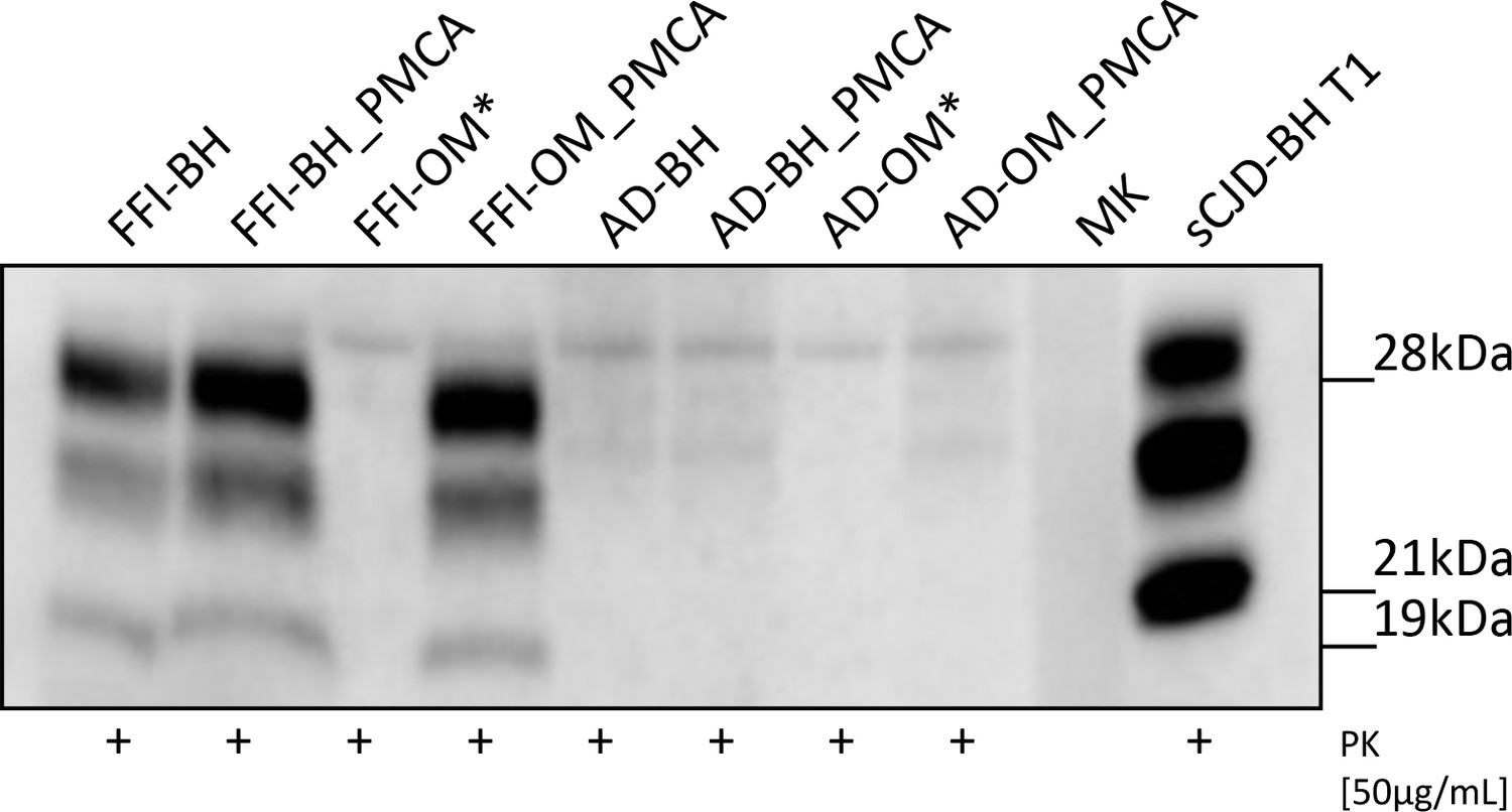
Western blot analysis of the inocula.
Prions were detected in all Fatal Familial Insomnia (FFI) samples except for the raw olfactory mucosa (FFI-OM). Notably, the glycoform ratio of all PrPres was identical and characterized by a predominance of the di-glycosylated band with the un-glycosylated one migrating at 19 kDa. No PrPres was found in samples collected from patients with AD. Three rounds of Protein Misfolding Cyclic Amplification (PMCA) were performed for each sample (brain homogenate [BH] or OM) before the analysis. BH of a sCJD-129MM1 patient (sCJD-BH T1) was used as migration control. Asterisks indicate samples that were not inoculated in mice. MK: molecular weight marker. Samples were immunoblotted with anti-PrP 6D11 antibody.
Figure 2
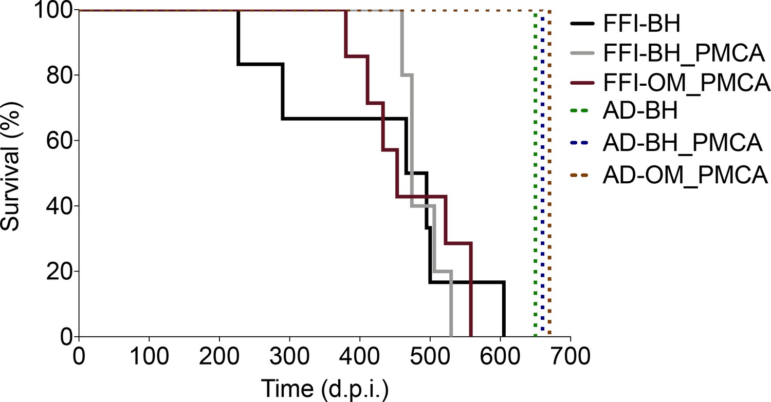
Survival time.
All animals inoculated with FFI-BH, FFI-BH_PMCA, and FFI-OM_PMCA succumbed to prion disease with similar survival time (431 ± 58 dpi, 489 ± 13 dpi, and 474 ± 27 dpi, respectively; p=0.085, log-rank test), while those inoculated with AD-BH, AD-BH_PMCA, and AD-OM_PMCA did not and were sacrificed at the end of the experiment. FFI: Fatal Familial Insomnia; PMCA: Protein Misfolding Cyclic Amplification; OM: olfactory mucosa; BH: brain homogenate.
Figure 3 with 1 supplement
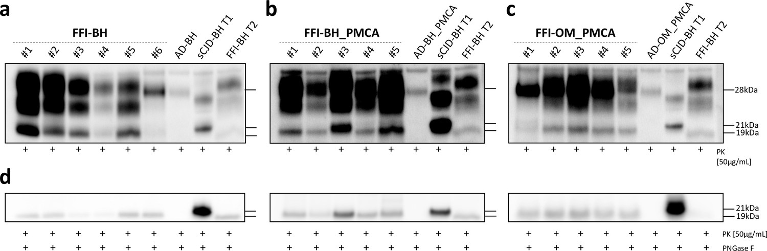
Western blot and PNGase F analysis of BvPrP-Tg407 brain homogenates.
BvPrP-Tg407 mice inoculated with FFI-BH (a), FFI-BH_PMCA (b), and FFI-OM_PMCA (c) showed the presence of PrPres while those inoculated with AD-BH, AD-BH_PMCA, and AD-OM_PMCA did not. All PrPres were characterized by the predominance of the di-glycosylated band except for the animal number #5 of the group FFI-BH, which showed a PrPres with the mono-glycosylated band predominant over the others. Samples were immunoblotted with anti-PrP 6D11 antibody. Similar findings have been observed using the Sha31 antibody (Figure 3—figure supplement 1). The brain homogenate of a FFI patient with PrPres migrating at 19 kDa (FFI-BH T2) and the brain homogenate of a sCJD patient with PrPres migrating at 21 kDa (sCJD-BH T1) were used as migration controls. PNGase F analyses showed that the un-glycosylated band migrated at 19 kDa in all cases, thus confirming the results obtained from animals with less clear migration pattern (e.g., animal number #3 of the FFI-BH_PMCA group and animal number #1 of the FFI-OM_PMCA group). Samples were immunoblotted with anti-PrP 6D11 antibody (d). FFI: Fatal Familial Insomnia; PMCA: Protein Misfolding Cyclic Amplification; OM: olfactory mucosa; BH: brain homogenate.
Figure 3—figure supplement 1
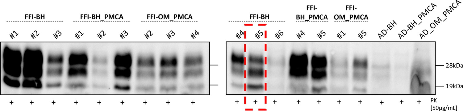
Western blot analysis of BvPrP-Tg407-inoculated mice.
BvPrP-Tg407-inoculated mice were analyzed with Sha31 antibody that recognizes a C-terminal region of the PrP (a.a. 145–152). All prion-inoculated animals, except the number #5 of the FFI-BH group, showed PrPres characterized by a prevalence of the di-glycosylated band. A PrPres characterized by a prevalent mono-glycosylated band was found in the brain of animal number #5 of the group FFI-BH (framed in red). No PrPres was found in the brain of animals inoculated with AD-related samples. FFI: Fatal Familial Insomnia; BH: brain homogenate.
Figure 4 with 2 supplements

Biochemical characterization of prions generated in BvPrP-Tg407 mice.
Proteinase K (PK) resistance assay (a) and conformational stability analysis (b) were performed to characterize the prions present in the brains of animals inoculated with FFI-BH, FFI-BH_PMCA, and FFI-OM_PMCA. These analyses revealed that prions found in the brain of FFI-BH-inoculated mice were more resistant to proteolytic digestion and less stable to guanidine hydrochloride (Gdn-HCl) treatment (black line) than those found in the brain of mice inoculated with FFI-BH_PMCA and FFI-OM_PMCA (gray and purple lines, respectively). In both cases, these differences were statistically significant (two-way ANOVA followed by Bonferroni post-tests; FFI-BH vs. FFI-BH_PMCA: *p<0.05; FFI-BH vs. FFI-OM_PMCA: ° p<0.05, °° p<0.01, °°° p<0.001; error bars: ± standard error of the mean [SEM]). In contrast, prions found in the brain of mice inoculated with FFI-BH_PMCA and FFI-OM_PMCA showed comparable PK-resistance profile and stability towards Gdn-HCl treatment. Separate PK and Gdn-HCl analysis were performed for animal number #5 that was excluded from the FFI-BH group (Figure 4—figure supplement 1). Analysis performed on FFI-BH sample and its PMCA product (FFI-BH_PMCA) showed that the amplification altered the PK and Gdn-HCl properties of PrPSc (Figure 4—figure supplement 2). FFI: Fatal Familial Insomnia; PMCA: Protein Misfolding Cyclic Amplification; OM: olfactory mucosa; BH: brain homogenate.
Figure 4—figure supplement 1
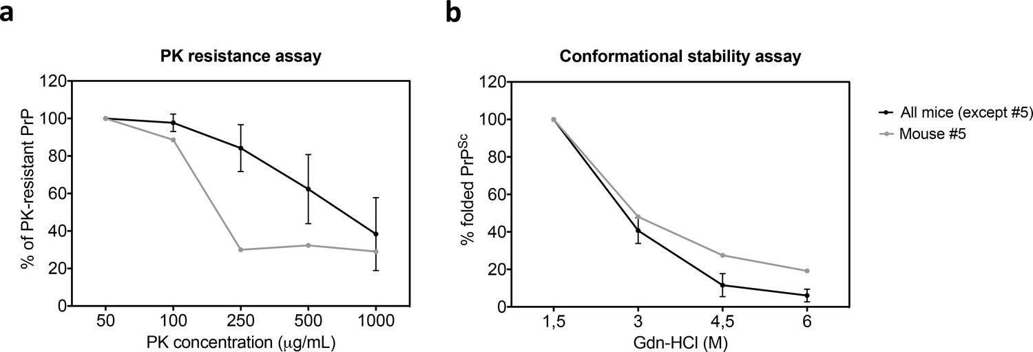
Proteinase K (PK)-resistance assay and conformational stability analysis of the prion present in the brains of animal number #5 excluded from the FFI-BH-injected group.
PK-resistance (a) and conformational stability (b) profiles of mouse #5 (gray line) was compared to those of all the other animals of the same group (black line). The PrPres found in the brain of mouse #5 appeared to be significantly more sensitive to PK digestion and more resistant to guanidine hydrochloride (Gdn-HCl) treatment compared to all the other prions, thus indicating that it might be a different isolate. Error bars:± standard error of the mean (SEM). FFI: Fatal Familial Insomnia; BH: brain homogenate.
Figure 4—figure supplement 2
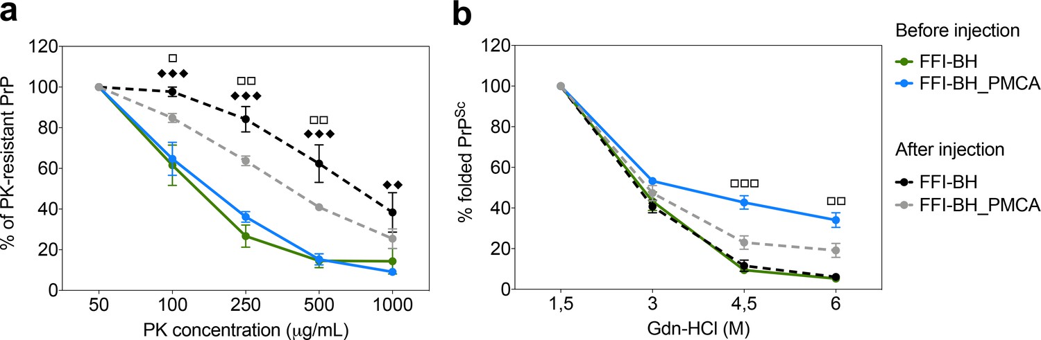
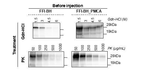
Proteinase K (PK)-resistance assay and conformational stability analysis of the prions present in FFI-BH and its product of amplification (FFI-BH_PMCA) before and after injection in mice.
PK-resistance (a) and conformational stability (b) of FFI-BH and FFI-BH_PMCA before (solid lines) and after (dashed lines) injection in BvPrP-Tg407 mice. Before the injection, the PK resistance of PrPSc associated with FFI-BH (green solid line) and FFI-BH_PMCA (blue solid line) was comparable. After the injection, both PrPSc became more resistant to PK digestion compared to the original inocula (FFI-BH, dashed black line; FFI-BH_PMCA dashed gray line) (a). Before the injection, the conformational stability of PrPSc associated with FFI-BH (green solid line) was significantly lower than that of FFI-BH_PMCA (blue solid line). After the injection, the stability of PrPSc associated with FFI-BH did not change (black dashed line), while that associated with FFI-BH_PMCA (gray dashed line) decreased in a statistically significant manner (b). Two-way ANOVA followed by Bonferroni post-tests; FFI-BH before vs. after injection: ♦♦♦ p<0.001, ♦♦ p<0.01; FFI-BH_PMCA before vs. after injection: □ p<0.05, □□ p<0.01, □□□p<0.001; error bars:± standard error of the mean (SEM). FFI: Fatal Familial Insomnia; PMCA: Protein Misfolding Cyclic Amplification; BH: brain homogenate.
Figure 5
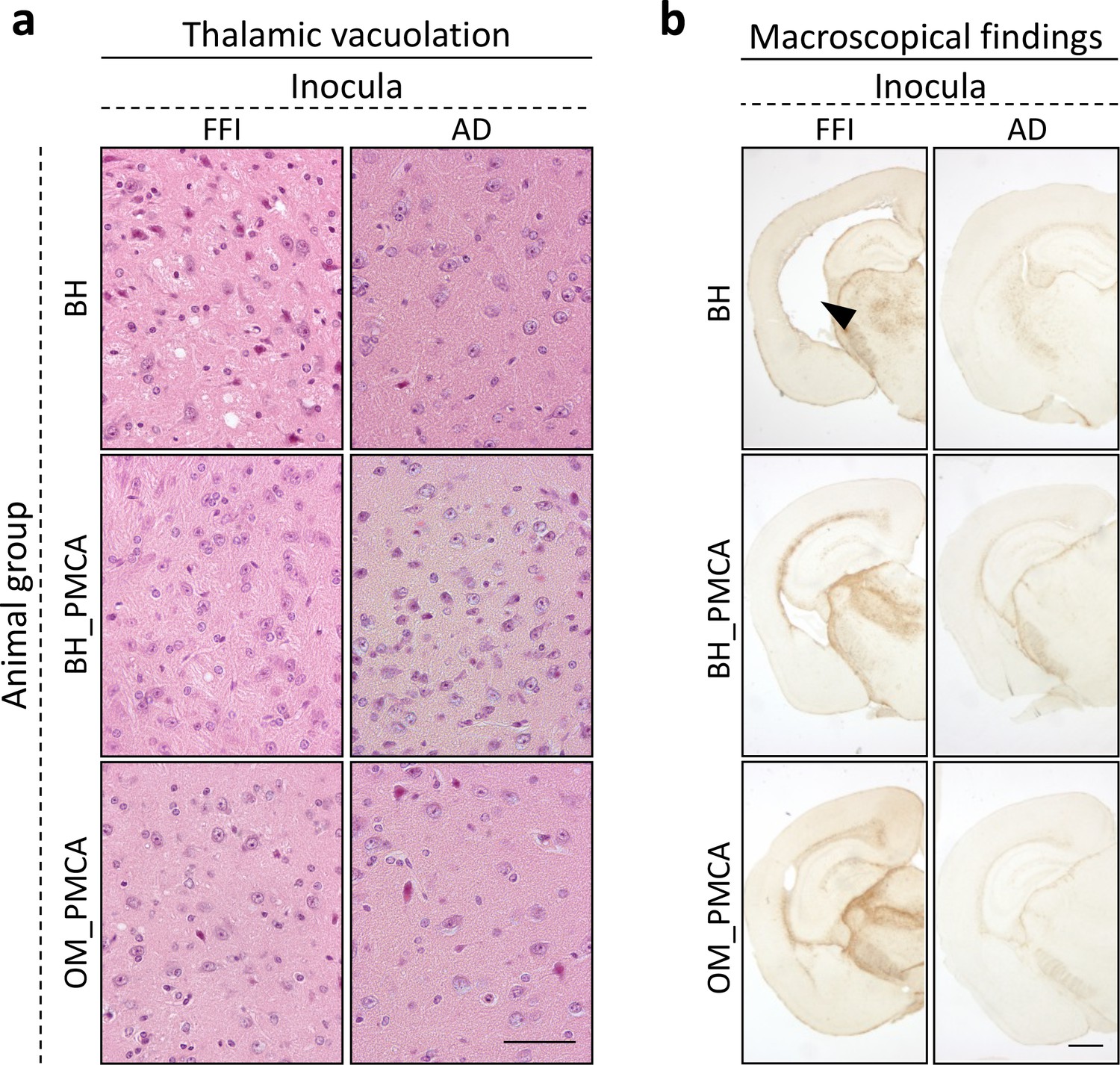
Histological findings.
Faint vacuolation was observed in the thalamus of BvPrP-Tg407 mice inoculated with FFI-BH, FFI-BH_PMCA, and FFI-OM_PMCA. No spongiform changes were found in the brain of animals inoculated with AD-BH, AD-BH_PMCA, and AD-OM_PMCA. Sections were stained with hematoxylin and eosin. Scale bar: 50 μM (a). Mice inoculated with FFI-BH showed significant enlargement of the ventricles (see black arrow) that was not observed in the brain of all the other inoculated mice. Severe astroglial activations were observed in the thalamus of all prion-inoculated mice. Sections were immunostained with anti-glial fibrillary acidic protein antibody. Scale bar: 500 μM (b). FFI: Fatal Familial Insomnia; PMCA: Protein Misfolding Cyclic Amplification; OM: olfactory mucosa; BH: brain homogenate.
Figure 6 with 3 supplements

Prion distribution in the brain of BvPrP-Tg407-inoculated mice.
BvPrP-Tg407 mice inoculated with FFI-BH showed synaptic distribution of PrPres with the presence of focal and plaque-like deposits mainly affecting the thalamus, striatum, and frontal cortex. Notably, animals #5 and #6 of this group did not show any PrPres (Figure 6—figure supplement 2). Animals inoculated with FFI-BH_PMCA showed only plaque-like deposits of PrPres mainly occurring in the thalamus and striatum. Similarly, but to a lesser extent, plaque-like deposits of PrPres were found in the thalamus, striatum, and frontal cortex of BvPrP-Tg407 mice inoculated with FFI-OM_PMCA (Figure 6—figure supplement 1). Focal deposits observed in the brain of FFI-BH, FFI-BH_PMCA, and FFI-OM_PMCA-injected mice were completely negative at ThS staining (Figure 6—figure supplement 3). No PrPres was found in the brain of mice inoculated with AD-BH, AD-BH_PMCA, and AD-OM_PMCA (Figure 6—figure supplement 1). Sections were immunostained with anti-PrP Saf34 antibody. Scale bar: 10 μm. FFI: Fatal Familial Insomnia; PMCA: Protein Misfolding Cyclic Amplification; OM: olfactory mucosa; BH: brain homogenate.
Figure 6—figure supplement 1
Schematic representation of prion distribution in the brain of BvPrP-Tg407-inoculated mice.
These cartoons summarize the PrPres distribution and the pattern of deposition in the brains of BvPrP-Tg407 mice inoculated with FFI-BH (left), FFI-BH_PMCA (central), and FFI-OM_PMCA (right). Synaptic and diffuse PrPres deposition pattern is represented with solid purple color while plaque-like deposits with purple dots. Animals inoculated with FFI-BH showed mixed synaptic-diffuse and plaque-like patterns of PrPres deposition mainly affecting the striatum, thalamus, and deep layers of the frontal cortex. No PrPres immunoreactivity was detectable in mice numbers #5 and #6. In the brain of animals inoculated with FFI-BH_PMCA, the deposition was characterized by focal plaque-like deposits of PrPres affecting the thalamus, striatum, and frontal cortex. A similar pattern of deposition was observed in the brain of animal number #2 inoculated with FFI-OM_PMCA. Surprisingly, animal number #1 of the same group did not show any detectable PrPres by immunohistochemistry (even if a PrPres signal was clearly detectable by western blot). All AD-BH, AD-BH_PMCA, and AD-OM_PMCA-inoculated animals did not show any PrPres signal. FFI: Fatal Familial Insomnia; PMCA: Protein Misfolding Cyclic Amplification; OM: olfactory mucosa; BH: brain homogenate.
Figure 6—figure supplement 2

Details of the prion distribution and astroglial activation found in the brain of mice numbers #5 and #6 inoculated with FFI-BH.
BvPrP-Tg407 mice numbers #5 and #6 inoculated with FFI-BH did not show any PrPres detectable by immunohistochemistry (anti-PrP Saf34 antibody) even in the presence of marked astroglial activation mainly affecting the thalamus, striatum, and frontal cortex (anti-glial fibrillary acidic protein antibody). Scale bar: 40 µm. FFI: Fatal Familial Insomnia; BH: brain homogenate.
Figure 6—figure supplement 3
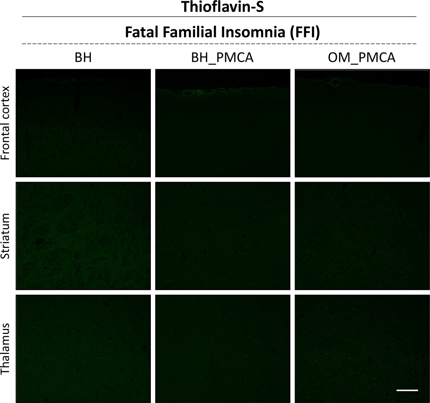
Evaluation of the amyloid tinctorial properties of the PrPres deposits.
The PrPres deposits found in the thalamus, striatum, and frontal cortex of many animals inoculated with FFI-BH, FFI-BH_PMCA, and FFI-OM_PMCA were negative at ThS staining and confirmed the lack of the typical amyloid properties. Scale bar: 40 µm. FFI: Fatal Familial Insomnia; PMCA: Protein Misfolding Cyclic Amplification; OM: olfactory mucosa; BH: brain homogenate.
Figure 7 with 1 supplement

Astroglial activation in the brain of BvPrP-Tg407-inoculated mice.
Animals inoculated with FFI-BH, FFI-BH_PMCA, and FFI-OM_PMCA showed severe glial activation in the thalamus and striatum. Moderate immunoreactivity was also found in the deep layer of the cerebral cortex of the animals inoculated with BH-FFI (Figure 7—figure supplement 1). Mild astroglial reactivity was observed in the striatum and thalamus of mice inoculated with AD-BH, AD-BH_PMCA, and AD-OM_PMCA. This activation is often detectable in healthy aged mice and is not an indicator of pathological processes (Figure 7—figure supplement 1). Sections were immunostained with anti-glial fibrillary acidic protein antibody. Scale bar: 10 μm. FFI: Fatal Familial Insomnia; PMCA: Protein Misfolding Cyclic Amplification; OM: olfactory mucosa; BH: brain homogenate.
Figure 7—figure supplement 1

Schematic representation of the astroglial activation in the brain of BvPrP-Tg407-inoculated mice.
These cartoons summarize the degree of the astroglial activation in the brains of BvPrP-Tg407 mice inoculated with FFI-BH (left), FFI-BH_PMCA (central), and FFI-OM_PMCA (right). Light green refers to a low degree of glial activation while dark green refers to a severe degree of glial activation. Regardless of the inocula, astroglial activation was mainly found in the thalamus, striatum, and frontal cortex of all challenged mice. Mild immunoreactivity was observed in the striatum, thalamus, and frontal cortex of AD-BH, AD-BH_PMCA, and AD-OM_PMCA-inoculated mice, indicating that this finding might be the result of physiological aging. FFI: Fatal Familial Insomnia; PMCA: Protein Misfolding Cyclic Amplification; OM: olfactory mucosa; BH: brain homogenate.
Figure 8

Real-Time Quaking-Induced Conversion (RT-QuIC) results of BvPrP-Tg407 brain homogenates.
All brain homogenates of the animals inoculated with FFI-BH, FFI-BH_PMCA, and FFI-OM_PMCA induced a rapid aggregation of the recBvPrP90-231 used as a RT-QuIC reaction substrate. Aggregation of the substrate was observed also in the presence of brain homogenates of mice inoculated with AD-BH, AD-BH_PMCA, and AD-OM_PMCA but occurred after 18 hr and were considered negative. Error bars:± standard error of the mean (SEM). FFI: Fatal Familial Insomnia; PMCA: Protein Misfolding Cyclic Amplification; OM: olfactory mucosa; BH: brain homogenate.
Author response image 1
Tables
Key resources table
| Reagent type (species) or resource | Designation | Source or reference | Identifiers | Additional information |
|---|---|---|---|---|
| Strain, strain background (mouse) | BvPrP-Tg407 mice | DOI:10.1128/JVI.01592-16 | ||
| Peptide, recombinant protein | recBvPrP90-231 | DOI:10.1038/srep46269 | ||
| Antibody | 6D11 | Covance | Catalog # SIG-399810 RRID:AB_2564735 | WB: 0.2 µg/mL |
| Antibody | Sha31 | SPI Bio | Catalog # A03213 | WB: 0.4 µg/mL |
| Antibody | Mouse IgG HRP Linked F(ab’)2 Fragment | GE | Catalog # NA9310V | |
| Antibody | Saf34 | Gently provided by Prof. Jacques Grassi DOI:10.1111/j.1471-4159.2004.02356.x DOI:10.1006/bbrc.1999.1730 IHC: 1.25 µg/mL | ||
| Antibody | GFAP antibody | DAKO | Catalog # Z0334 RRID:AB_10013382 | IHC: 0.5 µg/mL |
| Chemical compound, drug | PBS | Gibco | Catalog # 14200-067 | |
| Chemical compound, drug | Isofluorane | Isoba vet Schering-Plough S.A. (Merck company) | Catalog # 792632 | |
| Chemical compound, drug | Glucose | Gibco | Catalog # A2494001 | |
| Chemical compound, drug | Proteinase K | Invitrogen | Catalog # AM2542 | |
| Chemical compound, drug | Guanidine hydrochloride | Sigma-Aldrich | Catalog # 50950 | |
| Chemical compound, drug | NaCl | Carlo Erba | Catalog # 479686 | |
| Chemical compound, drug | EDTA | Sigma-Aldrich | Catalog # 03690 | |
| Chemical compound, drug | NP40 | BDH | Catalog # 56009 | |
| Chemical compound, drug | Deoxycholic acid, sodium salt | Millipore | Catalog # 264101 | |
| Chemical compound, drug | Tris-hydroxymethyl-aminomethane (Tris-HCl) | Carlo Erba | Catalog # 489973 | |
| Chemical compound, drug | Blotto, non-fat dry milk | Santa Cruz | Catalog # SC-2325 | |
| Chemical compound, drug | N-Lauroylsarcosine sodium salt (sarkosyl) | Sigma-Aldrich | Catalog # 61747 | |
| Chemical compound, drug | Phosphate buffered saline -RT-QuIC | Sigma-Aldrich | Catalog # P5493 | |
| Chemical compound, drug | Sodium dodecyl sulfate solution | Sigma-Aldrich | Catalog # 71736 | |
| Chemical compound, drug | Thioflavin T | Sigma-Aldrich | Catalog # T3516 | |
| Chemical compound, drug | Ethanol (for Carnoy fixative preparation) | Sigma-Aldrich | Catalog # 32221 | DOI:10.1111/j.1750-3639.2000.tb00240.x |
| Chemical compound, drug | Acetic acid glacial (for Carnoy fixative preparation) | Sigma-Aldrich | Catalog # 33209 | DOI:10.1111/j.1750-3639.2000.tb00240.x |
| Chemical compound, drug | Paraffin | Bio Optica | Catalog # 08-7920 | |
| Chemical compound, drug | Chloroform (for Carnoy fixative preparation) | Carlo Erba | Catalog # 438614 | DOI:10.1111/j.1750-3639.2000.tb00240.x |
| Chemical compound, drug | Guanidine thiocyanate | Merck-Millipore | Catalog # 1.04167.0250 | |
| Commercial assay or kit | Bolt 12%, Bis-Tris, 1.0 mm, Mini Protein Gel, 10-well | Invitrogen | Catalog # NW00120BOX | |
| Commercial assay or kit | Immobilon-P, PVDF, 0.45 µm | Millipore | Catalog # IPVH00010 | |
| Commercial assay or kit | 4X Bolt LDS Sample Buffer | Invitrogen | Catalog # B0007 | |
| Commercial assay or kit | 10X Bolt Sample Reducing Agent | Invitrogen | Catalog # B0009 | |
| Commercial assay or kit | ECL Prime Western Blotting System | Amersham | Catalog # RPN2232 | |
| Commercial assay or kit | ARK (Animal Research Kit) | DAKO | Catalog # K3954 | |
| Commercial assay or kit | Liquid DAB Substrate Chromogen System | DAKO | Catalog # K3468 | |
| Commercial assay or kit | PNGase F | New England Biolabs | Catalog # P0704S | |
| Other | Nanosep Centrifugal Devices | Pall Corporation | Catalog # OD100C34 | |
| Other | Hematoxylin | Bio Optica | Catalog # 05-06012 | |
| Other | Eosin | Bio Optica | Catalog # 05-10002 | |
| Other | Thioflavin S | Sigma-Aldrich | Catalog # T-1892 | |
| Software, algorithm | ImageJ | PMID:22743772 | RRID:SCR_003070 | |
| Software, algorithm | GraphPad PRISM 5.0 v | N/A | RRID:SCR_002798 | |
| Software, algorithm | Nikon ACT-1 acquisition software | N/A | https://www.nikon.com/products/microscope-solutions/support/download/software/camerasfor/act1_v263.htm | |
| Software, algorithm | G:BOX Chemi XT4 | N/A | http://www.alphametrix.de/page/index.php?category=geldoc&pageid=219 |
Additional files
Download links
A two-part list of links to download the article, or parts of the article, in various formats.
Downloads (link to download the article as PDF)
Open citations (links to open the citations from this article in various online reference manager services)
Cite this article (links to download the citations from this article in formats compatible with various reference manager tools)
PMCA-generated prions from the olfactory mucosa of patients with Fatal Familial Insomnia cause prion disease in mice
eLife 10:e65311.
https://doi.org/10.7554/eLife.65311


