Chronic pain induces generalized enhancement of aversion
Abstract
A hallmark feature of chronic pain is its ability to impact other sensory and affective experiences. It is notably associated with hypersensitivity at the site of tissue injury. It is less clear, however, if chronic pain can also induce a generalized site-nonspecific enhancement in the aversive response to nociceptive inputs. Here, we showed that chronic pain in one limb in rats increased the aversive response to acute pain stimuli in the opposite limb, as assessed by conditioned place aversion. Interestingly, neural activities in the anterior cingulate cortex (ACC) correlated with noxious intensities, and optogenetic modulation of ACC neurons showed bidirectional control of the aversive response to acute pain. Chronic pain, however, altered acute pain intensity representation in the ACC to increase the aversive response to noxious stimuli at anatomically unrelated sites. Thus, chronic pain can disrupt cortical circuitry to enhance the aversive experience in a generalized anatomically nonspecific manner.
https://doi.org/10.7554/eLife.25302.001Introduction
Chronic pain exerts a profound influence over daily life by impacting a range of sensory and affective behaviors. It is associated with enhanced response to noxious stimuli, leading to symptoms of allodynia and hyperalgesia at the site of tissue or nerve injury (Basbaum et al., 2009; Latremoliere and Woolf, 2009). The mechanisms for such sensory and affective hypersensitivity at the site of chronic pain have been well investigated. However, conditions such as fibromyalgia and persistent postoperative pain raise the possibility that chronic pain may also increase the aversive reaction towards noxious stimuli in an anatomically nonspecific distribution (Scudds et al., 1987; Petzke et al., 2003; Kehlet et al., 2006; Kudel et al., 2007; Scott et al., 2010). If confirmed, this generalized form of enhancement in pain aversion, as opposed to anatomically specific hypersensitivity, can greatly expand our understanding of the impact of chronic pain on behavior.
The anterior cingulate cortex (ACC) has a crucial role in the affective-aversive experience of pain (Lubar, 1964; Foltz and White, 1968; Turnbull, 1972; Talbot et al., 1995; Craig et al., 1996; Rainville et al., 1997; Koyama et al., 2000; Johansen et al., 2001; Koyama et al., 2001; LaGraize et al., 2006; Qu et al., 2011). The ACC receives nociceptive inputs from the medial thalamus as well as from other cortical regions (Vogt and Sikes, 2000; Shyu et al., 2010). Individual ACC neurons can respond to noxious stimuli by increasing firing rates (Sikes and Vogt, 1992; Yamamura et al., 1996; Hutchison et al., 1999; Kung et al., 2003; Iwata et al., 2005; Kuo and Yen, 2005; Zhang et al., 2011) to provide evaluation for the intensity of acute pain (Coghill et al., 1999; Büchel et al., 2002). While previous studies have demonstrated that the ACC is necessary and sufficient for the acquisition of stable aversive learning in the chronic pain condition (Johansen et al., 2001; Qu et al., 2011; Barthas et al., 2015; Navratilova et al., 2015), its role in the aversive response to transient acute pain signals is less well characterized. Furthermore, chronic pain has been shown to induce synaptic plasticity in ACC neurons, resulting in hypersensitivity at the site of injury through descending modulation (Wu et al., 2005; Li et al., 2010; Koga et al., 2015). It is unknown, however, if chronic pain can also impair ACC functions to alter the aversive response to noxious stimuli in an anatomically nonspecific manner. Using a multidisciplinary approach by combining optogenetics, in vivo electrophysiology and machine-learning decoding analysis, we found that chronic pain can disrupt acute pain representation in the ACC to induce generalized, anatomically nonspecific enhancement of aversion.
Results
Chronic pain can enhance the aversive response to noxious inputs at anatomically disparate sites
Conditioned place aversion (CPA) and conditioned place preference are well-established assays to test aversive learning as well as the negative reinforcement of analgesia in rodent chronic pain models (Johansen et al., 2001; King et al., 2009; Navratilova et al., 2012; Daou et al., 2013). These tests, however, have rarely been used to assess the aversive value presented by acute pain signals. Based on the concept of quantitative evaluation of aversion developed by standard CPA protocols (Johansen et al., 2001; Johansen and Fields, 2004), we constructed a brief 2-chamber CPA test for rats (Figure 1A). During the baseline (preconditioning) phase (10 min) of the test, rats were allowed free access to both chambers, and the time spent in either chamber was recorded as baseline. Next, during a brief conditioning period (10 min), we paired each chamber with a distinct laser stimulus directed at the hind paw of a freely-moving rat. The stimulus was classified as non-noxious (NS), low-intensity noxious (LS), or high-intensity noxious (HS). The intensity of the stimulus corresponded to the power output of the laser. Higher laser output transferred more heat to induce more intense thermal pain (Figure 1—figure supplement 1). At the behavioral level, NS did not typically elicit withdrawals within 5 s of stimulation (<5%), whereas HS and LS both elicited paw withdrawals 100% of the time. In addition, HS elicited withdrawals with a shorter latency than LS (Figure 1B). After this brief conditioning phase, we immediately tested the rat’s aversive response by measuring the time spent in each of the two treatment chambers during a test (postconditioning) phase (10 min). During the test phase, rats were again allowed free access to both chambers without any peripheral stimulation. We compared the amount of time spent during baseline (preconditioning) and test (postconditioning) phases in each chamber (Johansen et al., 2001; King et al., 2009; Navratilova et al., 2012). A statistically significant reduction in the amount of time spent in a treatment chamber during the test phase when compared with baseline indicates an avoidance of that chamber, which in turn indicates aversion towards the stimulus associated with that chamber. Thus, while similar in principle to the multi-day conditioning protocols using repeated or prolonged exposure to painful stimuli to measure the acquisition of stable aversive memory (Johansen et al., 2001; King et al., 2009; Navratilova et al., 2012), our paradigm allows the assessment of acute aversive reaction.

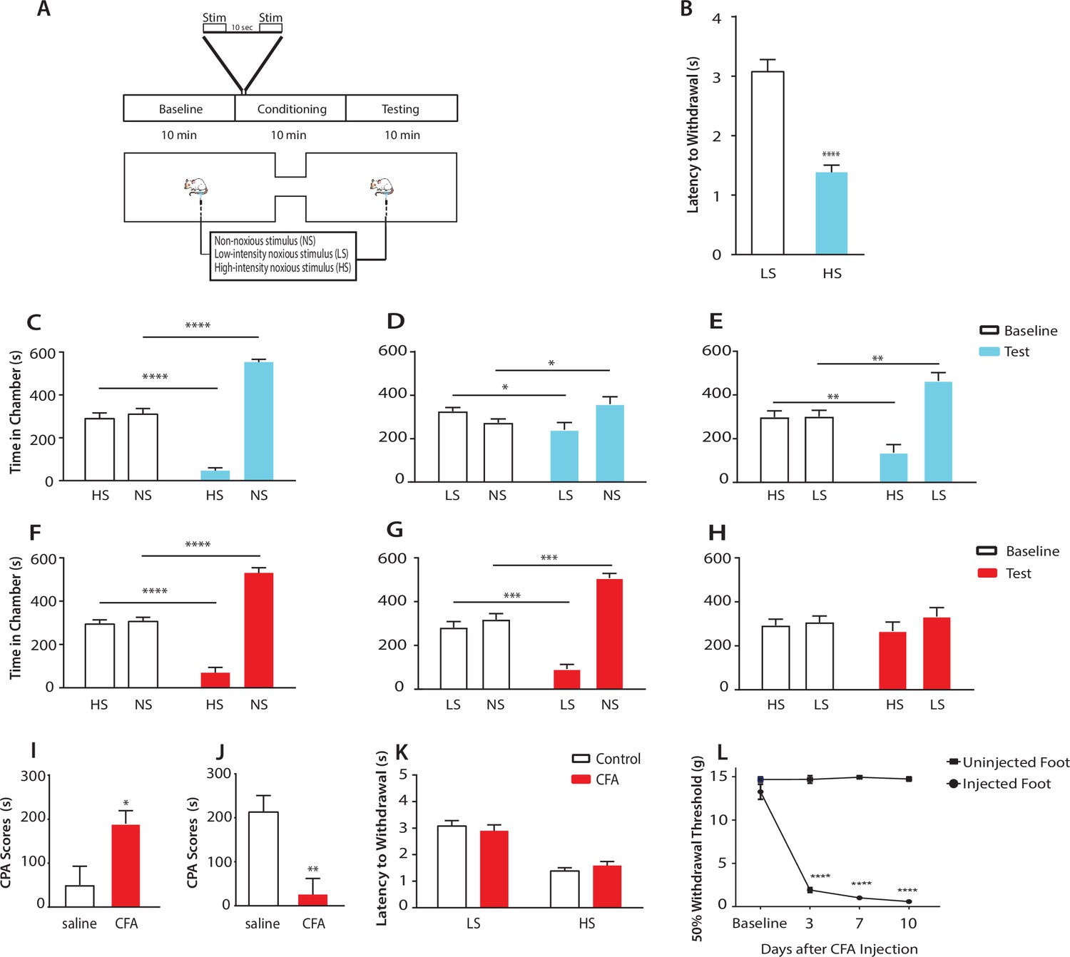
Chronic pain enhances the aversive response to acute pain at anatomically unrelated sites.
(A) Schematic for the conditioned place aversion (CPA) test. Each episode of peripheral stimulation lasted until paw withdrawal or in cases of no withdrawal (non-noxious stimulus or NS) a total of 5 s. (B) Latency to paw withdrawal was shorter with higher intensity pain stimulus (HS) than lower intensity stimulus (LS). n = 12; p<0.0001, Student’s t test. (C) During conditioning, rats received HS in one chamber and NS in the other chamber. After conditioning, rats spent less time in the chamber paired with HS during the test phase than at baseline, and more time in the chamber paired with NS. n = 14; p<0.0001, paired Student’s t test. (D) After conditioning with LS and NS in separate chambers, rats spent less time in the chamber paired with LS during the test phase than at baseline, and more time in the chamber paired with NS. n = 14; p=0.0131. (E) After conditioning with HS and LS, rats spent less time in the chamber paired with HS during the test phase than at baseline, and more time in the chamber paired with LS. n = 10; p=0.0012. (F) 10 days after CFA treatment, rats underwent conditioning by receiving HS stimulation of the uninjected paw in one chamber and NS stimulation of the same paw in the other chamber. During the test phase, rats spent less time in the chamber paired with HS than at baseline, and more time in the chamber paired with NS. n = 9; p<0.0001; paired Student’s t test. (G) After CFA treatment, after conditioning with LS and NS in separate chambers, rats spent less time in the chamber paired with LS stimulation of the uninjected paw during the test phase than at baseline, and more time in the chamber paired with NS. n = 9; p=0.0002. (H) After CFA treatment, rats could not differentiate between HS and LS treatments of the uninjected paws on the CPA test. n = 13; p=0.4923. (I) CFA treatment resulted in increased aversive response to LS. Rats were conditioned with LS and NS, and the CPA or aversion score was calculated by subtracting the amount of time rats spent during the test phase from baseline in the LS-paired chamber (see Materials and methods section). CPA scores for CFA-treated rats were significantly higher than saline control. n = 9–10; p=0.0179, unpaired Student’s t test. (J) After CFA treatment, rats could not distinguish between HS and LS. Rats were conditioned with HS in one chamber and LS in the other chamber. The CPA score for HS was calculated by subtracting the amount of time spent in the HS-paired chamber during the test phase from baseline. CFA-treated rats demonstrated a significant decrease in the CPA score, indicating an inability to distinguish between the aversive values of HS and LS. n = 8–13; p=0.0029, unpaired Student’s t test. (K) CFA treatment did not alter paw withdrawal latency in the uninjected paw. n = 12; p=0.8381, two-way ANOVA with repeated measures and post-hoc Bonferroni test. (L) CFA treatment caused mechanical allodynia in the injected but not uninjected paw. n = 12; p<0.0001. two-way ANOVA with repeated measures and post-hoc Bonferroni test.
First, we compared the aversive responses towards HS and NS. During conditioning, one of the chambers was paired with HS, and the other was paired with NS. We found that rats spent less time in the chamber paired with HS treatment during the test phase than at baseline (Figure 1C). Conversely, rats spent more time in the NS chamber during the test phase than at baseline. These results indicate that rats recognized and sought to avoid the aversive value associated with HS. Next, we measured the ability of the rats to distinguish between the aversive values of LS and NS. We conditioned rats by pairing LS with one chamber and NS in the other. After conditioning, rats spent less time in the chamber paired with LS relative to baseline, and more time in the chamber paired with NS (Figure 1D), indicating an ability to recognize the aversive value for LS. Finally, we conditioned rats with HS and LS, and during the test phase rats spent less time in the HS-paired chamber (and more time in the LS-paired chamber) than at baseline, suggesting that these rats displayed a greater aversive response to HS than LS (Figure 1E). Together, these results indicate that rats can detect not only the presence of acute pain, but also the aversive values of higher vs lower-intensity noxious stimuli. This quantitative distinction in aversive response correlates well with changes in nocifensive spinal reflex (Figure 1B).
Next, we tested if this quantifiable aversive response to acute pain is altered by the presence of chronic pain at an anatomically unrelated location. We injected Complete Freund’s Adjuvant (CFA) to induce persistent inflammatory pain in the opposite paw and performed CPA by conditioning with noxious stimulation of the uninjected paw. 10 days post-CFA injections, rats were conditioned with HS and NS to the healthy paw, and they demonstrated a preference for the NS chamber and avoidance of the HS chamber during the test phase compared with baseline (Figure 1F). These results suggest that rats in chronic pain were still able to distinguish between highly noxious from non-noxious stimulations. We then examined the response of rats in chronic pain to the low-intensity noxious stimuli. First, CFA-treated rats were conditioned with LS and NS in separate chambers. During the test phase these rats spent less time in the LS chamber than at baseline, indicating an aversive response to LS (Figure 1G). Interestingly, CFA-treated rats appeared to demonstrate an increased avoidance of the LS-paired chamber than rats that did not have chronic pain (compare Figure 1G with Figure 1D). We then examined the ability for these CFA-treated rats to distinguish between the aversive quality of LS vs HS. We found that CFA-treated rats did not seem able to clearly distinguish between LS and HS, as they failed to avoid the HS chamber after conditioning (Figure 1H), a striking difference from the avoidance of the HS chamber exhibited by rats that did not experience chronic pain (Figure 1E). These comparisons suggest qualitatively that while rats in chronic pain were still able to distinguish between non-noxious and noxious stimuli, they seemed to have developed a heightened aversive response to the low-intensity stimuli even at an anatomically distinct site.
To provide a quantitative analysis for the above findings, we calculated a CPA score (or aversion score) to measure the aversive value of peripheral stimulations. This CPA score was computed by subtracting the amount of time rats spent in a chamber paired with a specific noxious stimulus during the test phase from the time they spent in that chamber at baseline (Johansen et al., 2001; Johansen and Fields, 2004; De Felice et al., 2013). A higher CPA score (a greater difference in the time spent in the associated chamber between baseline and test phase) indicates a greater aversive value for that stimulus. We first calculated the CPA score for LS, after we conditioned rats with LS in one chamber and NS in the other. We found that this CPA score was significantly higher for CFA-treated rats than saline-treated (control) rats (Figure 1I). This quantitative analysis confirms that CFA-treated rats avoided the LS chamber more than control rats, and thus rats in chronic pain developed an increased aversive response to acute low-intensity noxious stimulations even at an anatomically distinct site. Next, we quantified the ability for the rats to distinguish between HS and LS. This time, we conditioned rats with HS and LS, and we computed the CPA score for HS, by subtracting the amount of time rats spent in the HS chamber during the test phase from the time they spent at baseline. In this case, a higher CPA score indicates a stronger aversive response to HS, and hence a greater ability to distinguish between HS and LS. Here, we found that CFA-treated rats demonstrated a dramatic reduction in their CPA score (Figure 1J), indicating that rats in chronic pain lost the ability to distinguish between the aversive values of HS and LS. Results from Figure 1I together indicate that after chronic pain, rats perceived both low-intensity and high-intensity noxious stimuli as highly aversive. Thus, the presence of chronic pain induced an alteration in the aversive evaluation of acute pain signals even at an anatomically distinct site. There have been reports suggesting that unilateral noxious stimulations can in some cases cause bilateral changes in spinal circuits regulating mechanical sensitivity (Gao and Ji, 2010, Gao et al., 2010). We tested whether the healthy paws in our study displayed any behavioral signs of spinal or peripheral hypersensitivity as the result of CFA injection in the opposite paws, but we did not find any abnormality in the withdrawal reflex to noxious stimulation or the presence of mechanical allodynia (Figure 1K). Thus, the effect of chronic pain on acute pain responses at anatomically unrelated sites is likely specific for the aversive component, sparing the sensory component. We termed this anatomically nonspecific increase in the aversive response to acute pain ‘generalized enhancement of pain aversion.’
Chronic pain disrupts the ACC representation of acute pain signals
Having established this phenomenon of generalized enhancement of pain aversion, we investigated whether a disruption in the ACC representation of acute pain contributes to the neural substrate for such behavior. We conducted extracellular recordings in the ACC in freely moving rats before, during and after peripheral stimulation by NS, LS or HS, and analyzed the firing rates of individual neurons (Figure 2A). We found that a number of neurons in the ACC increased their firing rates after an acute pain stimulus (see Materials and methods; Figure 2C), similar to reports from human studies (Hutchison et al., 1999). We also found a significant number of neurons that responded to noxious stimulation and at the same time showed increased firing rate after HS than LS (Figure 2D,E). The identification of such neurons that responded to noxious intensity is also compatible with previous reports (Sikes and Vogt, 1992; Yamamura et al., 1996; Hutchison et al., 1999; Kung et al., 2003; Iwata et al., 2005; Kuo and Yen, 2005; Zhang et al., 2011). In order to make an unbiased assessment of the contribution of individual ACC neurons in pain representation, we then applied a population-decoding analysis using a support vector machine (SVM) classifier (see Materials and methods). We analyzed groups of neurons across multiple recording sessions, each session comprising of >30 trials. We used some of the trials in training and the remaining trials for testing. Both pain-responsive and non-responsive neurons were used in unbiased decoding analysis, but the firing activity from pain-responsive neurons would contribute to a higher weight (i.e., being the ‘support vector’; see Materials and methods). Our decoding analysis yielded high accuracy in distinguishing between NS and HS (85% accuracy) or between LS and HS (76% accuracy) (Figure 2F, Figure 2—figure supplement 1). Therefore, our unbiased decoding analysis supported a critical role of ACC neurons in the representation of pain intensity.

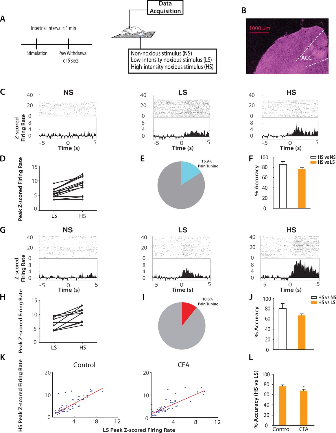
Chronic pain increases the responsiveness of ACC neurons to acute pain signals.
(A) Timeline and schematic for electrophysiological recordings in freely moving rats. Each trial of peripheral stimulation lasted until paw withdrawal or in cases of no withdrawal (non-noxious stimulus or NS) a total of 5 s. (B) Histology showing the location of ACC tetrode recordings. (C) Raster plots and peristimulus time histograms (PSTHs) before and after NS, LS and HS stimulations. Time zero denotes the onset of stimulus. Y-axis shows z-scored firing rates. To calculate the z-scored firing rate, we used the following equation: (FR – mean of FRb) / SD of FRb, where FR indicates firing rate and FRb indicates baseline firing rate prior to NS, LS or HS (see Materials and methods section). (D, E) A subset of neurons was identified among pain responsive neurons that showed higher firing rates after HS stimulation compared with LS (see Materials and methods for definition of pain responsive neurons). n = 14 out of a total of 88 neurons; for D, p<0.0001, paired Student’s t test. (F) Population-decoding analysis using a SVM classifier demonstrated decoding accuracy to distinguish between HS and NS (85%) or HS and LS (76%), n = 9. See Materials and methods. (G) Raster plots and PSTHs before and after NS, LS and HS stimulations in rats 10 days after CFA injections in the opposite paws. (H, I) A subset of neurons was found among pain responsive neurons that showed higher firing rates after HS stimulation compared with LS in the chronic pain condition. n = 10 out of a total of 93 neurons; for H, p<0.0001, paired Student’s t test. (J) Population-decoding analysis demonstrated decoding accuracy to distinguish between HS and NS (81%) or HS and LS (67%) in rats after chronic pain, n = 15. (K) A robust linear regression model was used to fit the peak z-scored firing rates and to calculate slope of the fit for all ACC neurons that demonstrated higher firing rates at HS compared with LS. Slope = 1.31 ± 0.09, R2 = 0.5937, n = 50 neurons. After chronic pain, neurons with higher firing rates at HS than LS showed a flatter tuning curve between LS and HS responses (see Materials and methods). Slope = 1.07 ± 0.07, R2 = 0.6904, n = 53 neurons. The two slope parameters are statistically different (p<0.05, unpaired Student’s t-test). (L) Decoding analysis showed that after chronic pain, there was a decrease in decoding accuracy to distinguish between HS and LS, n = 9 for pre-CFA, 15 for post-CFA; p<0.05, unpaired Student’s t test.
Our neurophysiological recordings were performed in freely moving animals, and thus baseline movement was unlikely to affect our data interpretation. However, noxious stimulation also induced paw withdrawals, and these movements are spinal reflexes in nature (Vardeh et al., 2016). To ensure that these spinal reflexes did not influence our neural findings, we quantitatively analyzed these motor responses. We found that while rats did not withdraw their paws in response to NS (<5%), the percentages of withdrawal responses to LS and HS were equal (~100%). To quantify the motor function of paw withdrawals, we then calculated the withdrawal velocity after LS and HS. We did not find any statistical difference in the velocity of withdrawals (Figure 2—figure supplement 2). These results indicate that there was no significant difference in the gross motor responses to LS and HS, in contrast to the dramatic difference in neural responses in the ACC seen in Figure 2. Thus, the neural responses we observed were less likely to be related to stimulus-induced movement. Such responses in the ACC more likely represented nociceptive processing, compatible with previous reports (Sikes and Vogt, 1992; Hutchison et al., 1999; Wang et al., 2003; Kuo and Yen, 2005; Zhang et al., 2011).
Next, we examined how the presence of chronic pain in the opposite limb affected ACC representation of acute pain (Figure 2G). After CFA injection in the opposite foot, we continued to find neurons in the ACC that responded to pain stimuli (Figure 2H). In addition, our SVM decoding algorithm continued to distinguish HS from NS (80% accuracy) and HS from LS (67% accuracy) (Figure 2J) – as compared to the chance level of 50% (Figure 2—figure supplement 3). We then examined the ‘tuning curves’ of all ACC neurons that showed increased peak firing rates after HS than LS. Such tuning curves demonstrate the degree of neural responsiveness to different intensities of noxious stimulation. In the chronic pain condition, however, neurons showed a flatter tuning curve, as indicated by a significant decrease in the slope of linear fit (Figure 2K). This suggests a decreased ability to distinguish between LS and HS at the level of individual neurons. Furthermore, our unbiased machine learning analysis also indicated that chronic pain caused a statistically significant decrease in the ability to decode the difference between LS and HS signals (control vs. CFA: 76% vs 67% accuracy, Figure 2L). Thus, our neural data indicate a disruption of ACC representation of acute pain intensity in the chronic pain state. This impaired distinction between LS and HS by ACC neurons correlates very well with behavioral findings that showed increases in the aversive valuation of LS in the chronic pain state (Figure 1G–J), suggesting that the ACC likely plays a role in the impact of chronic pain on the acute pain experience.
Chronic pain impairs the bidirectional regulation of acute pain by the ACC
To define the function of ACC in generalized enhancement of pain aversion, we temporally paired noxious stimuli with optogenetic control of ACC neurons (Figure 3A). First, we used channelrhodopsin (ChR2) to activate ACC pyramidal neurons, by expressing ChR2 linked to a CAMKII promotor in an AAV vector in the ACC region (Figure 3B; Figure 3—figure supplement 1). We did not observe any changes in paw withdrawal latency with optogenetic stimulation (Figure 3C), suggesting that brief ACC activation did not strongly modulate the acute nocifensive reflex. Next, we conditioned rats by coupling ACC activation with LS in one of the CPA chambers, and LS alone in the opposite chamber during the CPA test (Figure 3A). We found that ACC activation during the presentation of LS increased the aversive response to LS, as shown by an avoidance of the chamber associated with optogenetic activation during the test phase compared with baseline (Figure 3D). This pain-enhancing effect of ACC activation was not observed when the animal was presented with NS stimulation (Figure 3E), indicating that while transient ACC activation does not turn an acute non-noxious stimulus into a noxious one, it can significantly enhance the aversive quality of a noxious stimulus. Meanwhile, ACC activation during the presentation of HS increased the aversive response to HS on the CPA, but this effect was not statistically significant (Figure 3F). There are two possible interpretations for this apparent lack of effect of ACC activation during HS. First, our CPA test serves as a pain-scale report for rats, and with highly noxious stimulation (HS), there may be a maximal aversive response expressed by rats, similar to a maximal pain score experienced by humans. A second possible interpretation is that there may be a limit (ceiling effect) on the aversive response that could be elicited with our CPA assay, and the further aversive effect of ACC activation on HS was limited by such constraints. Nevertheless, our results clearly indicate that ACC activation during the presentation of a low intensity noxious input is capable of elevating the aversive value of that input, demonstrating that the ACC can indeed regulate the behavioral response to acute pain signals.

Optogenetic activation of the ACC has similar effects as chronic pain in enhancing the aversive response to acute pain.
(A) Schematic for a CPA test during optogenetic activation of the ACC. Light activation of the ACC was temporally coupled with peripheral stimulation to the paw. (B) Histologic expression of ChR2 in the ACC. (C) Light activation of the ACC did not alter paw withdrawal latency to noxious stimuli. n = 5; p=0.4654, paired Student’s t test. (D) ACC activation increased the aversive response to LS. One of the chambers was paired with optogenetic stimulation of the ACC and LS; the other chamber was paired with LS without ACC activation. Rats spent less time during the test phase than at baseline in the chamber paired with LS coupled with ACC activation and more time in the chamber paired with LS alone. n = 8; p=0.0026. (E) ACC activation did not elicit an aversive response to NS. One of the chambers was paired with ACC activation and NS; the other chamber was paired with NS alone. n = 9; p=0.7514. (F) ACC activation did not increase the aversive response to HS in a statistically significant manner. One of the chambers was paired with ACC activation and HS; the other chamber was paired with HS alone. n = 10; p=0.1584. (G) Coupling with ACC activation increased the aversive response of LS compared with NS. One of the chambers was paired with optogenetic stimulation of the ACC and LS; the other chamber was paired with NS alone. Rats spent significantly less time during the test phase than at baseline in the chamber paired with ACC activation and LS. n = 11; p=0.0075. (H) ACC activation caused a similar increase in the aversive response to LS as chronic pain. For comparison, rats were conditioned with LS and NS in separate chambers in both experiments. In one experiment (3G), LS was coupled with ACC activation, and the CPA score was calculated by subtracting the amount of time spent during the test phase from baseline in the chamber paired with simultaneous ACC activation and LS. In the second experiment, conditioning was performed in CFA-treated rats without ACC activation. A CPA score was calculated by subtracting the amount of time spent during the test phase from baseline in the chamber paired with LS in CFA-treated rats. The CPA scores from these two different experiments were similar. n = 9–11; p=0.9161, unpaired Student’s t test.
To ensure that activation of the ACC did not impact movement of the animals, we performed a locomotion test and found no changes in locomotor activity after ACC activation (Figure 3—figure supplement 2). Furthermore, as expected, shining light in the ACC injected with a YFP-only vector without opsin expression did not result in any changes in pain aversion phenotypes (Figure 3—figure supplement 3). These control experiments support the specific role of ACC neurons in the aversive response to pain.
To further understand the role of the ACC in generalized enhancement of pain aversion, we compared the effect of ACC activation with CFA treatment on the aversive evaluation of noxious stimulations. First, we conditioned rats by coupling ACC activation with LS in one chamber, and NS without ACC modulation in the opposite chamber. This experiment allowed us to assess the aversive value of LS during the activation of ACC relative to a non-noxious condition. When we compared the avoidance of the LS chamber in these rats that received ACC activation (Figure 3G) with naïve rats (Figure 1D), we noticed that ACC activation likely caused an additional increase in the aversive value of LS. To quantify this increase, we measured the CPA score for LS in this experiment by subtracting the amount of time rats spent during the test phase from baseline in the chamber paired with LS and ACC activation. This CPA score represents the aversive value for the LS in the presence of ACC activation. Next, we computed the aversive value for LS in the chronic pain state by calculating the CPA score in the experiment where we conditioned CFA-treated rats with LS vs NS without optogenetic modulation of the ACC. This second CPA score indicates the aversive value for the LS in the presence of chronic pain. We found that these two CPA scores were nearly identical (Figure 3H). Thus, the amplitude of the additional aversive effect provided by ACC activation during the presentation of LS is similar to the effect of chronic pain. This similarity supports the role for ACC activation in the generalized enhancement of pain aversion.
To further confirm the role of the ACC in pain regulation, we tested whether inhibition of ACC neurons could decrease the aversive response to noxious stimuli. We used halorhodopsin (NpHR) to inhibit ACC neurons during the presentation of noxious stimulation (Figure 4A; Figure 4—figure supplement 1). We conditioned rats by coupling ACC inhibition with a peripheral stimulus in one of the CPA chambers, and that stimulus alone in the opposite chamber (Figure 4A). We found that ACC inhibition decreased the aversive response to LS and HS, as shown by increased amounts of time spent in the chamber associated with LS or HS coupled with ACC inhibition during the test phase compared with baseline (Figure 4C,D). Inhibition of the ACC did not impact free movements of the animals (Figure 4—figure supplement 2). Furthermore, as expected, ACC inhibition had no effect on the aversive response to NS, which was not noxious (Figure 4—figure supplement 3). Next, we wanted to know if ACC inhibition could remove the aversive response to a noxious stimulus altogether. We paired one chamber with ACC inhibition and LS, and another chamber with NS. After conditioning, we found that ACC inhibition coupled with LS stimulation removed the avoidance of the LS-paired chamber (Figure 4E, p>0.05). Similarly, ACC inhibition during HS removed the higher aversive value of HS compared with LS, by eliminating the avoidance of HS-paired chamber (Figure 4F, p>0.05). These results indicate that ACC inhibition during the presentation of a more noxious stimulus can eliminate the greater aversive reaction towards that stimulus. Therefore, the ChR2 and NpHR data together demonstrate that neurons in the ACC can bidirectionally control the aversive response to acute pain.

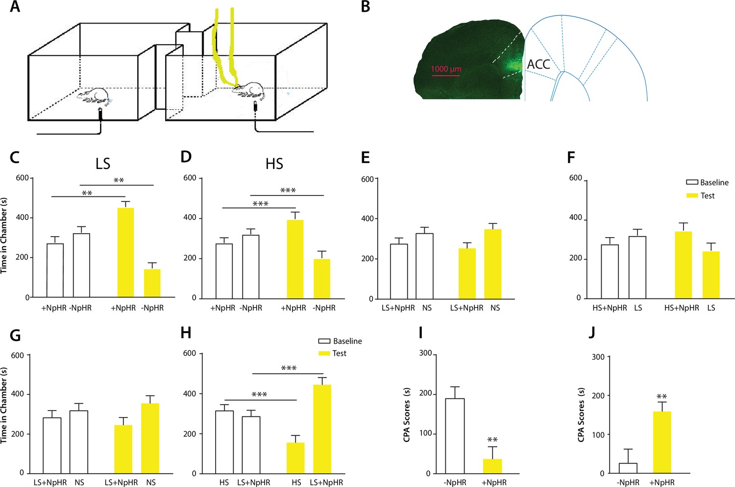
Optogenetic inhibition of the ACC diminishes the effect of chronic pain on the aversive response to acute pain.
(A) Schematic for a CPA test during optogenetic inhibition of the ACC. Optogenetic inhibition of the ACC was temporally coupled with peripheral stimulation to the paw. (B) Histologic expression of NpHR in the ACC. (C) ACC inhibition decreased the aversive response to LS. One of the chambers was paired with optogenetic inhibition of the ACC and LS; the other chamber was paired with LS alone. Rats spent more time during the test phase than at baseline in the chamber paired with light inhibition of the ACC coupled with LS and less time in the LS-alone chamber. n = 10; p=0.0043, paired Student’s t test. (D) ACC inhibition decreased the aversive response to HS. One of the chambers was paired with ACC inhibition and HS; the other chamber was paired with HS alone. Rats spent more time during the test phase than at baseline in the chamber paired with ACC inhibition coupled with HS and less time in the HS-alone chamber. n = 11; p=0.0006. (E) ACC inhibition abolished the aversive value of LS. One chamber was paired with ACC inhibition coupled with LS; the other chamber was paired with NS without ACC modulation. There was no statistically significant difference between test and baseline preference for either chamber. n = 9; p=0.5797. (F) ACC inhibition abolished the difference in aversive valuation between HS and LS. One chamber was paired with ACC inhibition coupled with HS; the other chamber was paired with LS. There was no statistically significant difference between test and baseline preference for either chamber. n = 12; p=0.1617. (G) ACC inhibition abolished the aversive value of LS even in CFA-treated rats. One chamber was paired with ACC inhibition coupled with LS; the other chamber was paired with NS. There was no statistically significant difference between test and baseline preference for either chamber. n = 10; p=0.2638. (H) ACC inhibition during the presentation of LS restored the normal differentiation between the aversive values of HS and LS in CFA-treated rats. One chamber was paired with ACC inhibition coupled with LS; the other chamber was paired with HS. Rats spent more time during the test phase than baseline in the chamber paired with ACC inhibition and LS, and less time in the HS-paired chamber. n = 10; p=0.0001. (I) ACC inhibition decreased the aversive value of LS in CFA-treated rats. CPA scores were compared in CFA-treated rats. In the control group (− NpHR), LS was not coupled with ACC inhibition during conditioning. In the test group (+ NpHR), LS was coupled with optogenetic inhibition of the ACC. In both groups, LS was conditioned against NS. CPA scores were calculated by subtracting the amount of time spent in the chamber paired with LS during the test phase from baseline. ACC inhibition reduced the CPA score for LS. n = 9–10; p=0.0026, unpaired Student’s t test. (J) ACC inhibition restored the normal difference in the aversive response to HS vs LS even after CFA treatment. In the control group (− NpHR), LS was not coupled with ACC inhibition during conditioning. In the test group (+ NpHR), LS was coupled with optogenetic inhibition of the ACC. In both groups, LS was conditioned against HS. CPA scores were calculated by subtracting the amount of time spent during the test phase in the chamber paired with HS from baseline. ACC inhibition increased the CPA score for HS. n = 10–13; p=0.0097.
Finally, we tested the effect of ACC inhibition specifically on the generalized enhancement of pain aversion. We have shown that CFA-treated rats demonstrated an increased aversive response to LS in the uninjected paw (Figure 1G). Here, we found that ACC inhibition during LS eliminated the avoidance of LS-paired chamber in CFA-treated rats when these rats were conditioned with NS (Figure 4G). Thus, ACC inhibition blocked the aversion-amplifying effect of chronic pain. Furthermore, rats in chronic pain have been shown to lose the ability to differentiate between LS and HS on the CPA test (Figure 1H). ACC inhibition during LS stimulations, however, restored the normal aversive scale in CFA-treated rats, by reinstating the avoidance of HS chamber (Figure 4H). To quantitatively confirm these findings, we calculated the CPA score for CFA-treated rats which were conditioned with LS with or without ACC inhibition and NS. This CPA score was computed by subtracting the amount of time rats spent during the test phase from baseline in the chamber paired with LS (Figure 4I). We found that ACC inhibition reduced this CPA score and hence the aversive response for LS even in rats with chronic pain. We also calculated the CPA score for CFA-treated rats which were conditioned with HS and LS in the presence or absence of ACC inhibition, by subtracting the amount of time rats spent during the test phase from baseline in the chamber paired with HS (Figure 4J). We found that ACC inhibition during LS elevated the CPA score for HS and hence reinstated the difference in aversive values between HS and LS in rats with chronic pain. Thus, ACC inhibition restored the normal aversive response to acute pain in those rats with chronic pain. These results strongly indicate that neural activities in the ACC play a vital role in the generalized enhancement of aversion in chronic pain conditions.
Discussion
The key finding in our study is that chronic pain causes a generalized enhancement in pain aversion. Previous studies have revealed the aversive state secondary to nociceptive inputs from the site of chronic pain (Johansen et al., 2001; Johansen and Fields, 2004; King et al., 2009; De Felice et al., 2013). The novel aspect of our results lies in the demonstration that chronic pain at one site in the body can also increase the aversive response to acute pain in a separate location. Interestingly, acute pain responses elicited by lower or intermediate intensity stimulus (such as LS) were more affected by chronic pain than maximal pain stimulus (HS), leading to a distortion of the pain-intensity scale. Epidemiological studies have shown that the presence of chronic pain coincides with increased pain severity and a similar distortion of pain intensity scale in a diffuse anatomic pattern (Scudds et al., 1987; Petzke et al., 2003; Kehlet et al., 2006; Kudel et al., 2007; Scott et al., 2010). Our study confirms these clinical findings by providing a causal link between chronic pain and a generalized enhancement of pain aversion. This generalized enhancement in aversion may be an important mechanism for chronic pain to influence normal sensory and affective processes.
Our study also provides a mechanistic basis for this generalized enhancement of pain aversion. Individual ACC neurons can respond to noxious stimuli by increasing firing rates (Sikes and Vogt, 1992; Yamamura et al., 1996; Hutchison et al., 1999; Kung et al., 2003; Iwata et al., 2005; Kuo and Yen, 2005; Zhang et al., 2011). Our study shows that this neural representation of acute pain intensity is profoundly altered by chronic pain, which causes ACC neurons to display a disproportional response to low-intensity pain stimuli. Previous studies have found that chronic pain can induce maladaptive synaptic plasticity in the ACC (Li et al., 2010; Koga et al., 2015). It is possible that such plasticity increases the response of ACC neurons to acute pain signals, and responses to low-intensity stimuli are disproportionally affected because the neuronal response is possibly saturated at high-intensity stimuli in the absence of chronic pain. At the network level, ACC neurons are known to project to or receive inputs from a number of regions important for pain processing, including the prefrontal cortex, medial thalamus, insular cortex, amygdala, nucleus accumbens, hippocampus, etc. Thus, acute pain likely triggers a concerted neural response in an interconnected network as suggested by fMRI studies (Schweinhardt and Bushnell, 2012; Wager et al., 2013). Chronic pain, however, has the capacity to disturb this network response.
Another important finding in our study is the ability of the ACC to exert bidirectional control of pain aversion. Prior lesion and pharmacological studies have shown that the rostral region of the ACC is required specifically for the acquisition of stable aversive learning induced by chronic pain (Johansen et al., 2001; Johansen and Fields, 2004; Qu et al., 2011). Our study indicates that this brain region is also necessary and sufficient for the temporal regulation of the aversive response to acute pain stimuli. Interestingly, in our study, brief activation of the ACC did not cause an aversive reaction to a non-noxious stimulus, whereas previous studies demonstrate that repeated activation of ACC neurons itself can be an aversive teaching signal (Johansen and Fields, 2004). This difference suggests that repeated and possibly prolonged activation of ACC is required for the acquisition of stable aversive memory, whereas transient activation provides context-specific aversive valuation and response. At the molecular and cellular level, persistent peripheral nociceptive inputs have been shown to trigger opioid signaling and synaptic plasticity in the ACC to regulate sensory and aversive components of chronic pain (Li et al., 2010; Navratilova et al., 2015). Our results here suggest that these mechanisms have the potential to impact ACC regulation of pain aversion in an input- and output-nonspecific fashion to exert a more generalized form of control for acute pain behavior.
In addition to pain regulation, the anterior cingulate cortex is also involved in a number of sensory, affective and cognitive processes (de Araujo et al., 2003; Rolls et al., 2003; Grabenhorst et al., 2008; Rolls et al., 2008). It plays an important role in reward-based learning, such as providing necessary evaluation for rewarding or aversive cues as well as for the interpretation of errors in predicting such cues (Bush et al., 2002). It also plays roles in attention and in conflict monitoring (Braver et al., 2001). While our experiment demonstrated that ACC changes are likely specific to pain instead of motor responses to pain, we cannot absolutely rule out all the potential behavioral covariance associated with noxious stimulation. However, given the complexity and diversity of its functions and anatomic connections, it is perhaps not surprising that plasticity within the ACC as the result of chronic pain can alter the regulation of general aversive responses. It should be noted, in addition, that given its rich functional connectivity, the ACC is likely to be an important node in a complex network of brain structures that regulate this generalized enhancement in pain aversion.
In summary, we have demonstrated that chronic pain can disrupt cortical function to increase the aversive response to acute noxious signals in an anatomically nonspecific manner. This mechanism of generalized enhancement of pain aversion may underpin the pathophysiology of diffuse pain syndromes such as fibromyalgia and chronic postoperative pain. It also raises the possibility that other conditions such as depression and anxiety may exert similar impact on acute pain or other sensory and affective processes in general.
Materials and methods
Animals
All procedures in this study were approved by the New York University School of Medicine (NYUSOM) Institutional Animal Care and Use Committee (IACUC) as consistent with the National Institute of Health (NIH) Guide for the Care and Use of Laboratory Animals to ensure minimal animal use and discomfort. Male Sprague-Dawley rats were purchased from Taconic Farms, Albany, NY and kept at Mispro Biotech Services Facility in the Alexandria Center for Life Science, with controlled humidity, temperature, and 12 hr (6:30 AM to 6:30 PM) light-dark cycle. Food and water were available ad libitum. Animals arrived to the animal facility at 250 to 300 grams and were given on average 10 days to adjust to the new environment prior to the onset of experiments.
Complete Freund's Adjuvant (CFA) administration
Request a detailed protocolTo produce chronic inflammatory pain, 0.1 ml of CFA (mycobacterium tuberculosis, Sigma-Aldrich) was suspended in an oil-saline (1:1) emulsion and injected subcutaneously into the plantar aspect of the hindpaw opposite to the paw that was stimulated by laser. Control rats received equal volume of saline injection.
Virus construction and packaging
Request a detailed protocolRecombinant AAV vectors were serotyped with AAV1 coat proteins and packaged by the viral vector core at the University of Pennsylvania. Viral titers were 5 × 10¹² particles/mL for AAV1.CAMKII.ChR2-eYFP.WPRE.hGH and AAV1.CAMKII.NpHR-eYFP.WPRE.hGH.
Stereotaxic optic fiber implantation and intracranial viral injections
Request a detailed protocolAs described previously (Goffer et al., 2013; Lee et al., 2015), rats were anesthetized with isoflurane (1.5% to 2%). In all experiments, virus was delivered to the anterior cingulate cortex (ACC) only. Rats were bilaterally injected with 0.5 µL of viral vectors at a rate of 0.1 µL/10 s with a 26-gauge 1 µL Hamilton syringe at anteroposterior (AP) +2.6 mm, mediolateral (ML) ±1.6 mm, and dorsoventral (DV) −2.25 mm, with tips angle 28° toward the midline. The microinjection needles were left in place for 10 min, raised 1 mm and left for another minute to allow for diffusion of virus particles away from injection site while minimizing spread of viral particles along the injection tract. Rats were then implanted with 200 μm optic fibers held in 1.25 mm ferrules (Thorlabs) in the ACC: AP +2.6 mm, ML ±1.6 mm, DV −1.25 mm. Fibers with ferrules were held in place by dental acrylic.
Electrode implantation and surgery
Request a detailed protocolTetrodes were constructed from four twisted 12.7 µm polyimide-coated microwires (Sandvik) and mounted in an eight tetrode VersaDrive (Neuralynx). Electrode tips were plated with gold to reduce electrode impedances to 100–500 kΩ at 1 kHz. Rats were anesthetized with isoflurane (1.5–2%). The skull was exposed and a 2.5-mm-diameter hole was drilled above the target region. A durotomy was performed before tetrodes were slowly lowered unilaterally into the ACC with the stereotaxic apparatus. Coordinates for ACC implants were: AP +2.7 mm, ML 0.8 mm, and DV 1.4 mm, with tetrode tips angled 10° toward the midline. The drive was secured to the skull screws with dental cement.
Following animal sacrifice, brain sections were collected at a thickness of 20 µm using Microm HM525 Cryostat machine, and sections were analyzed for viral expression and optic fiber localization with histological staining. Animals with improper fiber or electrode placements, low viral expression, or viral expression outside the ACC were excluded from the study.
In vivo electrophysiological recordings
Request a detailed protocolBefore stimulation, animals were given a 30 min period to habituate to a recording chamber over a mesh table. Noxious stimulation via a 1000 mW blue diode-pumped solid-state laser (Shanghai Dreams Laser Technology Co., LTD.) was applied 1 mm from the plantar surface of the hind paw contralateral to the brain recording site in freely moving rats (Chen et al., 2017). The laser output intensity could be NS (laser intensity of 50 mW), LS (150 mW) or HS (250 mW) (see below) in a single session with 200 µm core diameter fiber (M83L01, Thorlabs). The laser output power was calibrated by compact power and energy meter console (PM100D, Thorlabs) at the beginning of every recording session. In a single trial, the laser was turned on by a transistor-to-transistor (TTL) pulse generator (Doric) until paw withdrawal was observed (or for a total of 5 s if no withdrawal occurred). All recording sessions consisted of approximately 35 trials with variable inter-trial intervals (approximately 1 min) using one category of stimulation (NS, LS, or HS). A video camera (HC-V550, Panasonic) was used to record the experiment. Long inter-trial intervals between trials and the break between sessions were used to avoid sensitization. We did not identify any sensitization behavior nor physical damage to the paw during our experiment. The withdrawal latency was defined by the time between onset of laser and paw withdrawal.
In a subset of experiments intended for decoding analysis, we performed recordings using two different lasers to provide two different output intensities (NS&HS, or LS&HS). The stimulations were randomly applied to rat's hind paw for a total of approximately 60 trials (of equal number of trials for each stimulation intensity).
Optrode recordings were made when animals were anesthetized with 1% isoflurane and secured to a stereotaxic apparatus, as described previously (Lee et al., 2015). A 32-channel optrode (VersaDrive8 optical, Neuralynx) containing an optical fiber positioned 0.5 mm above the tips of 8 surrounding tetrodes was implanted after virus injection, using the coordinates mentioned above. The optical fiber was connected to a laser, which was connected to a TTL pulse-generating box (OTPG4, Doric Instruments). An extra output on the TTL box and the headstage were then connected to the data acquisition system in order to simultaneously record laser pulses and brain activity.
Data collection and preprocessing
Request a detailed protocolTetrodes were lowered in steps of 120 µm before each day of recording. The neuronal activity and the onset of noxious laser stimulation were simultaneously recorded with an acquisition equipment (Open Ephys) via an RHD2132 amplifier board (Intan Technologies). Signals were monitored and recorded from 32 low-noise amplifier channels at 30 kHz, band-passed filtered (0.3 to 7.5 kHz). To get spike activity, the raw data were high-pass filtered at 300 Hz with subsequent thresholding and offline sorting by commercial software (Offline Sorter, Plexon). The threshold was lower than the 3-Sigma peak heights line and optimized manually based on the signal to noise ratio. The features of three valley electrodes were used for spike sorting. Only clear spike clusters with good tetrode spike waveforms and ISI (inter spike interval) Poisson distribution were selected for analysis. Single units with peak firing rates lower than 1 Hz were excluded. Trials were aligned to the initiation of laser-on to compute the PSTH for each single unit using MATLAB (Mathworks).
Immunohistochemistry
Request a detailed protocolRats were deeply anesthetized with Isoflurane and transcardially perfused with ice-cold PBS followed by 4% paraformaldehyde (PFA) in PBS. Brains were fixed in PFA overnight and then transferred to 30% sucrose in PBS to equilibrate for three days as described (Lee et al., 2015). 20 µm coronal sections were made with a cryostat and washed with PBS for 10 min. Sections were washed in PBS and coverslipped with Vectashield mounting medium. Images containing tetrodes were stained with cresyl violet. These images were acquired using a Nikon eclipse 80i microscope with a DS-U2 camera head. Sections were also made after viral transfer for opsin verification, and these sections were stained with anti-rabbit GFP (1:500, Abcam, Cambridge, MA, #AB290), anti-mouse VGLUT 1/2 (1:200, Millipore, Temecula, CA, #MAB5502/5504), and DAPI (1:200, Vector Laboratories, Burlingame, CA) antibodies. Secondary antibodies were anti-rabbit IgG conjugated to AlexaFluor 488, and anti-mouse IgG conjugated to AlexaFluor 647 (1:200, Life Technologies, Carlsbad, CA). Images were acquired with a Zeiss LSM 700 Confocal Microscope (Carl Zeiss, Thornwood, NY).
Animal behavioral tests
For optogenetic experiments, optic fibers were connected to a 473 nm (for ChR2) or 589 nm (for NpHR) laser diode (Shanghai Dream Lasers) through a mating sleeve as described previously (Lee et al., 2015). Laser intensity was measured with a power meter (Thorlabs) prior to experiments. Laser was delivered using a TTL pulse-generator (Doric).
Conditioned place aversion (CPA)
View detailed protocolCPA experiments were conducted similar to what has been described previously (Johansen et al., 2001; Johansen and Fields, 2004; King et al., 2009; De Felice et al., 2013; Lee et al., 2015). The movements of rats in each chamber were automatically recorded by a camera and analyzed with the Any-maze software. The CPA protocol included preconditioning (baseline), conditioning, and testing phases (10 min during each phase). Animals spending more than 500 s or less than 100 s of the total time in either main chamber in the preconditioning phase were eliminated from further analysis (approximately 20% of total animals). Immediately following the pre-conditioning phase, the rats underwent conditioning for 10 min. Each of the two chambers was paired with a unique blue laser stimulus, which could be NS, LS or HS. For NS, the laser output power was approximately 50 mW, for LS, it was 150 mW, and for HS, it was 250 mW. Laser intensity was measured with a power meter (Thorlabs) prior to each experiment, and the same intensity was used consistently to generate LS and HS. A stimulus was terminated after paw withdrawal, and this occurred for LS and HS. In the case of NS, withdrawals occurred on less than 5% of stimulations, and in the case of non-withdrawals the stimulus was applied for a total of 5 s. The stimulus was repeated every 10 s. During a subset of the experiments, optogenetic activation was concurrent with laser stimulation in one of the treatment chambers. Laser stimulation, optogenetic stimulation and chamber pairings were counterbalanced. During the test phase, the animals did not receive any treatment and had free access to both compartments for a total of 10 min. Animal movements in each of the chambers were recorded, and the time spent in either of the treatment chambers was analyzed by the AnyMaze software. Decreased time spent in a chamber during the test phase as compared with the baseline indicates avoidance (aversion) for that chamber.
Paw withdrawal latency test
Request a detailed protocolA laser stimulation of 50 mW (NS), 150 mW (LS), or 250 mW (HS) was applied to the hind paw of rats. Less than 5% of the time 50 mW stimulation elicited a withdrawal response within 5 s, whereas 150 mW and 250 mW elicited withdrawal 100% of the time. A video was used to tape the procedure and analyze the withdrawal latency.
Mechanical allodynia test
Request a detailed protocolA Dixon up-down method with von Frey filaments was used to measure mechanical allodynia (Chaplan et al., 1994; Bourquin et al., 2006; Wang et al., 2011). Rats were individually placed into plexiglass chambers over a mesh table and acclimated for 20 min before testing. Beginning with 2.55 g, von Frey filaments in a set with logarithmically incremental stiffness (0.45, 0.75, 1.20, 2.55, 4.40, 6.10, 10.50, 15.10 g) were applied to the paws of rats. 50% withdrawal threshold was calculated as described previously (Wang et al., 2011).
Measurement of the velocity of paw withdrawals
Request a detailed protocolWe used a high speed camera (Sony Handycam FDR-AX53) to record frame by frame the movement of the paws after noxious stimulation with LS or HS. We measured the velocity of paw withdrawal by dividing the height of paw withdrawal by the time it took for the animal to reach this height with its affected paw. An average of 10 measurements were calculated for each rat.
Locomotion test
Request a detailed protocolWe recorded locomotion over 10 min for rats that received optogenetic activation or inhibition of the ACC. Either blue or yellow light was turned on for 3 s every 10 s during the locomotion test. In control rats, no light activation was provided. Total distance travelled was computed based on AnyMaze recordings.
Statistical analysis
Request a detailed protocolThe results of behavioral experiments were given as mean ± S.E.M. For mechanical allodynia, a two-way ANOVA with repeated measures and post hoc multiple pair-wise comparison Bonferroni tests were used to compare the time spent in chamber or 50% withdrawal threshold under various testing conditions. During the CPA test, a paired Student’s t test was used to compare the time spent in each treatment chamber before and after conditioning (i.e. baseline vs test phase for each chamber) (King et al., 2009). Decreased time spent in a chamber during the test phase as compared with the baseline indicates avoidance (aversion) for that chamber. A CPA score was computed by subtracting the time spent in the more noxious chamber during the test phase from the time spent in that chamber at baseline (Johansen et al., 2001; Johansen and Fields, 2004; De Felice et al., 2013). Thus, for rats that were conditioned with LS and NS, CPA for LS was computed by subtracting the time spent in the LS chamber during the test phase from the time spent in that chamber at baseline. Meanwhile, for rats that were conditioned with HS and LS, CPA for HS was computed by subtracting the time spent in the HS chamber during the test phase from the time spent in that chamber at baseline. A two-tailed unpaired Student’s t test was used to compare differences in CPA scores under various testing conditions.
For neuronal spike analysis, to define a neuron that altered its firing rate in response to a peripheral stimulus, we calculated peri-stimulus time histograms (PSTH), using a 5 s range before and after laser stimulus and a bin size of 200 ms. The baseline mean and standard deviation was calculated from the five second interval prior to stimulus. To calculate z-scored firing rate, we used the following equation: Z = (FR – mean of FRb) / standard deviation of FRb, where FR indicates firing rate and FRb indicates baseline firing rate prior to NS, LS or HS. To define a pain responsive neuron, we used the following criteria: (1) The absolute value of the z score firing rate of least two time bins after stimulation must be ≥2.33; and (2) If the first criterion is passed, the lower bound z-score as defined by (Z –ZSEM) at least two bins after stimulation must be greater than 1.645. ZSEM is defined by the following equations: ZSEM (bin) = FRSEM (bin)/standard deviation of FRb (baseline/bin size), and FRSEM (bin) = (standard error of FR over all laser trials)/bin size. For ACC neurons that demonstrated increased firing rates after HS than LS, we also used a robust linear regression model to fit the peak z-scored firing rates in response to HS and LS and to calculate the slope of fit. This provided a ‘tuning curve’ to differentiate between HS and LS. For comparing the slopes of two regression lines, we used a Student’s t-test (Andrade and Estévez-Pérez, 2014).
For all tests, a p value<0.05 was considered statistically significant. All data were analyzed using the GraphPad Prism Version 7 software (GraphPad) and MATLAB (MathWorks).
Population-decoding analysis using machine learning
Request a detailed protocolAfter spike sorting, we obtained population spike trains from simultaneously recorded ACC neurons. For each single neuronal recording, we binned spikes into 100 ms to obtain spike count data in time. To simulate the online decoding, we used a 100 ms moving window to accumulate spike count statistics from laser onset (time 0) until 5 s (i.e., 50 bins). We assessed the decoding accuracy at each time bin based on the cumulative spike count statistics. Therefore, for a total of C neurons, the input dimensionality ranged from C (the first bin) to 50C (all bins). In these experiments where we randomly mixed different laser intensities (NS and HS or LS and HS), we assumed that we have n1 trials under laser intensity 1, and n2 trials under laser intensity 2. We split the total (n1 +n2) trials into two groups, 80% used for training, and 20% used for testing. The goal of population-decoding analysis was to classify the trial labels of different stimulation intensities (e.g., LS vs. HS) based on population spike data. We used a support vector machine (SVM) classifier (Bishop, 2007). The SVM is a discriminative supervised learning model that constructs the classification boundary by a separating hyperplane with maximum margin. Specifically, the SVM can map the input x into high-dimensional feature spaces which allows nonlinear classification.
where denote the class label for the training sample xi (some of which associated with nonzero are called support vectors), b denotes the bias, and K(•,•) denotes the kernel function. We used a polynomial kernel and trained the nonlinear SVM with a sequential minimal optimization algorithm (MATLAB Machine Learning Toolbox: ‘fitcsvm’ function). Finally, the decoding accuracy was assessed by 5-fold cross-validation from 100 Monte Carlo simulations. We report the mean ± S.E.M. decoding accuracy.
As a control, we also computed the chance-level decoding accuracy. We randomly permuted class labels between two classes and repeated the decoding analysis. This shuffling procedure was repeated 500 times, and we reported the chance level by the averaged classification accuracy based on shuffled data with permuted labels. In theory, when the sample sizes from both classes are perfectly balanced, the chance level should be close to 50%. Based on our experimental data, we obtained a chancel-level ~51–53%.
In all population-decoding analyses (pre- and post-CFA), we only used the recording sessions with five or more simultaneously recorded ACC units, independent of the cell firing properties. For NS vs HS, we have 6 and 4 sessions in pre-CFA and post-CFA conditions, respectively. For NS vs LS, we have 5 and 4 sessions in pre-CFA and post-CFA conditions, respectively. For LS vs HS, we have 9 and 15 sessions in pre-CFA and post-CFA conditions, respectively.
References
-
Statistical comparison of the slopes of two regression lines: A tutorialAnalytica Chimica Acta 838:1–12.https://doi.org/10.1016/j.aca.2014.04.057
-
The anterior cingulate cortex is a critical hub for pain-induced depressionBiological Psychiatry 77:236–245.https://doi.org/10.1016/j.biopsych.2014.08.004
-
Dissociable neural responses related to pain intensity, stimulus intensity, and stimulus awareness within the anterior cingulate cortex: a parametric single-trial laser functional magnetic resonance imaging studyJournal of Neuroscience 22:970–976.
-
Quantitative assessment of tactile allodynia in the rat pawJournal of Neuroscience Methods 53:55–63.https://doi.org/10.1016/0165-0270(94)90144-9
-
Deciphering neuronal population codes for acute thermal painJournal of Neural Engineering 14:036023.https://doi.org/10.1088/1741-2552/aa644d
-
Pain intensity processing within the human brain: a bilateral, distributed mechanismJournal of Neurophysiology 82:1934–1943.
-
Remote optogenetic activation and sensitization of pain pathways in freely moving miceJournal of Neuroscience 33:18631–18640.https://doi.org/10.1523/JNEUROSCI.2424-13.2013
-
Representation of umami taste in the human brainJournal of Neurophysiology 90:313–319.https://doi.org/10.1152/jn.00669.2002
-
Capturing the aversive state of cephalic pain preclinicallyAnnals of Neurology 74:275–265.https://doi.org/10.1002/ana.23922
-
The role of rostral cingulumotomy in "pain" reliefInternational Journal of Neurology 6:353–373.
-
Pain-related neurons in the human cingulate cortexNature Neuroscience 2:403–405.https://doi.org/10.1038/8065
-
Anterior cingulate cortical neuronal activity during perception of noxious thermal stimuli in monkeysJournal of Neurophysiology 94:1980–1991.https://doi.org/10.1152/jn.00190.2005
-
Unmasking the tonic-aversive state in neuropathic painNature Neuroscience 12:1364–1366.https://doi.org/10.1038/nn.2407
-
Anterior cingulate activity during pain-avoidance and reward tasks in monkeysNeuroscience Research 39:421–430.https://doi.org/10.1016/S0168-0102(01)00197-3
-
Predictors and consequences of multiple persistent postmastectomy painsJournal of Pain and Symptom Management 34:619–627.https://doi.org/10.1016/j.jpainsymman.2007.01.013
-
Activation of corticostriatal circuitry relieves chronic neuropathic painJournal of Neuroscience 35:5247–5259.https://doi.org/10.1523/JNEUROSCI.3494-14.2015
-
Effect of medial cortical lesions on the avoidance behavior of the catJournal of Comparative and Physiological Psychology 58:38–46.https://doi.org/10.1037/h0041014
-
Endogenous opioid activity in the anterior cingulate cortex is required for relief of painJournal of Neuroscience 35:7264–7271.https://doi.org/10.1523/JNEUROSCI.3862-14.2015
-
Neuroimaging of pain: insights into normal and pathological pain mechanismsNeuroscience Letters 520:129–130.https://doi.org/10.1016/j.neulet.2012.06.014
-
Predicting dissatisfaction following total knee replacement: a prospective study of 1217 patientsJournal of Bone and Joint Surgery - British Volume 92:1253–1258.https://doi.org/10.1302/0301-620X.92B9.24394
-
Pain perception and personality measures as discriminators in the classification of fibrositisThe Journal of Rheumatology 14:563–569.
-
Nociceptive processing by anterior cingulate pyramidal neuronsJournal of Neurophysiology 103:3287–3301.https://doi.org/10.1152/jn.00024.2010
-
Nociceptive neurons in area 24 of rabbit cingulate cortexJournal of Neurophysiology 68:1720–1732.
-
Evaluation of pain perception after anterior capsulotomy: a case reportSomatosensory & Motor Research 12:115–126.https://doi.org/10.3109/08990229509101503
-
Bilateral cingulumotomy combined with thalamotomy or mesencephalic tractotomy for painSurgery, Gynecology & Obstetrics 134:958–962.
-
Toward a Mechanism-Based approach to pain diagnosisThe Journal of Pain 17:T50–T69.https://doi.org/10.1016/j.jpain.2016.03.001
-
The medial pain system, cingulate cortex, and parallel processing of nociceptive informationProgress in Brain Research 122:223–235.https://doi.org/10.1016/S0079-6123(08)62141-X
-
An fMRI-based neurologic signature of physical painNew England Journal of Medicine 368:1388–1397.https://doi.org/10.1056/NEJMoa1204471
-
Upregulation of forebrain NMDA NR2B receptors contributes to behavioral sensitization after inflammationJournal of Neuroscience 25:11107–11116.https://doi.org/10.1523/JNEUROSCI.1678-05.2005
Decision letter
-
David D GintyReviewing Editor; Howard Hughes Medical Institute, Harvard Medical School, United States
In the interests of transparency, eLife includes the editorial decision letter and accompanying author responses. A lightly edited version of the letter sent to the authors after peer review is shown, indicating the most substantive concerns; minor comments are not usually included.
[Editors’ note: a previous version of this study was rejected after peer review, but the authors submitted for reconsideration. The first decision letter after peer review is shown below.]
Thank you for choosing to send your work, "Chronic pain induces generalized enhancement of pain aversion", for consideration at eLife. Your initial submission has been reviewed by a Senior Editor and three reviewers, one of whom is a member of our Board of Reviewing Editors. Although the work is of interest, we regret to inform you that the findings at this stage are too preliminary for further consideration at eLife.
While the reviewers expressed a high degree of interest in your study, several issues raised prevent us from moving forward with the manuscript in its present form. To summarize, main concerns include a lack of detailed descriptions of how each experiment was performed, including whether the same or different cohorts of rats were used for multiple measurements, and issues with how each experiment was analyzed with statistical methods; each of the three reviewers struggled with the statistical analysis, including how CPA values in Figure 1 were calculated. Reviewers also agreed that there is a missing control experiment (effects of optogenetic inhibition of the ACC on CPA by itself (or paired with NS)). Also, the degree of viral infection of ACC neurons was not measured and the cell types that are infected and express the optogenetic probes was also lacking. As for the concern noted in individual reviews regarding whether brain regions other than the ACC are involved, the consensus following discussion amongst the reviewers was that the finding of ACC involvement is of sufficient interest even in the absence of evidence implicating other brain regions. These consensus views are noted for your consideration in preparation for submission of a new manuscript to eLife or elsewhere.
Reviewer #1:
This study reports that chronic pain caused by CFA injected into the paw causes a generalized enhancement in pain aversion, as measured by laser stimulation of the uninjured paw of the opposite limb. The authors make clever use of CPA to examine aversive values presented by acute pain signals, delivered by the laser. They found that a submaximal acute pain behavior elicited by a low-intensity noxious laser pulse (LS) is more affected by chronic pain than is maximal pain behavior elicited by high-intensity stimulus (HS). Presumably the HS is already maximally aversive. Physiologically, the neural representation of acute pain in the ACC elicited by the LS to the uninjured paw is altered in the chronic pain state. Although I AM not an expert in the decoding analysis used to conclude that chronic pain disrupts the ACC representation of LS, I find this study to have several interesting findings.
Concerns that would need to be addressed:
In the CPA measurement, the extent to which rats avoid the chamber in which LS is delivered in the CFA group is said to be increased compared to LS treatment of the uninjured rats (Figure 1D vs 1G). This observation forms the basis of the entire study, and yet I am not clear on the statistical test used to draw this conclusion. In general, the way in which CPA measurements were calculated and how statistical calculations and comparisons were made are confusing. This needs clarification.
Whether alterations of central representation of LS in the chronic pain state is unique to the ACC or whether the ACC is one component of an interconnected network of brain regions similarly altered is not clear. That is, does optogenetic perturbation of any component of an interconnected network (medial thalamus, etc) lead to similar findings? Is the ACC the locus of altered neural representation of acute pain to an uninjured limb or one component of many?
Reviewer #2:
The manuscript by Zhang et al. uses a clever experimental design to uncover enhanced aversion to noxious input in the context of ongoing pain, and then uses optogenetic approaches to provide evidence for a role of the ACC for these behavioral changes. The paper has many strengths including novelty of both the approaches and the findings.
Although this study is not the first to show bilateral changes in mechanical sensitivity associated with persistent unilateral inflammation, it is the first, to my knowledge, to measure the aversive aspect of this change in a CPA test.
The authors provide intriguing correlative data supporting a role for the ACC in the responses to noxious stimulation through tetrode recordings. I do not feel fit to assess the experiments presented in Figure 2 because they are beyond my area of expertise.
The most interesting finding in this manuscript is that optogenetic activation of ACC neurons increases the aversiveness to noxious stimulation, whereas inhibition of ACC neurons decreases the aversiveness to noxious stimulation.
Major concerns.
1) I feel that the descriptions of how the data were analyzed are insufficient for me to determine whether the analyses were done correctly. For instance, many graphs show the time spent on each side of the chamber before and after conditioning treatment. In each case, the data presented in the first two columns are the mirror image of the data presented in the second two columns (since the rats are either in one chamber or the other, so this is not an independent variable.) To analyze this type of data, the authors do a 2-way ANOVA, which I find confusing. In other experiments, they calculate a CPA score, which is defined as the difference between the time spent in either treatment chamber. Again, I'm not exactly sure which numbers were subtracted to give the resulting data. I think these experiments would benefit from a clearer description as well as careful analysis by a statistical expert (not me). The authors should also clarify whether these experiments were done on the same cohort of rats, or whether naive rats were used for each experiment.
2) Conceptually, I find it hard to wrap my head around the difference between the low intensity noxious stimulus and the high intensity noxious stimulus. In each case, the rat is exposed to a laser until (and only until) the stimulus triggers a nociceptive reflex. Granted, the authors show that in CPA tests the mice can distinguish LS from HS. And they also show that a declassifying code can distinguish responses from LS and HS. Nevertheless, I feel that, in the spectrum of possible pain, the two types of stimuli would be rather similar. For this reason, I find it peculiar that the optogenetic stimulation of the ACC affects behavioral responses to LS but not HS. The reason given "HS presumably already elicited maximal ACC response, resulting in a less pronounced effect of optogenetic activation" is both unsatisfying and overinterpreted (note that there is actually no effect of optogenetic stimulation in the HS condition).
3. It seems to me that a key control is the effect of optogenetic inhibiton of the ACC on CPA by itself (or paired with NS). This control is missing.
Reviewer #3:
The report by Zhang et al. addresses an important issue in pain biology; how do discrete injuries to one portion of the body develop into wide-spread pain syndromes that cause patients to have hypersensitivity/allodynia in areas that were unaffected by the original insult? The answer to this question will have a direct and profound impact on a number of chronic pain conditions for which there are currently no treatments.
To address this issue, Zhang et al. used state of the art techniques to probe the role of the anterior cingulate cortex (ACC) in modulating the sensitivity of the hindlimb contralateral to the hindlimb in which inflammation had been induced with CFA 10 days prior to testing. Virally-expressed channelrhodopsin (ChR2) or halorhodopsin (NpHR) were used to modulate ACC activity. Extracellular recording tetrodes were also used to record ACC activity in response to noxious stimulation of the limbs. These techniques were combined with a conditioned place preference assay (CPA) to quantify the "aversive" responses from rats.
The paper begins by attempting to demonstrate that the paradigm induces "generalized enhancement of pain aversion". To do this the authors used a high power blue laser (50- to 250- mW) shined on the hindlimb to deliver a noxious heat stimulus to the hindpaw. Using three stimulus intensities they demonstrate that at the lowest intensity (50mW) mice do not develop a preference for either chamber of the PCA arena. In contrast, a 150mW (LS) or 250mW (HS) stimulus induces avoidance of the chamber in which the stimulus is applied. The authors show that the latency to a nocifensive response is significantly shorter at HS than LS. They interpret this result to mean that the HS stimulus is more noxious than the LS stimulus. While this might make sense intuitively, it is not consistent with what we know about spinally-mediated reflexes. Assuming that the spot size is consistent at LS and HS intensities (and it should be for this laser), the only difference to the rats should be the time it takes for the heat sensitive neuron to reach threshold. Once this threshold is reached, the spinal reflex is triggered and the movement should occlude further stimulation. Rats receiving the two different stimulus strengths should exhibit the same number of nocifensive behaviors (multiple presentations were made during the training period), the only difference being when during the stimulus application the behavior is triggered. If this is not the case (i.e. the 250mW stimulus is more noxious), the authors need to demonstrate this by showing greater activation of spinal circuits (e.g. via pERK stainining in dorsal horn neurons).
The authors build on this observation by injecting CFA into one hindpaw and 10 days later, testing rats in the PCA arena by applying the HS and LS stimuli to the hindlimb contralateral to the CFA-inflamed limb. Evidence that CFA has induced "generalized enhancement of pain aversion" is that the rats now spend even less time in the chamber associated with the LS stimulation (compared to pre-CFA), although they exhibit no change in response to be exposure to the HS stimulation. The authors conclude that the difference in response to the LS stimulus is due to an central mechanism (i.e., CNS) that has caused body-wide hypersensitivity. There are a number of problems with this conclusion. First, the difference in response to the LS stimulus, pre- and post- CFA is not very large and similar in magnitude to differences in responses time spent in the two chambers in the absence of any stimulus (e.g., Figure 1C). Also making assessment of these results is difficult in that the number it is not clear whether the same rats were used for multiple experiments. Were the same rats tested over and over with the various stimuli, before and after CFA? Repeated use of the same rats will almost certainly complicate behavioral analysis. Finally, if the reason that there was no difference in the post-CFA response to the HS represented a ceiling affect, this would mean that whatever is regulating the "generalized enhancement of pain aversion" has a surprisingly limited range in being able to influence the response to painful sensations.
In the next experiment, tetrodes are used to record activity in the ACC (the proposed sited responsible for the "generalized enhancement of pain aversion") during hindpaw stimulation. Applying a population-decoding analysis using a support vector machine classifier the author's found that 53% of ACC neurons were tuned for the stimuli in that they exhibited greater firing for the HS vs. the LS. Given that these recordings are made in freely moving animals that may or may not have been previously stimulated in the CPA arena, it is probably not surprising that 53% of their neurons exhibited changes in firing frequency between the LS and HS. However, to conclude that all of these ACC neurons can code painful stimuli is difficult without more information. Following CFA-induced inflammation, there is no change in the percent of neurons identified as tuned (although the authors state there is a decrease (from 53% to 51%). The major difference identified by the authors was that there was a decrease in the accuracy of the decoding analysis with respect to its ability to detect differences between the LS and HS stimulus. This was attributed to the increase in response to the LS stimulus combined with no change in the response to the HS stimulus. The stability of the HS responses was attributed to a ceiling response to the stronger stimuli. As above, this interpretation is based on assumption that the HS stimulus caused more pain than the LS, for which there is no independent confirmation. Another issue raised by these results is the surprisingly high number of ACC neurons identified by the SVM as being "tuned" to pain intensity. Given all of the processes that have been attributed to the ACC it is surprising that half of the neurons would have this property. The major concern with this analysis is that in order to determine pain tuning, the authors used a 5 s window before and after application of the laser to determine which ACC neurons were part of pain circuit. However, the average response latency to the stimulus was ca. 1.5 sec for the HS and 3.0 for the LS stimulus. This means that for the two stimuli, different amounts of time following the pain reflex was used to determine the peak response (and calculate the Z-score). Thus, it is not surprising that the HS stimuli induced more activity in ACC neurons given there was more time for activation of CNS circuits. Had more time been allowed following the LS, it is possible that response would be equal between the two stimuli.
The final portion of the paper uses AAV to virally express either ChR2 or NpHR in the ACC and pairs activation of these opsins with hindlimb stimulation. No information is provided indicating the extent of expression in terms of number or types of neurons that express ChR2 or NpHR, other than a statement that pyramidal neurons were activated (a low power image is provided that is not particularly helpful other than indicating that the ACC was hit by the viral injection). ChR2 activation did not affect latency in response to stimulation of the hindpaw, but did increased aversion to LS, but not HS stimulation and occlude the difference between LS and HS in the CFA-treated rats. NpHR activation had the opposite affect.
Summary
The questions asked in this report are important and timely and the techniques employed are cutting edge. The way the bar graphs were presented was confusing and statistical analysis was hard to follow. More information is required on whether animals were used for multiple tests. The major problem concerned the assumption that the two stimuli produced different levels of pain and the sampling method used to compare the response to the two stimuli. Thus, the conclusion that the ACC is "necessary and sufficient" is premature. One would also like to see whether other areas that have been similarly implicated – insula, prefrontal cortex – had similar or different responses.
[Editors’ note: what now follows is the decision letter after the authors submitted for further consideration.]
Thank you for submitting your article "Chronic pain induces generalized enhancement of aversion" for consideration by eLife. Your article has been reviewed by three peer reviewers, one of whom is a member of our Board of Reviewing Editors, and the evaluation has been overseen by a Senior Editor. The reviewers have opted to remain anonymous.
The reviewers have discussed the reviews with one another and the Reviewing Editor has drafted this decision to help you prepare a revised submission.
Two of the reviewers of the original submission note that the new paper is improved, and that there is sufficient interest to merit publication in eLife. In order to assess the ACC electrophysiological and ontogenetic analysis, a new reviewer was invited to comment on the ACC physiology and technical aspects of the paper and its merits. This reviewer has raised serious concerns about the interpretation of the ACC physiological analysis and the absence of key controls for these experiments and certain statistical measures. A major concern the conclusion that ACC has a direct relationship to pain and pain responses. The electrophysiology section is problematic because the neurons are described as "pain-tuned". However, pain likely correlates strongly with movements of the mouse. Thus, similar results may be obtained with ephys in motor cortex. Therefore, whether the neurons exhibit "pain tuning" is considered premature due to lack of controls for movement. The bidirectional optogenetics experiments are more convincing than the ephys results. We would be happy to consider a revision that addresses the major concerns about interpretation of the ACC physiology and optogenetic manipulations as well as some of the statistical analyses.
Major points:
1) Electrophysiology is performed in ACC, and the authors are able to distinguish NS, LS, and HS conditions based on firing rates using an SVM classifier. They also show that manipulation of ACC can influence some of their behavioral measures of pain responses. My main issue with these results is that there are likely many behavioral variables that correlate with pain and that influence the behavioral readouts. Just because pain is what is considered here, it does not mean that is what ACC is encoding or influencing. Although there are many possible correlated variables, I will give an example using a single one: movement / motor efference copy. When the laser is applied to the forelimb, this will result in forelimb movement (withdrawal or smaller movements) and these movements might vary substantially between NS, LS, and HS conditions. It is well established that much of cortex (even sensory areas) receives motor efference copies. It therefore seems entirely possible that the spiking activity measured in Figure 2 could be related to movement and not pain. The authors have not done any controls to rule out movement. They would need to show that movement is the same between conditions or would need to show that movements outside a pain context do not trigger ACC activity. Relatedly, it is possible that activating or inactivating ACC causes changes in locomotor behavior. ACC is interconnected with motor regions and thus it might be possible that movement (or many other behavioral variables) could be perturbed rather than pain coding. Have the authors measured any features of locomotion in their experiments? Without at least ruling out movement cases, I am not convinced it is fair to conclude that ACC is encoding features directly related to pain. ACC could very well be encoding a different variable that just happens to covary with pain here.
2) The optogenetic experiments seem to need additional controls. First, there are no measurements of what the ChR2 and NpHR stimuli do to neural activity. It is assumed that they activate and inactive ACC, respectively. However, it seems important to show evidence that this is the case. It is dangerous to assume this just because of behavioral effects. For example, it is well established with microstimulation that inhibition can be rapidly recruited through synaptic connections and actually shut down excitatory activity. It seems essential to have some validation of the tools.
Also, it is common to do control experiments with laser light and a virus lacking the opsin. This controls for potential effects, like heating the brain or the visible light from the laser. For example, in Figure 3, the mice could be learning a paired association between the blue light (which is easy for them to see compared to the yellow light in Figure 4) and the pain from the laser to the forelimb (like in traditional fear conditioning). This pairing could drive their behavior in a more robust way than just the forelimb stimulus. Together these experiments seem important to verify that the effects are due to bidirectional modulation of ACC firing and not things like seeing the blue light.
3) The authors have quantified latency to withdrawal for the LS and HS stimuli. Have they also looked at the fraction of trials with a withdrawal?
4) In all experiments, how was the laser stimulus calibrated for the LS and HS stimuli? Was the latency to withdrawal for LS and HS similar for all experiments in Figures 1–4?
5) There are many places where statistics are missing. Statements are made about differences between figure panels but no statistics are provided. Some cases include:
– Comparing Figure 1D in Results paragraph 3
– Comparing Figure 1E in the same section
– Comparing Figure 2E, in subsection “Chronic pain disrupts the ACC representation of acute pain signals, paragraph two”
– Comparing Figures 1G,2D, in subsection “Chronic pain impairs the bidirectional regulation of acute pain by the ACC”
– Comparing Figures 1E,4D, same section, paragraph three
– Comparing Figures 1F,4E, same section, paragraph three
6) The authors mention a change in slope between the plots in Figure 2K. In the legend, the slopes are noted, but no statistics are provided to test if these slopes are significantly different.
7) I am not convinced by the occlusion results from Figure 3I. Both CFA and ChR2 on their own cause a higher CPA score. This is due to a decrease in time spent in the conditioned chamber. With both of these cases, the time spent in the conditioned chamber approaches zero. When CFA and ChR2 are done together, there is no chance of ever seeing an additive effect because each one individually already approaches the floor (zero time in the conditioned chamber). Given that there is no chance of seeing an additive effect due to floor effects, this result is not meaningful. I suggest removing Figure 3I.
8) For the SVM analyses, it would be good to show a chance level of decoding. For example, if the labels for the NS and HS trials are randomized in Figure 2J (for example with 1000 runs of different random assignments of labels), what are the bounds of the chance level of decoding achieved? Do these values fall outside the chance levels?
https://doi.org/10.7554/eLife.25302.016Author response
[Editors’ note: a previous version of this study was rejected after peer review, but the authors submitted for reconsideration. The first decision letter after peer review is shown below.]
Reviewer #1:
This study reports that chronic pain caused by CFA injected into the paw causes a generalized enhancement in pain aversion, as measured by laser stimulation of the uninjured paw of the opposite limb. The authors make clever use of CPA to examine aversive values presented by acute pain signals, delivered by the laser. They found that a submaximal acute pain behavior elicited by a low-intensity noxious laser pulse (LS) is more affected by chronic pain than is maximal pain behavior elicited by high-intensity stimulus (HS). Presumably the HS is already maximally aversive. Physiologically, the neural representation of acute pain in the ACC elicited by the LS to the uninjured paw is altered in the chronic pain state. Although I AM not an expert in the decoding analysis used to conclude that chronic pain disrupts the ACC representation of LS, I find this study to have several interesting findings.
Concerns that would need to be addressed:
In the CPA measurement, the extent to which rats avoid the chamber in which LS is delivered in the CFA group is said to be increased compared to LS treatment of the uninjured rats (Figure 1D vs 1G). This observation forms the basis of the entire study, and yet I am not clear on the statistical test used to draw this conclusion. In general, the way in which CPA measurements were calculated and how statistical calculations and comparisons were made are confusing. This needs clarification.
We sincerely apologize for the lack of detailed explanation for our experimental design and statistical analysis. In our CPA test (Figure 1A), rats underwent three phases of experiments. During the first preconditioning (baseline) phase, they were left in the CPA chambers with free access, and the amount of time they spent in each chamber was measured to indicate baseline chamber preference. Next, rats were conditioned with a distinct stimulus in each treatment chamber for a total of 10 min. Thus, in Figure 1D, during the conditioning phase, in one chamber rats received LS (low-intensity noxious stimulus), and in the other chamber they received NS (non-noxious stimulus). Finally, in the third “test” phase, conditioning stimuli were removed, and rats were allowed free access, and their movements were again recorded. We then calculated the time spent in each designated chamber at baseline (preconditioning) vs the test (postconditioning) phase. The treatment chambers were counterbalanced against treatment conditions to avoid innate preference. To present our data more clearly, we have reorganized the figures for the CPA data in our revised manuscript (Figures 1,3,4). In the new figures, the first two bars (white bars) represented the time spent in the designated chamber associated with each stimulus at baseline, and the next two bars (colored bars) showed the time spent for each chamber during the test phase. Thus, in Figure 1D, at baseline, rats displayed no overt preference for either treatment chamber. During the test phase, however, naïve rats spent less time in the chamber paired with LS treatment, and more time in the chamber paired with NS. After a review of literature on the use of CPA in pain studies (Johansen et al., 2001; Johansen and Fields, 2004) and consultation with a statistician, Dr. Zhe Chen, a co-author in our study, in our revised manuscript we have used a paired Student’s t test to compare the time spent in each treatment chamber before and after conditioning (i.e. baseline vs test phase for each chamber). Our analysis yielded a statistically significant difference between baseline and test conditions for both LS and NS chambers, suggesting that rats spent significantly more time in the NS chamber and less time in the LS chamber during the test phase compared with baseline. We described this phenomenon as conditioned place aversion for the LS treatment. We then repeated this assay for rats after CFA injections to assess the conditioned place aversion for LS in the chronic pain condition, and the result is shown in Figure 1G. Similarly we analyzed the rats’ ability to distinguish between HS (high-intensity noxious stimuli) and LS in the absence (Figure 1E) or presence of chronic pain (Figure 1H). We have expanded the Results section and modified our Figure 1 legend to provide a clear description of these data. Such method of data analysis was also applied to Figures 1 and 4.
We also sincerely apologize for not providing a clear explanation in our previous manuscript on how we calculated the CPA scores or aversion scores for Figure 1I and J. For such analysis, we adopted the methods from Fields and Porreca groups, who pioneered such studies (Johansen et al., 2001; Johansen and Fields, 2004; De Felice et al., 2013). For Figure 1I, we analyzed the rats’ aversive response to LS when conditioned against NS. To do this, we subtracted the time rats spent in the LS chamber during the test phase from the time spent in that chamber at baseline. We called this difference the CPA score or aversion score for LS (when it is compared with NS). This CPA score indicates how much each rat avoided the LS chamber after conditioning with LS against NS, and this score gives us a quantitatively measure for how the rats could distinguish between LS and NS. This is how the Fields and Porreca groups computed their CPA scores (Johansen et al., 2001; Johansen and Fields, 2004; De Felice et al., 2013). We then compared this CPA score from saline-treated (control) rats with the CPA score from CFA-treated rats. This comparison is shown in Figure 1I. We used an unpaired Student’s t testto calculate the statistical difference in the CPA scores between saline (control) and CFA treated rats. We found that CFA- treated rats (rats that experienced chronic pain) demonstrated a statistically significant increase in this CPA score, suggesting an increased avoidance of the LS chamber. Thus, rats in chronic pain demonstrate an increased aversion to a low-intensity noxious stimulus when compared with naïve rats. We then performed a similar analysis to compare the ability to distinguish between LS and HS stimuli in saline- vs CFA-treated rats (Figure 1J). In this analysis, we subtracted the time spent in the HS chamber during the test phase from the time spent in that chamber at baseline.
We called this difference the CPA score for HS (when it is compared with LS). Effectively this CPA score reflects how much a rat avoids the HS chamber when it has to choose between HS and LS chambers after a short conditioning period. We then compared this CPA score for HS for saline-treated vs CFA-treated rats, using an unpaired Student’s t test. We found that CFA-treated rats, compared with saline-treated rats, demonstrated a decrease in the avoidance of the HS chamber as evidenced by a decreased CPA score. Thus, chronic pain causes a decrease in the rats’ ability to distinguish between the aversive values of HS and LS. As the reviewer astutely pointed out, Figure 1I formed an important behavioral basis for our study. These panels (Figure 1I) demonstrate that rats after chronic pain show an increased ability to distinguish between non-noxious and low-intensity noxious signals, and a decreased ability to distinguish between low-intensity and high-intensity noxious signals. Thus, there is a disturbance in pain scale in these rats, likely indicating that rats in chronic pain interpret even low-intensity stimulus as highly noxious. These results are remarkably compatible with results from clinical studies. We have used similar methods to calculate CPA scores under slightly different experimental conditions in Figures 3,4 as well.
Again, we would like to sincerely apologize for the lack of clarity in explaining our behavior data. We have modified the text in the Results section and figure legends to provide greater clarity for our use of the CPA score. We have also expanded the Results and Materials and methods section to provide a clearer explanation of our study methods and statistical analysis (see revised Materials and methods section, particularly the expanded Statistical Analysis subsection).
Whether alterations of central representation of LS in the chronic pain state is unique to the ACC or whether the ACC is one component of an interconnected network of brain regions similarly altered is not clear. That is, does optogenetic perturbation of any component of an interconnected network (medial thalamus, etc) lead to similar findings? Is the ACC the locus of altered neural representation of acute pain to an uninjured limb or one component of many?
We appreciate this astute comment from the reviewer. We did not perform experiments on other brain regions in this study. We believe that the present finding of ACC involvement is of sufficient interest to the pain research community, as we are the first group to identify this generalized enhancement of aversion and a potential neural substrate for this behavior. We also hypothesize that the ACC is likely one crucial component in an interconnected network of brain regions that fully regulate this important behavior in the chronic pain state. We have included such discussion in our Discussion section (second to last paragraph of the new manuscript). We also plan to perform future studies to investigate the role of other cortical regions such as the prelimbic prefrontal cortex in this behavioral condition, but such studies are beyond the scope of our current manuscript.
Reviewer #2:
[…]
Major concerns.
1) I feel that the descriptions of how the data were analyzed are insufficient for me to determine whether the analyses were done correctly. For instance, many graphs show the time spent on each side of the chamber before and after conditioning treatment. In each case, the data presented in the first two columns are the mirror image of the data presented in the second two columns (since the rats are either in one chamber or the other, so this is not an independent variable.) To analyze this type of data, the authors do a 2-way ANOVA, which I find confusing. In other experiments, they calculate a CPA score, which is defined as the difference between the time spent in either treatment chamber. Again, I'm not exactly sure which numbers were subtracted to give the resulting data. I think these experiments would benefit from a clearer description as well as careful analysis by a statistical expert (not me). The authors should also clarify whether these experiments were done on the same cohort of rats, or whether naive rats were used for each experiment.
We sincerely apologize for the lack of detailed explanation for our experimental design and statistical analysis. In our CPA test (Figure 1A), rats underwent three phases of experiments. During the first preconditioning (baseline) phase, they were left in the CPA chambers with free access, and the amount of time they spent in each chamber was measured to indicate baseline chamber preference. Next, rats were conditioned with a distinct stimulus in each treatment chamber for a total of 10 min. Thus, in Figure 1D, during the conditioning phase, in one chamber rats received LS (low-intensity noxious stimulus), and in the other chamber they received NS (non-noxious stimulus). Finally, in the third “test” phase, conditioning stimuli were removed, and rats were allowed free access, and their movements were again recorded. We then calculated the time spent in each designated chamber at baseline (preconditioning) vs the test (postconditioning) phase. The treatment chambers were counterbalanced against treatment conditions to avoid innate preference. To present our data more clearly, we have reorganized the figures for the CPA data in our revised manuscript (Figures 1,3,4). In the new figures, the first two bars (white bars) represented the time spent in the designated chamber associated with each stimulus at baseline, and the next two bars (colored bars) showed the time spent for each chamber during the test phase. Thus, in Figure 1D, at baseline, rats displayed no overt preference for either treatment chamber. During the test phase, however, naïve rats spent less time in the chamber paired with LS treatment, and more time in the chamber paired with NS.
After a review of literature on the use of CPA in pain studies (Johansen et al., 2001; Johansen and Fields, 2004) and consultation with a statistician, Dr. Zhe Chen, a co-author in our study, in our revised manuscript we have used a paired Student’s t test to compare the time spent in each treatment chamber before and after conditioning (i.e. baseline vs test phase for each chamber). Thus, in Figure 1, for example, our analysis yielded a statistically significant difference between baseline and test conditions for both LS and NS chambers, suggesting that rats spent significantly more time in the NS chamber and less time in the LS chamber during the test phase compared with baseline. We described this phenomenon as conditioned place aversion for the LS treatment. We then repeated this assay for rats after CFA injections to assess the conditioned place aversion for LS in the chronic pain condition, and the result is shown in Figure 1G. Similarly, we analyzed the rats’ ability to distinguish between HS (high-intensity noxious stimuli) and LS in the absence (Figure 1E) or presence of chronic pain (Figure 1H). We have expanded the Results section and modified our Figure 1 legend to provide a clear description of these data. Such method of data analysis was also applied to Figure 1 and 4.
We also sincerely apologize for not providing a clear explanation in our previous manuscript on how we calculated the CPA scores or aversion scores for Figure 1I and J. For such analysis, we adopted the methods from Fields and Porreca groups, who pioneered such studies (Johansen et al., 2001; Johansen and Fields, 2004; De Felice et al., 2013). For example, in Figure 1I, we analyzed the rats’ aversive response to LS when conditioned against NS. To do this, we subtracted the time rats spent in the LS chamber during the test phase from the time they spent in that chamber at baseline. We called this difference the CPA score or aversion score for LS (when it is compared with NS). This CPA score indicates how much each rat avoided the LS chamber after conditioning with LS against NS, and this score gives us a quantitatively measure for how the rats could distinguish between LS and NS. This is how the Fields and Porreca groups computed their CPA scores (Johansen et al., 2001; Johansen and Fields, 2004; De Felice et al., 2013). We then compared this CPA score from saline-treated (control) rats with the CPA score from CFA-treated rats. This comparison is shown in Figure 1I. We used an unpaired Student’s t test to calculate the statistical difference in the CPA scores between saline (control) and CFA treated rats. We found that CFA-treated rats (rats that experienced chronic pain) demonstrated a statistically significant increase in this CPA score, suggesting an increased avoidance of the LS chamber. Thus, rats in chronic pain demonstrate an increased aversion to a low-intensity noxious stimulus when compared with naïve rats. We then performed a similar analysis to compare the ability to distinguish between LS and HS stimuli in saline- vs CFA-treated rats (Figure 1J). In this analysis, we subtracted the time spent in the HS chamber during the test phase from the time spent in that chamber at baseline. We called this difference the CPA score for HS (when it is compared with LS). Effectively this CPA score reflects how much a rat avoids the HS chamber when it has to choose between HS and LS chambers after a short conditioning period. We then compared this CPA score for HS for saline-treated vs CFA-treated rats, using an unpaired Student’s t test. We found that CFA-treated rats, compared with saline-treated rats, demonstrated a decrease in the avoidance of the HS chamber as evidenced by a decreased CPA score. Thus, chronic pain causes a decrease in the rats’ ability to distinguish between the aversive values of HS and LS. We have used similar methods to calculate CPA scores under slightly different experimental conditions in Figure 3 and 4.
Again, we would like to sincerely apologize for the lack of clarity in explaining our behavior data. We have modified the text in the Results section and figure legends to provide greater clarity for our use of the CPA score. We have also expanded the Results and Materials and methods section to provide a clearer explanation of our study methods and statistical analysis (see revised Materials and methods section, particularly the expanded Statistical Analysis subsection).
Finally, in terms of the cohorts of animals, some (but by no means all) of the cohorts were used for more than one behavior test, such as the tests between LS vs NS, LS vs HS and HS vs NS. The purpose for using the same rats for these three tests is to confirm that the same rat could demonstrate the ability to differentiate between LS and NS, LS and HS and HS and NS. The confirmation of this behavioral consistency is important for our study, as we seek to establish that rats could distinguish between different pain intensities. However, in those cases of repeated testing, the following measures were taken to avoid behavioral sensitization. 1) We avoided testing in any particular order. Thus, different rats underwent different behavior tests at different time points (some rats underwent LS vs NS first, whereas other rats underwent LS vs HS first, etc). 2) A period of several days was designated for rest between different test conditions. 3) Stimulations were paired with different chambers during different assays for the same rat so as to avoid chamber association over time. As the results of the above measures, we did not observe any behavioral sensitization in our assays. We also did not observe any long lasting aversive memory, as rats returned to baseline non-preference for any chamber at the start of CPA on any given day. It should be noted that to further ensure the overall reproducibility of our data, we have taken the following additional measures. 1) Multiple cohorts were used for each behavior test, and results as shown in each behavior data panel in Figures 1,3,4 come from 2-4 cohorts of rats. For additional safeguard against any possible interference on behavior tests, different cohorts underwent testing in different orders. 2) We have used saline controls whenever appropriate (Figures 1I,3J). 3) Finally, in order to confirm the reproducibility of our data, we have repeated each of the CPA tests again with new rats, increasing the n for each experiment by approximately 25%. All additional data points have been included in the revised submission.
2) Conceptually, I find it hard to wrap my head around the difference between the low intensity noxious stimulus and the high intensity noxious stimulus. In each case, the rat is exposed to a laser until (and only until) the stimulus triggers a nociceptive reflex. Granted, the authors show that in CPA tests the mice can distinguish LS from HS. And they also show that a declassifying code can distinguish responses from LS and HS. Nevertheless, I feel that, in the spectrum of possible pain, the two types of stimuli would be rather similar. For this reason, I find it peculiar that the optogenetic stimulation of the ACC affects behavioral responses to LS but not HS. The reason given "HS presumably already elicited maximal ACC response, resulting in a less pronounced effect of optogenetic activation" is both unsatisfying and overinterpreted (note that there is actually no effect of optogenetic stimulation in the HS condition).
We appreciate this thoughtful comment from the reviewer. In fact, there are several lines of evidence that support a significant difference in the noxious intensity between LS and HS. First, LS and HS are driven by different power outputs from our blue laser. LS corresponds to 150mW of power output, whereas HS corresponds to 250mW. In response to the reviewer, we measured the temperature generated by LS and HS using a temperature sensor. The temperature generated by LS after 3s (average time of paw withdrawal) was 53.9+2.1 oC, whereas the temperature generated by HS after 1.5s (time of paw withdrawal) was 61.42+1.6 oC. We have added this data as Figure 1—figure supplement 1. The reason that HS achieved higher temperature than LS at the time of paw withdrawal is that heat transfer from the laser to the tissue happens on a nonlinear time scale. This transfer occurs faster than the physical movement of paw withdrawal, is faster for HS than for LS due to the higher power output of HS, and can persist even after the immediate removal of the direct heat source (laser). This, in fact, is not surprising. Human studies have not equated withdrawal responses with pain intensity. In most human studies, even though subjects withdraw in response to a variety of noxious stimuli, the actual pain intensity experienced with each stimulus is different and graded by self-report. In this study we try to show that we can use CPA as a similar test to assess pain intensity in rodents. Secondly, unlike a CO2 laser which has focused tissue penetration, the laser we use emits visible light and has a diffuse tissue penetration. Hence, in addition to an increase in peak temperature, an increase in laser output power can also cause increased depth and area of tissue penetration. Thus, HS effectively activated more TRP channels on more nociceptive afferent neurons than LS. A third line of evidence comes from our behavior data. As pointed out by the reviewer, both the latency to withdrawal and CPA results indicate that naïve rats could distinguish between LS and HS (Figure 1B). Finally, as the reviewer pointed out, our machine learning decoding analysis, which was unbiased and independent from behavioral phenotypes, was also able to decode the difference between HS and LS based on neural data.
In terms of the optogenetic effect on LS and HS, our data indicates that stimulation of the ACC increased the aversive response to LS in a way that is statistically significant (Figure 3D).
After we have repeated our experiments on additional rats, we found that stimulation of the ACC can indeed increase the aversive response to HS, but such a response is not statistically significant (Figure 3F). There are two possible interpretations for such findings. First, at the physiological level, clinical postoperative studies have shown that the pain scale (0-10) can be disrupted in chronic pain patients, but the maximal pain score does not always change. We believe that our CPA test serves as a similar pain scale report for rats, and thus with very highly noxious stimulation (HS), there could be a maximal aversive response expressed by the rats in our experiments, similar to a maximal pain score experienced by patients. A secondpossible interpretation for this data is that there may be a limit on the aversive response that could be tested with our CPA assay. Thus, it is the ceiling effect of the assay, rather than native physiology, which was responsible for these results. As we could not distinguish between these two possibilities, we agree with the reviewer that our statement of “maximal ACC response” would be an over-interpretation. We have revised our interpretation in the Results section of our manuscript accordingly. Please note, however, that inhibition of the ACC does reduce the aversive effect of HS (Figure 4D). Thus, the aversive response to HS on our CPA assay is modifiable uni-directionally by alterations of ACC functions. We would like to emphasize, in addition, that these results do not affect the central thesis of our study, which is that aversive response to low-intensity stimulation is elevated by the presence of chronic pain and that the ACC plays an important role in such an altered aversive response.
3. It seems to me that a key control is the effect of optogenetic inhibiton of the ACC on CPA by itself (or paired with NS). This control is missing.
We appreciate this astute comment and have performed this control experiment. The results are shown in Figure 4—figure supplement 2, and they demonstrate that inhibition of the ACC does not have any effect on the CPA when paired with NS.
Reviewer #3:
[…]
The paper begins by attempting to demonstrate that the paradigm induces "generalized enhancement of pain aversion". To do this the authors used a high power blue laser (50- to 250- mW) shined on the hindlimb to deliver a noxious heat stimulus to the hindpaw. Using three stimulus intensities they demonstrate that at the lowest intensity (50mW) mice do not develop a preference for either chamber of the PCA arena. In contrast, a 150mW (LS) or 250mW (HS) stimulus induces avoidance of the chamber in which the stimulus is applied. The authors show that the latency to a nocifensive response is significantly shorter at HS than LS. They interpret this result to mean that the HS stimulus is more noxious than the LS stimulus. While this might make sense intuitively, it is not consistent with what we know about spinally-mediated reflexes. Assuming that the spot size is consistent at LS and HS intensities (and it should be for this laser), the only difference to the rats should be the time it takes for the heat sensitive neuron to reach threshold. Once this threshold is reached, the spinal reflex is triggered and the movement should occlude further stimulation. Rats receiving the two different stimulus strengths should exhibit the same number of nocifensive behaviors (multiple presentations were made during the training period), the only difference being when during the stimulus application the behavior is triggered. If this is not the case (i.e. the 250mW stimulus is more noxious), the authors need to demonstrate this by showing greater activation of spinal circuits (e.g. via pERK stainining in dorsal horn neurons).
We appreciate this careful analysis from the reviewer. However, we believe that there are several lines of evidence that support a difference in the noxious intensity between LS and HS. First, in terms of the physical property of the heat stimuli, LS corresponds to 150mW of power output from the blue laser that we use, whereas HS corresponds to 250mW. In response to the reviewer, we measured the temperature generated by LS and HS using a temperature sensor. The temperature generated by LS after 3s (average time of paw withdrawal) was 53.9+2.1 oC, whereas the temperature generated by HS after 1.5s (time of paw withdrawal) was considerably higher at 61.42+1.6 oC. We have added this data as an independent demonstration of the greater noxious intensity of HS in Figure 1—figure supplement 1. It is important to note that a typical laser generates heat on a nonlinear time scale. The energy generated by the laser (E) = W*t, where W is laser power and t is time. If the energy can be absorbed totally by skin, Q (heat) =E. Due to the higher power output, HS can achieve a steeper increase in temperature over time compared with LS. According to the heat transfer formula Q=mCΔT, the change of temperature ΔT depends on the skin specific heat capacity (C) and mass (m) that is being heated. Thus, ΔT=Q/mC=Wt/mC. It should be noted that m and C are dependent on the properties of the rat skin, and these values may be slightly different from those derived from the temperature sensor. Importantly, the temperature in the rat paws after laser stimulation increases in a non-linear fashion, and heat transfer from the laser to tissue happens on a faster scale than the physical movement of withdrawal, and faster for HS than for LS. Hence HS can achieve a higher peak temperature than LS. This is the same case with human studies which use lasers to trigger pain. In human studies, even though subjects withdraw in response to a variety of noxious stimuli, the actual pain intensity experienced with each stimulus is different and has to be graded by self- report. In this study we try to show that we can use CPA as a similar report to assess pain intensity in rodents.
Secondly, unlike a CO2 laser which has focused tissue penetration, the blue laser we used emits visible light and has a diffuse tissue penetration. We apologize if we did not make this point clear. Thus, an increase in laser power causes not only an increase in temperature at a focal point (as measured by our temperature sensor) but also an increased depth and area of tissue penetration. As a result, HS effectively activated more TRP channels on more nociceptive afferent neurons than LS.
A thirdline of evidence comes from our behavior data. Both the latency to withdrawal and CPA results indicate that naïve rats could distinguish between LS and HS very well (Figure 1B). It is particularly important to note that naïve rats withdrew their paws to both LS and HS, as the reviewer pointed out, and yet they still avoided the HS chamber when it was compared with LS during conditioning on the CPA test, strongly indicating that HS is more aversive than LS (Figure 1E).
Finally, our machine learning decoding analysis, which is unbiased and independent from behavioral results, is also able to detect the difference between HS and LS based on the firing rates of ACC neurons.
Based on these four lines of evidence, we believe that HS provides a greater noxious value than LS.
With regards to the experiment suggested by the reviewer, we think that it is a very interesting idea. Phosphorylation of ERK is important for synaptic plasticity and hence a key step in central sensitization. In a landmark paper, staining of pERK in dorsal horn neurons was used to demonstrate that “nociceptive-specific activation of ERK in spinal neurons contributes to pain hypersensitivity” (Ji et al., 1999). Subsequent papers have also show ERK to be a key link between peripheral nociceptive inputs and long lasting spinal activation (Kawasaki et al., 2004). However, there are key differences between our current study and these important studies. First, thematically, our study intends to show that chronic pain alters the aversive valuation of transient acute pain signals. In contrast to the studies cited above, the acute aversive responses elicited by LS and HS are meant to be temporary, and they were not meant to result from spinal sensitization. Indeed, we did not observe any behavioral or electrophysiological evidence for sensitization. In our study, we did not observe a decrease in paw withdrawal latency after LS or HS during the CPA or electrophysiological recordings. In addition, our electrophysiological data did not demonstrate time-dependent increases in spike rate responses. Second, at the technical level, in most of the studies examining the role of pERK in spinal sensitization, the peripheral stimulation was either intense (direct electrical stimulation of c-fiber) or had lasting qualities such as nerve injury or persistent inflammation. However, the stimulations in our study were transient and did not last more than 3 seconds in the case of LS or 1.5 seconds in the case of HS. Any stimulus was also followed by 10 seconds of no stimulation, and the whole conditioning phase lasted 10 minutes. This means a total of (600/(10+3)x3=138 seconds of LS stimulation or (600/(10+1.5)x1.5=78 seconds of HS stimulation. These calculations suggest that HS and LS are not likely to cause persistent spinal activation, further supporting our idea that the ACC can regulate transient or acute aversive signals, not just chronic pain. Therefore, we believe that in our study, neither LS nor HS was sufficient to trigger phosphorylation of ERK in dorsal horn neurons, and hence the proposed experiment is not likely to work. However, we feel that the four reasons above provided sufficient rationale for why HS was more noxious than LS.
The authors build on this observation by injecting CFA into one hindpaw and 10 days later, testing rats in the PCA arena by applying the HS and LS stimuli to the hindlimb contralateral to the CFA-inflamed limb. Evidence that CFA has induced "generalized enhancement of pain aversion" is that the rats now spend even less time in the chamber associated with the LS stimulation (compared to pre-CFA), although they exhibit no change in response to be exposure to the HS stimulation. The authors conclude that the difference in response to the LS stimulus is due to an central mechanism (i.e., CNS) that has caused body-wide hypersensitivity. There are a number of problems with this conclusion. First, the difference in response to the LS stimulus, pre- and post- CFA is not very large and similar in magnitude to differences in responses time spent in the two chambers in the absence of any stimulus (e.g., Figure 1C). Also making assessment of these results is difficult in that the number it is not clear whether the same rats were used for multiple experiments. Were the same rats tested over and over with the various stimuli, before and after CFA? Repeated use of the same rats will almost certainly complicate behavioral analysis. Finally, if the reason that there was no difference in the post-CFA response to the HS represented a ceiling affect, this would mean that whatever is regulating the "generalized enhancement of pain aversion" has a surprisingly limited range in being able to influence the response to painful sensations.
We appreciate these careful observations from the reviewer. To address the reviewer’s firstconcern, we have reorganized our data presentation in the new manuscript. This organization allows a better visualization for our CPA data (Figures 1,3,4). In the new panels, the first two bars represented the time spent in the designated chamber associated with each stimulus at baseline (prior to conditioning), and the next two bars showed the time spent for each chamber during the test phase (after conditioning). Thus, in Figure 1D, at baseline, rats displayed no overt preference for either treatment chamber. During the test phase, however, naïve rats spent less time in the chamber paired with LS treatment, and more time in the chamber paired with NS. Comparing Figure 1Dwith 1G, however, it is clear that CFA-treated rats displayed a significant increase in the avoidance of LS chamber. In order to more rigorously analyze the difference between LS and NS or LS and HS, we have also compared rats that received saline injection with CFA-treated rats.
These data are shown in Figure 1I and J. Figure 1I demonstrate the difference in the aversive response to the LS stimulus between saline-treated and CFA-treated rats. To make such comparisons, we adopted the methods developed by the Fields and Porreca groups to calculate a CPA score or aversion score (Johansen et al., 2001; Johansen and Fields, 2004; De Felice et al., 2013). For Figure 1I, we analyzed the rats’ aversive response to LS when they were conditioned with LS and NS. To do this, we calculated a CPA score by subtracting the time spent in the LS chamber during the test phase from the time spent in that chamber at baseline. This CPA score indicates how much each rat avoided the LS chamber after conditioning with LS against the NS stimulus, and it gives us a quantitatively measure for how the rats could distinguish between LS and NS. We then compared CPA scores from saline- treated (control) rats with CPA scores from CFA-treated rats. This comparison is shown in Figure 1I. We used an unpaired Student’s t test to calculate the statistical difference between control and CFA-treated rats and found that CFA rats (rats that experienced chronic pain) demonstrated a statistically significant increase in the CPA scores, suggesting an increased avoidance of the LS chamber compared with the NS chamber. Thus, rats in chronic pain demonstrate an increased aversion to a low-intensity noxious stimulus when compared with naïve rats. We then performed a similar analysis to compare the ability to distinguish between LS and HS stimuli in saline- vs CFA-treated rats (Figure 1J). In this analysis, we calculated a different CPA score by subtracting the time spent in the HS chamber during the test phase from the time spent in that chamber at baseline. This CPA score quantifies how much a rat avoids the HS chamber when it is conditioned with HS and LS. We then compared these CPA scores for saline-treated vs CFA- treated rats, using an unpaired Student’s t test. We found that CFA-treated rats, compared with saline-treated rats, demonstrated a decrease in the avoidance of the HS chamber when conditioned against LS. Thus, chronic pain causes a decrease in the rats’ ability to distinguish between the aversive values of HS and LS. Figure 1I formed the important behavioral basis for our study. These panels (Figure 1I) demonstrate that rats after chronic pain show an increased ability to distinguish between non-noxious and low-intensity noxious signals, and a decreased ability to distinguish between low-intensity and high-intensity noxious signals. Thus, there is a disturbance in pain scale in these rats, indicating that rats in chronic pain interpret even low-intensity stimulus as highly noxious. These results are remarkably compatible with results from clinical studies (Scudds et al., 1987; Petzke et al., 2003; Kehlet et al., 2006; Kudel et al., 2007; Scott et al., 2010). We sincerely apologize for the lack of clarity in explaining our behavior data. We have expanded the Results and Materials and methods section to provide a clear explanation of our study methods and statistical analysis.
To address the reviewer’s secondconcern, in terms of the cohorts of animals, some (but by no means all) of the cohorts were used for more than one behavior test, such as the tests between LS vs NS, LS vs HS or HS vs NS. The purpose for using the same rats for these three tests is to confirm that the same rat could demonstrate the ability to differentiate between LS and NS, LS and HS and HS and NS. The confirmation of this behavioral consistency is important for our study, as we seek to establish that rats could distinguish between different pain intensities.
However, in those cases of repeated testing, the following measures were taken to avoid behavioral sensitization. 1) We avoided testing in any particular order. Thus, different rats underwent different behavior tests at different time points (some rats underwent LS vs NS first, whereas other rats underwent LS vs HS first, etc). 2) A period of several days was designated for rest between different test conditions. 3) Stimulations were paired with different chambers during different assays for the same rat so as to avoid chamber association over time. As the results of the above measures, we did not observe any behavioral sensitization in our assays. We also did not observe any long lasting aversive memory, as rats returned to baseline non- preference for any chamber at the start of CPA on any given day. It should be noted that to further ensure the overall reproducibility of our data, we have taken the following additional measures. 1) Multiple cohorts were used for each behavior test, and results as shown in each behavior data panel in Figures 1,3,4 come from 2-4 cohorts of rats. For additional safeguard against any possible interference on behavior tests, different cohorts underwent testing in different orders. 2) We have used saline controls whenever appropriate (Figures 1I,3J). 3) Finally, in order to confirm the reproducibility of our data, we have repeated each of the CPA tests again with new rats, increasing the n for each experiment by approximately 25%. All additional data points have been included in the revised submission.
Finally, in terms of the ceiling effect of the aversive response to noxious stimuli, we feel that such findings are not surprising. There are two possible interpretations for such findings. First, at the physiological level, clinical postoperative studies have shown that the pain scale (0-10) can be disrupted in chronic pain patients, but the maximal pain score does not always change. We believe that our CPA test serves as a similar pain scale report for rats, and thus with very highly noxious stimulation (HS), there could be a maximal aversive response expressed by the rats in our experiments, similar to a maximal pain score experienced by patients. A secondpossible interpretation for this data is that there may be a limit on the aversive response that could be tested with our CPA assay. Thus, it is the ceiling effect of the assay, rather than native physiology, which was responsible for these results. We have provided this explanation in the Results sections of our revised manuscript. Please note, however, that such ceiling effects do not affect the central thesis of our study, which is that aversive response to low-intensity stimulation is elevated by the presence of chronic pain and that the ACC plays a role in such an altered aversive response.
In the next experiment, tetrodes are used to record activity in the ACC (the proposed sited responsible for the "generalized enhancement of pain aversion") during hindpaw stimulation. Applying a population-decoding analysis using a support vector machine classifier the author's found that 53% of ACC neurons were tuned for the stimuli in that they exhibited greater firing for the HS vs. the LS. Given that these recordings are made in freely moving animals that may or may not have been previously stimulated in the CPA arena, it is probably not surprising that 53% of their neurons exhibited changes in firing frequency between the LS and HS. However, to conclude that all of these ACC neurons can code painful stimuli is difficult without more information.
We apologize for the lack of clear explanation for our encoding and decoding analyses. As the reviewer astutely pointed out, it is not surprising that >50% of ACC neurons which responded to pain signals showed changes in firing frequency. After consultation with Dr. Zhe Chen, a co-author on this manuscript who is a statistician and computational neuroscientist, we have decided to use a very stringent set of criteria to define pain responsive neurons. These criteria are now carefully explained in the (Statistical Analysis subsection of) Materials and methods section of our revised manuscript. Based on these criteria, fewer neurons could be categorized as pain responsive. In addition, pain tuning neurons were identified as those pain responsive neurons which further displayed increased firing rate in response to HS compared with LS. For the sake of clarification, we have revised Figure 2, including the pie charts in Figure 2E, and we have provided additional specific information in the Results section and Figure 2 legend regarding the fraction of pain tuning neurons.
Secondly, as indicated in the Materials and methods section of our manuscript, a support vector machine analysis was used to analyze a group of neurons during a recording session comprised of 60 trials. Importantly, the contribution of individual neurons to decoding was weighed during the training trials. Some neurons provided high degrees of coding information; others did not.
Those neurons that did not report high coding efficacy were assigned smaller weights in our support vector machine algorithm, whereas neurons that displayed higher coding efficacy had higher weights. We did not a prioriassign the weights, but our algorithm assigned the weights based on actual data during the training trials. In other words, our machine learning algorithm “learned” that individual neurons could encode varying degrees of information regarding pain intensity and took this information into consideration during the test runs. The purpose of this decoding analysis is to provide unbiased assessment of the importance of ACC neurons in decoding pain intensity, and so naturally it is vital that we used all the neurons in our decoding algorithm. As our results show (Figure 2F), our algorithm analyzed all the neurons in a test session and used this composite information to predict if the stimulus was HS or LS with a high degree of accuracy in naïve rats. In agreement with the reviewer, we cannot conclude, based on such analysis, that all the neurons in the ACC are important for pain decoding. What we can say, however, is that pain tuning and non-tuning neurons together provide enough information for the computer to detect pain intensity. In fact, this demonstrates the power of machine learning analysis: it is able to achieve very accurate pain decoding even when the neurons provide highly heterogeneous information (with a large number of neurons responding very little to noxious stimuli). We apologize for the lack of explanation of our decoding methods, and we have expanded the decoding section in our Results section to explain our approach and our data more clearly.
Following CFA-induced inflammation, there is no change in the percent of neurons identified as tuned (although the authors state there is a decrease (from 53% to 51%). The major difference identified by the authors was that there was a decrease in the accuracy of the decoding analysis with respect to its ability to detect differences between the LS and HS stimulus. This was attributed to the increase in response to the LS stimulus combined with no change in the response to the HS stimulus. The stability of the HS responses was attributed to a ceiling response to the stronger stimuli. As above, this interpretation is based on assumption that the HS stimulus caused more pain than the LS, for which there is no independent confirmation.
We appreciate this comment from the reviewer. There are several lines of evidence that support a significant difference in the noxious intensity between LS and HS. First, LS and HS are driven by different power outputs from our laser. LS corresponds to 150mW of power output, whereas HS corresponds to 250mW. The temperature generated by LS after 3s (time of paw withdrawal) is approximately 53.9+2.1 oC by measurement from a temperature sensor, whereas the temperature generated by HS after 1.5s (time of paw withdrawal) is approximately 61.42+1.6 oC. We have added this data as an independentdemonstration of the greater noxious intensity of HS in Figure 1—figure supplement 1. In addition, heat transfer from the laser to the tissue happens on a nonlinear time scale, and it can happen faster than the actual movement of paw withdrawal. Thus, despite the common time stamp of paw withdrawal, HS achieved a substantially higher peak temperature than LS in our study. Secondly, unlike a CO2 laser which has focused tissue penetration, the laser we use emits visible light and has a diffuse tissue penetration. Hence, in addition to an increase in peak temperature, an increase in laser output power can also cause increased depth and area of tissue penetration. Thus, HS effectively activated more TRP channels on more nociceptive afferent neurons than LS. A thirdline of evidence comes from our behavior data. Both our latency to withdrawal and CPA results indicate that naïve rats could distinguish between LS and HS (Figure 1B).
Another issue raised by these results is the surprisingly high number of ACC neurons identified by the SVM as being "tuned" to pain intensity. Given all of the processes that have been attributed to the ACC it is surprising that half of the neurons would have this property.
We apologize for the lack of clear explanation for our methods to identify pain responsive and tuning neurons. In our new revised manuscript, we used a very stringent set of criteria for identifying neurons that responded to pain. These criteria are now carefully explained in the (Statistical Analysis subsection of) Materials and methods section of our manuscript. Based on these criteria, few neurons could be categorized as pain responsive. In addition, pain tuning neurons were identified as those pain responsive neurons which displayed increased firing rate in response to HS compared with LS. There are approximately 15% of neurons in the ACC which are pain-tuning by our criteria. To provide further clarification, we have revised our Figure 2, including the pie charts in Figure 2E to clearly indicate the fraction of neurons that were pain responsive and at the same time displayed tuning properties. Meanwhile, we would like to emphasize that SVM was notused to identify pain responsive or tuning neurons. SVM was used to decode whether a random sample of ACC neurons, which contains a combination of pain responsive and non-responsive neurons, could predict the noxious intensity. Due to the unbiased nature of this analysis, we did not tell the algorithm which neurons were pain-tuning, and which ones were not. Our SVM algorithm assigned a weight to each individual neuron, depending on the relevance of the firing rate changes in that neuron in response to a noxious stimulus (please refer to the Materials and methods section for details on how this weight was calculated). We then used spike information from all neurons in a test session to decode noxious intensity. Thus, the goal of our SVM analysis is to demonstrate an unbiased assessment for the capability of neurons in the ACC for pain-intensity decoding. The purpose for such analysis is to provide another way to confirm that an ensemble of ACC neurons carries meaningful information regarding the aversive value of noxious stimulation. We have revised our Results section to make this point more clear.
The major concern with this analysis is that in order to determine pain tuning, the authors used a 5 s window before and after application of the laser to determine which ACC neurons were part of pain circuit. However, the average response latency to the stimulus was ca. 1.5 sec for the HS and 3.0 for the LS stimulus. This means that for the two stimuli, different amounts of time following the pain reflex was used to determine the peak response (and calculate the Z-score). Thus, it is not surprising that the HS stimuli induced more activity in ACC neurons given there was more time for activation of CNS circuits. Had more time been allowed following the LS, it is possible that response would be equal between the two stimuli.
We appreciate this comment from the reviewer. In our encoding analysis, in order to define a pain responsive neuron, we used 5s window before and after laser stimulation. While there is a difference in withdrawal latency to LS and HS, we purposely used a long enough time window to minimize any potential bias. The calculated z-score is based on baseline recording prior to stimulus and thus should not be affected by the timing of withdrawals. After stimulus, neural responses typically occurred within 3 seconds (similar to the timing of withdrawals but in many cases earlier than withdrawals), and we did not observe multiple peaks of spike rate increases. Thus when we used peak z-score over 5 seconds after stimulation to identify pain responsive neurons, we did not bias in favor of HS. To confirm the impartiality of our approach, we have also performed unbiased decoding analysis using a shorter time window (3 seconds) after stimulus. We have shown this data in Figure 2—figure supplement 1 of our revised manuscript. As this figure shows, in fact, a 3 second time window is sufficient to provide decoding with similar accuracy as 5 seconds. This additional result provided confirmation that most of the neural changes peaked well within a 5 second period after peripheral stimulation, and that based on such information we could reliably predict the intensity of stimulation.
The final portion of the paper uses AAV to virally express either ChR2 or NpHR in the ACC and pairs activation of these opsins with hindlimb stimulation. No information is provided indicating the extent of expression in terms of number or types of neurons that express ChR2 or NpHR, other than a statement that pyramidal neurons were.
Since we used a CAMKII promotor, we expect that excitatory pyramidal neurons are most likely infected. We have done additional staining experiments to verify this finding, using VGLUTs as excitatory neuronal marker and DAPI as cellular marker. These control experiments are shown in Figure 3—figure supplement 1 and Figure 4—figure supplement 1. As can be seen in these figures, we achieved high expression of opsins in excitatory neurons. We have also assessed the efficacy of opsin expression using these markers. Up to 90% of YFP positive neurons (for both ChR2 and NpHR constructs) stained positively for VGLUTs.
[Editors’ note: what now follows is the decision letter after the authors submitted for further consideration.]
Major points:
1) Electrophysiology is performed in ACC, and the authors are able to distinguish NS, LS, and HS conditions based on firing rates using an SVM classifier. They also show that manipulation of ACC can influence some of their behavioral measures of pain responses. My main issue with these results is that there are likely many behavioral variables that correlate with pain and that influence the behavioral readouts. Just because pain is what is considered here, it does not mean that is what ACC is encoding or influencing. Although there are many possible correlated variables, I will give an example using a single one: movement / motor efference copy. When the laser is applied to the forelimb, this will result in forelimb movement (withdrawal or smaller movements) and these movements might vary substantially between NS, LS, and HS conditions. It is well established that much of cortex (even sensory areas) receives motor efference copies. It therefore seems entirely possible that the spiking activity measured in Figure 2 could be related to movement and not pain. The authors have not done any controls to rule out movement. They would need to show that movement is the same between conditions or would need to show that movements outside a pain context do not trigger ACC activity.
We appreciate this comment from the reviewer. Our experiments are performed on freely moving animals, and our recordings have two phases – a baseline phase and a phase after noxious stimulation. Since we are analyzing changes in firing rates from baseline after peripheral stimulation, baseline locomotion is unlikely to affect our data interpretation. As the reviewer pointed out, however, pain does elicit additional movements. In our case, acute pain-induced movements are in the form of paw withdrawals. It should be noted that these paw withdrawals are well-known spinal reflexes (Negus et al., J Pharmacol Exp Ther. 2006; Vardeh et al., 2016), and hence are not movements directly produced by the motor system in the brain. As a result, compared with purposeful movements, spinal withdrawal reflexes are less likely to be accompanied by a motor efference copy in the brain. Nevertheless, as the reviewer inferred, these movements can potentially confound the interpretation of our neural findings, and we agree that control experiments are necessary to rule out such confounds.
To ensure that paw withdrawals did not influence our neural recordings, we performed the following control experiments. First, as the reviewer suggested, we analyzed the motor function in response to peripheral stimuli. We found that the percentages of withdrawal responses to LS and HS were both 100% (in contrast, the percentage of withdrawal responses after NS was <5%). We reported this data in the revised Results section. Next, as the reviewer suggested, we compared the motor aspect of paw withdrawals in response to LS and HS. To quantify this motor response, we calculated the velocity of paw withdrawals after laser stimulation using a high speed camera. We measured the paw withdrawal velocity by dividing the highest point each paw reached by the time it took to reach this point (see Materials and methods). We did not find any statistical difference in the withdrawal velocity between the responses to LS and HS, and this result was reported in Figure 2—figure supplement 2. This experiment suggests that there is no significant difference in the gross motor response to LS and HS, in contrast to the dramatic difference in neural spiking rates in the ACC seen in Figure 2.
To provide further support that ACC activities are specific to pain rather than movement, we also examined the effect of bidirectional modulation of ACC on movements, as the reviewer suggested. In Figure 3C, we have shown that activation of the ACC neurons does not alter the latency to paw withdrawals, suggesting that ACC activation is unlikely to disrupt motor activities during our experiments. As suggested by the reviewer, we have done an additional experiment to measure locomotion over 10 minutes for rats that received optogenetic activation or inhibition of the ACC. The optogenetic protocol we used in this control experiment mirrors the ones used for CPA tests and neurophysiological recordings. Thus, light was turned on for 3 seconds every 10 seconds during the locomotion tests. We did not observe any difference in locomotion when the ACC was activated or inhibited. These results are reported in Figure 3—figure supplement 2 and Figure 4—figure supplement 2. The results of these control experiments suggest that ACC modulations do not alter locomotion.
In the context of these control experiments, the simplest interpretation of our neurophysiological data is that neural activities in the ACC correlated with nociceptive information, and not pain-induced movements. This interpretation is also compatible with findings from previous fMRI and animal studies that suggest a crucial role for the ACC in decoding the aversive component of pain.
At the same time, however, we acknowledge that we cannot absolutely rule out all the potential behavioral covariance associated with noxious stimulation, as the reviewer has suggested. As a result, we felt that it would be prudent to limit our interpretation and to avoid the broader claim for the identification of “pain-tuning neurons.” We have thus removed this term throughout our revised manuscript. In addition, we have put forth a statement limiting the breadth of interpretation of our neural data in the Discussion section of our revised manuscript.
Relatedly, it is possible that activating or inactivating ACC causes changes in locomotor behavior. ACC is interconnected with motor regions and thus it might be possible that movement (or many other behavioral variables) could be perturbed rather than pain coding. Have the authors measured any features of locomotion in their experiments? Without at least ruling out movement cases, I am not convinced it is fair to conclude that ACC is encoding features directly related to pain. ACC could very well be encoding a different variable that just happens to covary with pain here.
We appreciate this comment from the reviewer. In Figure 3C, we have shown that activation of the ACC neurons does not alter the latency to paw withdrawals, suggesting that ACC activation is unlikely to disrupt locomotor activities. However, we agree with the reviewer that an additional locomotion control experiment is necessary. As suggested by the reviewer, we have measured locomotion over 10 minutes for rats that received optogenetic activation or inhibition of the ACC. The optogenetic protocol we used in this control experiment mirrors the ones used for CPA experiments. Thus, light was turned on for 3 seconds every 10 seconds during the locomotion tests. We did not observe any differences in locomotion when the ACC was activated or inhibited. These results are reported in Figure 3—figure supplement 2 and Figure 4—figure supplement 2. The results of these control experiments suggest that ACC modulations do not alter locomotion. Hence, they provide additional support for our interpretation that the ACC likely encodes features directly related to pain rather than movements.
2) The optogenetic experiments seem to need additional controls. First, there are no measurements of what the ChR2 and NpHR stimuli do to neural activity. It is assumed that they activate and inactive ACC, respectively. However, it seems important to show evidence that this is the case. It is dangerous to assume this just because of behavioral effects. For example, it is well established with microstimulation that inhibition can be rapidly recruited through synaptic connections and actually shut down excitatory activity. It seems essential to have some validation of the tools.
We would like to thank the reviewer for the suggestion for these controls. We have performed these control experiments. Specifically, we have performed in vivo optrode recordings with both ChR2 and NpHR. The results are shown in revised Figure 3—figure supplement 1 and Figure 4—figure supplement 1. As shown in these figures, ChR2 stimulation produces faithful neural activation, whereas NpHR stimulation results in depressed neuronal spiking.
Also, it is common to do control experiments with laser light and a virus lacking the opsin. This controls for potential effects, like heating the brain or the visible light from the laser. For example, in Figure 3, the mice could be learning a paired association between the blue light (which is easy for them to see compared to the yellow light in Figure 4) and the pain from the laser to the forelimb (like in traditional fear conditioning). This pairing could drive their behavior in a more robust way than just the forelimb stimulus. Together these experiments seem important to verify that the effects are due to bidirectional modulation of ACC firing and not things like seeing the blue light.
As suggested by the reviewer, we have performed a control experiment by injecting a viral vector that expressed only YFP without any opsins into the ACC. We applied laser light to the ACC in rats injected with this virus during the CPA tests. We paired one chamber with light treatment and LS, and the opposite chamber with LS alone. We also repeated this test with HS. We did not observe any preference or aversion for the chamber paired with light treatment. The results from these control experiments are reported in Figure 3—figure supplement 3. These results provide support for the interpretation that behavioral findings from Figures 3,4 are due to bilateral modulations of ACC firing as the result of functional activation of the opsins.
3) The authors have quantified latency to withdrawal for the LS and HS stimuli. Have they also looked at the fraction of trials with a withdrawal?
We have calculated the percentage of paw withdrawals in response to LS and HS. We found that the percentage of withdrawal responses in both cases were 100%. In contrast, the percentage of withdrawal responses after NS was <5%. We reported this data in the Results section.
4) In all experiments, how was the laser stimulus calibrated for the LS and HS stimuli? Was the latency to withdrawal for LS and HS similar for all experiments in Figures 1–4?
We calibrated our laser with a power meter prior to each behavior test or electrophysiological recording. LS stimulation corresponded to a power of 150mW, whereas HS stimulation corresponded to a power of 250mW from our laser. The latency to withdrawal for LS and HS is similar for all experiments. We have revised our manuscript to include this information in the Materials and methods section.
5) There are many places where statistics are missing. Statements are made about differences between figure panels but no statistics are provided. Some cases include:
– Comparing Figure 1D in Results paragraph 3
For this comparison, we did not provide statistics because we wanted to show qualitatively that there appears to be a difference in the avoidance of the LS paired chamber (compared with NS) between CFA-treated rats and rats without chronic pain. We then quantified this phenotypic difference by comparing saline- with CFA-treated rats in Figure 1I. Thus, Figure 1I provides a statistical analysis for the difference suggested by Figure 1D.
– Comparing Figure 1E in the same section
Please see the reply above. Figure 1J provides the statistical analysis for the qualitative difference suggested by Figure 1E.
– Comparing Figure 2E, in subsection “Chronic pain disrupts the ACC representation of acute pain signals, paragraph two”
We have omitted this comparison, as it was not central to our study. We have changed the Results section accordingly.
– Comparing Figures 1G,2D, in subsection “Chronic pain impairs the bidirectional regulation of acute pain by the ACC”
For this comparison, we did not provide statistics because we wanted to show qualitatively that there appears to be an increase in the avoidance of the LS paired chamber (compared with NS) in rats that received optogenetic treatment in the ACC. We then quantified this increased avoidance in Figure 3H by a calculation of the aversion score, and compared this aversion score with the aversion score for rats that experienced chronic pain (CFA-treated rats).
– Comparing Figures 1E,4D, same section, paragraph three
We have provided statistics for this analysis in the revised manuscript.
– Comparing Figures 1F,4E, same section, paragraph three
We have provided statistics for this analysis in the revised manuscript.
In some of the above referenced instances, we tried to demonstrate qualitatively the difference in pain aversion between two different conditions (either in rats that experienced chronic pain vs no chronic pain or in rats that received optogenetic modulation of the ACC vs rats that did not receive such modulation). In a sense, we called the reader’s attention to such qualitative differences, and we then performed more rigorous statistical analyses using appropriate controls. To avoid any confusion, we have clarified our approach in the Results section of the revised manuscript to indicate the distinction between qualitative comparison and quantitative analyses in the referenced sections of our revised manuscript.
6) The authors mention a change in slope between the plots in Figure 2K. In the legend, the slopes are noted, but no statistics are provided to test if these slopes are significantly different.
We apologize for this oversight. We have reported the mean/SE estimate of the slope parameter in the revised manuscript. We have also provided the statistical analysis in the legend to Figure 2K as well as in the Materials and methods section.
7) I am not convinced by the occlusion results from Figure 3I. Both CFA and ChR2 on their own cause a higher CPA score. This is due to a decrease in time spent in the conditioned chamber. With both of these cases, the time spent in the conditioned chamber approaches zero. When CFA and ChR2 are done together, there is no chance of ever seeing an additive effect because each one individually already approaches the floor (zero time in the conditioned chamber). Given that there is no chance of seeing an additive effect due to floor effects, this result is not meaningful. I suggest removing Figure 3I.
We have now removed Figure 3I and the corresponding text in the revised manuscript.
8) For the SVM analyses, it would be good to show a chance level of decoding. For example, if the labels for the NS and HS trials are randomized in Figure 2J (for example with 1000 runs of different random assignments of labels), what are the bounds of the chance level of decoding achieved? Do these values fall outside the chance levels?
We appreciate this comment from the reviewer. We have now computed the chance level based on randomly permutated labels. The procedure is described in the revised Materials and methods section. One representative example is shown in Figure 2—figure supplement 3. The mean and the error bar of the chance level are shown in the figure. The decoding accuracy derived from the true labels is significantly greater than the chance levels in all cases.
https://doi.org/10.7554/eLife.25302.017Article and author information
Author details
Funding
National Institute of Neurological Disorders and Stroke (NS100065)
- Jing Wang
- Zhe Chen
National Science Foundation (IIS-1307645)
- Zhe Chen
National Institute of General Medical Sciences (GM102691)
- Jing Wang
National Institute of General Medical Sciences (GM115384)
- Jing Wang
The funders had no role in study design, data collection and interpretation, or the decision to submit the work for publication.
Acknowledgements
This work was supported by the National Institute of General Medical Sciences (GM102691, GM115384, JW), National Institute of Neurological Disorders and Stroke (NS100065, ZC and JW), (Bethesda, MD, USA), the National Science Foundation (IIS-1307645, Arlington, VA, ZC) and the Anesthesia Research Fund of New York University Department of Anesthesiology (New York, NY, JW).
Ethics
Animal experimentation: All procedures in this study were approved by the New York University School of Medicine Institutional Animal Care and Use Committee (IACUC) as consistent and in strict accordance with the National Institute of Health (NIH) Guide for the Care and Use of Laboratory Animals to ensure minimal animal use and discomfort. The protocol (170315-01) was approved by the ethics committee at New York University School of Medicine. All surgeries were performed under isoflurane anesthesia, and every effort was made to minimize suffering.
Reviewing Editor
- David D Ginty, Howard Hughes Medical Institute, Harvard Medical School, United States
Version history
- Received: January 20, 2017
- Accepted: May 1, 2017
- Version of Record published: May 19, 2017 (version 1)
Copyright
© 2017, Zhang et al.
This article is distributed under the terms of the Creative Commons Attribution License, which permits unrestricted use and redistribution provided that the original author and source are credited.
Metrics
-
- 2,773
- Page views
-
- 528
- Downloads
-
- 64
- Citations
Article citation count generated by polling the highest count across the following sources: Crossref, Scopus, PubMed Central.
Download links
Downloads (link to download the article as PDF)
Open citations (links to open the citations from this article in various online reference manager services)
Cite this article (links to download the citations from this article in formats compatible with various reference manager tools)
Further reading
-
- Neuroscience
Postsynaptic mitochondria are critical for the development, plasticity, and maintenance of synaptic inputs. However, their relationship to synaptic structure and functional activity is unknown. We examined a correlative dataset from ferret visual cortex with in vivo two-photon calcium imaging of dendritic spines during visual stimulation and electron microscopy reconstructions of spine ultrastructure, investigating mitochondrial abundance near functionally and structurally characterized spines. Surprisingly, we found no correlation to structural measures of synaptic strength. Instead, we found that mitochondria are positioned near spines with orientation preferences that are dissimilar to the somatic preference. Additionally, we found that mitochondria are positioned near groups of spines with heterogeneous orientation preferences. For a subset of spines with a mitochondrion in the head or neck, synapses were larger and exhibited greater selectivity to visual stimuli than those without a mitochondrion. Our data suggest mitochondria are not necessarily positioned to support the energy needs of strong spines, but rather support the structurally and functionally diverse inputs innervating the basal dendrites of cortical neurons.
-
- Neuroscience
Several discrete groups of feeding-regulated neurons in the nucleus of the solitary tract (nucleus tractus solitarius; NTS) suppress food intake, including avoidance-promoting neurons that express Cck (NTSCck cells) and distinct Lepr- and Calcr-expressing neurons (NTSLepr and NTSCalcr cells, respectively) that suppress food intake without promoting avoidance. To test potential synergies among these cell groups we manipulated multiple NTS cell populations simultaneously. We found that activating multiple sets of NTS neurons (e.g., NTSLepr plus NTSCalcr (NTSLC), or NTSLC plus NTSCck (NTSLCK)) suppressed feeding more robustly than activating single populations. While activating groups of cells that include NTSCck neurons promoted conditioned taste avoidance (CTA), NTSLC activation produced no CTA despite abrogating feeding. Thus, the ability to promote CTA formation represents a dominant effect but activating multiple non-aversive populations augments the suppression of food intake without provoking avoidance. Furthermore, silencing multiple NTS neuron groups augmented food intake and body weight to a greater extent than silencing single populations, consistent with the notion that each of these NTS neuron populations plays crucial and cumulative roles in the control of energy balance. We found that silencing NTSLCK neurons failed to blunt the weight-loss response to vertical sleeve gastrectomy (VSG) and that feeding activated many non-NTSLCK neurons, however, suggesting that as-yet undefined NTS cell types must make additional contributions to the restraint of feeding.