Zygotic gene activation in the chicken occurs in two waves, the first involving only maternally derived genes
Abstract
The first wave of transcriptional activation occurs after fertilisation in a species-specific pattern. Despite its importance to initial embryonic development, the characteristics of transcription following fertilisation are poorly understood in Aves. Here, we report detailed insights into the onset of genome activation in chickens. We established that two waves of transcriptional activation occurred, one shortly after fertilisation and another at Eyal-Giladi and Kochav Stage V. We found 1544 single nucleotide polymorphisms across 424 transcripts derived from parents that were expressed in offspring during the early embryonic stages. Surprisingly, only the maternal genome was activated in the zygote, and the paternal genome remained silent until the second-wave, regardless of the presence of a paternal pronucleus or supernumerary sperm in the egg. The identified maternal genes involved in cleavage that were replaced by bi-allelic expression. The results demonstrate that only maternal alleles are activated in the chicken zygote upon fertilisation, which could be essential for early embryogenesis and evolutionary outcomes in birds.
https://doi.org/10.7554/eLife.39381.001eLife digest
The early stages of animal development involve a handover of genetic control. Initially, the egg cell is maintained by genetic information inherited from the mother, but soon after fertilization it starts to depend on its own genes instead. Activating genes inside the fertilized egg cell (zygote) so that they can take control of development is known as zygotic genome activation.
Despite the fact that birds are often used to study how embryos develop, zygotic genome activation in birds is not well understood. Fertilization in birds, including chickens, is different to mammals in that it requires multiple sperm to fertilize an egg cell. As such, zygotic genome activation in birds is likely to differ from that in mammals.
By examining gene expression in embryos from mixed-breed chickens, Hwang, Seo et al. showed that there are two stages of zygotic genome activation in chickens. The genes derived from the mother become active in the first stage, while genes from the father become active in the second stage. Genome activation in birds is therefore very different to the same process in mammals, which involves genome activation of both parents from the first stage. This extra level of control may help to prevent genetic complications resulting from the presence of multiple sperm, each of which carries a different set of genes from the father.
https://doi.org/10.7554/eLife.39381.002Introduction
The genetic events of early embryogenesis are initiated by zygotic genome activation (ZGA) (Lee et al., 2014; Tadros and Lipshitz, 2009). The timing and mechanisms of ZGA have been investigated in various species (Aanes et al., 2011; Baugh et al., 2003; Harvey et al., 2013; Karr et al., 1985; Lee et al., 2013b; Leichsenring et al., 2013; Liang et al., 2008; Newport and Kirschner, 1982; Poccia et al., 1985; Tan et al., 2013). In mammals, the first wave (1st wave) of transcriptional activation (also known as minor ZGA) occurs after fertilisation, during pronucleus (PN) formation. The subsequent second wave (2nd wave) of transcriptional activation (major ZGA) occurs during the two-cell stage of mice and the eight-cell stage of humans (Aoki et al., 1997; Braude et al., 1988; Xue et al., 2013). In avian species, reports in chicken and quail embryos have described gene activation during early cell cleavage (Nagai et al., 2015; Olszańska et al., 1984), but transcriptional activation has not been investigated during fertilisation. Recent studies suggest that there are two waves of ZGA in chickens based on mRNA profile (Hwang et al., 2018aHwang et al., 2018c). However, it is necessary to examine features such as de novo transcription in order to determine the timing and mechanisms of ZGA precisely.
The 1st wave of ZGA exhibits numerous characteristics that are species-dependent. In mice, the most distinctive feature of the 1st wave in the PN stage is that transcription from the paternal PN is greater than that from the maternal PN, due to the epigenetic regulation of the latter (Aoki et al., 1997; Aoshima et al., 2015; Bouniol et al., 1995; Wu et al., 2016; Zhang et al., 2016). In addition, the 1st wave is highly promiscuous, in that the expression of untranslatable mRNAs and intergenic regions is observed (Abe et al., 2015). In zebrafish, the mitochondrial genome is activated in the one-cell embryo (Heyn et al., 2014). In plants, the zygotic genome is activated soon after fertilisation, and rice zygotes show asymmetric activation of parental genomes (Anderson et al., 2017; Chen et al., 2017). As the earliest expressed genes in ZGA are species-specific (Heyn et al., 2014), the patterns of transcription during the 1st wave should be examined so that we can understand early embryogenesis in each species. However, no detailed investigation of transcription at fertilisation in avian species has been reported. As polyspermy is a distinctive feature in birds (Snook et al., 2011; Iwao, 2012), we hypothesised that the 1st wave derived from the parental genome would exhibit unique characteristics. Here, we conducted a genome-wide study of primary transcripts to clarify which genes undergo transcriptional activation during embryogenesis in chicken. We identified avian-specific expression patterns of the parental genome during the 1st wave. The results provide intriguing insights into initial the genome activation associated with physiological characteristics upon fertilisation in birds.
Results and discussion
Detection of de novo transcription after fertilisation is difficult because of the large number of mRNAs that are being processed in the oocyte. We examined primary transcripts toassess the existence and timing of transcriptional activation accurately, using previously generated bulked embryonic whole-transcriptome sequencing (WTS) data (Hwang et al., 2018aHwang et al., 2018c) (Figure 1A). Hierarchical clustering of precursor mRNA (pre-mRNA) expression demonstrated that zygotes differed from oocytes, suggesting dynamic changes in primary transcripts after fertilisation (Figure 1B). Phosphorylated RNA polymerase II C-terminal domain first appeared during the late EGK.II to early EGK.III (Nagai et al., 2015), but the expression of pre-mRNA differed between EGK.III and EGK.VI (Figure 1B). The number of upregulated pre-mRNAs that are found in the zygote when compared to the oocyte provides evidence of a 1st wave (Figure 1C). In addition, a large number of pre-mRNAs were upregulated between EGK.III and EGK.VI, revealing the presence of a 2nd wave. This result is more direct evidence of the existence and timing of two waves of ZGA in chicken.

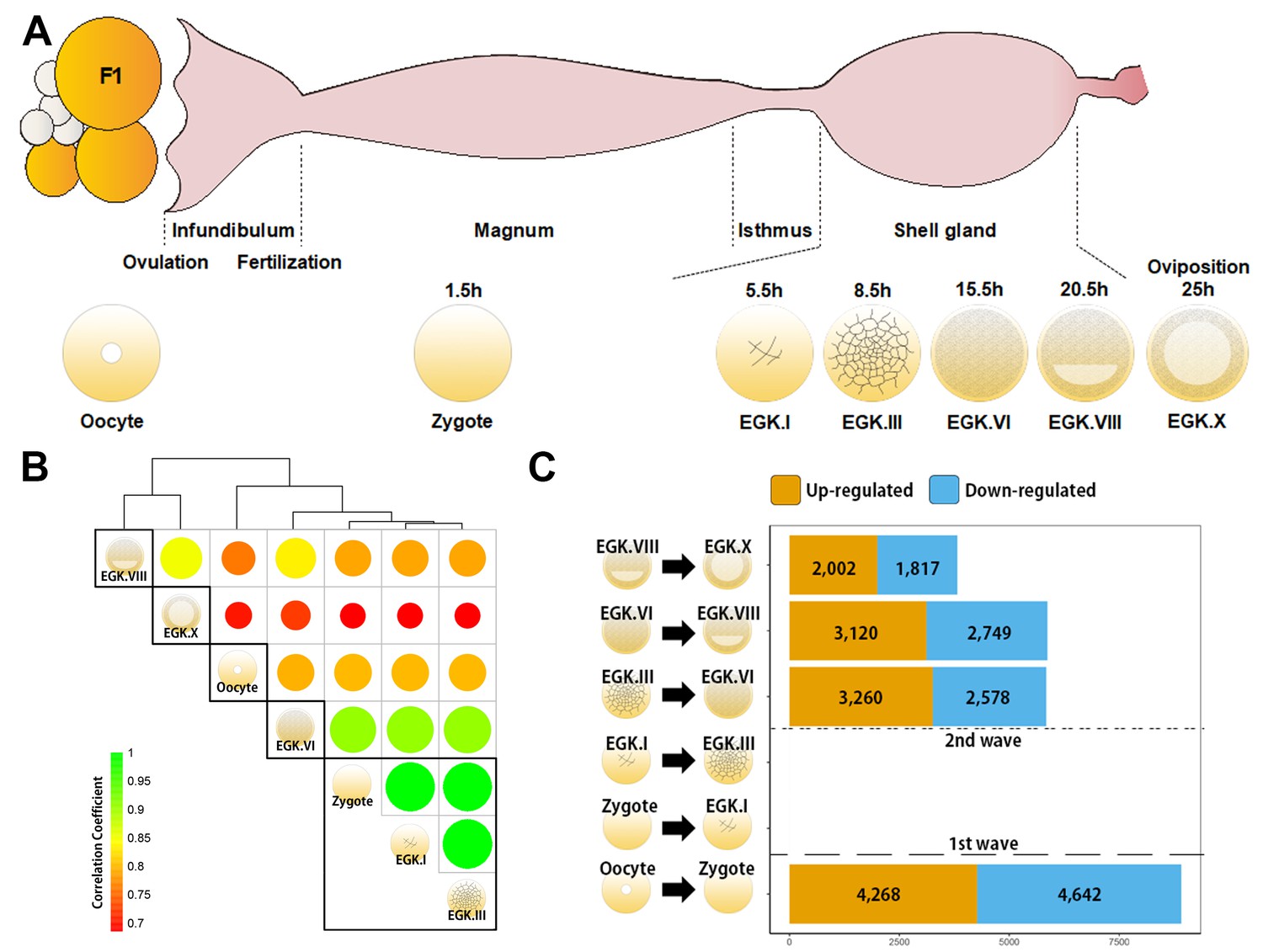
Genome-wide transcriptional activation during chicken early development.
(A) Representative images of early embryos from oocyte to Eyal-Giladi and Kochav X (EGK.X) used for RNA-Seq and acquisition in the chicken oviduct. All embryos were classified following the morphological criteria of EGK. h, hours after fertilisation for each stage of embryos. (B) Hierarchical clustering of the whole transcriptome during early development in chicken. The size and colour of each circle represents the strength of the correlation coefficients based on whole-transcriptome expression. The black rectangle represents optimal clusters (k = 5) based on the Silhouette score. The transcriptomic changes between consecutive stages, including oocyte vs. zygote and EGK.III vs. EGK.VI, are shown. Zygote, EGK.I and EGK.III had similar transcriptome profiles. (C) Number of differentially expressed intronic regions in consecutive stages. The orange and blue colors represent up- and downregulated genes at 5% significance level after false discovery rate (FDR) multiple testing adjustment. The 1st wave of transcriptional activation between oocyte and zygote and the 2nd wave between EGK.III and EGK.VI are shown.
A number of expressed regions exhibited significant differences between the oocyte and zygote and between EGK.III and EGK.VI (Figure 1—figure supplement 1). The number of expressed regions was reduced during EGK.I and EGK.III but increased after EGK.VI. Of all of the genomic regions that are expressed, the proportion of expressed intronic regions decreased after fertilisation and increased gradually after EGK.VI (Figure 1—figure supplement 2). Unlike the expression patterns seen during the minor ZGA in mammals (Abe et al., 2015), the proportion of expressed intergenic regions was constant regardless of transcriptional activation, indicating no expression of these regions during the 1st wave in chickens. In genic regions, large numbers of up- and downregulated mRNAs and long intergenic noncoding RNAs (lincRNAs) were observed during the 1st wave, while other RNAs were mostly downregulated after fertilisation (Figure 1—figure supplement 3), suggesting a potential role for long transcripts in the early cleavage stages. All RNA types were significantly upregulated during the 2nd wave.
We examined the candidate genes affected by the two waves using reverse transcription PCR (RT-PCR). Six upregulated genes in each wave were selected as representative genes (Supplementary file 1): DLX6, GATA2, ZIC4, LYPD2, IFITM5 and NKX6-3 for the 1st wave, and WNT11, WNT3A, C8ORF22, NAT8L, PCOLCE2 and AKAP2 for the 2nd wave. We successfully demonstrated two waves of transcriptional activation for all of the selected genes (Figure 2 and Figure 2—figure supplement 1). The validated genes belonging to the 2nd wave of activation indicated a lack of transcriptional activity during rapid cellularisation in the cleavage period, and showed that the 2nd wave of transcriptional activation in chicken occurred not between EGK.II and EGK.III, but between EGK.IV and EGK.V. The existence and timing of the two distinct waves of transcriptional activation were also confirmed experimentally and were consistent with the results of the bulked embryonic WTS analyses.

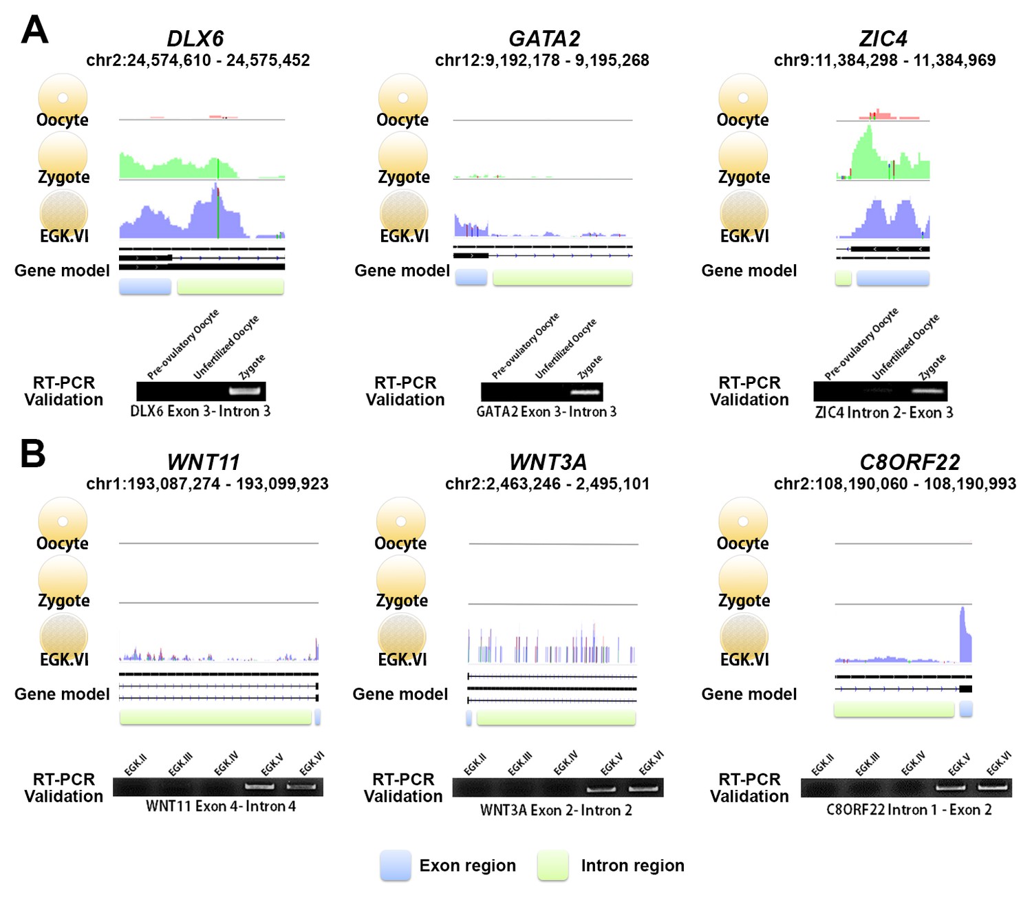
Exonic and intronic mapped reads on candidate genes related to the 1st and 2nd wave of transcriptional activation in chickens.
(A, B) The pooled mapped reads based on the stage (three samples in each stage) were visualised using the Integrative Genomics Viewer tool. Detection with RT-PCR of gene activation via the appearance of primary transcripts based on whole-transcriptome sequencing and validation of the intronic expression of three genes (DLX6, GATA2 and ZIC4) during the 1st wave (A) and of three different genes (WNT11, WNT3A and C8ORF22) during the 2nd wave (B). The following figure supplements are available for Figure 2.
We hypothesised that the haploid nucleus of supernumerary sperm could be substantially induced during the 1st wave in addition to paternal and maternal PN activation because polyspermic fertilisation occurs in avian species. To assess this hypothesis, we generated multiomics data including whole-genome sequencing (WGS) and WTS. We completed WGS of six parents (three male Korean Oge (mKO) and three female White Leghorn (fWL) chickens) to identify breed-specific single-nucleotide polymorphisms (SNPs) (Figure 3A). We also generated single embryonic WTS data from hybrid oocyte, zygote and EGK.X blastoderms derived from the WGS-sequenced parents to examine the characteristics of the 1st wave-activated transcripts and of allelic expression. After confirming hybrid embryo formation between mKO and fWL (Figure 3—figure supplement 1), we collected oocytes, zygotes and EGK.X blastoderms from hens on the same day (Figure 3—figure supplement 2). Each embryo contained an average of 2.1 µg of total RNA (Supplementary file 2A). We performed the same analysis used in bulked embryonic WTS on single embryonic WTS to further establish the characteristics of the 1st wave. The WTS samples generated from the single embryos were clustered according to their respective stages (Figure 3B). A total of 4275 differentially expressed mRNAs were detected (Figure 3C; FDR-adjusted p<0.05), among which 1883 were upregulated and 2392 were downregulated in the zygote stage compared to the oocyte. We also observed that 118 and 786 lincRNAs were up- and downregulated, respectively. Owing to the dramatic changes in early development between fertilisation and oviposition, 10,298 mRNAs and 2507 lincRNAs were differentially expressed between the zygote and EGK.X stages (Figure 3C). We also observed a large number of primary transcripts that are upregulated in the zygote stage when compared to the oocyte stage(Supplementary file 2B; FDR-adjusted p<0.05). These results once again demonstrate that primary transcriptional activation occurs as developmental stage moves from oocytes to zygotes at single-embryo resolution, in terms of the numbers of differentially expressed pre-mRNAs and long transcripts.

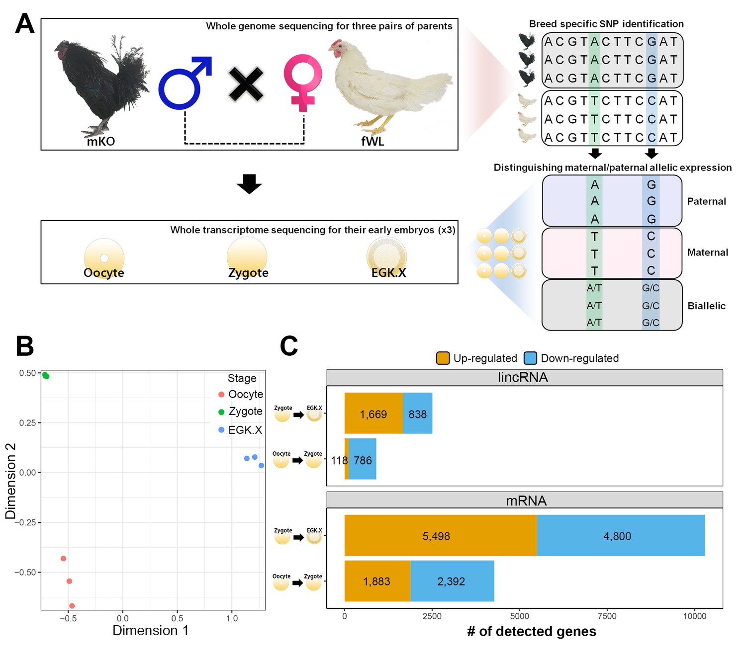
Whole-transcriptome analysis of single early chicken embryos.
(A) Schematic diagram of the experimental design using a multiomics approach to assess allelic expression. Three pairs of parental male Korean Oge (mKO) and female White Leghorn (fWL) chickens were subjected to whole-genome sequencing. Hybrid single embryos between mKO and fWL at the oocyte, zygote and EGK.X stages from each parent were subjected to whole-transcriptome sequencing. Allelic expression in the hybrid embryos was examined on the basis of breed-specific SNPs. (B) Multidimensional scaling (MDS) plot based on log2 trimmed mean of M-value (TMM) normalised gene expression of the whole transcriptome in pre-oviposited chicken embryos. Biological triplicates of single embryos were clustered, and three developmental stages were distinct. (C) Number of significantly detected long transcripts (mRNAs and lincRNAs) detected by comparing gene expression among single oocytes, zygotes and EGK.X embryos (FDR-adjusted p<0.05).
Next, we identified parental allele-specific expression patterns during the 1st wave of transcriptional activation. A total of 1544 parentally derived SNPs were detected, distributed across 424 transcripts including mRNAs and lincRNAs (Supplementary file 3A). Interestingly, all of the transcripts that were identified in the zygote stage exhibited maternally derived expression during the 1st wave (Figure 4A and Supplementary file 3A). Most of the maternally derived transcripts, except for seven mRNAs and two lincRNAs, were replaced as bi-allelic expression occurred in the EGK.X stage. These nine transcripts could be interpreted as residual maternal transcripts that were not activated during the 2nd wave, rather than as genomic-imprinted genes, which are not conserved in avian species (Frésard et al., 2014). To verify this observation, we selected six pre-mRNAs (MAP7D1, ESCO1, CCNB3, SYTL1, GRHL1 and LLGL1) that are upregulated during the 1st wave as representatives and validated the genotypes using Sanger sequencing (Figure 4B and Supplementary file 3B). All of the selected genes showed maternal allelic expression in the zygote until the EGK.VI stage, except for the GRHL1 gene. These maternally derived genes converted to bi-allelic expression after the maternal-to-zygotic transition (MZT) at EGK.X. This phenomenon is distinguished from that in mammals, in which transcriptional activity in the paternal PN is two times greater than that in the maternal PN (Aoki et al., 1997). These results indicate that there is no possibility that the activated transcripts are derived from the supernumerary sperm nuclei and paternal PN, in contrast to the data from mammals (Aoki et al., 1997; Bouniol et al., 1995).

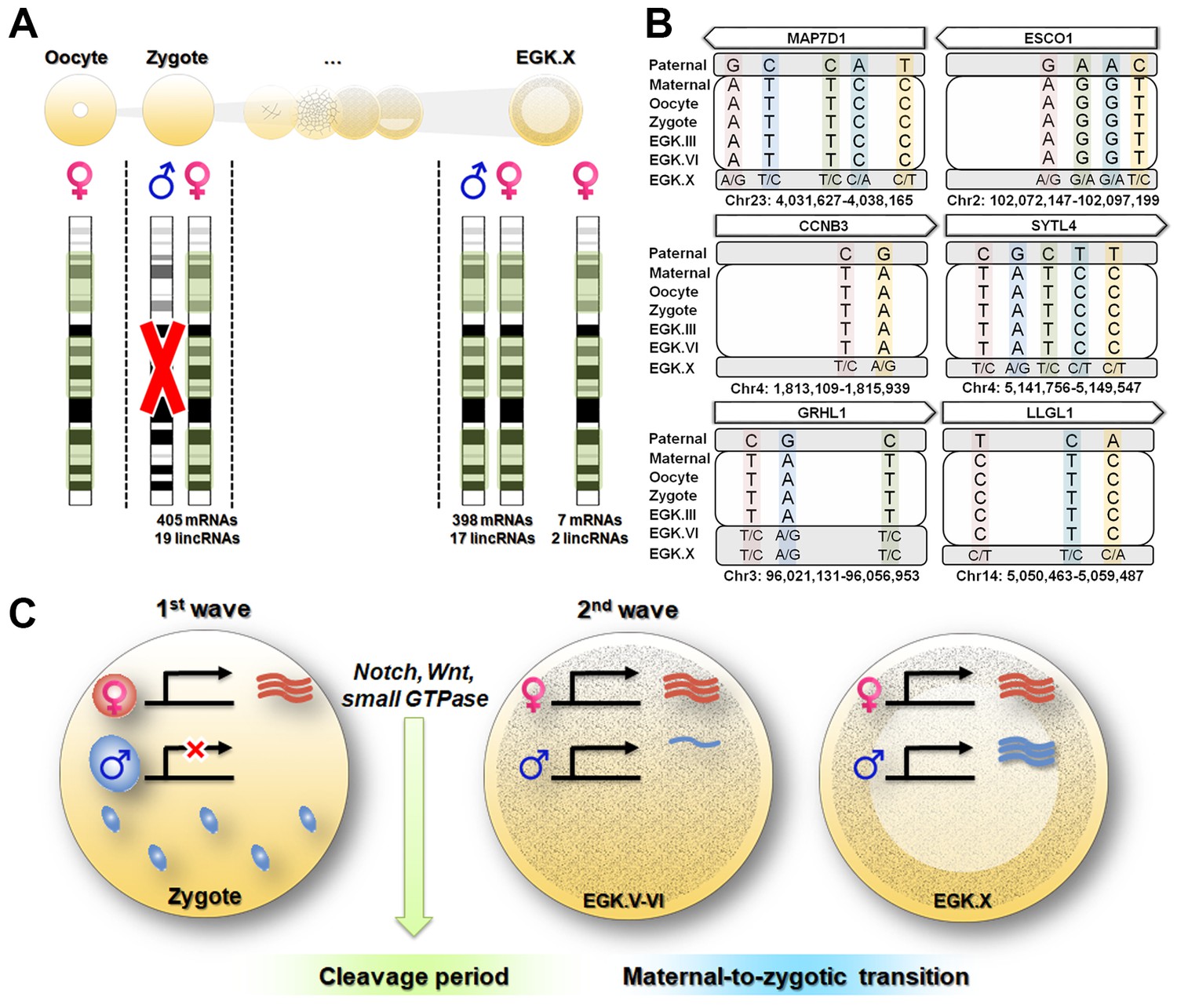
Maternal genome activation (MGA) during the 1st wave of transcriptional activation in chicken zygote.
(A) Determination of parental allelic expression from the zygote stage. Only maternal alleles were observed in transcripts induced by stst activation. These maternally derived upregulated genes showed bi-allelic expression after EGK.X. (B) Validation of 1st wave transcription-induced maternal allelic expression by Sanger sequencing. The maternal transcription profile after the 1st wave changed to bi-allelic expression between EGK.VI and EGK.X after the 2nd activation. (C) Schematic summary of genome activation during chicken early development. Only MGA occurred after fertilisation and this wave of gene activation may regulate the cleavage period.
We examined the functional characteristics of the maternal genes that are activated during the 1st wave of transcriptional activation identified from the single embryonic WTS data. The analysis revealed that the 1st wave-activated maternal transcripts were enriched in the following pathways: cell cycle; Notch signalling pathway; Wnt signalling pathway; regulation of transcription, DNA-templated; and regulation of small GTPase-mediated signal transduction (Figure 4—figure supplement 1 and Supplementary file 4). These pathways were activated from the maternal genome and are involved in rapid asymmetric cellularisation during the cleavage period in chickens (Hwang et al., 2018c) and other species (Castanon et al., 2013; Huang et al., 2015; Priess, 2005; Tse et al., 2012; Zhang et al., 2014). While the 1st wave in mice promotes the low-level expression of numerous non-functional genes (Abe et al., 2015), the maternal genes activated during the 1st wave in chickens seem to be related to early cell division in embryogenesis.
As demonstrated in previous studies, the characteristics of the 1st wave vary among species (Abe et al., 2015; Anderson et al., 2017; Chen et al., 2017; Heyn et al., 2014). Our results suggest the exclusive activation of maternal alleles after fertilisation in chicken (Figure 4C). However, after MZT, most expressed genes were derived from both paternal and maternal genomes. Functionally, transcripts affected by the 1st wave were involved in asymmetric rapid cellularisation and in the fundamental regulation of further development (Figure 4C). We speculate that this evolved by necessity in animals following physiological polyspermy (Figure 4—figure supplement 2). Polyspermic animals require a number of sperm to activate large eggs (Iwao, 2012). In addition to pathological mitosis (Snook et al., 2011), polyspermic embryos of sea urchin demonstrated that transcriptional activation after fertilisation was greatly stimulated by the PN of supernumerary sperm (Poccia et al., 1985). Such a disproportionate genome contribution could result in an excessive amount of transcription. The total polyspermy number reportedly varies (Hemmings and Birkhead, 2015; Lee et al., 2013a) and is positively correlated with egg size (Birkhead et al., 1994). Individual sperm provide genomic diversity (Wang et al., 2012) but could result in genomic instability if different types of transcripts are expressed by various sperm nuclei. Therefore, polyspermic animals may have evolved means of inhibiting the activation of the paternal PN to control gene expression levels from the 1st wave. Taken together, our results suggest that the maternally derived 1st wave is essential for early development and evolutionary outcomes in avian species.
Materials and methods
Reagent type (species) or resource | Designation | Source or reference | Identifiers | Additional information |
---|---|---|---|---|
Sequence-based reagent | Breed-specific primers | (Choi et al., 2007) | See elsewhere in 'Materials and methods' | |
Sequence-based reagent | RT-PCR primers | This paper | See Supplementary file 6 | |
Commercial assay or kit | DNeasy Mini Kit | Qiagen | Qiagen:69504 | |
Commercial assay or kit | TRIzol reagent | Invitrogen | Invtirogen:15596026 | |
Commercial assay or kit | SuperScript III First-Strand Synthesis System | Invitrogen | Invitrogen:18080051 | |
Commercial assay or kit | pGEM-T Easy Vector Systems | Promega | Promega:A1360 | |
Software, algorithm | Code used for RNA-seq quantification analysis | This paper | The python code used for RNA-seq quantification analysis. See Source code 1 |
Experimental animals and animal care
Request a detailed protocolThe experimental use of chickens was approved by the Institute of Laboratory Animal Resources, Seoul National University (SNU-150827–1). The experimental animals were cared for according to a standard management program at the University Animal Farm, Seoul National University, Korea. The procedures for animal management, reproduction and embryo manipulation adhered to the standard operating protocols of our laboratory.
Identification of differentially expressed regions during early developmental stages of chickens
Request a detailed protocolTo detect de novo transcription, the analytical approach to primary transcripts used in previous studies of other species (Abe et al., 2015; Graf et al., 2014; Lee et al., 2013b; Paranjpe et al., 2013) was followed. In the quantification step, four types of genomic regions were considered: transcripts, exons, introns and intergenic regions. Although quantification of the transcript and exon level can be achieved directly without any pre-processing steps by using the galGal4 gene annotation file (GTF), the genomic position needs to be defined in order to estimate the expression levels of the intron and intergenic regions. When defining intron area, overlapping annotation of the exon within the associated gene makes it difficult to define intron regions from the reference genome. In addition, information from different strands should be considered when defining intron regions between each exon. To address these issues, intron region was defined using custom python script (Source code 1). As in the method used to define the intron region between exons within the associated gene, python script was used to define intergenic regions between genes within the same chromosome. After defining intronic and intergenic regions, a GTF was generated using the coordinate information. Expression levels were measured with HTSeq-count (v 0.6.1) on the basis of the the GTFs (Anders et al., 2015b).
To explore gene expression changes during early developmental stages, pre-existing bulked embryonic WTS data covering the oocyte, zygote, EGK.I, EGK.III, EGE.VI, EGK.VIII and EGK.X stages (GSE86592) (Hwang et al., 2018bHwang et al., 2018c) were employed. Three types of matrix data were generated, and these data were employed in statistical analyses. Six statistical tests, oocyte vs. zygote, zygote vs. EGK.I, EGK.I vs. EGK.III, EGK.III vs. EGK.VI, EGK.VI vs. EGK.VIII and EGK.VIII vs. EGK.X, were performed using the edgeR package (Robinson et al., 2010) in the matrix data derived from intron and intergenic regions separately. More detailed contrast tests were performed on the generalised linear model. In this study, a result was considered significant at a FDR-adjusted p-value of p<0.05 (Benjamini and Hochberg, 1995).
Genomic DNA isolation and DNA sequencing library preparation for WGS data
Request a detailed protocolGenomic DNA was isolated from blood collected from the wing vein of six parental chickens (three mKO and three fWL) using 1 mL 30-gauge syringes (Shina Corporation, Seoul, Korea). The blood samples were transferred into EDTA tubes (BD Biosciences, San Jose, CA, USA) immediately after collection. Blood (10 µL) was used for isolation of genomic DNA using a DNeasy Mini Kit (Qiagen, Valencia, CA, USA). The quality of the extracted genomic DNA was determined using the Trinean DropSense96 system (Trinean, Gentbrugge, Belgium), RiboGreen (Invitrogen, Carlsbad, CA, USA) and an Agilent 2100 Bioanalyzer (Agilent Technologies, Santa Clara, CA, USA). Genomic DNA was used for the construction of cDNA libraries using a TruSeq Nano DNA LT Library Preparation Kit (Illumina Inc., San Diego, CA, USA). The resulting libraries were subjected to chicken genome resequencing (30 × coverage) using the Illumina Nextseq 500 platform to produce paired 150 bp reads. The raw sequencing data were deposited in BioProject under accession number PRJNA393895.
RT-PCR for confirmation of hybrid embryos
Request a detailed protocolBefore collecting early embryos, EGK.X blastoderms formed from crosses between mKO and fWL were incubated in a chamber at 37.5°C under 80% humidity for 18 hr. Genomic DNA was isolated from Hamburger and Hamilton stage 4 (HH4) (Hamburger and Hamilton, 1951) embryos using a DNeasy Mini Kit (Qiagen). RT-PCR was performed to confirm hybridisation between KO and WL using breed-specific primers (AS3554-I9/P5FWD WL F: 5′-AGC AGC GGC GAT GAG CGG TG-3′; WL R: 5′-CTG CCT CAA CGT CTC GTT GGC-3’; AS3554-WT/P5FWD KO F: 5’-AGC AGC GGC GAT GAG CAG CA-3′; KO R: 5′-CTG CCT CAA CGT CTC GTT GGC-3′) (Choi et al., 2007), with an initial incubation at 95°C for 10 min, followed by 35 cycles of 95°C for 30 s, 69°C for 30 s and 72°C for 30 s. The reaction was terminated after a final incubation at 72°C for 10 min.
Alignment and variant calling for WGS data
Request a detailed protocolThe paired-end reads for six chickens (three biological replications of mKO and fWL breeds) were generated using the Illumina Nextseq 500 platform. In total, 8.38 billion reads or ~2.53 Gbp of sequences were generated. Paired-read sequences were selected for quality using Trimmomatic (v0.33) (Bolger et al., 2014). Using Bowtie 2 (v2.2.5) (Langmead and Salzberg, 2012), reads were aligned to the reference genome sequence galGal4 (Build v 4.82) with an average alignment rate of 91.61%. After potential PCR duplicates were filtered and misalignments resulting from the presence of insertions and deletions (INDELs) were corrected, SNPs were detected using GATK v3.4.46 (McKenna et al., 2010). More detailed, potential PCR duplicates were filtered using the option ‘REMOVE_DUPLICATES = true’ in the ‘MarkDuplicates’ open-source tool of Picard (v 1.138) (https://broadinstitute.github.io/picard/). SAMtools (v1.2) (Li et al., 2009) was then employed to create index files for reference and Binary Alignment/Map (BAM) files. In the variant-calling step with GATK v3.1, local realignment of reads to correct misalignments was performed because of the presence of INDELs (‘RealignerTargetCreator’ and ‘IndelRealigner’ arguments). In the GATK tool, two types of arguments, ‘UnifiedGenotyper’ and ‘SelectVariants’ were employed for variant calling. In addition, ‘VariantFiltration’ was applied to filter bad variants on the basis of the following criteria: (1) variants with a Phred-scaled quality score <30 were filtered; (2) SNPs with ‘mapping quality zero (MQ0) >4’, ‘quality depth <5’ and ‘(MQ0 / (1.0*DP))>0.1’ were filtered; and (3) SNPs with ‘Phred-scaled P value using Fisher’s exact test >200’ were filtered. As a result, 10,529,469 variants were detected, of which 9,805,997 variants (93.129%) were previously known variants (Supplementary file 5A, B).
Chicken early hybrid embryo preparation, RNA isolation and library preparation for single embryonic WTS data
Request a detailed protocolThe egg-laying times of three fWLs, which were mated with mKOs, were recorded. A single hybrid EGK.X blastoderm was collected from WL hens after oviposition. To collect single oocytes and hybrid zygotes, WL hens were sacrificed and their follicles were harvested. Oocytes and hybrid zygotes were collected simultaneously from one WL hen. Owing to the small transcriptomic differences between pre- and post-ovulatory oocytes observed in the previous study (Elis et al., 2008) and the infeasibility of simultaneous acquisition of post-ovulatory oocytes and zygotes from a single hen, only the pre-ovulatory large F1 oocyte was isolated. Only zygote embryos not showing cleavage and located in the magnum were collected within 1.5 hr after fertilisation, according to the recorded egg-laying times (Figure 3—figure supplement 2). All embryos were classified according to morphological criteria (Figure 1A). Shortly after collection, the embryos were separated from the egg using sterile paper, and the shell membrane and albumen were detached from the yolk. A piece of filter paper (Whatman, Maidstone, UK) with a hole in the centre was placed over the germinal disc. After cutting around the paper containing the embryo, it was gently turned over and transferred to saline to further remove the yolk and vitelline membrane to allow embryo collection. Total RNA was isolated from early embryos using TRIzol reagent (Invitrogen). The quality of the extracted total RNA was determined using the Trinean DropSense96 system (Trinean), RiboGreen (Invitrogen) and an Agilent 2100 Bioanalyzer (Agilent Technologies). Total RNA was used for construction of cDNA libraries using a TruSeq Stranded Total RNA Sample Preparation Kit (Illumina, Inc.). The resulting libraries were subjected to whole-transcriptome analysis using the Illumina Nextseq 500 platform to produce paired 150 bp reads. The raw sequencing data were deposited in Gene Expression Omnibus (GEO) under accession number GSE100798.
Quality control, alignment and quantification of mapped reads for single embryonic WTS data
Request a detailed protocolTrimmomatic (v 0.33) (Bolger et al., 2014) was used to generate clean reads. Per-base sequence qualities were checked using FastQC (v 0.11.2) (Andrews, 2010) and filtered fastq files. Trimmed reads were aligned to the galGal4 genome files using the HISAT2 alignment software (v 2.0.0) (Kim et al., 2015) with the following alignment option: ‘--rna-strandness RF’. Sequence Alignment/Map (SAM) files were converted into compressed and sorted BAM files using SAMtools (v 1.4.1) (Li et al., 2009). The mapped reads were quantified using HTSeq-count (Anders et al., 2015a) with the merged GTF, with total RNAs and lincRNAs derived from Ensembl and ALDB (Li et al., 2015), respectively. The quantification of mapped reads on intronic regions for single embryonic WTS data was performed using the procedure also used for bulked embryonic WTS data.
Variant calling RNA-Seq
Request a detailed protocolUsing the alignment file (.BAM), potential PCR duplicates were removed using the Picard (v 1.138) software with ‘REMOVE_DUPLICATES = true’ in the ‘MarkDuplicates’ option. After that, the SplitNCigarReads tool implemented in GATK was performed with the ‘-rf ReassignOneMappingQuality -RMQF 255 -RMQT 60 U ALLOW_N_CIGAR_READS’ option. In the variant-calling step with GATK, local realignment of reads was performed to correct misalignments (using the ‘RealignerTargetCreator’ and ‘IndelRealigner’ options). Finally, base-recalibration was performed using BaseRecalibrator implemented in GATK with known variant sites in galGal4. Using HaplotypeCaller in the GATK tool, variant calling was performed with the ‘-dontUseSoftClippedBases -stand_call_conf 20.0 -stand_emit_conf 20.0’ option. Finally, bad variants were filtered using the VariantFiltration tool with ‘-window 35 -cluster 3 -filterName FS -filter ‘FS >30.0’ -filterName QD -filter ‘QD <2.0’’ option. At the end of this process, 265,788 variants were detected, of which 248,030 variants (93.319%) were previously known sites (Supplementary file 5C, D).
Identification of the maternally and paternally expressed genes through detection of breed-specific variants
Request a detailed protocolMaternal and paternal samples were genotyped using WGS, and their offspring, including maternal oocytes, were genotyped using WTS (variant calling on the RNA-Seq data). After pre-processing, there were two types of genotype data (DNA and RNA sequencing data) available for the mother, father, oocyte, zygote and EGK.X. In two types of SNP data, 10,529,469 and 265,788 variants were detected in DNA and RNA sequencing data, respectively. First, breed-specific SNPs (such as , first, SNPs ‘0/0’ and ‘1/1’ genotype for maternal and paternal groups, respectively; and second, SNPs ‘1/1’ and ‘0/0’ genotype for maternal and paternal groups, respectively) were identified and annotated using SnpSift (Cingolani et al., 2012) in parental SNP data. As a result, 216,003 SNPs were identified as breed-specific SNPs. After that, two SNP datasets (breed-specific SNPs and their offspring genotypes derived from the RNA-Seq data) were combined to detect maternally and paternally expressed genes, and 14,817 SNPs were commonly identified in breed-specific SNPs and those derived from RNA-Seq data. Using these combined genotype data, three types of filtering steps were carried out. First, mismatched genotypes of the reference and alternative allele between breed-specific SNPs and SNPs derived from the RNA-Seq were removed; two variants were removed in this step. Second, different genotypes within the biological replicates were removed; 9,143 SNPs were removed in this step. Finally, mismatched genotypes between maternal samples and oocyte samples were removed; six SNPs were removed in this step. The remaining 5,666 SNPs were annotated using the SnpSift tool with galGal4 and ALDB GTFs. To find the most conservative evidence of parental expression, if a single SNP was found within the gene or genotype pattern that was not consistent among the SNPs, it was filtered out. In addition, unannotated SNPs in both databases, Ensembl and ALDB, were removed to facilitate biological interpretation. At the end of this process, 1,544 SNPs were detected as parental expression markers, all of which showed a maternal expression pattern (Supplementary file 5E).
Identification of functional characteristics of differentially expressed genes
Request a detailed protocolOn the basis of the biological process terms (BP terms) of the GO and KEGG pathways, functional enrichment tests using DAVID (Dennis et al., 2003) were performed on the differentially expressed genes.
Exon–intron RT-PCR and validation of allelic expression
Request a detailed protocolTotal RNA (1 µg) was used as the template for cDNA synthesis using the SuperScript III First-Strand Synthesis System (Invitrogen). The cDNA was serially diluted 5-fold and equalised quantitatively for PCR amplification. To validate allelic expression, additional single hybrid embryos at EGK.III and VI were collected from parents with identical genotypes as confirmed by WGS, and their total RNA isolation and cDNA synthesis were performed as described above. Primers for exon–intron PCR of 12 genes and for allelic expression of six genes were designed using the program Primer3 (Untergasser et al., 2012) (Supplementary file 6A, B). RT-PCR was performed with an initial incubation at 95°C for 5 min, followed by 35 cycles of 95°C for 30 s, 59°C for 30 s and 72°C for 30 s. The reaction was terminated after a final incubation at 72°C for 5 min. PCR products were cloned into the pGEM-T Easy Vector (Promega, Madison, WI, USA) for sequencing with an ABI 3730xl DNA Analyzer (Applied Biosystems, Foster City, CA, USA).
Data availability
Generated WGS of parental chickens has been deposited in BioProject under accession number PRJNA393895 (https://www.ncbi.nlm.nih.gov/bioproject/?term=PRJNA393895). Generated single hybrid embryonic WTS data has been deposited in GEO under accession number GSE100798 (https://www.ncbi.nlm.nih.gov/geo/query/acc.cgi?acc=GSE100798). Published bulked embryonic WTS data are available under accession number GSE86592 (https://www.ncbi.nlm.nih.gov/geo/query/acc.cgi?acc=GSE86592).
-
NCBI Gene Expression OmnibusID GSE100798. Avian zygote activates only maternal allele to disburden high variation of supernumerary sperms contrary to mammal.
-
NCBI BioProjectID PRJNA393895. Avian zygote activates only maternal allele to disburden high variation of supernumerary sperms contrary to mammal.
-
NCBI Gene Expression OmnibusID GSE86592. Developmental programs in chicken early embryos by whole transcriptome analysis.
References
-
SoftwareA quality control tool for high throughput sequence dataFastQC.
-
Controlling the false discovery rate: a practical and powerful approach to multiple testing.Journal of the Royal Statistical Society Series B pp. 289–300.
-
A comparative study of sperm-egg interactions in birdsReproduction 101:353–361.https://doi.org/10.1530/jrf.0.1010353
-
Trimmomatic: a flexible trimmer for illumina sequence dataBioinformatics 30:2114–2120.https://doi.org/10.1093/bioinformatics/btu170
-
Endogenous transcription occurs at the 1-cell stage in the mouse embryoExperimental Cell Research 218:57–62.https://doi.org/10.1006/excr.1995.1130
-
Anthrax toxin receptor 2a controls mitotic spindle positioningNature Cell Biology 15:28–39.https://doi.org/10.1038/ncb2632
-
Zygotic genome activation occurs shortly after fertilization in maizeThe Plant Cell 29:2106–2125.https://doi.org/10.1105/tpc.17.00099
-
Identification of breed-specific DNA polymorphisms for a simple and unambiguous screening system in germline chimeric chickensJournal of Experimental Zoology Part A: Ecological Genetics and Physiology 307:241–248.https://doi.org/10.1002/jez.373
-
Transcriptome-wide investigation of genomic imprinting in chickenNucleic Acids Research 42:3768–3782.https://doi.org/10.1093/nar/gkt1390
-
A series of normal stages in the development of the chick embryoJournal of Morphology 88:49–92.https://doi.org/10.1002/jmor.1050880104
-
Polyspermy in birds: sperm numbers and embryo survivalProceedings of the Royal Society B: Biological Sciences 282:20151682.https://doi.org/10.1098/rspb.2015.1682
-
Egg activation in physiological polyspermyREPRODUCTION 144:11–22.https://doi.org/10.1530/REP-12-0104
-
HISAT: a fast spliced aligner with low memory requirementsNature Methods 12:357–360.https://doi.org/10.1038/nmeth.3317
-
Fast gapped-read alignment with Bowtie 2Nature Methods 9:357–359.https://doi.org/10.1038/nmeth.1923
-
Zygotic genome activation during the maternal-to-zygotic transitionAnnual Review of Cell and Developmental Biology 30:581–613.https://doi.org/10.1146/annurev-cellbio-100913-013027
-
SMART 7: recent updates to the protein domain annotation resourceNucleic Acids Research 40:D302–D305.https://doi.org/10.1093/nar/gkr931
-
The sequence alignment/Map format and SAMtoolsBioinformatics 25:2078–2079.https://doi.org/10.1093/bioinformatics/btp352
-
Transcription and polyadenylation processes during early development of quail embryoJournal of Embryology and Experimental Morphology 79:11–24.
-
RNA synthesis in male pronuclei of the sea urchinBiochimica et Biophysica Acta (BBA) - Gene Structure and Expression 824:349–356.https://doi.org/10.1016/0167-4781(85)90042-9
-
The maternal-to-zygotic transition: a play in two actsDevelopment 136:3033–3042.https://doi.org/10.1242/dev.033183
-
Primer3--new capabilities and interfacesNucleic Acids Research 40:e115.https://doi.org/10.1093/nar/gks596
Decision letter
-
Claudio D SternReviewing Editor; University College London, United Kingdom
-
Patricia J WittkoppSenior Editor; University of Michigan, United States
In the interests of transparency, eLife includes the editorial decision letter and accompanying author responses. A lightly edited version of the letter sent to the authors after peer review is shown, indicating the most substantive concerns; minor comments are not usually included.
[Editors’ note: a previous version of this study was rejected after peer review, but the authors submitted for reconsideration. The first decision letter after peer review is shown below.]
Thank you for submitting your work entitled "The only maternal genome is activated in avian zygotes after fertilization" for consideration by eLife. Your article has been reviewed by three peer reviewers, and the evaluation has been overseen by a Reviewing Editor and a Senior Editor. The following individual involved in review of your submission has agreed to reveal their identity: Claudio D Stern (Reviewer #1).
Our decision has been reached after consultation between the reviewers. Based on these discussions and the individual reviews below, we regret to inform you that your work will not be considered further for publication in eLife at this time.
As you will see in the reviews below, the reviewers found many positive aspects of the study and think it may still be suitable for publication in eLife; however, ambiguity about which findings are novel here relative to other recent papers must be addressed before this work can be considered further. For example, you'll see one of the reviewers interpreted the two waves of zygotic activation as being reported for the first in this study. I appreciate that you have delineated the differences among your recent papers to the eLife staff, but the paper itself will also need to be substantially revised to more clearly focus only on the novel findings that are uniquely presented here. The reviewers need to see this revised version before they can assess suitability for eLife, thus we are rejecting this work now, but leaving the door open for resubmission of a revised version as a new manuscript. This revision must also address the other concerns raised in the reviews below, including verification of the sequence analysis pipeline. The manuscript should also be carefully evaluated for proper English, as grammatical problems make it hard to understand in some places; even the title is confusing as written.
Reviewer #1:
This is potentially an important paper exploring the timing of first activation of the zygotic genome in an avian embryo, the chick (Gallus gallus). Using a carefully designed set of experiments (mainly transcriptomics, also taking advantage of crosses between two different strains of parents with distinctive SNPs), the authors reveal that there are two separate waves of zygotic gene activation (ZGA): a first major wave occurring soon after fertilization, in which only genes located in maternally-derived chromosomes are activated, and this is followed some hours later (stage EGK V) where genes on both maternally- and paternally-derived chromosomes become active.
Overall I think that this is a very carefully conducted study and that it reveals very important information which was lacking until now about when the zygotic genome is activated in the chick embryo, an important model system. A peculiarity of avian species is that fertilization is highly polyspermic and the authors speculate that these two waves of genome activation, where the paternally-derived genes are activated comparatively late, is a consequence of polyspermy. In anamniotes (including invertebrates as well as Amphibians and fishes), the zygotic genome is only activated late, after about 10 synchronous cell divisions (at the mid-blastula stage, a process generally known as the mid-blastula transition MBT); therefore any patterning and cell commitment events until this time must depend on inherited maternal determinants since there cannot be differential gene expression before MBT. This paper is important because it shows that, as in mammals, the bird genome is indeed activated very early (but with the peculiarity that only maternally-derived genes are turned on first), which allows differential gene expression almost from the start. Interestingly, among the genes/pathways enriched in the first wave of zygotic gene expression, are genes involved in intercellular cell signalling, especially Notch and Wnt pathways. This suggests again that early patterning events in the amniote embryo rely on zygotic, rather than maternal gene products.
I only have one major comment: the paper is very poorly written and the English needs a lot of attention. In places it is almost impossible to understand (even the title makes no sense, as are parts of the Abstract). It is essential that the authors seek help from native English speakers who could help to make this paper readable. I find myself unable to criticise it more deeply because there are several parts which I don't understand.
When re-writing, please be careful to distinguish very clearly between transcription from "maternally-derived genes", and "maternal determinants" (i.e. gene products inherited from the mother, as in anamniotes before ZGA) – because the concept of MBT and late activation of the zygotic genome in many model species is so engrained in the developmental literature, there is some danger than some readers may be confused by these to quite different concepts/findings.
Reviewer #2:
In this manuscript the authors analyse the transcriptome of chicken embryos at pre-oviposition stages. Whole genome RNA-seq analysis was carried out on mature follicles and early EGK stages. The authors found abundant maternal RNAs and increases in transcripts at two separate developmental stages: the zygote and EG V. In previous work by these authors, they used the RNA from these developmental stages to define expression clusters that changed during early uterine development (Hwang, 2018). In this manuscript the authors used interbreed crosses to assess paternal and maternal transcripts. They found that maternal transcripts only increased after the oocyte state and conclude that the paternal genome is not activated at early stages. The authors hypothesise that polyspermy in bird species has led to the transcription silencing of the paternal genome during early development. The hypothesis is intriguing but the paper needs revision and statistical analysis to determine if the hypotheses are proven.
1) Not enough information is given on the bioinformatics analysis of variants presented for the data in Supplementary file 5E. Allele depth, read depth and SNP quality needs to be presented.
2) How are the 8 gene expression clusters identified in the author's previous paper (Huang, 2018) related to the DEGs identified in this paper?
3) My concern is the ability to distinguish between maternal transcripts in the early zygote and de novo RNA transcription. This is premised by the author's statement:
'We also investigated primary transcripts throughout early development to obtain definitive evidence of gene activation as the massive alteration of maternally stored RNAs after fertilization may have masked the smaller effects of the 1st activation.'
The presence of maternal RNAs is clouding the analysis of de novo transcription. Can the authors identify maternal transcripts through looking at unmapped reads from the 3' end of follicle transcripts and either de novo transcript assembly on these follow by a molecular biology analysis of the transcripts to verify the maternal 3' UTR patterns?
4) I believe the title is miswritten. 'Only the maternal genome is transcribed in avian zygotes after fertilisation'.
5) There are several problems with nomenclature. Referring to avian zygotes suggests that several avian species were examined. The authors only examined chicken (Gallus gallus). We do not know if this mechanism is conserved in other bird species.
6) The English needs to be improved. Sections of the paper are difficult to understand.
7) Figure 1A: A time scale is needed. Chicken oviposition requires ~20-24 hours. An approximate stages of EGK should be given.
Reviewer #3:
The authors Hwang et al., have submitted a manuscript entitled 'The only maternal genome is activated in avian zygotes after fertilization'. In regards to the mechanisms of the zygotic genome activation, very few reports are available in avian species comparatively to the mammalian ones. This report provides significant data and advances to enlight the transcription activation of both maternal and paternal genomes with two waves of activation.
The manuscript is clear and the hypothesis well supported by the data even if few questions remain to be answered by the authors.
- On Figure 1, (Results and Discussion, second paragraph) some of the observed changes of the number of expressed regions are hypothesized to be due to large-scale degradation of maternal transcripts. How the authors do support this hypothesis in the absence of functional tests? What would be the mechanisms and the signal on the transcripts to be recognized for being degraded ? A specific signature?
- Similarly the hypothesis of the stable maternal transcripts (Results and Discussion, seventh paragraph) should be detailed.
- On Figure 2, the two waves of transcriptional activation are detailed and illustrated by the choice of 3 genes for each stage transition. However, the choice of those genes is not clearly justified and why 'only' 3 ones? Some genes associated to defined lineages (epiblast or germ lineage for example) could be also chosen and compared during the establishment of those events.
- As illustrated on Figure 4, some of the signaling pathways – (Results and Discussion, eighth paragraph) were primarily upregulated during the first stages but then down regulated in contrast to the other identified GO terms. Could the authors detail the signaling pathways that emerge in the second wave.
- In the Supplementary file 4, several GO terms are linked to the DNA replication, cell cycle and active proliferation of the cells, with a high p value. Even if it could be expected as the embryo is engaged into an active cleavage and cellularization process, the authors do not mention those facts in the manuscript. Could it be corrected ? Is the embryonic transcription silent for those specific genes?
- Similarly, in Supplementary file 5B, the list of TFs should perhaps be presented completely with the gene names, in particular, the Zn-C2H2 genes as only few of them appear to be expressed (on the 721 identified through HGNC list. More details should be provided to illustrate the specificity – or not – of those first transition.
- As a general question, the repetitive sequences are present in the avian genome as in other genomes and their regulation is also highly dependent on the developmental stages as demonstrated in several species. The authors should at least mention those facts and explain why they excluded them from their analysis.
As a conclusion the manuscript is highly important for the avian field and only few comments have probably to be addressed before being considered for publication.
[Editors’ note: what now follows is the decision letter after the authors submitted for further consideration.]
Thank you for submitting your article "Only the maternal genome is transcribed in chicken zygote upon fertilisation" for consideration by eLife. Your article has been reviewed by three peer reviewers, one of whom is a member of our Board of Reviewing Editors, and the evaluation has been overseen by Patricia Wittkopp as the Senior Editor. The reviewers have opted to remain anonymous.
The reviewers have discussed the reviews with one another and the Reviewing Editor has drafted this decision to help you prepare a revised submission.
Summary:
Overall, you will see that two of the reviewers essentially raise the same points as each other. They mainly relate to the clarity of the writing style and how previous work, both from your lab and from others (including other species) is treated. There is some question as to the degree to which the findings described are novel or unusual enough to merit publication in a journal of the calibre of eLife. For this reason, we have decided to invite you to submit a final revised version of your paper provided that you can really address these concerns. On balance and in agreement with the reviewers, I feel that the resulting paper could be both more scholarly and shorter, emphasizing the aspects that are truly novel and interesting in a more direct way.
We agree that If this early wave is real, biologically meaningful, RNA polymerase-II driven transcription that only occurs from the maternal genome and not simply noise, this would be a fascinating new discovery. However, with the current presentation of the work, the reviewers were not quite convinced. They felt that the conclusion the authors wish to draw is too important to be presented in this difficult to decipher and experimentally incomplete format.
Essential revisions:
Major re-writing and re-structuring. The literature on the maternal/zygotic transition and differential use of maternal and paternal genomes both in avian and in other species must be incorporated as past work/background and the novel aspects clearly emphasized.
Reviewer #1:
I read the previous version of this paper and I feel that the manuscript has improved. However some problems remain. The main one is that the English can still do with some improvement. Specifically, even the main message of the paper, that the maternal (but not paternal) genome is activated in a first wave of ZGA in chicken, is difficult to extract from the writing style. Here are two examples:
Abstract: "Surprisingly, maternal genome activation was exclusively found in the zygote stage.…". The grammar is very misleading. It should read "Surprisingly, only the maternal genome was found to be activated in the zygote stage.…" (and they could even add, for greater clarity "…, the paternal genome remaining silent until the XXX stage").
Title: "Only the maternal genome is transcribed in chicken zygote upon fertilisation". This is somewhat clearer than the Abstract, but the latter part of the title does not make it clear that this is a transient situation – it sounds as if the paternal genome is never activated. A possible alternative might be: "Zygotic gene activation in chicken occurs in two waves, the first involving only the maternally-derived genome".
I understand that this is a complex situation to describe for non-native English speakers but it is crucial for the message of this paper to come across clearly. More editing is still required throughout the manuscript to improve clarity.
Lastly, I still feel that the novelty of the work does not emerge very clearly from what has already been published by this group and by others. Findings reported in previous work should not be repeated as being new but only referred to with a literature reference. When previously published data were re-analyzed to make new findings, this should be stated explicitly. The methods should not repeat the description of how previously published data were generated but state that they were the same set, reanalyzed and then describe how. Doing these things will probably help to condense the paper substantially but also to focus the main message more clearly.
Overall this will emerge as a short and sharp paper merely describing that during the first wave of ZGA (previously described), only the maternal genome is activated whereas both maternal and paternal genomes are activated during the second wave. This is not a major conceptual advance, but it is an interesting finding whose uniqueness or otherwise may emerge as more studies of ZGA are done in other species in avians and other phyla.
Reviewer #2:
The authors have provided satisfactory replies to the seven points that were mentioned in the initial manuscript mainly by adding new elements such as supplementary data and/or changes in the text.
Reviewer #3:
The major findings are that zygotic transcription in chicks begins in two waves, similar to findings in other organisms, and, more surprisingly, that transcripts detected in the first wave appear to arise exclusively from the maternal genome.
These preliminary findings could form the basis of a more substantial body of work. However, limitations of this study include:
1) This is entirely descriptive work without experiments to address the biological significance of the early wave of transcription, the significance of transcribing from the maternal genome only, or sufficient alternative approaches to validate the proposed early transcription of the maternal genome. Descriptive work is certainly important but, in this case, is perhaps better suited for an archival journal, as the data as presented do not provide a conceptual advance. With respect to the biological relevance of their findings, it should also be noted that Abe et al., 2015, identified promiscuous transcription of many low abundance RNAs in the 1-cell mouse embryo, including transcription of intergenic regions lacking clear promoters (Abe et al., 2015); are the authors here making a similar observation of spurious new transcripts or is there something biologically significant about the early transcripts they report here?
2) Concerns about novelty given prior work on ZGA in avians (chick and quail) and prior work showing an early wave of new transcripts in mice, nematodes, sea urchins, and other organisms. The onset of zygotic transcription in avians has been addressed in at least two other publications that are not mentioned by the authors when they claim that "no detailed investigation of the dynamic transcriptional events occurring at fertilisation in avian species has been reported." This leaves out the work of Nagai et al., who suggested that transcription begins at the 7th to 8th cell division (64-128 cell stage) in chick, based on the levels of actively transcribing RNA polymerase II, and Olszanska et al., who reported that zygotic transcription begins in quail during early cleavage stages. It would help this manuscript to point out these findings explicitly and explain why their findings differ (largely in the identification of the putative early wave of new transcripts).
3) Difficult to read text, written primarily for a highly specialized audience. The Introduction, for example, is a poorly organized, assortment of topics on early gene expression, and also lacks any mention of the major contributions to understanding of zygotic genome activation from work in Drosophila, Xenopus, sea urchins, C. elegans, and other model organisms, where much of the essential work on ZGA was initially done.
With respect to specific experimental concerns: It would help to provide a more detailed analysis of early gene expression for multiple candidates (especially those expressed in the first wave of transcription), using alternatives to RNA-Seq, as they have done by RT-PCR for a very limited number of genes at a limited number of early stages. In addition, Figure 1C refers to intronic sequences. Are the mature mRNAs corresponding to these intronic sequences expressed? Importantly, what is the level of expression of 1st wave transcripts compared to the 2nd wave of activation? Are they expressed at levels that could have a biological impact? Are these transcripts similar in abundance to the low abundance random expression of unclear significance reported in mice?
[Editors' note: further revisions were requested prior to acceptance, as described below.]
Thank you for resubmitting your work entitled "Zygotic gene activation in the chicken occurs in two waves, the first involving only maternally derived genes" for further consideration at eLife. Your revised article has been favorably evaluated by Patricia Wittkopp (Senior Editor) and a Reviewing Editor.
The manuscript has clearly been improved significantly but there are 3 remaining issues that need to be addressed before acceptance, as outlined below:
1) The description of the method (transcriptomics) and how the study in this paper differs from the previous study, which was the subject of criticism by one of the reviewers but clearly also confused the others, is still extremely confusing. It now reads:
"This study was conducted using one type of WGS data and two kinds of WTS data. We declare here to avoid confusion of two different WTS data. Of two types of WTS data, one is bulk embryonic RNA sequencing data that is generated in previous studies (Hwang et al., 2018b, Hwang et al., 2018c) for investigating expression profile of protein coding genes (GSE86592). In this study, we defined this data as "bulked WTS data". Other data is newly generated data for this study and is WTS data for single embryos, which is a descendant of whole genome sequenced samples. Here we defined "single embryonic WTS data" for this dataset. In this study, bulk embryonic RNA sequencing data was reused and analysed, but other analyses were performed."
This is extremely badly written and confusing to the point of being almost incomprehensible. I think the authors are trying to say that while Hwang et al., 2018b, c published RNA-seq data derived from RNA pooled from several embryos, the present study uses new RNA-seq datasets from single embryos. WGS and WTS also needs to be explained more clearly. It also needs to be more explicit in terms of which data are derived from the previously published studies and which are from the new single-embryo RNA-seq. This is very important. A shorter, clearer description will help considerably!
2) As above, the English still needs considerable revision throughout the manuscript.
The first thought that comes to mind is that the company that has been "helping" the authors to polish their English is unable to do so properly either because of their lack of understanding of the science, or because of lack of care. Recurring problems with the use of the definite article, and many other problems with the grammar of the manuscript throughout, suggests that it is the latter and I would strongly advise the authors to find other sources of advice on the language for the future. This is not just a problem that can be solved with an automated spell checker. Whoever advises on the English must take particular care to ensure that the text is absolutely clear. I think this requires the authors to work directly with the advisors. Please have another pass at ensuring clarity and simplicity of the writing.
3) At the same time the length of this article is about 1000 words beyond the limit for a Short Report. Please shorten it to the 2000 word limit. It should not be impossible to do this, especially because at present the English is too convoluted. The paper will greatly benefit from being more punchy, less speculative and more direct.
https://doi.org/10.7554/eLife.39381.030Author response
[Editors’ note: the author responses to the first round of peer review follow.]
We believe that all raised issues were resolved in the submitted draft. Although we used the RNA-seq data used in previous papers again (Hwang et al., 2018; Hwang et al., 2018,), we performed intron region analysis for the first time to examine pre-mRNA signals that were not previously conducted. A new hypothesis was established through discovery in this initial analysis, and multi-omics data including whole genome sequencing and whole transcriptome sequencing data were newly generated to examine the hypothesis. This newly produced data was used only for this paper except for the data deposit in the repository.
We also revised our manuscript according to the comments and suggestions, and prepared point-by-point responses to the reviewers. Finally, we edited the English in this revised manuscript with two native speakers of an English editing company.
Reviewer #1:
[…] I only have one major comment: the paper is very poorly written and the English needs a lot of attention. In places it is almost impossible to understand (even the title makes no sense, as are parts of the Abstract). It is essential that the authors seek help from native English speakers who could help to make this paper readable. I find myself unable to criticise it more deeply because there are several parts which I don't understand.
We are thankful of the comments for improving our manuscript by the reviewer. As reviewer suggested, we checked the revised manuscript with two native English speakers from a professional English editing company.
When re-writing, please be careful to distinguish very clearly between transcription from "maternally-derived genes", and "maternal determinants" (i.e. gene products inherited from the mother, as in anamniotes before ZGA) – because the concept of MBT and late activation of the zygotic genome in many model species is so engrained in the developmental literature, there is some danger than some readers may be confused by these to quite different concepts/findings.
Thanks for this valuable feedback for improving our paper’s readability. In order to distinguish clearly between two concepts, we mentioned about “maternally stored RNAs” in addition to induced transcripts derived by ZGA at the beginning of Introduction(first paragraph).
Reviewer #2:
[…] 1) Not enough information is given on the bioinformatics analysis of variants presented for the data in Supplementary file 5E. Allele depth, read depth and SNP quality needs to be presented.
We are thankful to the considerate comments on our manuscript. As the reviewer’s comment, we included the quality control information for detected SNPs in Supplementary file 5B, D (Please find second and fourth taps for WGS and WTS, respectively).
2) How are the 8 gene expression clusters identified in the author's previous paper (Huang, 2018) related to the DEGs identified in this paper?
Single embryonic RNA-seq generated in this study covered oocyte, zygote, and EGK.X stages from each hen. Thus, original clusters in seven patterns can be classified into five clusters (Original cluster 1, 2, 3, 4/5/6, and 7), because of the absence from EGK.I to EGK.VIII. Many genes seem to be included in DEGs, which are in similar pattern to our previous paper (Results and Discussion, fifth paragraph). Also, we provided gene expression information involved in the clusters as Supplementary file 3.
3) My concern is the ability to distinguish between maternal transcripts in the early zygote and de novo RNA transcription. This is premised by the author's statement:
'We also investigated primary transcripts throughout early development to obtain definitive evidence of gene activation as the massive alteration of maternally stored RNAs after fertilization may have masked the smaller effects of the 1st activation.'
The presence of maternal RNAs is clouding the analysis of de novo transcription. Can the authors identify maternal transcripts through looking at unmapped reads from the 3' end of follicle transcripts and either de novo transcript assembly on these follow by a molecular biology analysis of the transcripts to verify the maternal 3' UTR patterns?
Thanks for your valuable feedback on our study. In this study, we tried to find the definite evidence of 1st wave transcription after fertilisation in chicken zygotes because maternally stored RNAs are enriched in early embryos. We analysed newly expressed intronic region to investigate the definite evidence of de novo transcription after fertilisation, based on whole-transcriptome RNA-seq (revised Figure 1), which is same strategy with previous studies (Lee et al., 2013; Paranjpe et al., 2013, etc.)(Results and Discussion, first paragraph). Furthermore, candidate regions identified in this study was validated using Exon-intron PCR method (Figure 2). As reviewer’s suggestion, the translation of maternally stored mRNA is controlled depending on the difference of 3’ UTR during oocyte maturation (Yang et al., Genes Dev 2017). The diverse 3’UTR pattern between maternally stored and newly expressed transcripts could be found after fertilization. This topic is much interesting, but seems to be little out-side in main aim of this paper.
4) I believe the title is miswritten. 'Only the maternal genome is transcribed in avian zygotes after fertilisation'.
As the reviewer’s suggestion, the title is changed as ‘Only the maternal genome is transcribed in chicken zygote upon fertilisation’.
5) There are several problems with nomenclature. Referring to avian zygotes suggests that several avian species were examined. The authors only examined chicken (Gallus gallus). We do not know if this mechanism is conserved in other bird species.
We greatly agree with the reviewer’s comment. We have used the term chicken zygote to describe our results throughout the revised manuscript.
6) The English needs to be improved. Sections of the paper are difficult to understand.
As reviewer suggested, we checked the revised manuscript with two native English speakers from a professional English editing company.
7) Figure 1A: A time scale is needed. Chicken oviposition requires ~20-24 hours. An approximate stages of EGK should be given.
According to the reviewer’s suggestion, we added the time scale (hours after fertilisation) for each stage in revised Figure 1A.
Reviewer #3:
[…] The manuscript is clear and the hypothesis well supported by the data even if few questions remain to be answered by the authors.
- On Figure 1, (Results and Discussion, second paragraph) some of the observed changes of the number of expressed regions are hypothesized to be due to large-scale degradation of maternal transcripts. How the authors do support this hypothesis in the absence of functional tests? What would be the mechanisms and the signal on the transcripts to be recognized for being degraded ? A specific signature?
We are thankful of the helpful comments for improving our manuscript by the reviewer. The massive degradation of maternal RNA starts during maturation of oocyte, and continues after fertilisation (Alizadeh et al., Mol Reprod Dev 2005). These degraded genes are appeared to be required for meiosis, but not for early embryonic development. In addition, down-regulated genes after fertilisation were observed in mouse and human early embryos also (Xue et al., 2013). This discussion will be helpful for the potential readers, so we mentioned it in Results and Discussion (second paragraph).
- Similarly the hypothesis of the stable maternal transcripts (Results and Discussion, seventh paragraph) should be detailed.
After 2nd wave of transcriptional activation, the induced genes would be expressed from both alleles in chicken based on our transcriptomic and Sanger sequencing analysis shown in revised Figure 4, and biallelic expression of mammalian orthologues of imprinting genes in chicken embryos validated by a previous study (Frésard et al., 2014). The genes showing only maternal SNP pattern in EGK.X could be considered to be residual maternal transcripts without zygotic expression by 2nd wave. We added a detailed description in Results and Discussion(sixth paragraph).
- On Figure 2, the two waves of transcriptional activation are detailed and illustrated by the choice of 3 genes for each stage transition. However, the choice of those genes is not clearly justified and why 'only' 3 ones? Some genes associated to defined lineages (epiblast or germ lineage for example) could be also chosen and compared during the establishment of those events.
Firstly, we investigated genome-wide intronic expression using whole-transcriptome analysis and observed 1st and 2nd wave of transcriptional activation based on primary transcripts including intronic sequence after fertilisation and EGK.VI (revised Figure 1). Then, the chosen genes are clearly up-regulated intron sequence between oocyte and zygote, and between EGK.III and EGK.VI, shown in Supplementary file 1. Also, these genes are well known to be involved in various biological processing such as transcription factor (DLX6 [Gitton et al., Development 2011], ZIC4 [Chervenak et al. Dev Dyn 2013]), lineage segregation and differentiation marker (GATA2 [Sheng and Stern, Mech Dev 1999], C8ORF22 [Jiang et al., PLoS Biol 2008]), and signalling-related genes (WNT11, WNT3A [van Amerongen and Nusse, Development 2009]). In this regard, we chose these six candidate genes for the investigation of 1st and 2nd wave of transcriptional activation as representatives. We have stated the detailed description in Results and Discussion (third paragraph) of revised manuscript.
- As illustrated on Figure 4, some of the signaling pathways – (Results and Discussion, eighth paragraph) were primarily upregulated during the first stages but then down regulated in contrast to the other identified GO terms. Could the authors detail the signaling pathways that emerge in the second wave.
In this study, we only generated whole-transcriptome sequencing on oocyte, zygote, and EGK.X embryo from each hen. Also, we focused on the maternally expressed functional genes induced by 1st wave, because we covered signalling pathways by 2nd wave in our previous studies (Hwang et al., 2018 and Hwang et al., 2018). According to the previous study, 1st wave-activated Notch, Wnt, and small GTPase signalling are decreased during MZT, while another Wnt ligands such as WNT8C and TGF-β signalling are induced by 2nd wave. As the reviewer’s suggestion, we have added related discussion and mentioned our previous studies in Results and Discussion (seventh paragraph).
- In the Supplementary file 4, several GO terms are linked to the DNA replication, cell cycle and active proliferation of the cells, with a high p value. Even if it could be expected as the embryo is engaged into an active cleavage and cellularization process, the authors do not mention those facts in the manuscript. Could it be corrected ? Is the embryonic transcription silent for those specific genes?
In Supplementary file 4A, GO terms involved in active proliferation, such as DNA replication (4.26E-06) and cell cycle (5.62E-04), appeared to be lowest p-value, indicating active cleavage and cellularsation process after fertilisation in chicken.
- Similarly, in Supplementary file 5B, the list of TFs should perhaps be presented completely with the gene names, in particular, the Zn-C2H2 genes as only few of them appear to be expressed (on the 721 identified through HGNC list. More details should be provided to illustrate the specificity – or not – of those first transition.
Our TFs analysis based on gene list in AnimalTFDB including total 817 genes. Among them, ZnF_C2H2 is involved in all three representative patterns (5, 7, and 15 TFs each). Also, the enrichment tests for each pattern showed significantly different P-value (3.42E-03, 5.13E-05, and 1.17E-09 each), which means that the TFs containing ZnF_C2H2 domain are involved in early embryogenesis in chicken. As the reviewer’s suggestion, we added whole list of TFs subjected for SMART domain analysis in Supplementary file 5B and the related sentences were mentioned in Results and Discussion (seventh paragraph).
- As a general question, the repetitive sequences are present in the avian genome as in other genomes and their regulation is also highly dependent on the developmental stages as demonstrated in several species. The authors should at least mention those facts and explain why they excluded them from their analysis.
Thank you for your valuable feedback on our study. Our whole-genome sequencing of parents and whole-transcriptome sequencing of their embryos were analysed to identify expressed allele from male or female after fertilisation. However, we believe it is not practically possible to distinguish allelic expression based on repeat sequences in our data, which recently relied on short-read sequencing technology. Also, only long transcripts such as mRNAs and lincRNAs were shown to be induced by 1st wave. We added related sentences in Results and Discussion (sixth paragraph) of revised manuscript.
As a conclusion the manuscript is highly important for the avian field and only few comments have probably to be addressed before being considered for publication.
[Editors' note: the author responses to the re-review follow.]
Reviewer #1:
I read the previous version of this paper and I feel that the manuscript has improved. However some problems remain. The main one is that the English can still do with some improvement. Specifically, even the main message of the paper, that the maternal (but not paternal) genome is activated in a first wave of ZGA in chicken, is difficult to extract from the writing style. Here are two examples:
Abstract: "Surprisingly, maternal genome activation was exclusively found in the zygote stage.…". The grammar is very misleading. It should read "Surprisingly, only the maternal genome was found to be activated in the zygote stage.…" (and they could even add, for greater clarity "…, the paternal genome remaining silent until the XXX stage").
We are thankful of the comments for improving our manuscript by the reviewer. We modified and added the suggested sentences by the reviewer in Abstract for clarity.
Title: "Only the maternal genome is transcribed in chicken zygote upon fertilisation". This is somewhat clearer than the Abstract, but the latter part of the title does not make it clear that this is a transient situation – it sounds as if the paternal genome is never activated. A possible alternative might be: "Zygotic gene activation in chicken occurs in two waves, the first involving only the maternally-derived genome".
We also agree with reviewer’s comment. According to the reviewer’s suggestion, title was changed to “Zygotic gene activation in the chicken occurs in two waves, the first involving only maternally derived genes”.
I understand that this is a complex situation to describe for non-native English speakers but it is crucial for the message of this paper to come across clearly. More editing is still required throughout the manuscript to improve clarity.
We agree with the reviewer’s opinion. We made extensive modifications in the whole manuscript including Introduction and Results and Discussion. Also, we checked the revised manuscript with two native English speakers from a professional English editing company.
Lastly, I still feel that the novelty of the work does not emerge very clearly from what has already been published by this group and by others. Findings reported in previous work should not be repeated as being new but only referred to with a literature reference. When previously published data were re-analyzed to make new findings, this should be stated explicitly. The methods should not repeat the description of how previously published data were generated but state that they were the same set, reanalyzed and then describe how. Doing these things will probably help to condense the paper substantially but also to focus the main message more clearly.
As the reviewer pointed out, we have resolved an issue that exists throughout the article. In particular, we added the mentions about the limitation on 1st wave ZGA in previous works in avian species in Introduction (first paragraph). In addition, we restructured Materials and methods to describe the analysis used in this study. Also, we added a paragraph about previously generated bulked embryonic whole-transcriptome sequencing (WTS) data and single embryonic WTS data at the beginning of Materials and methods (first paragraph). This paragraph contains a clearer description of the used data.
Reviewer #3:
The major findings are that zygotic transcription in chicks begins in two waves, similar to findings in other organisms, and, more surprisingly, that transcripts detected in the first wave appear to arise exclusively from the maternal genome.
These preliminary findings could form the basis of a more substantial body of work. However, limitations of this study include:
1) This is entirely descriptive work without experiments to address the biological significance of the early wave of transcription, the significance of transcribing from the maternal genome only, or sufficient alternative approaches to validate the proposed early transcription of the maternal genome. Descriptive work is certainly important but, in this case, is perhaps better suited for an archival journal, as the data as presented do not provide a conceptual advance. With respect to the biological relevance of their findings, it should also be noted that Abe et al., 2015, identified promiscuous transcription of many low abundance RNAs in the 1-cell mouse embryo, including transcription of intergenic regions lacking clear promoters (Abe et al., 2015); are the authors here making a similar observation of spurious new transcripts or is there something biologically significant about the early transcripts they report here?
We are thankful of the helpful comment for improving our manuscript by the reviewer. As the reviewer pointed out, the expressed intergenic regions was not changed during pre-ovipositional development in chicken regardless of transcriptional activation (Figure 1—figure supplement 2), unlike the minor ZGA in mammals (Abe et al., 2015). Also, we found that 1st wave-activated maternal genes were functionally enriched in Notch, Wnt, and GTPase signalling (Figure 4—figure supplement 1), which could be involved in early cleavage (Figure 4C), as suggested by previous studies in chicken (Hwang et al., 2018c) and other species (Castanon et al., 2013, Huang et al., 2015, Priess, 2005, Tse et al., 2012, Zhang et al., 2014). We added such sentences in Results and Discussion (subsection “Identification of two waves of ZGA from primary transcript expression measured by intron-spanning mapped reads on bulked WTS data”, last paragraph).
2) Concerns about novelty given prior work on ZGA in avians (chick and quail) and prior work showing an early wave of new transcripts in mice, nematodes, sea urchins, and other organisms. The onset of zygotic transcription in avians has been addressed in at least two other publications that are not mentioned by the authors when they claim that "no detailed investigation of the dynamic transcriptional events occurring at fertilisation in avian species has been reported." This leaves out the work of Nagai et al., who suggested that transcription begins at the 7th to 8th cell division (64-128 cell stage) in chick, based on the levels of actively transcribing RNA polymerase II, and Olszanska et al., who reported that zygotic transcription begins in quail during early cleavage stages. It would help this manuscript to point out these findings explicitly and explain why their findings differ (largely in the identification of the putative early wave of new transcripts).
We also agree with the reviewer’s comment. Previous studies covered ZGA during early cleavage in chicken and quail, but our study is focused on the very first transcription upon fertilization in zygote. We revealed the first wave of ZGA after fertilization prior to early cleavage, based on genome-wide pre-mRNA expression and PCR validation in chicken. For more clarity of our novelty, we added the references you mentioned and related sentences, and modified our description in Introduction (first paragraph).
3) Difficult to read text, written primarily for a highly specialized audience. The Introduction, for example, is a poorly organized, assortment of topics on early gene expression, and also lacks any mention of the major contributions to understanding of zygotic genome activation from work in Drosophila, Xenopus, sea urchins, C. elegans, and other model organisms, where much of the essential work on ZGA was initially done.
We have modified the article to be more straightforward so that a broad range of potential readers can better understand our findings. We also added more examples about ZGA in many species, together with previous studies as references, and made focusing on 1st wave ZGA inIntroduction (first two paragraphs).
With respect to specific experimental concerns: It would help to provide a more detailed analysis of early gene expression for multiple candidates (especially those expressed in the first wave of transcription), using alternatives to RNA-Seq, as they have done by RT-PCR for a very limited number of genes at a limited number of early stages.
As the reviewer’s suggestion, we validated more candidates (revised supplementary file 1) in Figure 2—figure supplement 1using exon-intron RT-PCR and added related sentences in Results and Discussion (subsection “Verification of de novo transcripts using exon-intron reverse transcription-PCR”). In addition, we believe that the two different WTS data we generated are good materials to validate each other. The revised draft explained this fact more clearly. Please see Results subsection “Reaffirmation of the transcriptional activation using WTS data from single embryo”.
In addition, Figure 1C refers to intronic sequences. Are the mature mRNAs corresponding to these intronic sequences expressed? Importantly, what is the level of expression of 1st wave transcripts compared to the 2nd wave of activation? Are they expressed at levels that could have a biological impact? Are these transcripts similar in abundance to the low abundance random expression of unclear significance reported in mice?
We thanks for pointing out an important issues. First, intronic mapped reads represent the expression of pre-mRNA. Many previous studies on ZGA were performed based on this approach to define de novo expression (Abe et al., 2015, Graf et al., 2014, Lee et al., 2013b, Paranjpe et al., 2013).
Second, mean logFC of 1st wave activated genes was 2.187 between oocyte and zygote, and that of 2nd wave activated genes was 2.489 between EGK.III and VI, based on the expression level of pre-mRNAs. This implied that the relative transcriptional activity between two waves were similar and the expression levels could have enough for a biological impact.
Finally, as we mentioned in our first response to reviewer #3, these transcripts seem to be from a genic region which could be involved in cleavage period functionally, not from an intergenic region (Figure 1—figure supplement 2 and Figure 4—figure supplement 1). Although the validation studies in mouse for inefficient splicing and 3’ processing by 1st wave are not able in avian species, these early expression are not shown to be the low abundance in random expression and unclear functions.
* The affiliation #1 and #2 were updated.
* Figure 3—figure supplement 2 was corrected and simplified to present the scheme of single embryo acquisition.
* Key resources table was added at the start of Materials and methods.
* An unnecessary content, Supplementary file 3, was removed in revised draft.
* The custom python script was attached as Source code file 1.
[Editors' note: further revisions were requested prior to acceptance, as described below.]
The manuscript has clearly been improved significantly but there are 3 remaining issues that need to be addressed before acceptance, as outlined below:
1) The description of the method (transcriptomics) and how the study in this paper differs from the previous study, which was the subject of criticism by one of the reviewers but clearly also confused the others, is still extremely confusing. It now reads:
"This study was conducted using one type of WGS data and two kinds of WTS data. We declare here to avoid confusion of two different WTS data. Of two types of WTS data, one is bulk embryonic RNA sequencing data that is generated in previous studies (Hwang et al., 2018b, Hwang et al., 2018c) for investigating expression profile of protein coding genes (GSE86592). In this study, we defined this data as "bulked WTS data". Other data is newly generated data for this study and is WTS data for single embryos, which is a descendant of whole genome sequenced samples. Here we defined "single embryonic WTS data" for this dataset. In this study, bulk embryonic RNA sequencing data was reused and analysed, but other analyses were performed."
This is extremely badly written and confusing to the point of being almost incomprehensible. I think the authors are trying to say that while Hwang et al., 2018b, c published RNA-seq data derived from RNA pooled from several embryos, the present study uses new RNA-seq datasets from single embryos. WGS and WTS also needs to be explained more clearly. It also needs to be more explicit in terms of which data are derived from the previously published studies and which are from the new single-embryo RNA-seq. This is very important. A shorter, clearer description will help considerably!
We are thankful of the helpful comment to improve our manuscript by editors. As editors pointed out, we modified the explanation for datasets, including published WTS (subsection “Identification of differentially expressed regions during early developmental stages of chickens”, last paragraph), WGS (Line 225-238) and newly generated WTS (subsection “Chicken early hybrid embryo preparation, RNA isolation and library preparation for single embryonic WTS data”) in Materials and methods, and subsection “Data availability”, to state used datasets clearly.
2) As above, the English still needs considerable revision throughout the manuscript.
The first thought that comes to mind is that the company that has been "helping" the authors to polish their English is unable to do so properly either because of their lack of understanding of the science, or because of lack of care. Recurring problems with the use of the definite article, and many other problems with the grammar of the manuscript throughout, suggests that it is the latter and I would strongly advise the authors to find other sources of advice on the language for the future. This is not just a problem that can be solved with an automated spell checker. Whoever advises on the English must take particular care to ensure that the text is absolutely clear. I think this requires the authors to work directly with the advisors. Please have another pass at ensuring clarity and simplicity of the writing.
As editors’ suggestion, we have checked the revised manuscript from another English editing company.
3) At the same time the length of this article is about 1000 words beyond the limit for a Short Report. Please shorten it to the 2000 word limit. It should not be impossible to do this, especially because at present the English is too convoluted. The paper will greatly benefit from being more punchy, less speculative and more direct.
As editors suggested, we have shortened the main text to 1,866 word count. In this course, we have reduced repetitive contents in Introduction and throughout the manuscript, and have excluded the contents out of the key finding (Figure 4—figure supplement 1B, C and Supplementary file 4B).
https://doi.org/10.7554/eLife.39381.031Article and author information
Author details
Funding
National Research Foundation of Korea (NRF-2015R1A3A2033826)
- Jae Yong Han
The funders had no role in study design, data collection and interpretation, or the decision to submit the work for publication.
Ethics
Animal experimentation: The experimental use of chickens was approved by the Institute of Laboratory Animal Resources, Seoul National University (SNU-150827-1). The experimental animals were cared for according to a standard management program at the University Animal Farm, Seoul National University, Korea. The procedures for animal management, reproduction and embryo manipulation adhered to the standard operating protocols of our laboratory.
Senior Editor
- Patricia J Wittkopp, University of Michigan, United States
Reviewing Editor
- Claudio D Stern, University College London, United Kingdom
Publication history
- Received: June 20, 2018
- Accepted: October 29, 2018
- Accepted Manuscript published: October 30, 2018 (version 1)
- Version of Record published: November 19, 2018 (version 2)
- Version of Record updated: November 20, 2018 (version 3)
Copyright
© 2018, Hwang et al.
This article is distributed under the terms of the Creative Commons Attribution License, which permits unrestricted use and redistribution provided that the original author and source are credited.
Metrics
-
- 2,422
- Page views
-
- 361
- Downloads
-
- 10
- Citations
Article citation count generated by polling the highest count across the following sources: Crossref, PubMed Central, Scopus.
Download links
Downloads (link to download the article as PDF)
Open citations (links to open the citations from this article in various online reference manager services)
Cite this article (links to download the citations from this article in formats compatible with various reference manager tools)
Further reading
-
- Developmental Biology
- Stem Cells and Regenerative Medicine
Cell-free DNA (cfDNA) present in the bloodstream or other bodily fluids holds potential as a non-invasive diagnostic for early disease detection. However, it remains unclear what cfDNA markers might be produced in response to specific tissue-level events. Organoid systems present a tractable and efficient method for screening cfDNA markers. However, research investigating the release of cfDNA from organoids is limited. Here, we present a scalable method for high-throughput screening of cfDNA from cardiac organoids. We demonstrate that cfDNA is recoverable from cardiac organoids, and that cfDNA release is highest early in differentiation. Intriguingly, we observed that the fraction of cell-free mitochondrial DNA appeared to decrease as the organoids developed, suggesting a possible signature of cardiac organoid maturation, or other cardiac growth-related tissue-level events. We also observe alterations in the prevalence of specific genomic regions in cardiac organoid-derived cfDNA at different timepoints during growth. In addition, we identify cfDNA markers that were increased upon addition of cardiotoxic drugs, prior to the onset of tissue demise. Together, these results indicate that cardiac organoids may be a useful system towards the identification of candidate predictive cfDNA markers of cardiac tissue development and demise.
-
- Developmental Biology
- Neuroscience
Fenestrated and blood-brain barrier (BBB)-forming endothelial cells constitute major brain capillaries, and this vascular heterogeneity is crucial for region-specific neural function and brain homeostasis. How these capillary types emerge in a brain region-specific manner and subsequently establish intra-brain vascular heterogeneity remains unclear. Here, we performed a comparative analysis of vascularization across the zebrafish choroid plexuses (CPs), circumventricular organs (CVOs), and retinal choroid, and show common angiogenic mechanisms critical for fenestrated brain capillary formation. We found that zebrafish deficient for Gpr124, Reck, or Wnt7aa exhibit severely impaired BBB angiogenesis without any apparent defect in fenestrated capillary formation in the CPs, CVOs, and retinal choroid. Conversely, genetic loss of various Vegf combinations caused significant disruptions in Wnt7/Gpr124/Reck signaling-independent vascularization of these organs. The phenotypic variation and specificity revealed heterogeneous endothelial requirements for Vegfs-dependent angiogenesis during CP and CVO vascularization, identifying unexpected interplay of Vegfc/d and Vegfa in this process. Mechanistically, expression analysis and paracrine activity-deficient vegfc mutant characterization suggest that endothelial cells and non-neuronal specialized cell types present in the CPs and CVOs are major sources of Vegfs responsible for regionally restricted angiogenic interplay. Thus, brain region-specific presentations and interplay of Vegfc/d and Vegfa control emergence of fenestrated capillaries, providing insight into the mechanisms driving intra-brain vascular heterogeneity and fenestrated vessel formation in other organs.