Structure of Vps4 with circular peptides and implications for translocation of two polypeptide chains by AAA+ ATPases
Abstract
Many AAA+ ATPases form hexamers that unfold protein substrates by translocating them through their central pore. Multiple structures have shown how a helical assembly of subunits binds a single strand of substrate, and indicate that translocation results from the ATP-driven movement of subunits from one end of the helical assembly to the other end. To understand how more complex substrates are bound and translocated, we demonstrated that linear and cyclic versions of peptides bind to the S. cerevisiae AAA+ ATPase Vps4 with similar affinities, and determined cryo-EM structures of cyclic peptide complexes. The peptides bind in a hairpin conformation, with one primary strand equivalent to the single chain peptide ligands, while the second strand returns through the translocation pore without making intimate contacts with Vps4. These observations indicate a general mechanism by which AAA+ ATPases may translocate a variety of substrates that include extended chains, hairpins, and crosslinked polypeptide chains.
https://doi.org/10.7554/eLife.44071.001Introduction
The large and diverse family of AAA+ ATPases (ATPases Associated with various Activities) (Erzberger and Berger, 2006) includes multiple members that form hexamers and are believed to unfold protein substrates by translocating them through their central pore (Nyquist and Martin, 2014). The structures of several AAA+ ATPases have been determined by electron cryo-microscopy (cryo-EM) in the presence of engaged substrate, including the mitochondrial inner membrane protease YME1 (Puchades et al., 2017), the disaggregase Hsp104 (Gates et al., 2017), the chaperone ClpB (Deville et al., 2017; Yu et al., 2018), the TRIP13 mitotic checkpoint regulator (Alfieri et al., 2018), the SNARE disassembly machine NSF (White et al., 2018), the VAT archaeal homolog of Cdc48/p97 (Ripstein et al., 2017), the proteasome (de la Peña et al., 2018; Dong et al., 2019), and Vps4 (Han et al., 2017; Monroe et al., 2017), which associates with the positive regulator Vta1 (Azmi et al., 2006; Lottridge et al., 2006; Scott et al., 2005) to drive the ESCRT pathways that mediate multiple membrane fission events in eukaryotic cells by disassembling filaments comprised of ESCRT-III subunits (McCullough et al., 2018). Structures of all of these complexes have been determined in an asymmetric, lock-washer conformation in which four or five of the six subunits in the hexamer form a helix that adopts a right-handed helical configuration, while the other one or two subunits are displaced from the helical axis, as if transitioning between ends of the helix.
These AAA+ ATPase complexes bind the substrate polypeptide in the central pore in an extended conformation, which in the case of Vps4 has been modeled as a β-strand-like conformation whose right-handed helical symmetry (60° rotation and ~6.5 Å displacement every two amino acid residues) matches the symmetry of the helical AAA+ ATPase subunits (Han et al., 2017; Monroe et al., 2017). Although the resolution of currently available AAA+ ATPase substrate complexes makes it challenging to model precise details of the substrate structure, this conformation is appealing because it allows the substrate to bind the helical AAA+ ATPase subunits with successive dipeptides of the substrate making equivalent interactions with the enzyme and because it is accessible for almost all amino acid residues. Some variations from the canonical conformation are likely to occur, especially for sequences that contain proline, which has a fixed −60° phi angle, and glycine, which is flexible and lacks a side chain, which seems to be important for binding. Interfaces between the helical AAA+ ATPase subunits are stabilized by binding of ATP at the active site of the first subunit and contacts with the ‘finger’ arginine residues of the following subunit. These observations suggest a model in which ATP hydrolysis at the last interface in the helix promotes disengagement to an open, transitioning conformation that allows nucleotide exchange, with ATP binding to the transitioning subunit allowing it to rejoin the growing end of the helical assembly and bind the next dipeptide of the extended substrate polypeptide. Similar structures for multiple AAA+ ATPase peptide complexes (Alfieri et al., 2018; de la Peña et al., 2018; Deville et al., 2017; Dong et al., 2019; Gates et al., 2017; Han et al., 2017; Monroe et al., 2017; Puchades et al., 2017; Ripstein et al., 2017; White et al., 2018) support a general model in which cycles of this process cause the AAA+ ATPase to ‘walk’ along its substrate and thereby translocate the substrate through its central hexameric pore (Han and Hill, 2019).
This model explains how an extended polypeptide substrate might be translocated, but it does not explain the translocation of more complex substrates. For example, the proteasome can process substrates starting from internal loops (Kraut and Matouschek, 2011), substrates that are crosslinked (Lee et al., 2002), and substrates that are conjugated to ubiquitin (Shabek and Ciechanover, 2010). Similarly, Cdc48 can process substrates that are covalently ligated to ubiquitin chains (Bodnar and Rapoport, 2017), and ClpXP can process disulfide-cross-linked dimers (Burton et al., 2001).
Here, we show that the mechanism proposed for linear, extended polypeptides is also compatible with translocation of more complex substrates. Peptides that include a known Vps4-binding sequence were synthesized in linear and circular configurations and shown to bind Vps4 with similar affinities. Structure determination showed that a primary segment of the circular peptide binds indistinguishably from the isolated linear peptide, while a secondary segment packs against it in a β-ladder hairpin configuration that passes through the hexamer pore without distorting the Vps4 structure or making intimate contacts with Vps4. These observations indicate that AAA+ ATPases can translocate two chains of a substrate, such as would occur in crosslinked chains or in ubiquitin conjugates, using the same mechanism as for an extended polypeptide.
Results and discussion
Linear and circular peptides bind Vps4 with similar affinity
The experimental design was guided by our previously reported biochemical studies (Han et al., 2015; Monroe et al., 2014) and cryo-EM structure (Han et al., 2017; Monroe et al., 2017) of Vps4 in complex with an 8-residue peptide (DEIVNKVL; peptide F) that was derived from the yeast ESCRT-III subunit, Vps2 (Han et al., 2015). Although this peptide was originally discovered as a relatively tight-binding sequence, we subsequently found that its binding affinity is comparable to a diverse range of peptide sequences (data not shown). This indicates that its complex with Vps4 reflects a canonical translocating state, as does its structural similarity with multiple other AAA+ ATPase complexes (Han and Hill, 2019). ADP·BeFx was used as the non-hydrolysable ATP analog because our earlier studies indicated that it stabilizes the Vps4 hexamer and supports peptide binding to a greater extent than AMPPNP or ATPγS, presumably because it is a better mimic of ATP at the Vps4 active site (Han et al., 2015).
The following peptides were synthesized using Fmoc solid-phase peptide synthesis with acetylated N-termini and amidated C-termini: F12 (peptide F flanked on both ends by two glycine residues); F30 (peptide F extended by four residues at the N-terminus and 18 residues at the C-terminus; FF30 (F30 but including a second copy of peptide F) (Figure 1, Figure 1—figure supplements 1–9). Most of the additional residues in F30 and FF30 were glycine, alanine, or serine, which are not expected to bind strongly to Vps4 (Han et al., 2017). Lysine was included at position 2 to allow labeling (not used in this study) and at position 29 to promote solubility. An N-terminal cysteine was included in F30 and FF30, and versions of these peptides were also synthesized with a C-terminal hydrazide to facilitate synthesis of the circular cF30 and cFF30 peptides, which are identical to F30 and FF30 except for cyclization through a peptide bond between the N and C terminal residues (Figure 1).

Linear and circular peptides bind Vps4 with similar affinities.
Sequences of the linear and circular peptides used in this study are shown, together with competition fluorescence polarization binding isotherms and calculated KD and Ki values.
-
Figure 1—source data 1
Binding of peptides to Vps4 in a fluorescence polarization competition assay related to Figure 1 and Figure 1—figure supplement 1.
- https://doi.org/10.7554/eLife.44071.013
Competitive fluorescence polarization showed that F12, F30, FF30, cF30, and cFF30 all bound Vps4 with similar affinities, with the cyclized peptide cF30 and cFF30 showing slightly weaker binding (~3 fold) (Figure 1). Essentially identical binding constants were determined for binding to Vps4 and to Vps4-Hcp1 (Figure 1—figure supplement 1), which is the stable hexamer construct used for structural studies. These data indicate that linear and circular versions of the same peptide bind Vps4 with similar affinities, and that structure determination with the Hcp1 fusion will provide a good representation of the association with the isolated Vps4 AAA+ ATPase cassette.
Structure of Vps4-circular peptide complexes
We determined cryo-EM structures of cF30 and cFF30 complexes using the same approach as for the previously reported linear peptide complex (Han et al., 2017; Monroe et al., 2017). Constructs of Vps4-Hcp1 and the VSL domain of the activator protein Vta1 were the same as the earlier studies, as were the concentrations of Vps4-Hcp1, ADP·BeFx and peptide, and the glutaraldehyde crosslinking procedures. The only difference was a 10-fold higher concentration of Vta1VSL, which was increased because the earlier cryo-EM reconstructions showed low Vta1VSL occupancy.
Density maps were reconstructed for the cF30 and cFF30 complexes at 3.8 Å and 4.0 Å resolution, respectively (Figure 2, Figure 2—figure supplements 1–8, Figure 2—videos 1–3, Table 1). No differences are apparent in the refined models, except that the Vta1VSL cofactor protein is better defined in the cFF30 complex structure, probably because Spotiton (Dandey et al., 2018) was used to prepare the cFF30 grids (below). Because other regions of the cF30 and cFF30 reconstructions are essentially identical, we used particles from both datasets to reconstruct a combined map at 3.6 Å. The overall structure superimposes closely with the previously reported linear peptide complex (Han et al., 2017), including the same helical arrangement of five Vps4 subunits (subunits A-E). Although details of nucleotide configuration are not definitively resolved, consistent with the earlier structure, the subunit AB, BC, and CD interfaces appear to bind ADP·BeFx (ATP), while the DE interface density is ambiguous, but could be ADP or an ADP/ADP·BeFx mixture (Figure 2C, Figure 2—video 3). Density at the subunit E active site is consistent with binding to ADP, and the subunit F active site has such weak density that it does not indicate whether or not nucleotide is bound.

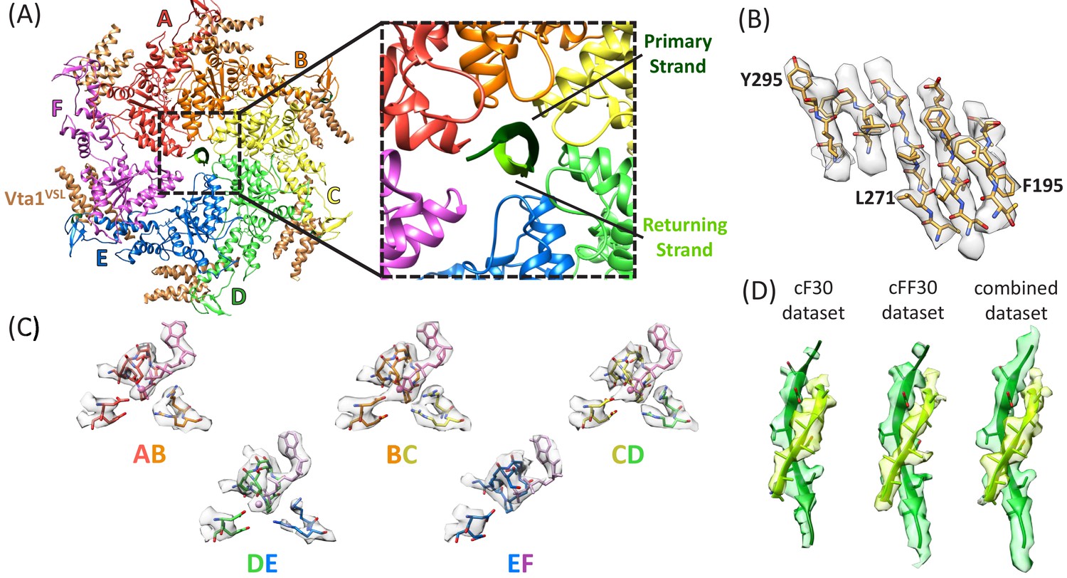
Structure determination.
(A) Overall structure of the Vps4-cyclic peptide complex. The close up view of the pore region shows the primary strand (dark green) and returning strand (light green) of the cyclic peptide. (B) Representative section of density in the large domain of the Vps4 B subunit. (C) Density around the nucleotides and coordinating residues for the active sites of Vps4 subunits A-E. These binding sites occur at the interface with the following subunit. (D) Density around the circular peptides. Shown as side views separately for the cF30, cFF30, and combined maps.
Reconstruction, refinement, and validation statistics.
https://doi.org/10.7554/eLife.44071.026Reconstruction | |
---|---|
Number of particle images | 237,480 |
Resolution (0.143 FSC) (Å) | 3.6 |
Map sharpening B-factor (Å2) | −157 |
EMDB accession number | EMD-0443 |
Model refinement of Vps4 subunits A-E | |
PDB accession number | 6NDY |
Resolution used for refinement (Å) | 3.6 |
Number of atoms | 12531 |
RMSD: Bond length (Å) | 0.003 |
RMSD: Bond angles (°) | 0.739 |
Ramachandran: Favored (%) | 94.1 |
Ramachandran: Allowed (%) | 5.9 |
Ramachandran: Outlier (%) | 0 |
Validation | |
Molprobity score/percentile (%) | 1.64 (100%) |
Clashscore/percentile (%) | 4.67 (100%) |
EMRinger score | 1.04 |
Circular peptides bind the Vps4 pore in a hairpin conformation
The density and refined models for the cF30 and cFF30 peptides are essentially identical (Figure 2D), with two polypeptide strands passing through the Vps4 pore (Figure 3A, Figure 3—video 1). The pore loop positions and the substrate strand with the strongest density (primary strand) superimpose closely on the earlier peptide F complex structure, except that density for the peptide now extends both N-terminally and C-terminally for two additional residues (Figure 3B, Figure 3—video 2). The density is consistent with the peptide F sequence binding in the same register as the earlier peptide F complex, with odd-numbered residues binding to an array of class I pockets and even-numbered residues binding to an array of class II pockets (Han et al., 2017) (Figure 3C, Figure 3—video 3). We attempted to discern the orientation of this peptide strand by comparing the assigned orientation with a peptide model built and refined optimally in the reverse orientation (Figure 2—figure supplement 4). This analysis showed that the map-model correlation coefficients and the EMRinger scores (Barad et al., 2015) slightly favor the assigned orientation, but are not definitive. This ambiguity is expected for the current 3.6 Å resolution, and the peptide orientation remains an important question for future studies.

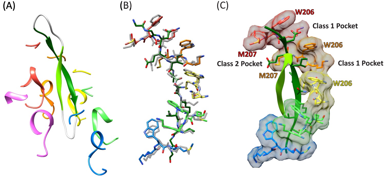
Cyclic peptide structure and coordination.
(A) Model of the entire cyclic peptide, including the residues that lack density (gray), with the Vps4 pore loop 1 and 2 residues. (B) Superposition of the cyclic peptide structure (colors) on the previously determined structure of the linear peptide F (gray). The returning strand of the cyclic peptide is omitted for clarity. (C) Ordered residues of the cyclic peptide are shown as green ribbons. Pore loop one residues K205, W206, and M207 of the five Vps4 subunits that form the helical assembly that binds the substrate peptide are shown as sticks and molecular surfaces. Alternating side chains bind to class I pockets between pairs of W206 residues of adjacent subunits (two examples labeled), and to class II pockets between pairs of M207 side chains from adjacent subunits (one example labeled).
The returning strand of the circular peptides has weaker density, indicating that it is more mobile (Figure 2D). Density for these side chains is not strongly defined, although the sequence of the cFF30 peptide and the presence of two residues on either side of the primary strand F-peptide motif means that at least 5 of the returning strand residues in the pore region must have relatively large side chains. Nevertheless, the density indicates that the returning strand adopts an extended conformation in which eight residues are reasonably modeled as forming a β-ladder interaction with the primary strand. The 10 residues of the circular peptides that lack experimental density can be reasonably modeled in hairpin turn conformations (Figure 3A).
Insights into substrate translocation
The second strand of the circular peptide is accommodated within the Vps4 pore by the displacement of subunit F from the substrate-binding groove and by the peptide adopting the same helical symmetry as Vps4 subunits A-E. The pseudo two-fold axis along the length of the circular peptide β-ladder aligns with the helical axis of Vps4 subunits A-E (Figure 4, Figure 4—video 1), thereby maximizing the distance of the second strand away from the helical Vps4 subunits (A-E) that bind the primary strand, and maximizing the space available for the second peptide strand. An open question is whether or not it is possible to accommodate a third strand without distorting the Vps4 structure.

Cyclic peptide aligns with the helical axis.
Side and top views of the cyclic peptide and Vps4 pore loop one residues. The pseudo two-fold axis that relates the path of the two peptide strands to each other (albeit with opposite direction) aligns with the helical axis of Vps4 subunits A-E (gray). This ensures that the second strand is maximally distant from subunits A-E, thereby explaining the lack of contacts between Vps4 and the second strand.
The hinge angle between the large and small ATPase domains (Gonciarz et al., 2008) varies by just 2° (126–128°) for subunits A-E, but is more open and variable (126–141°) for the three subunit F models derived from focused classification of the combined cF30 and cFF30 particles (Figure 5A). The more variable and open subunit F structure, and the lack of close contacts between the large ATPase domain of subunit F and the large ATPase domains of its neighboring subunits A and E (Figure 5B), is consistent with nucleotide exchange occurring during transit from the subunit-E state to the subunit-A state.

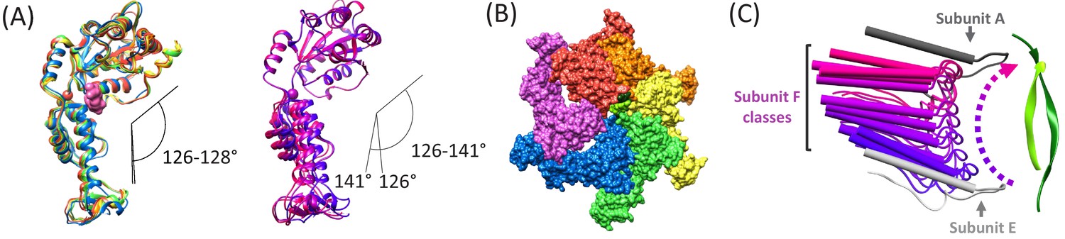
Subunit F conformation and contacts.
(A) Overlap of subunits showing variation in the hinge angle between large and small domains, as defined in Gonciarz et al. (2008). The overlaps were performed on the large domains. Left, Subunits A-E. Right, three classification structures of subunit F. (B) The large domain interfaces for AB, BC, CD, and DE subunit pairs are closely associated. These interfaces are much more open for the EF and FA subunit pairs, as seen by the openings in this slightly tilted view. (C) Side view of the Vps4 circular peptide complex with all of the classified subunit F models from all structures: cF30, cFF30, and combined cF30 and cFF30, plus the two previously reported data sets of Vps4 with the linear peptide F (Han et al., 2017; Monroe et al., 2017). Subunit F is colored based on position along its proposed trajectory.
The proposed ~30 Å transition of subunit F from the subunit-E end of the Vps4 helix to the subunit-A end of the helix is consistent with the results of focused classification of the various Vps4 datasets, including those of cF30, cFF30, combined cF30 + cFF30, and the two linear peptide structures (Han et al., 2017; Monroe et al., 2017). These classifications each provide two or three maps that show distinct positions for subunit F, all of which avoid contact with the returning strand of the cyclic peptides and together span the path traversed during cycle of the proposed translocation mechanism (Figure 5C, Figure 5—video 1).
The focused classification may indicate a mechanism to trigger ATP hydrolysis preferentially at the subunit D active site (DE interface), which is thought to give directionality the Vps4 translocation cycle (Han et al., 2017; Monroe et al., 2017). Specifically, the subunit F state closest to docking against subunit A (F3), correlates with the subunit E small domain rotating 7° and shifting 3 Å relative to the primary position that is seen in all other reconstructions. Thus, the final stage of the subunit F transition is coupled to conformational changes that may propagate through an extended 10-residue strand to the subunit E finger arginine residues that complete coordination of ATP at the subunit D active site (Figure 6). This proposal extends the model that ATP is stably bound at the AB, BC, CD interfaces, while ATP hydrolysis is promoted at the DE interface by the ATP-dependent binding of subunit F against subunit A.

Transition of subunit F is coupled to movement of subunit E.
The maps reconstructed by focused classification over subunit F show that the uppermost subunit F position from the cyclic peptide data correlates with displacement of the subunit E small domain (cyan). This domain is connected by an extended 10-residue stretch of residues to the finger arginines that complete the subunit D active site.
Structure and functional implications of the Vta1 activator protein
The Vta1VSL domains dimerize in an antiparallel orientation through formation of a four-helix bundle (Xiao et al., 2008). Each dimer binds to adjacent Vps4 subunits, yielding a stoichiometry of 12 Vta1 subunits (six dimers) to 6 Vps4 subunits. The Vta1VSL density is clearer than our earlier reconstructions with the extended Vps2 peptide (Han et al., 2017; Monroe et al., 2017), presumably because of its 10-fold higher concentration in the current study and consequently higher occupancy in the particles imaged. Moreover, the Vta1VSL density is better for the cFF30 structure compared to the cF30 dataset, presumably because the use of Spotiton to prepare cFF30 grids reduced the time to vitrification, which reduced the contacts with the air-water interface that cause complex dissolution and preferred orientation (Dandey et al., 2018; Noble et al., 2018).
In a substantial increase over our previous reconstructions using conventional blotting methods (Han et al., 2017; Monroe et al., 2017), ~17% of the particles derived from Spotiton grids contribute density to all six Vta1VSL sites on the Vps4 hexamer (Figure 2—figure supplement 8). Reconstruction of these particles yielded an overall resolution of 4.4 Å. Distinct densities at each Vps4 subunit interface allowed unambiguous assignment of all four helices in the Vta1 dimers, although their location at the periphery of the Vps4 complex remains a region of relatively low local resolution (~5–8 Å). This supports the conclusion that Vps4 and Vta1 associate with 6:12 stoichiometry in the fully assembled complex (Sun et al., 2017).
Vta1 promotes Vps4 oligomerization and increases the ATPase activity (Azmi et al., 2006; Lottridge et al., 2006; Scott et al., 2005), and enhances ESCRT-III disassembly in vitro (Azmi et al., 2008). These effects likely result from multiple interconnected mechanisms: Association of Vta1’s N-terminal MIT domains with ESCRT-III polymers will reinforce Vps4 recruitment to ESCRT-III complexes; binding of Vta1VSL dimers in bridging interactions between adjacent Vps4 subunits will promote formation of the active hexamer; and Vta1VSL support the contacts of subunits E and F that may promote ATP hydrolysis at the subunit D active site (above).
Comparison with other AAA+ ATPase peptide complexes
Several structures of AAA+ ATPases that translocate protein substrates have been reported with coordinates of bound peptides deposited in the Protein Data Bank, including Hsp104 (Gates et al., 2017), YME1 (Puchades et al., 2017), TRIP13 (Alfieri et al., 2018), the proteasome (de la Peña et al., 2018; Dong et al., 2019), and NSF (White et al., 2018). In all cases the bound peptides are single, extended strands that pass through the pore and overlap closely with the structure of peptide F in the Vps4 complex, albeit sometimes in the opposite orientation or with local differences in phi/psi angles. Superposition on the pore loop 1 residues of Vps4 shows that the circular Vps4-bound cF30/cFF30 peptide fits reasonably into the YME1, Hsp104, and NSF structures (Figure 7, Figure 7—video 1). In contrast, superposition on the TRIP13 and proteasome structural models show overlap of pore loop two residues with the returning strand of the cyclic peptides, although in general the pore loop two residues appear to be relatively mobile, which raises the possibility that they may reposition to accommodate a two-stranded substrate. Thus, regardless of whether or not Vps4 binds its substrates in a hairpin conformation in vivo, it seems possible that this mechanism will also be accessible to other AAA+ ATPases.

Overlap of cyclic peptide structure suggests compatibility with YME1, Hsp104, and NSF.
(A) Vps4 color, YME1 (Puchades et al., 2017) gray. Overlap is on the Cα atoms of the peptide and pore loop 1 residues of the five helical subunits. (B) Same as (A) but without Vps4 and showing the zoomed in region of panels (C), (D), and (E). (C) Vps4 cyclic peptide after overlap on YME1. (D) Same as (C) for Hsp104 ring 1 (Gates et al., 2017). (E) Same as (C) for Hsp104 ring 2. (F) Same as (C) for NSF (White et al., 2018).
Implications for mechanism and function
Given the model that substrate translocation results from the pore loops acting on just one of the strands, it will be interesting to determine the extent to which the two chains of dual-stranded substrates slip with respect to each other, and the extent to which they pass through the pore at the same rate. Our structures also raise interesting questions about directionality. Although it is not yet definitively resolved, the Vps4 structures are most consistent with binding of the primary strand N-terminal residues at the A-end of the Vps4 helix and C-terminal residues at the E-end. This orientation is consistent with the biological role of Vps4 in translocating toward the ESCRT-III N-terminal domain, but other AAA+ ATPases apparently translocate their protein substrates in the opposite direction (N-to-C) (Alfieri et al., 2018; Puchades et al., 2017) or in either direction (Augustyniak and Kay, 2018). Indeed, the same mechanism of translocation could be applied to substrates bound with their primary strand in either orientation because side chains make a major contribution to binding, and forward and reversed β-strands can superimpose their Cα atoms and side chains. Regardless, our structures suggest that substrates might bind the AAA+ pore in a hairpin conformation that is translocated in both the N-to-C and C-to-N directions at the same time.
Contexts in which translocation of two polypeptide chains through a AAA+ ATPase pore might be functionally important include the initiation of translocation from an internal loop, crosslinked substrates, and ubiquitin adducts, as have been indicated for the proteasome (Kraut and Matouschek, 2011; Lee et al., 2002; Shabek and Ciechanover, 2010), Cdc48 (Bodnar and Rapoport, 2017), and ClpX (Burton et al., 2001). The model for translocation from an internal loop is illustrated in Figure 7—video 2 and the model for translocation of a ubiquitylated substrate is illustrated in Figure 7—video 3. As shown in these animations, the model for simultaneous translocation of two polypeptide chains through the pore of a AAA+ ATPases will cause them to rotate with respect to each other, with one complete rotation anticipated for every 12 residues of primary strand translocated. This is because successive dipeptides of the primary strand bind successive Vps4 subunits, which are rotated 60° with respect to each other, at the lining of the pore, while the secondary strand continually lies along the center of the pore. The twist that this introduces to the substrate when long stretches of double chains are translocated might be resolved by relative rotation of the AAA+ ATPase or by periodically switching which strand engages the pore loops and which lies along the open channel. The extent to which simultaneous binding and translocation of two polypeptide chains or a polypeptide with some other adduct actually occurs for the various different AAA+ ATPases remains to be determined.
Materials and methods
Resource type | Designation | Source or reference | Identifiers |
---|---|---|---|
Recombinant protein | Vps4-HCP1 | PMID: 28379137 | Addgene (RRID:SCR_002037): 87737 |
Recombinant protein | Vta1VSL | PMID: 28379137 | Addgene (RRID:SCR_002037): 87738 |
Software, algorithm | GraphPad Prism | GraphPad Software, Inc, La Jolla, C | RRID:SCR_002798 |
Software, algorithm | MotionCor2 | PMID: 28250466 | RRID:SCR_016499 |
Software, algorithm | RELION | PMID: 23000701 | RRID:SCR_016274 |
Software, algorithm | UCSF Chimera | PMID: 15264254 | RRID:SCR_004097 |
Software, algorithm | Coot | PMID: 20383002 | RRID:SCR_014222 |
Software, algorithm | Phenix | PMID: 20124702 | RRID:SCR_014224 |
Materials used for peptide synthesis
Request a detailed protocol2-chlorotrityl resin and 1-[Bis(dimethylamino)methylene]−1 H-1,2,3-triazolo[4,5-b]pyridinium-3-oxidhexafluorophosphate (HATU) were purchased from ChemPep. TentaGel R RAM resin was purchased from Rapp Polymere. Boc-Cys(Trt)-OH, Fmoc-L-Cys(Trt)-OH, Fmoc-L-Lys(Boc)-OH, Fmoc-L-Asp(tBu)-OH, Fmoc-L-Glu(tBu)-OH, Fmoc-L-Ser(tBu)-OH, Fmoc-L-Asn(Trt)-OH, Fmoc-Gly-OH, Fmoc-L-Ala-OH, Fmoc-L-Val-OH, Fmoc-L-Leu-OH, and Fmoc-Gly-Ser(psiMe,Mepro)-OH were purchased from Gyros Protein Technologies. Fmoc-Lys(Ac)-OH was purchased from Anaspec. Fmoc-Lys(Dde)-OH was purchased from AAPPTec. Synthesis grade trifluoracetic acid (TFA), ACS grade dimethylformamide (DMF), peptide synthesis grade n-methylmorpholine (NMM), synthesis grade n-methylpyrrolidinone (NMP), ACS grade anhydrous diethyl ether, HPLC grade acetonitrile (ACN), HPLC grade methanol, LC-MS grade ACN with 0.1% formic acid, and LC-MS grade water with 0.1% formic acid were purchased from Fisher Scientific (all reagent brands from Fisher Scientific were Fisher Chemical). Piperidine, triisopropylsilane (TIS), 1,2-ethanedithiol (EDT), 5 (6)-carboxyfluorescein, N,N′-Diisopropylcarbodiimide, Oxyma Pure, anhydrous hydrazine, acetic anhydride, and 4-mercaptophenylacetic acid (MPAA) were purchased from Sigma Aldrich.
Peptide synthesis
Request a detailed protocolPeptides were synthesized on a Prelude X instrument (Gyros Protein Technologies) using Fmoc solid-phase peptide synthesis at 30 μmol scale. Deprotection cycles employed three treatments of 2 mL 20% piperidine in DMF for 3 min followed by three washes for 30 s using 2 mL DMF. Coupling cycles consisted of addition of 0.65 mL 200 mM amino acid in NMP, 0.65 mL 195 mM HATU in DMF, and 0.5 mL 600 mM NMM in DMF. Resin and coupling reagents were then mixed using nitrogen for 25 min at room temperature before being washed three times with 2 mL DMF. Tentagel R RAM resin (loading density 0.19 mmol/g) was utilized for the synthesis of C-terminal amides on peptides F, F12, F30, and FF30. To generate C-terminal hydrazides for cF30 and cFF30, 2-chlorotrityl chloride resin was converted to 2-chlorotrityl hydrazine at 0.2 mmol/g density according to published protocol (Zheng et al., 2013). To improve synthesis quality of peptides, the pseudoproline Fmoc-Gly-Ser(ΨMe,Mepro)-OH was introduced. Labeled peptide F was generated through the coupling of 5-(6)-carboxyfluorescein at the N-terminus. After completion of syntheses, peptide resins were thoroughly washed with DCM and dried under vacuum. Cleavage of peptide resins was achieved after 180 min agitation with 4 mL TFA containing 2.5% each of water, TIS, and EDT per 30 μmol peptide resin. The TFA solution was then precipitated into ice-cold diethyl ether and centrifuged at 4696 g (5,000 RPM) for 10 min. Supernatant was decanted while pellets were triturated with ether before being dried under vacuum.
Peptide cyclization by native chemical ligation
Request a detailed protocolThe approach to peptide cyclization was adapted from previous work (Zheng et al., 2012). 1.5 μmol of HPLC purified linear peptide with C-terminal hydrazide was dissolved in 9 mL deionized (DI) water and stored on ice for 30 min. 1 mL of 200 mM NaNO2 (pH 3.75 in DI water) was then added and allowed to react for 20 min at 4°C to convert the hydrazide into an acyl azide. Conversion of the acyl azide into a 4-mercaptophenylacetic acid (MPAA) thioester was achieved through addition of 10 mL 100 mM MPAA in ligation buffer (6 M GnHCl, 200 mM PO4, pH 7.2). The reaction was then nutated at room temperature. At 15 min, 2 mL (1:1 ligation buffer, DI water, pH 7) 0.5 M TCEP was added to reduce oxidized MPAA and peptide. The reaction was quenched after 1 h with 1 mL 100% AcOH before centrifugation at 4,696 g for 15 min. This solution was filtered with a 0.2 μm syringe filter before HPLC purification.
Peptide purification and analysis by HPLC and LC-MS
Request a detailed protocol30 μmol of crude peptide was dissolved in 20% ACN 0.1% TFA and sonicated for 5 min before centrifugation at 4696 g for 10 min to remove precipitated material. Supernatant was filtered using a 0.2 μm filter before injection onto HPLC. Mobile phases for purification were 0.1% TFA in water (Buffer A) and 0.1% TFA in 90% ACN (Buffer B). Purification was performed on an Agilent 1260 or Beckman Gold 126 HPLC while analytical traces were collected on an Agilent 1260 HPLC. Conditions for each peptide purification are detailed in Table 2. Analytical traces were collected at 214 nm over a 20 min gradient of 10% to 55% Buffer B at 1 mL/min using a Phenomenex C18 Kinetix column (100 Å, 5 μm, 4.6 × 150 mm) heated to 45°C, except where noted within figure supplement legends (Figure 1—figure supplements 2A, 3A and 4A, 5A/C, 6A, and 7A/C).
HPLC purification conditions for the peptides used in this study.
https://doi.org/10.7554/eLife.44071.040Peptide | HPLC column | Flow rate | Gradient |
---|---|---|---|
F | Phenomenex C4 Luna (100 Å, 10 μm, 10 × 250 mm) | 5 mL/min | 20 to 80% Buffer B |
F12 | Phenomenex C12 Jupiter (90 Å, 10 μm, 21.2 × 250 mm) | 5 mL/min | 25 to 45% Buffer B |
F30 | Phenomenex C12 Jupiter (90 Å, 10 μm, 21.2 × 250 mm) | 10 mL/min | 20 to 45% Buffer B |
cF30 (pre-cyclization) | Phenomenex C12 Jupiter (90 Å, 10 μm, 21.2 × 250 mm) | 10 mL/min | 20 to 45% Buffer B |
cF30 | Phenomenex C4 Jupiter (100 Å, 10 μm, 10 × 250 mm) | 5 mL/min | 10 to 35% Buffer B |
FF30 | Phenomenex C12 Jupiter (90 Å, 10 μm, 21.2 × 250 mm) | 10 mL/min | 29 to 37% Buffer B |
cFF30 (pre-cyclization | Phenomenex C12 Jupiter (90 Å, 10 μm, 21.2 × 250 mm) | 10 mL/min | 20 to 45% Buffer B |
cFF30 | Phenomenex C4 Jupiter (100 Å, 10 μm, 10 × 250 mm) | 5 mL/min | 27 to 38% Buffer B |
For LC-MS analysis, mobile phases were 0.1% FA in water (Buffer A) and 0.1% FA in ACN (Buffer B). Data were collected on an Agilent 6120 single quadrupole mass spectrometer with an Agilent 1260 front-end. HPLC traces were collected over a 7 min gradient of 5% to 90% Buffer B at 0.75 mL/min using an Agilent Poroshell C18 column (120 Å, 3.6 μm, 4.6 × 50 mm) heated to 50°C. Mass spectra were obtained over a window of 400 to 2,000 m/z in fast scan and positive ion mode (Figure 1—figure supplements 2B, 3B and 4B, 5B/D, 6B and 7B/D). Deconvoluted masses were determined using Agilent Chemstation with averaged scans across the major ion signal.
Trypsin digestion of peptides and high-resolution LC-MS
Request a detailed protocolLinear and cyclic peptides from lyophilized powder were dissolved in alkylating buffer (40 mM 2-chloroacetamide, 10 mM TCEP, 100 mM Tris, pH 8) to a concentration of ~30 μM before rotating at 37°C for 60 min. Before sample loading, Pierce C18 tips were equilibrated with three treatments of 100 µL 0.1% FA in ACN and three treatments of 100 µL 0.1% FA in water. 40 µL of each sample loaded onto Pierce C18 tips through five repeats of aspiration and dispensing. C18 tips were washed with 100 µL 0.1% FA in water before elution of the peptide using 0.1% FA in 70% ACN. The eluent was concentrated to 10 µL by speed-vac before addition of 50 µL trypsin (1:10, Pierce Trypsin Protease MS Grade, Thermo Fisher Scientific) in 50 mM ammonium bicarbonate buffer (ABC, pH 7.5). Trypsin treated samples were incubated at 37°C for 90 min before quenching through addition of FA to a final concentration of 1%. Trypsin was removed from the samples using Vivacon 30 k MWCO filters (Sartorius) and centrifugation at 14,000 x g for 20 min. Samples were then diluted 1:1000 using 0.1% FA in water before transferring to MS vials and storage at −80°C.
For mass spectrometry analysis, 2 µL of sample was injected onto a Thermo Fisher EASY-nanoLC 1000 with a Picofrit column (New Objectives, 360 µm OD x 75 µm ID, 150 mm, packed with 3 µm Reprosil-PUR) coupled to an Orbitrap Velos Pro. Mobile phases consisted of 0.1% FA 5% DMSO in water (Buffer A) and 0.1% FA 5% DMSO in ACN (Buffer B). Gradient conditions were 5% to 45% Buffer B over 30 min at 400 nL/min. MS1 spectra were collected using the Orbitrap analyzer from 350 to 1550 m/z at 60,000 resolution (FWHM as defined at m/z 400). The top two most intense ions from the MS1 scan were selected for HCD fragmentation using a normalized collision energy of 40 eV. MS2 spectra were collected at a resolution of 15,000.
To identify peptides in an unbiased manner, raw data files were converted to mgf format for analysis in SearchGUI (Barsnes and Vaudel, 2018) using the OMSSA search algorithm. Peptide sequences were added to a modified FASTA file containing ~2000 decoy human proteins. Spectrum match settings used trypsin digestion with one missed cleavage allowed, carbamidomethylation of Cys as a fixed modification (denoted as ‘Am’ in peptide sequences), acetylation of K as a variable modification, precursor m/z tolerance of 10 ppm, and fragment m/z tolerance of 0.2 Da. Post-processing utilized PeptideShaker (Vaudel et al., 2015) to identify peptide fragments present in each sample. The false discovery rate (FDR) for peptide identification was set to 0.01. Identified peptides were exported from PeptideShaker as an MZID file to Skyline (MacLean et al., 2010). Visualization of all peptide fragments (Figure 1—figure supplement 8) and MS2 spectra (Figure 1—figure supplement 9) were produced using Skyline.
Fluorescence polarization assay for peptide binding
Request a detailed protocolBinding of unlabeled peptides to Vps4 and Vps4-Hcp1 hexamer was quantified using competitive binding assays. Briefly, a dilution series of unlabeled peptide was made in fluorescence polarization assay buffer (20 mM HEPES, pH 7.4, 100 mM NaCl, 1 mM ADP·BeFx, 10 mM MgCl2, 10 mM TCEP) with 200 nM Vps4 hexamer and 1 nM fluorescein-labeled peptide F. Reaction equilibrium was reached with 3 h incubation at room temperature. Fluorescence polarization was measured on a Biotek Synergy Neo HTS Multi-Mode Microplate Reader using 485/528 nm excitation/emission wavelengths. IC50 values were calculated using GraphPad Prism 7 (RRID:SCR_002798) by fitting raw polarization data to Equation 1 with the FPmin manually constrained to the polarization value of labeled peptide F alone. Ki values, which correspond to the dissociation constants for the unlabeled peptides, were calculated with Equation 2 (Nikolovska-Coleska et al., 2004) using previously published KD values for labeled peptide F (0.253 ± 0.015 µM with Vps4 and 0.230 ± 0.010 µM with Vps4-Hcp1; Monroe et al., 2017) and IC50 values from Equation 1.
Grid preparation and vitrification
Request a detailed protocolComplexes were prepared for cryo-EM analysis as described (Monroe et al., 2017) except that Vta1VSL was included at 10-fold higher concentration for crosslinking. cF30 complex was vitrified using a Vitrobot (Thermo Fisher Scientific), as described (Monroe et al., 2017).
cFF30 complex samples were vitrified using the Spotiton robot as described (Dandey et al., 2018; Jain et al., 2012; Razinkov et al., 2016), starting from a Vps4 complex at 18 μM (hexamer). Briefly, the Spotiton device uses piezo dispensing to apply small (50 pL) drops of sample across a ‘self-blotting’ nanowire grid as it flies past en route to plunge into liquid ethane. Nanowire grids for use with Spotiton were manufactured in-house, backed by lacey carbon film supports, and prepared as described (Razinkov et al., 2016; Wei et al., 2018), including plasma cleaning for 10 s (O2 + H2) using a Solarus 950 (Gatan, Inc). The time between sample application to the grid and plunging into liquid ethane (spot-to-plunge time) was ~145 ms. Spotiton was operated at ~85% relative humidity and ambient temperature (~21°C). Under these conditions, evaporation is estimated to be 300 Å/s.
Single-particle cryoEM data collection
Request a detailed protocolSingle-particle micrographs were collected on a Titan Krios (Thermo Fisher Scientific) equipped with an energy filter and a K2 BioQuantum counting camera (Gatan, Inc); the microscope was operated at 300 kV at a nominal magnification of 130,000x, with a calibrated pixel size of 1.09 Å. Exposure was set to 10 s (50 frames/movie, detector operated at counting mode), for a total dose of 76.68 e–/Å2 with a defocus range of 1.6 to 2.2 µm. Each dataset was collected over one session using Leginon. Frames were aligned and dose weighted using MotionCor2 RRID:SCR_016499 (Zheng et al., 2017).
Image processing
Request a detailed protocolcF30 and cFF30 complex datasets were initially processed separately with essentially the same workflow (Figure 2—figure supplements 2 and 3). Movie frames were aligned, dose weighted, and summed using MotionCor2 RRID:SCR_016499 (Zheng et al., 2017). CTF parameters were determined on non-dose-weighted sums using gctf (Zhang, 2016). Micrographs with poor CTF cross correlation scores (<0.04) were excluded from downstream analyses. A total of 2838 (cF30) and 1855 (cFF30) dose-weighted sums were used for all subsequent image processing steps.
529,807 (cF30) and 356,705 (cFF30) particles were extracted and used as input for full CTF-corrected image processing. After multiple rounds of 2D classification in RELION (RRID:SCR_016274) (Scheres, 2012), 204,636 (cF30) and 155,900 (cFF30) particles were retained, based on visual inspection of classes with high-resolution Vps4 features, and used for an initial round of 3D classification. After 3D classification, 157,775 (cF30) and 144,776 (cFF30) particles were used for RELION (RRID:SCR_016274) auto-refinement (Scheres, 2012), which in each case generated an ~4.5 Å density map of the Hcp1-Vps4 fusion complex based on the gold-standard FSC criterion. To improve the resolution of Vps4, we performed signal subtraction of Hcp1 densities, as described (Bai et al., 2015; Monroe et al., 2017). For the cF30 complex dataset, an additional round of 3D classification post-Hcp1-subtraction assigned 92,704 particles into a single class with high-resolution Vps4 features. These particles were used for a final round of RELION (RRID:SCR_016274) auto-refinement, producing a 3.8 Å resolution density map of Vps4. For the cFF30 complex dataset, the Hcp1-subtracted dataset was used for RELION (RRID:SCR_016274) auto-refinement without further classification, producing a 4.0 Å resolution density map.
To improve the resolution of the density map, the final datasets for cF30 and cFF30 complexes were combined for further processing (Figure 2—figure supplements 1 and 4). Merging the datasets was justified based on the similar binding affinities and densities (Figure 2D). The merged dataset comprised a total of 237,480 particles (92,704 from cF30 complex, 144,776 from the cFF30 complex). RELION (RRID:SCR_016274) auto-refinement was performed using the --solvent_correct_fsc option in combination with a soft-edge mask encompassing Vps4. B-factor sharpening of −157 Å2 was applied to the final 3.56 Å map using an automated procedure in RELION (RRID:SCR_016274) postprocessing (Rosenthal and Henderson, 2003). Local resolutions were estimated using ResMap (Kucukelbir et al., 2014).
As observed in our previous structures of Vps4 bound to linear peptides, subunit F was poorly resolved due to its relative flexibility (Monroe et al., 2017). We therefore performed focused 3D classification by applying a spherical mask over subunit F density (Figure 2—figure supplement 4). Classification was performed without re-alignment (i.e., using the --skip_align flag in RELION, K = 6), leading to three classes with ordered density and three classes with disordered density (Figure 2—figure supplement 5). Particles from classes with ordered density were used for separate RELION auto-refinement. The resulting maps were then used for rigid-body fitting of Vps4 coordinates into each subunit F position using UCSF Chimera (RRID:SCR_004097) (Pettersen et al., 2004).
Focused 3D classification of Vta1VSL was performed by applying a single custom mask that covered each of the six possible Vta1 binding sites (Figure 2—figure supplement 8). Particles were classified without re-alignment (i.e., using the --skip_align flag in RELION, K = 10). 24,778 particles from the cFF30 dataset were sorted into a single class that showed Vta1VSL densities at all six Vps4 interfaces. These particles were used for RELION 3D auto-refinement, resulting in a 4.4 Å resolution reconstruction. Vta1VSL models were fitted into the reconstruction using rigid-body fitting as previously described (Monroe et al., 2017).
Model building, refinement, and validation
Request a detailed protocolThe model of AAA+ ATPase cassettes for Vps4 subunits A-E and the substrate from our previous structure (PDBID: 6AP1) were fit to the 3.6 Å map as rigid bodies and subjected to real-space refinement using Phenix (RRID:SCR_014224) (Adams et al., 2010) following the same approach as for the earlier structure of Vps4 in complex with a linear substrate (Han et al., 2017). The returning chain was built manually in Coot (RRID:SCR_014222) (Emsley et al., 2010) followed by real-space refinement using Phenix with restraints to β-strand conformation and its starting position. The refined model was assessed using MolProbity (RRID: SCR_014226) (Chen et al., 2010).
To test for overfitting, the refined model (subunits A-E of Vps4 and the main and returning chains of the cyclic substrate) were randomly displaced by 0.2 Å and re-refined against one of the RELION half-maps used to generate the 3.6 Å map. FSC curves were generated for the re-refined model against the half map used for re-refinement (FSCwork) and against the other half map (FSCtest) (Figure 2—figure supplement 1E). The agreement between FSCwork and FSCtest indicates that the model has not been overfit.
Structure deposition
Request a detailed protocolThe refined model comprising the Vps4 ATPase domains of subunits A-E and the cyclic peptide is accessible via the PDB (RRID: SCR_012820; PDB ID: 6NDY) together with the 3.6 Å map from the combined dataset (RRID: SCR_003207, EMDB Accession Number EMD-0443). The complete model, including regions not subjected to atomic refinement such as the 12 Vta1VSL domains and subunit F, is also available via the PDB (PDB ID: 6OO2), together with the map containing Vta1VSL densities at all six Vps4 interfaces (RRID: SCR_003207, EMDB Accession Number EMD-20142). The two maps derived from the cF30 and cFF30 complex datasets individually, and the three maps for subunit F, have been deposited to the EMDB (RRID: SCR_003207, EMDB Accession Numbers EMD-20144, EMD-20147, EMD-20139, EMD-20140, EMD-20141).
Data availability
The refined model comprising the Vps4 ATPase domains of subunits A-E and the cyclic peptide is accessible via the PDB (RRID: SCR_012820; PDB ID: 6NDY) together with the 3.6 Å map from the combined dataset (RRID: SCR_003207, EMDB Accession Number EMD-0443). The complete model, including regions not subjected to atomic refinement such as the 12 Vta1VSL domains and subunit F, is also available via the PDB (PDB ID: 6OO2), together with the map containing Vta1VSL densities at all six Vps4 interfaces (RRID: SCR_003207, EMDB Accession Number EMD-20142). The two maps derived from the cF30 and cFF30 complex datasets individually, and the three maps for subunit F, have been deposited to the EMDB (RRID: SCR_ 003207, EMDB Accession Numbers EMD-20144, EMD-20147, EMD-20139, EMD-20140, EMD-20141).
-
Protein Data BankID 6NDY. Vps4 with Cyclic Peptide Bound in the Central Pore.
-
EMDataBankID EMD-0443. Vps4 with Cyclic Peptide Bound in the Central Pore.
-
Protein Data BankID 6OO2. Vps4 with Cyclic Peptide Bound in the Central Pore.
-
EMDataBankID EMD-20142. Vps4 with Cyclic Peptide Bound in the Central Pore.
-
EMDataBankID EMD-20144. Vps4 with Cyclic Peptide Bound in the Central Pore (Map for Peptide cF30).
-
EMDataBankID EMD-20147. Vps4 with Cyclic Peptide Bound in the Central Pore (Map for Peptide cFF30).
-
EMDataBankID EMD-20139. Vps4 with Cyclic Peptide Bound in the Central Pore (Focused Classification of Subunit F, State1).
-
EMDataBankID EMD-20140. Vps4 with Cyclic Peptide Bound in the Central Pore (Focused Classification of Subunit F, State2).
-
EMDataBankID EMD-20141. Vps4 with Cyclic Peptide Bound in the Central Pore (Focused Classification of Subunit F, State3).
References
-
PHENIX: a comprehensive Python-based system for macromolecular structure solutionActa Crystallographica Section D Biological Crystallography 66:213–221.https://doi.org/10.1107/S0907444909052925
-
Recycling of ESCRTs by the AAA-ATPase Vps4 is regulated by a conserved VSL region in Vta1The Journal of Cell Biology 172:705–717.https://doi.org/10.1083/jcb.200508166
-
SearchGUI: a highly adaptable common interface for proteomics search and de novo enginesJournal of Proteome Research 17:2552–2555.https://doi.org/10.1021/acs.jproteome.8b00175
-
MolProbity: all-atom structure validation for macromolecular crystallographyActa Crystallographica Section D Biological Crystallography 66:12–21.https://doi.org/10.1107/S0907444909042073
-
Spotiton: new features and applicationsJournal of Structural Biology 202:161–169.https://doi.org/10.1016/j.jsb.2018.01.002
-
Features and development of cootActa Crystallographica. Section D, Biological Crystallography 66:486–501.https://doi.org/10.1107/S0907444910007493
-
Evolutionary relationships and structural mechanisms of AAA+ proteinsAnnual Review of Biophysics and Biomolecular Structure 35:93–114.https://doi.org/10.1146/annurev.biophys.35.040405.101933
-
Biochemical and structural studies of yeast Vps4 oligomerizationJournal of Molecular Biology 384:878–895.https://doi.org/10.1016/j.jmb.2008.09.066
-
Structure and mechanism of the ESCRT pathway AAA+ ATPase Vps4Biochemical Society Transactions 47:37–45.https://doi.org/10.1042/BST20180260
-
Spotiton: a prototype for an integrated inkjet dispense and vitrification system for cryo-TEMJournal of Structural Biology 179:68–75.https://doi.org/10.1016/j.jsb.2012.04.020
-
Quantifying the local resolution of cryo-EM density mapsNature Methods 11:63–65.https://doi.org/10.1038/nmeth.2727
-
Concurrent translocation of multiple polypeptide chains through the proteasomal degradation channelJournal of Biological Chemistry 277:34760–34765.https://doi.org/10.1074/jbc.M204750200
-
Structures, functions, and dynamics of ESCRT-III/Vps4 membrane remodeling and fission complexesAnnual Review of Cell and Developmental Biology 34:85–109.https://doi.org/10.1146/annurev-cellbio-100616-060600
-
The oligomeric state of the active Vps4 AAA ATPaseJournal of Molecular Biology 426:510–525.https://doi.org/10.1016/j.jmb.2013.09.043
-
Marching to the beat of the ring: polypeptide translocation by AAA+ proteasesTrends in Biochemical Sciences 39:53–60.https://doi.org/10.1016/j.tibs.2013.11.003
-
UCSF Chimera?A visualization system for exploratory research and analysisJournal of Computational Chemistry 25:1605–1612.https://doi.org/10.1002/jcc.20084
-
A new method for vitrifying samples for cryoEMJournal of Structural Biology 195:190–198.https://doi.org/10.1016/j.jsb.2016.06.001
-
RELION: implementation of a bayesian approach to cryo-EM structure determinationJournal of Structural Biology 180:519–530.https://doi.org/10.1016/j.jsb.2012.09.006
-
Structural and mechanistic studies of VPS4 proteinsThe EMBO Journal 24:3658–3669.https://doi.org/10.1038/sj.emboj.7600818
-
PeptideShaker enables reanalysis of MS-derived proteomics data setsNature Biotechnology 33:22–24.https://doi.org/10.1038/nbt.3109
-
Optimizing "self-wicking" nanowire gridsJournal of Structural Biology 202:170–174.https://doi.org/10.1016/j.jsb.2018.01.001
-
Gctf: real-time CTF determination and correctionJournal of Structural Biology 193:1–12.https://doi.org/10.1016/j.jsb.2015.11.003
Decision letter
-
James M BergerReviewing Editor; Johns Hopkins University School of Medicine, United States
-
John KuriyanSenior Editor; University of California, Berkeley, United States
In the interests of transparency, eLife includes the editorial decision letter and accompanying author responses. A lightly edited version of the letter sent to the authors after peer review is shown, indicating the most substantive concerns; minor comments are not usually included.
[Editors’ note: this article was originally rejected after discussions between the reviewers, but the authors were invited to resubmit after an appeal against the decision.]
Thank you for submitting your work entitled "Implications for AAA ATPase processing of protein substrate loops from the structure of Vps4 bound to a circular peptide" for consideration by eLife. Your article has been reviewed a Senior Editor, a Reviewing Editor, and two reviewers. The reviewers have opted to remain anonymous.
Our decision has been reached after consultation between the reviewers. Based on these discussions and the individual reviews below, we very much regret to inform you that we cannot accommodate this work in eLife. There were no issues with the technical aspects of the findings. However, both referees were in agreement that the presented structure likely represents a specialized scenario (an initiation complex that is determined by the tightly binding F-peptide), and felt that it constitutes a variation of prior published peptide-bound structures of Vps4 which does not provide additional insights into the translocation process. There was also concern that the low complexity of the secondary strand may allow its return through the central pore without making any motor contacts, and that this may not therefore reflect a normal two-strand operation mode of AAA peptide translocases on more complex sequences. During the consultation process, the referees did note that if new conformations or substrate interactions could be identified which expand on the motor mechanism, or if an argument could be made that this the state seen here is perhaps equivalent to how Vps4 acts on Vps2 during disassembly of ESCRT-III (accompanied by appropriate functional evidence), then the impact could strengthened. We hope that you will find their comments useful in moving forward.
Reviewer #1:
In this manuscript, Hill and coworkers describe the cryo-EM structure of cyclic peptide-bound Vps4 in complex with ADP·BeFx and the VSL domain of Vta1, suggesting how this AAA+ motor may translocate loops or two strands of protein substrates. The authors used circularized 30-mer peptides with one or two copies of the 8-residue segment DEIVNKVL (F peptide) that was derived from the ESCRT-II subunit Vps2, specifically binds to the Vps4 pore, and whose structure bound to Vps4 has previously been solved by the same group. This primary segment of the circular peptides was found to bind indistinguishably from the linear peptide, whereas the secondary segment forms β-ladder interactions with the primary segment and passes through the Vps4 pore without making any significant contacts.
The presented data are of good quality and provide insights into how Vsp4 may simultaneously translocate two polypeptide chains. However, the strong agreement with the previously published structure of linear peptide-bound Vps4 limits the extent of conceptually advances. Due to the use of the specifically-binding F-peptide combined with a low-complexity (G, A, and S-rich) secondary segment, it is not too surprising that affinities and peptide-motor interactions are highly similar or identical to those previously described. The authors strongly generalize their findings and suggest throughout the manuscript, including in the title and abstract, that other AAA+ ATPases may process substrate loops or multiple chains in a very similar manner. However, it is unclear to what extent the observed conformations and interactions are relevant for other AAA+ motors, as the presented structure likely reflects an initiation complex in which a specifically recognized peptide binds with micro-molar affinity to a static, ATP-hydrolysis-deficient Vps4. As acknowledged by the authors, other substrate-bound AAA+ motors do not show peptides in the same β-strand conformation with highly ordered subunit contacts, which also makes it unlikely that the secondary strand of a substrate loop forms similar β-ladder interactions as observed here for Vps4. Many of these other structures, including the recently published substrate-bound 26S proteasome, suggest less regular, steric interactions of pore-loops with the substrate polypeptide, which may then also involve contacts with a secondary strand when substrate loops are translocated.
The authors favor a model in which the primary strand binds in C-to-N terminal orientation to the Vps4 subunits A – E, with the secondary strand spared from any interactions. This model would imply that folded domains N-terminal of the motor-bound primary strand would get preferentially unfolded. Biochemical studies of substrate processing by the 26S proteasome (e.g. Piwko and Jentsch, 2006) indicate that initiation on an internal loop can lead to processing of the N-terminal, C-terminal, or both segments, speaking against selective interactions with only the primary strand. It is thus questionable whether the model presented here applies to other AAA+ motors including the proteasome.
The authors suggest (or imply through their wording) that they identified a new mechanisms of substrate engagement: "Our data indicate a third potential mechanism in which substrate engagement and translocation initiate from an internal segment by binding of a folded hairpin directly within the hexamer pore." Even though structural data have so far been missing, internal initiation and the translocation of multiple chains had already been well established by extensive biochemical studies, not only for the 26S proteasome, but also for members of the Clp family. These previous studies should be cited and the presented Vps4 structure discussed accordingly.
Regarding the coupling of ATP hydrolysis and substrate translocation, a similar coordination of conformational changes as described here for subunits F and E has also been observed for subunits of the substrate-engaged 26S proteasome in various stages of the ATPase cycle (de la Pena et al., 2018) and should be discussed.
The presented structures add some details, but no groundbreaking new findings about the interaction of Vps4 with the VSL domain of the Vta1 activator.
In summary, even though it is interesting to visualize Vps4 bound to a circular peptide, conceptually new findings are somewhat limited due to the strong similarities to the previously published structures of the linear peptide-bound motor. Since the presented structures likely represent an initiation complex with a tightly binding peptide, it remains unclear to what extent observed interactions apply to processively translocating Vps4 or even other AAA+ motors, especially in light of existing substrate-bound structures that already show deviations in peptide conformations and orientations within the central pore. The authors should try to focus their manuscript primarily on Vps4 and reduce their generalization about the mechanisms of other AAA+ motors. Contrary to the author's suggestion, these structures did not identify a novel mechanism for internal initiation of substrate processing, however, they do represent the first visualization of such a complex. I'll have to leave it to the reviewing editor to decide whether the presented advances are significant enough to consider publication in eLife.
Reviewer #2:
In this manuscript Han, et al. determined a 3.6-Å structure of the Vps4 AAA+ ATPase, which is essential for dissociating ESCRT complexes, bound to a cyclic peptide containing the Vps2 binding sequence. The significant results are that the peptide is bound in a β-ladder hairpin conformation with two strands spanning the Vps4 translocation channel. The Vps2 sequence is bound in the same arrangement with defined pore-loop contacts as previous structures while the returning strand is more flexible and runs along the helical interface without making contact with the hexamer. The structure is important because it shows that two strands can be accommodated in the translocation channel; all previous structures of AAA+ translocases show a single unfolded strand in the channel. The authors model this β-hairpin into these previous structures and propose this as evidence that related AAA+s can initiate and translocate internal loops of substrates in addition accessible termini – although this is tenuous given that no experiments directly address this functionally and other AAA+s are not tested. Additionally, the authors are able to increase the occupancy of the Vta1 cofactor subunit and thereby establish its stoichiometry in the complex and improve the reconstruction and modeling of this region compared to previous structures. The work in this manuscript is technically very sound with compelling substrate binding analyses, cryo-EM structure determination and molecular modeling methods. However, two points of concern regarding the functional significance and novelty of these results reduce enthusiasm for publication in eLife: (1) These results may be a specific consequence of the experimental setup which includes the use of a cyclic peptide with low-complexity Gly, Ala and Ser residues outside Vps2 that may be required to fit in the channel, use of ADP·BeFx, glutaraldehyde crosslinking, and a truncated Vps4 with the VSL domain added for stability. Thus, it is unclear whether a native Vps4 complex could bind the full-length Vps2 substrate by this mechanism or if other AAA+s can translocate multiple strands through the channel. (2) The Vps4 hexamer and bound Vps2 peptide are in an identical configuration and nucleotide state as the two previously published structures (one at higher resolution) by this group and the second strand of the substrate is flexible and passively bound without any specific contacts with the Vps4 channel, thus the insight into the AAA+ translocation mechanism, that Vps4 can accommodate two strands under these conditions, has modest impact to the field.
Specific comments:
-Were other nucleotides tested for binding to the cyclic peptide? Hydrolysable nucleotides, such as ATP or ATPyS, would be worth testing to potentially capture different nucleotide states of the subunits or different translocation intermediates.
-What is the solution structure of the cyclic peptide? Does it form the β-hairpin structure or is this a consequence of Vps4 binding? Perhaps CD spectra of the peptide could be measured.
-Were other cyclic peptides tested with difference sequences for the hairpin? It seems that this two-stranded β-ladder complex may require low complexity or specific amino acids with minimal side chains for the returning strand that is adjacent the Vps2 sequence in order to fit in the channel. Functional significance would be improved if a solution-state β-sheet or the full α-helix that contains the Vps2 sequence were tested.
-The occupancy and nucleotide state of the subunits is discussed, but no data is shown in support of this.
-In their translocation model the authors propose that subunit F, which is disconnected from the substrate and asymmetric with respect to the helical arrangement of the hexamer, moves ~30 Å to the subunit-A end of the helix during a translocation step. By focussed classification of this region of the hexamer they identify different positions of subunit F. Local resolution for this region is stated to be 4-7 Å for these classes, however no data is shown. How was the focussed classification performed? Is the cryo-EM density for F improved by focussed classification? Only the Vta1 focussed classification is discussed in the methods. From Figure 4B it is difficult to tell how these positions/conformations of F fit with their translocation model or that "they span a substantial fraction of the path" that is proposed for a translocation step. Please show the range of motion and relative positions of subunit A and E along the channel axis.
-As rationale for a potential conserved function of AAA+s binding a two-strand β-ladder structure the authors make claims that are questionable and not referenced. It is stated "AAA+ ATPases are often thought to initiate substrate engagement from a protein terminus". Please provide supporting studies for this claim. For AAA+ disaggregases it has been proposed for a number of years that engagement can occur from internal segments (see Haslberger et al., 2008).
Additionally it is stated that AAA+ hexamerization could occur "around a linear portion of their substrate", but "this possibility is relevant for family members that show inherently weak hexamerization in the absence of bound substrate but is unlikely to be applicable for robust hexamers like the proteasome and Hsp104". This claim is inaccurate, and no supporting references are provided. In fact, the "lock washer" helical conformation has only been observed in the presence of substrate for these AAA+s. Crystal structures show a continuous helix, hexamerization is dynamic and dependent on nucleotide state (see DeSantis et al., 2012, Aguado et al., 2015 and Uchihashi et al., 2018) and the substrate-free cryo-EM structures of VAT, ClpB and Hsp104 show an open helical spiral that is an entirely different arrangement and likely incompatible with translocation. Thus, while hexamerization/oligomerization can occur without substrate, these complexes are highly dynamic. Therefore, hexamerization around the substrate or, more likely, passing an internal segment though the seam interface are highly plausible models for engagement of internal segments by the proteasome, Hsp104 and other AAA+s.
[Editors’ note: what now follows is the decision letter after the authors submitted for further consideration.]
Thank you for resubmitting your work entitled "Structure of Vps4 with circular peptides and implications for translocation of two polypeptide chains by AAA+ ATPases" for further consideration at eLife. Your revised article has been favorably evaluated by John Kuriyan (Senior Editor), a Reviewing Editor, and two reviewers. Although one referee is now in favor of publication, the other (Reviewer 2) raises a few salient points that we feel are worth considering before making a final decision. Please respond to these comments using your best judgement. Once your revised manuscript is received, we will make an editorial decision, with no further input required from the reviewers.
Reviewer #2:
I appreciate the authors' further discussion about the F-peptide conformation and its similarities to structures observed for related AAA motors. I agree that the overall arrangement and mode of peptide interaction is similar to other motors, as this is largely dictated by the very consistent helical-staircase arrangement of ATPase subunits. The question is about the high regularity of those interactions in Vps4, whether substrates in general have to adopt a defined β structure in the pore, and to what extent the observed conformation originates from the tight binding (KD = 250 nM) and consequent potential energy minimum of the characterized state. A comparison with peptide conformations in other AAA motors is certainly warranted, but needs careful phrasing, especially because the intermediate resolution of those structures makes an assessment of phi/psi angles difficult. Even for the Vps4 structure presented here, the authors' strong claims about β strand conformation is oddly contrasted by their uncertainty about N-to-C or C-to-N directionality of the bound peptide.
Independent of whether the F-peptide complex is indeed an initiation complex, its high affinity is a bit surprising given the non-specific nature of interactions during substrate translocation. If peptides in general bound with similarly high affinity, as suggested by the authors, I am wondering how Vps4 can maintain a high enough selectivity, and whether the autoinhibitory effect of its MIT domains in the absence of MIM binding would be sufficient to prevent promiscuous, non-specific interactions of all kinds of partially or fully unstructured proteins with the central pore. However, this will have to be addressed in future studies.
Relevant for the conclusions of this study is to what extent the F-peptide determines the overall conformation and leads to the strong agreement between the linear and circular peptide-bound structures. If this F-peptide binding represents a very general mode with no specific interactions, why do the extended and circular peptides all bind in exactly the same register?
Some concerns thus remain about the general advance of this study. It is true that a structure of a AAA motor with two polypeptides in the pore has not been described before. Yet, since it perfectly overlays with the previously published linear peptide-bound structure, why is this a "surprising finding for the field", especially considering that it may largely be determined by the tightly binding F-peptide? The accommodation of a second chain in the central pore is itself not unexpected. If the F-peptide sequence is indeed not "special", the authors should provide some explanation for why it stays aligned with the motor in exactly the same way for 1 or 2 strands in the pore and all structures analyzed.
The mechanistic insight of this story would for instance be significantly increased if the authors could reveal whether the motor always selectively interacts with only one strand of particular directionality and spares the other etc.
Regarding the proposed mechanism for stimulating ATP hydrolysis in subunit D, the authors propose that a subunit F-induced rotation of subunit E's small AAA domain may propagate to the arginine residues to complete coordination of ATP at the subunit D active site. However, this model of arginine engagement does in my opinion not agree with the ambiguous nucleotide density observed at the D-E interface, which was interpreted as ADP or an ADP/ADP·BeFx mixture. It is expected that active sites bound to ADP vs. ATP (or ADP·BeFx) show significantly different arrangement and distances for the arginine fingers.
In summary, even though a AAA motor structure with two peptide chains in the central pore has not been presented before, I am not sure whether the mechanistic insight and advance of this manuscript in its current form are high enough for publication in eLife.
Reviewer #3:
Han et al., have adequately addressed all reviewer concerns and have made appropriate adjustments to the manuscript text and figures. Some concerns remain about the possibility that the position of the returning strand and hairpin structure in the channel may be specific to the use of these cyclic peptides and Vps4, thereby limiting the potential that this serves as a general translocation mechanism for AAA+s. Nonetheless, I agree with the authors that this work provides the first key structural insight into how AAA+s could potentially initiate from internal segments or translocate through conjugated or crosslinked sites in proteins. Thus, with these changes, I feel this work is sufficient for publication.
https://doi.org/10.7554/eLife.44071.060Author response
[Editors’ note: the author responses to the first round of peer review follow.]
Our decision has been reached after consultation between the reviewers. Based on these discussions and the individual reviews below, we very much regret to inform you that we cannot accommodate this work in eLife. There were no issues with the technical aspects of the findings. However, both referees were in agreement that the presented structure likely represents a specialized scenario (an initiation complex that is determined by the tightly binding F-peptide), and felt that it constitutes a variation of prior published peptide-bound structures of Vps4 which does not provide additional insights into the translocation process. There was also concern that the low complexity of the secondary strand may allow its return through the central pore without making any motor contacts, and that this may not therefore reflect a normal two-strand operation mode of AAA peptide translocases on more complex sequences. During the consultation process, the referees did note that if new conformations or substrate interactions could be identified which expand on the motor mechanism, or if an argument could be made that this the state seen here is perhaps equivalent to how Vps4 acts on Vps2 during disassembly of ESCRT-III (accompanied by appropriate functional evidence), then the impact could strengthened. We hope that you will find their comments useful in moving forward.
This summary presents three concerns, each of which is addressed below:
1) Concern #1: The presented structure likely represents a specialized scenario (an initiation complex that is determined by the tightly binding F-peptide).
There is little evidence to suggest that peptide F corresponds to an initiation site, whereas considerable evidence indicates that the Vps4-peptide F complex represents a canonical translocating conformation.
The history of identifying this peptide may have led to some confusion on this point. Phyllis Hanson’s lab showed that the human ESCRT-III proteins CHMP2A and CHMP1B contain a segment N-terminal to the MIM sequence that can associate with VPS4 (Shim et al., 2008) and promote ATPase activity (Merrill et al., 2010). We subsequently mapped a relatively tight binding sequence to a 20-residue segment of the yeast CHMP2 homolog, Vps2p (Han et al., 2015), and later found an 8-residue peptide (the F peptide sequence) that maintained similar affinity (Monroe et al., 2017). Although this path would be consistent with having identified an especially tight-binding initiation site, there are multiple reasons to think that the structures do in fact represent the canonical translocating conformation:
i) Our unpublished studies have now identified multiple unrelated peptide sequences that bind with similar affinity to peptide F, and even one that binds 30 times more strongly. Thus, despite the path that led to its identification, peptide F is not an unusually tight binding sequence.
ii) The structures show that the residues of peptide F do not make specific interactions. Instead, the structures show that very different side chains bind to a series of essentially identical binding sites (Han et al., 2017). This is exactly the sort of binding mode expected for binding and translocation of the highly diverse sequences found in ESCRT-III subunits.
iii) The peptide F sequence is not conserved in other ESCRT-III proteins that are substrates for Vps4.
iv) Finally, all of the other AAA ATPase peptide complex structures that have been reported bind peptide in the same manner as we reported for Vps4 (Figure 1). There are a number of points to clarify in this regard:
Reviewer 1 stated: “Many of these other structures, including the recently published substrate-bound 26S proteasome, suggest less regular, steric interactions of pore-loops with the substrate polypeptide, which may then also involve contacts with a secondary strand when substrate loops are translocated”. This is not true. Indeed, the most recent publication of a substrate-bound proteasome cryo-EM structure (Dong et al., 2019), which includes two conformational states that correspond to translocation (third mode), cites our earlier Vps4-subsrate papers in eLife in support of their statement: “The third mode is consistent with the proposed ATP hydrolysis mechanism of several other hexameric ATPase motors”. Moreover, overlap on both of the translocating proteasome structures of Dong et al. (PDB 6MSJ and 6MSK) shows very close agreement with the conformation and binding of peptide F by Vps4 (Figure 1). Similarly, overlap on all four of the models deposited for the stalled proteasome complex of de la Peña et al., (2018) are in close agreement with Vps4substrate coordination (Figure 1). The only deviation from a clear one-to-one match of amino acid residues occurs for one of the four del la Peña et al. structures (6EF2), where two glycines are modeled in place of one residue of the Vps4-peptide F and the other proteasome structures. This may reflect difficulty of modeling glycine conformations in extended polypeptides at worse than 4Å resolution, or it may reflect an authentic deviation from the canonical substrate conformation, as might be expected for glycine residues that lack side chains and hence engage only weakly with the AAA ATPase pore (our unpublished data show that Vps4 does not bind polyglycine).
Even structures that were modeled with the peptide bound in the opposite orientation [YME1 (Puchades et al., 2017; 6AZ0) and TRIP13 (Alferi et al., 2018; PDB 6F OX)] overlap closely with Vps4-peptide F. Although these substrate sequences run in opposite directions, their Cα atoms and side chains occupy equivalent positions with respect to the AAA pore loops (Figure 1). Because binding is mediated through side chain interactions, and because Cα atoms are superimposable when a β strand is modeled in N-to-C or C-to-N directions, the binding geometry and inferred translocation mechanisms are essentially the same as for Vps4.
Another point of confusion may be that some of the deposited structures [ClpB (Yu et al., 2018, HSP104 (Gates et al., 2017), YME1, and one of the four de la Peña et al., proteasome models] show one or two residues of the substrate in a non-β-strand conformation. Nevertheless, those structures do in fact align closely with Vps4, with the Cα atoms and side chains in the equivalent positions (Figure 1). The difference is that one or two non-glycine residues have been built with a positive phi angle in those models, which leaves residues in essentially the same place but does not conform to a β conformation. Moreover, given that positive phi angles are improbable for non-glycine residues, we suspect that these structures should be rebuilt and refined in this detail to resemble Vps4 even more closely in details of main chain conformation, in addition to the equivalent positions of side chains that are already apparent.
In short, there is no reason to think that peptide F binding to Vps4 represents a specialized scenario, and abundant reason to think that it represents a canonical translocating conformation.

The Vps4-peptide F complex superimposes closely with other AAA+ ATPase-substrate complexes.
Overlap on AAA domains shows that pore loop 1 residues and the C α trace of the bound peptides superimpose closely. Left, side view. Right, top view. Vps4, gray. Other structures, colors: proteasome (5 structures), HSP104 (2 structures), YME1, NSF, TRIP13, ClpB (2 structures).
In order to avoid confusion over this point, the first paragraph of the Results section now includes: “Although peptide F was originally discovered as a relatively tight-binding sequence, we subsequently found that its binding affinity is comparable to a range of peptide sequences (data not shown). This indicates that its complex with Vps4 reflects a canonical translocating state, as does its structural similarity with multiple other AAA+ ATPase complexes”.
This is followed by a citation to a recent review that emphasizes the structural similarity between the Vps4 peptide complex and the other AAA+ substrate complexes.
2) Concern #2: It constitutes a variation of prior published peptide-bound structures of Vps4 which does not provide additional insights into the translocation process.
Our manuscript presents two major conceptual advances. One important new insight is the finding that hairpins can be bound, and presumably translocated, using the same mechanism as for a single strand. This is important because, as cited in our manuscript, a number of biochemical experiments [(Lee et al., 2002), (Shabek and Ciechanover, 2010), (Bodnar and Rapoport, 2017), (Burton et al., 2001)] have indicated that two (or more) polypeptide strands can be engaged in the pore of a AAA+ ATPase. Despite this long-standing interest, the mechanism by which this might happen was not indicated by the previous structural studies.
In order to emphasize this point, the final sentence of the abstract now reads:
“These observations indicate a general mechanism by which AAA+ ATPases may translocate a variety of substrates that include extended chains, hairpins, and crosslinked polypeptide chains.”
Another important conceptual advance follows from our finding that the small domain of subunit E tracks with positions seen by focused classification for subunit F. This indicates a mechanism to trigger ATP hydrolysis specifically at the active site in the final helical subunit interface. Hydrolysis at this site is a fundamental element of the sequential mechanism that has now been proposed by multiple groups, but thus far without an apparent structural basis. Thus, our observation that movement of F (coupled to completion of ATP binding at the subunit F-A interface) displaces the subunit E small domain and hence the subunit E trigger arginine residues that complete the subunit D active site, suggests an elegant structural mechanism that can be explored in future studies.
3) Concern #3: The low complexity of the secondary strand may allow its return through the central pore without making any motor contacts, and that this may not therefore reflect a normal two-strand operation mode of AAA peptide translocases on more complex sequences.
This concern is valid for one of the two structures that we reported (cF30), but it does not apply to the other structure (cFF30) described in our manuscript. The circular cFF30 peptide includes two complex “F” sequences separated from each other by 6 residues on one side and 8 residues on the other. The 6residue linker is of low complexity, but the 8-residue linker contains two lysines and a cysteine. Our structure shows the primary strand comprising an F peptide flanked by two additional residues on both sides. Thus, the configurations that most closely match the reviewers concern have a tight, reverse turn starting at the last residue (or going into the first residue) of the primary strand. As shown in Figure 2, these would still put five or six “high complexity” residues within the central region of the pore on the secondary strand. Moreover, the structure shows ample space for large side chains to be accommodated on the secondary strand of the bound cyclic peptide, and their lack of side chain density is explained by high mobility in the unconstrained environment and expectation that multiple sequence registers are represented. In short, the assertion that the secondary strand passes through the pore with low complexity residues is not correct.

The cFF30 peptide displays “high complexity” residues on both strands in the pore.
The map shows that a peptide F sequence (red font) binds in the central region of the primary strand and that this is flanked by two ordered glycine residues on each side. The four possible configurations that display a reverse turn between the first/last ordered primary strand residue are shown. The secondary strand residues that would lie in the central region of the pore are indicated with a red box. In all cases these include at least five “high complexity” residues.
To clarify this point, the subsection “Circular peptides bind the Vps4 pore in a hairpin conformation” of the revised manuscript now includes the sentence:
“Density for these side chains is not strongly defined, although the sequence of the cFF30 peptide and the presence of two residues on either side of the primary strand F-peptide motif means that at least 5 of the returning strand residues in the pore region must have relatively large side chains.”
Reviewer #1:
In this manuscript, Hill and coworkers describe the cryo-EM structure of cyclic peptide-bound Vps4 in complex with ADP·BeFx and the VSL domain of Vta1, suggesting how this AAA+ motor may translocate loops or two strands of protein substrates. The authors used circularized 30-mer peptides with one or two copies of the 8-residue segment DEIVNKVL (F peptide) that was derived from the ESCRT-II subunit Vps2, specifically binds to the Vps4 pore, and whose structure bound to Vps4 has previously been solved by the same group. This primary segment of the circular peptides was found to bind indistinguishably from the linear peptide, whereas the secondary segment forms β-ladder interactions with the primary segment and passes through the Vps4 pore without making any significant contacts.
The presented data are of good quality and provide insights into how Vsp4 may simultaneously translocate two polypeptide chains. However, the strong agreement with the previously published structure of linear peptide-bound Vps4 limits the extent of conceptually advances.
As noted above, an important conceptual advance is that two strands can bind, and presumably be translocated, using the same mechanism as employed for a linear-bound peptide. This insight was not anticipated in previous publications.
Due to the use of the specifically-binding F-peptide combined with a low-complexity (G, A, and S-rich) secondary segment, it is not too surprising that affinities and peptide-motor interactions are highly similar or identical to those previously described.
As discussed under concern #3, above, the secondary segment is not low complexity in the case of peptide cFF30.
The authors strongly generalize their findings and suggest throughout the manuscript, including in the title and abstract, that other AAA+ ATPases may process substrate loops or multiple chains in a very similar manner. However, it is unclear to what extent the observed conformations and interactions are relevant for other AAA+ motors, as the presented structure likely reflects an initiation complex in which a specifically recognized peptide binds with micro-molar affinity to a static, ATP-hydrolysis-deficient Vps4. As acknowledged by the authors, other substrate-bound AAA+ motors do not show peptides in the same β-strand conformation with highly ordered subunit contacts, which also makes it unlikely that the secondary strand of a substrate loop forms similar β-ladder interactions as observed here for Vps4. Many of these other structures, including the recently published substrate-bound 26S proteasome, suggest less regular, steric interactions of pore-loops with the substrate polypeptide, which may then also involve contacts with a secondary strand when substrate loops are translocated.
As discussed under concern #1, above, it seems unlikely that our structure represents an initiation complex. Moreover, our structure is in fact very similar to essentially all other substrate-bound AAA+ motors, and that deviations in the other substrate complexes likely represent sloppy model building (positive phi angles) or special cases such as localized glycine residues. As indicated under Concern #1, the point has been clarified in the revised manuscript by adjusting wording in the first paragraph of the Results section and Discussion section and by adding a citation to a review that was published since our original submission.
The authors favor a model in which the primary strand binds in C-to-N terminal orientation to the Vps4 subunits A – E, with the secondary strand spared from any interactions. This model would imply that folded domains N-terminal of the motor-bound primary strand would get preferentially unfolded. Biochemical studies of substrate processing by the 26S proteasome (e.g. Piwko and Jentsch, 2006) indicate that initiation on an internal loop can lead to processing of the N-terminal, C-terminal, or both segments, speaking against selective interactions with only the primary strand. It is thus questionable whether the model presented here applies to other AAA+ motors including the proteasome.
We do favor one orientation of peptide binding for Vps4, but our manuscript makes it clear that we have are unable to definitively determine the orientation of binding from the current data. For example, the “Circular peptides bind the Vps4 pore in a hairpin conformation” section includes the sentences: “The map-model correlation coefficients and the EMRinger scores (Barad et al., 2015) all slightly favor the assigned orientation, but are not definitive. This ambiguity is expected for the current 3.6 Å resolution, and the peptide orientation remains an important question for future studies.”
We also make it clear that other AAA+ ATPases appear to bind substrate in the opposite orientation and that the same mechanism can apply in either orientation. For example, the subsection “Implications for mechanism and function” now includes:
“This orientation is consistent with the biological role of Vps4 in translocating toward the ESCRT-III N-terminal domain, but other AAA+ ATPases apparently translocate their protein substrates in the opposite direction (N-to-C) (Alfieri et al., 2018; Puchades et al., 2017) or in either direction (Augustyniak and Kay, 2018). Indeed, the same mechanism of translocation could be applied to substrates bound with their primary strand in either orientation because side chains make a major contribution to binding, and forward and reversed β-strands can superimpose their Cα atoms and side chains.”
The authors suggest (or imply through their wording) that they identified a new mechanisms of substrate engagement: "Our data indicate a third potential mechanism in which substrate engagement and translocation initiate from an internal segment by binding of a folded hairpin directly within the hexamer pore." Even though structural data have so far been missing, internal initiation and the translocation of multiple chains had already been well established by extensive biochemical studies, not only for the 26S proteasome, but also for members of the Clp family. These previous studies should be cited and the presented Vps4 structure discussed accordingly.
This is a good point. Accordingly, we have deleted discussion of initiation of translation from an internal loop in the revised manuscript. As suggested, we have retained the discussion of implications for translation of more than one polypeptide chain at a time, and have cited literature reporting this activity of the 26S proteasome, ClpX, and Cdc48.
Regarding the coupling of ATP hydrolysis and substrate translocation, a similar coordination of conformational changes as described here for subunits F and E has also been observed for subunits of the substrate-engaged 26S proteasome in various stages of the ATPase cycle (de la Pena et al., 2018) and should be discussed.
The beautiful paper by de la Pena et al., is cited in the first paragraph of the Introduction, where the similarity between this proteasome structure(s) and structures of other AAA+ ATPase complexes with substrate peptides is used to set up the argument in favor of the sequential, hand-over-hand, conveyer-belt model. The de la Pena et al. paper is also cited in the section “Comparison with other AAA+ ATPase-peptide complexes”. The de la Pena et al., paper is not cited, however, in the section “Insights to coupling of ATPase activity and substrate translocation”. This is because this section focuses on the question of how ATP hydrolysis is selectively triggered at the subunit D active site given that the subunit A, B, C, and D active sites appear to be essentially identical. Our Monroe et al., 2017 paper was the first to propose subunit D as the site of ATP hydrolysis and, indeed, the directionality of translocation seems to make selective hydrolysis at this site a mechanistic requirement. While the de la Pena et al. paper provides multiple important insights, at our reading it does not seem to address the question of how hydrolysis is selectively favored at the site equivalent to Vps4 subunit D.
The presented structures add some details, but no groundbreaking new findings about the interaction of Vps4 with the VSL domain of the Vta1 activator.
We agree that these details are not as important as the finding that two polypeptides can bind the pore in the same manner as a single extended chain, and by implication can be translocated using the same mechanism. Nevertheless, they do advance understanding and merit inclusion in the context of the whole manuscript, as is done quite briefly in our revised manuscript.
In summary, even though it is interesting to visualize Vps4 bound to a circular peptide, conceptually new findings are somewhat limited due to the strong similarities to the previously published structures of the linear peptide-bound motor. Since the presented structures likely represent an initiation complex with a tightly binding peptide, it remains unclear to what extent observed interactions apply to processively translocating Vps4 or even other AAA+ motors, especially in light of existing substrate-bound structures that already show deviations in peptide conformations and orientations within the central pore. The authors should try to focus their manuscript primarily on Vps4 and reduce their generalization about the mechanisms of other AAA+ motors. Contrary to the author's suggestion, these structures did not identify a novel mechanism for internal initiation of substrate processing, however, they do represent the first visualization of such a complex. I'll have to leave it to the reviewing editor to decide whether the presented advances are significant enough to consider publication in eLife.
This is a clear summary of reviewer #1 concerns, each of which was addressed individually above.
Reviewer #2:
In this manuscript Han, et al. determined a 3.6-Å structure of the Vps4 AAA+ ATPase, which is essential for dissociating ESCRT complexes, bound to a cyclic peptide containing the Vps2 binding sequence. The significant results are that the peptide is bound in a β-ladder hairpin conformation with two strands spanning the Vps4 translocation channel. The Vps2 sequence is bound in the same arrangement with defined pore-loop contacts as previous structures while the returning strand is more flexible and runs along the helical interface without making contact with the hexamer. The structure is important because it shows that two strands can be accommodated in the translocation channel; all previous structures of AAA+ translocases show a single unfolded strand in the channel. The authors model this β-hairpin into these previous structures and propose this as evidence that related AAA+s can initiate and translocate internal loops of substrates in addition accessible termini – although this is tenuous given that no experiments directly address this functionally and other AAA+s are not tested. Additionally, the authors are able to increase the occupancy of the Vta1 cofactor subunit and thereby establish its stoichiometry in the complex and improve the reconstruction and modeling of this region compared to previous structures. The work in this manuscript is technically very sound with compelling substrate binding analyses, cryo-EM structure determination and molecular modeling methods.
We agree with this summary of the primary findings in our manuscript. We also agree that the modeling with other AAA+ ATPase is necessarily speculative, although we are more confident in the relevance of this comparative analysis than the reviewer, whose concerns may be influenced by misunderstanding about the nature of the Vps4 complexes. As discussed elsewhere, the Vps4 complexes do not appear to be specialized initiation states, but are instead highly representative of translocating complexes of AAA+ ATPases in general.
However, two points of concern regarding the functional significance and novelty of these results reduce enthusiasm for publication in eLife: (1) These results may be a specific consequence of the experimental setup which includes the use of a cyclic peptide with low-complexity Gly, Ala and Ser residues outside Vps2 that may be required to fit in the channel, use of ADP·BeFx, glutaraldehyde crosslinking, and a truncated Vps4 with the VSL domain added for stability. Thus, it is unclear whether a native Vps4 complex could bind the full-length Vps2 substrate by this mechanism or if other AAA+s can translocate multiple strands through the channel.
The issues raised in this paragraph are each addressed separately:
(i) Use of a cyclic peptide with low-complexity Gly, Ala and Ser residues outside of Vps2 that may be required to fit in the channel.
As discussed above under Concern #3, this concern is not valid for the cFF30 peptide whose binding affinities and Vps4 complex structure are reported in our manuscript.
(ii) Use of ADP·BeFx.
We don’t agree that this should be a concern. It is usual for nonhydrolyzable analogs such as ATPγS, AMPPNP and ADP·BeFx to be used to visualize the active state of ATPases. If not inhibited with a non-hydrolyzeable nucleotide, ATPases are almost invariably inhibited by mutagenesis or by some other inhibitor (such as inhibition of deubiquitylation by the proteasome. It is curious that we observe peptide binding in the presence of ADP·BeFx or ADP·AlFx but not in the presence of ATPγS or AMPPNP (Han et al., 2015). However, a good explanation is provided in the same Han et al. 2015 study, which found that ADP·BeFx and ADP·AlFx are more effective at stabilizing the Vps4 hexamer. This implies that ADP metal fluorides are better mimics of ATP in the Vps4 active site than ATPγS or AMPPNP, which is quite reasonable give the extensive coordination of phosphates in Vps4 active site.
(iii) Glutaraldehyde crosslinking.
The use of glutaraldehyde crosslinking is commonly employed in many cryo-EM structure determinations to protect the complex against disruptive interactions with the air interface immediately prior to vitrification. Importantly, our structure determination of Vps4 in the presence of glutaraldehyde is very similar to structures of Vps4 determined in the absence of glutaraldehyde (Su et al., 2017 and Sun et al., 2017) of intact particles.
(iv) Truncated Vps4 with the VSL domain added for stability.
This is not a concern because structures of full length Vps4 in the presence and absence of the VSL domain are essentially identical to our structure (Su et al., 2017; Sun et al., 2017).
(v) It is unclear whether a native Vps4 complex could bind the full-length Vps2 substrate by this mechanism.
We agree that it is unclear whether or not Vps4 binds full-length Vps2 with two strands within the translocation pore. We do not intend for that to be taken as a conclusion of this study, and have clarified this point by modifying the subsection “Comparison with other AAA+ ATPase-peptide complexes” to include the text:”Thus, regardless of whether or not Vps4 binds its substrates in a hairpin conformation in vivo, it seem likely that at least some of the AAA+ ATPases may use the same mechanism to translocate linear polypeptides and more complex substrates, such as protein loops, crosslinked substrates, and ubiquitin adducts, as have been indicated for the proteasome (Kraut and Matouschek, 2011; Lee et al., 2002; Shabek and Ciechanover, 2010), CDC48 (Bodnar and Rapoport, 2017), and ClpX (Burton et al., 2001).
(vi) … or if other AAA+s can translocate multiple strands through the channel.
Biochemical studies have already established that other AAA+ ATPase can translocate multiple strands through the channel. These studies are cited in our manuscript for the proteasome (Lee et al., 2002; Shabek and Ciechanover, 2010; Kraut and Matouschek 2011), ClpX (Burton et al., 2001), and Cdc48 (Bodnar and Rapoport 2017). A key point of our paper, therefore, is not that Vps4 does translocate a hairpin structure for any specific substrate, but rather that
Vps4 and presumably other AAA+ ATPases appear to have the potential to translocate two-strand substrates using the same mechanism as inferred for single strand substrates.
2) The Vps4 hexamer and bound Vps2 peptide are in an identical configuration and nucleotide state as the two previously published structures (one at higher resolution) by this group and the second strand of the substrate is flexible and passively bound without any specific contacts with the Vps4 channel, thus the insight into the AAA+ translocation mechanism, that Vps4 can accommodate two strands under these conditions, has modest impact to the field.
We believe that the observation that two strands can bind and (presumably) be processed in the same way as a single strand is a surprising finding for the field. The point is not that a different mechanism of translocation has been found, but that the same mechanism likely applies to a substantially different substrate.
Specific comments:
-Were other nucleotides tested for binding to the cyclic peptide? Hydrolysable nucleotides, such as ATP or ATPyS, would be worth testing to potentially capture different nucleotide states of the subunits or different translocation intermediates.
We did not test different nucleotides in this study. We previously surveyed binding of linear peptides in the presence of multiple different nucleotides (ADP, ATP, ATPγS, AMPPNP, ADP·BeFx, and ADP·BeFx), and only detected binding to ADP·AlFx and ADP·BeFx (Han et al., 2015). The absence of binding to ATP makes sense because, unlike a number of the other recently reported AAA+ ATPase structures, the enzyme we are using has the wild type sequence and an intact active site. Therefore, as soon as a short peptide binds in the presence of ATP is expected to be translocated off the enzyme. The absence of binding to the other nucleotides likely reflects the lower stability of the Vps4 hexamer that we see in the presence of those nucleotides as judged by size exclusion chromatography (Han et al., 2015). Our preferred interpretation, therefore, is that ADP·AlFx and ADP·BeFx are better mimics of non-hydrolysable ATP at the Vps4 active sites. It is possible that altered biochemical conditions would reveal biochemical binding of peptide in the presence of different nucleotides. But we don’t consider that possibility to be of sufficient importance to merit further study at this time, especially because our structure of the Vps4 complex resembles structures of multiple other AAA+ ATPases (including Vps4; Su et al., 2017; Sun et al., 2017) determined in a variety of active, inactive, and nucleotide-bound states (Han and Hill, 2019).
In order to reduce potential confusion over this issue, the first paragraph of the Results section in the revised manuscript now includes “ADP·BeFx was used as the non-hydrolysable ATP analog because our earlier studies indicated that it stabilizes the Vps4 hexamer and supports peptide binding to a greater extent than ADPPNP or ATPγS, presumably because it is a better mimic of ATP at the Vps4 active site”
-What is the solution structure of the cyclic peptide? Does it form the β-hairpin structure or is this a consequence of Vps4 binding? Perhaps CD spectra of the peptide could be measured.
We do not believe that this proposed experiment would provide an important insight. It seems likely that the cyclic peptide adopts a variety of conformations in solution, include those with some β-hairpin character. And it seems inevitable that interaction with Vps4 further defines the peptide conformation. Importantly, the β-hairpin conformation is an energetically accessible conformation for essentially all sequences.
-Were other cyclic peptides tested with difference sequences for the hairpin? It seems that this two-stranded β-ladder complex may require low complexity or specific amino acids with minimal side chains for the returning strand that is adjacent the Vps2 sequence in order to fit in the channel. Functional significance would be improved if a solution-state β-sheet or the full α-helix that contains the Vps2 sequence were tested.
Two different peptides are presented in our manuscript, one of which (cF30) has an extended stretch of low complexity residues, the other of which (cFF30) avoids an extended stretch of low complexity residues specifically to mitigate this concern. We do not believe it would be highly informative to survey a few more individual sequences. Rather, now that we have established the possibility of hairpin structures binding in the same manner as single chains, our focus will be on determining whether or not this can and does occur with the authentic substrates of a variety of AAA+ ATPases. Although resolving that important question is beyond the scope of the current manuscript.
-The occupancy and nucleotide state of the subunits is discussed, but no data is shown in support of this.
We did not emphasize this in the submitted manuscript because the observations are essentially identical to our findings with Vps4 in complex with a linear peptide (Han et al., 2017). As requested, we now show the relevant data by including figures of density at the nucleotide sites as Figure 2C and the associated supplemental video in the revised manuscript.
-In their translocation model the authors propose that subunit F, which is disconnected from the substrate and asymmetric with respect to the helical arrangement of the hexamer, moves ~30 Å to the subunit-A end of the helix during a translocation step. By focussed classification of this region of the hexamer they identify different positions of subunit F. Local resolution for this region is stated to be 4-7 Å for these classes, however no data is shown. How was the focussed classification performed? Is the cryo-EM density for F improved by focussed classification? Only the Vta1 focussed classification is discussed in the methods. From Figure 4B it is difficult to tell how these positions/conformations of F fit with their translocation model or that "they span a substantial fraction of the path" that is proposed for a translocation step. Please show the range of motion and relative positions of subunit A and E along the channel axis.
The Materials and methods section has been updated to describe how the subunit F data were processed. Furthermore, the revised manuscript includes new figures that show how focused classification over subunit F was performed (Figure 2—figure supplement 4) and validation of the results (Figure 2—figure supplement 5). This includes local resolution heat maps, and gold standard, corrected FSC plots. We also show the structural relationship of F1, F2, and F3 to each other (Figure 2—figure supplement 6), their variation in hinge angle (Figure 5A), and their relationship to focused classification models of subunit F from other Vps4 datsets along the proposed transition of subunit F during the translocation cycle (Figure 5C).
-As rationale for a potential conserved function of AAA+s binding a two-strand β-ladder structure the authors make claims that are questionable and not referenced. It is stated "AAA+ ATPases are often thought to initiate substrate engagement from a protein terminus". Please provide supporting studies for this claim. For AAA+ disaggregases it has been proposed for a number of years that engagement can occur from internal segments (see Haslberger et al., 2008).
This is a good point. We have removed the claim that AAA+ ATPases are thought to initiate from a protein terminus from the revised manuscript.
Additionally it is stated that AAA+ hexamerization could occur "around a linear portion of their substrate", but "this possibility is relevant for family members that show inherently weak hexamerization in the absence of bound substrate but is unlikely to be applicable for robust hexamers like the proteasome and Hsp104". This claim is inaccurate, and no supporting references are provided. In fact, the "lock washer" helical conformation has only been observed in the presence of substrate for these AAA+s. Crystal structures show a continuous helix, hexamerization is dynamic and dependent on nucleotide state (see DeSantis et al., 2012, Aguado et al., 2015 and Uchihashi et al., 2018) and the substrate-free cryo-EM structures of VAT, ClpB and Hsp104 show an open helical spiral that is an entirely different arrangement and likely incompatible with translocation. Thus, while hexamerization/oligomerization can occur without substrate, these complexes are highly dynamic. Therefore, hexamerization around the substrate or, more likely, passing an internal segment though the seam interface are highly plausible models for engagement of internal segments by the proteasome, Hsp104 and other AAA+s.
We have removed the qualifier about robust hexamers from the revised manuscript.
[Editors’ note: the author responses to the re-review follow.]
Reviewer #2:
I appreciate the authors' further discussion about the F-peptide conformation and its similarities to structures observed for related AAA motors. I agree that the overall arrangement and mode of peptide interaction is similar to other motors, as this is largely dictated by the very consistent helical-staircase arrangement of ATPase subunits. The question is about the high regularity of those interactions in Vps4, whether substrates in general have to adopt a defined β structure in the pore, and to what extent the observed conformation originates from the tight binding (KD = 250 nM) and consequent potential energy minimum of the characterized state. A comparison with peptide conformations in other AAA motors is certainly warranted, but needs careful phrasing, especially because the intermediate resolution of those structures makes an assessment of phi/psi angles difficult. Even for the Vps4 structure presented here, the authors' strong claims about β strand conformation is oddly contrasted by their uncertainty about N-to-C or C-to-N directionality of the bound peptide.
The revised text notes the observed similarity between reported structures, why we favor a canonical β conformation for bound substrate, the limits of the current structural models, and the likelihood that some variation from the standard β conformation. Specifically, we have included the following text in the introduction: “These AAA+ ATPase complexes bind the substrate polypeptide in the central pore in an extended conformation, which in the case of Vps4 has been modeled as a β-strand-like conformation whose right-handed helical symmetry (60° rotation and ~6.5 Å displacement every two amino acid residues) matches the symmetry of the helical AAA+ ATPase subunits (Han et al., 2017; Monroe et al., 2017). Although the resolution of currently available AAA+ ATPase substrate complexes makes it challenging to model precise details of the substrate structure, this conformation is appealing because it allows the substrate to bind the helical AAA+ ATPase subunits with successive dipeptides of the substrate making equivalent interactions with the enzyme and because it is accessible for almost all amino acid residues. Some variation from the canonical conformation is likely to occur, especially for sequences that contain proline, which has a fixed -60° phi angle, and glycine, which is flexible and lacks a side chain, which seems to be an important for binding.”.
The reason we can make stronger claims about β conformation than about peptide directionality is two-fold. Primarily, it is much easier to tell from medium resolution maps whether or not a segment is in a β conformation than its direction. Unlike helices, for which the direction of the side chain gives a clear indication of directionality, strands lack any such indicator. Indeed, at medium resolution the density for β strands is almost exactly superimposable in N to C vs C to N directions. The distinction only becomes apparent when the resolution is sufficient to resolve main chain carbonyl groups. Second, in all of the reported structures, the AAA+ ATPase pore loops that mediate binding display the same symmetry as a β strand, thereby providing a chemical rationale for why substrate should bind in a β conformation. No equivalent rationale is strongly apparent for a preferred orientation.
Independent of whether the F-peptide complex is indeed an initiation complex, its high affinity is a bit surprising given the non-specific nature of interactions during substrate translocation. If peptides in general bound with similarly high affinity, as suggested by the authors, I am wondering how Vps4 can maintain a high enough selectivity, and whether the autoinhibitory effect of its MIT domains in the absence of MIM binding would be sufficient to prevent promiscuous, non-specific interactions of all kinds of partially or fully unstructured proteins with the central pore. However, this will have to be addressed in future studies.
We note that Vps4 selectivity is currently understood to be maintained by (at least) two mechanisms: MIT autoinhibition (as noted by the review) and assembly. In the former case, our published biochemical studies (Han et al., JBC, 2015) have demonstrated that MIT-mediated autoinhibition is sufficient to block binding of peptides that have comparable affinity to peptide F. In the latter case, binding of the Vps4 N-terminal MIT domain to MIM motifs on ESCRT-III subunits recruits Vps4 to high local concentration at filaments of ESCRT-III subunits. This high local concentration, coupled with the associated high local concentration of the Vta1 cofactor protein, promotes Vps4 assembly to the active hexameric state only when the enzyme is associated with the ESCRT-III substrate.
Relevant for the conclusions of this study is to what extent the F-peptide determines the overall conformation and leads to the strong agreement between the linear and circular peptide-bound structures. If this F-peptide binding represents a very general mode with no specific interactions, why do the extended and circular peptides all bind in exactly the same register?
Register is not rigorously defined because, as acknowledged in the text, we can’t confidently determine whether the peptide binds in the N to C or the C to N directions.
Nevertheless, we can see that all of peptide F is ordered in the previously reported structures with the linear 8-residue peptide. We are confident that this binds in the register modeled – or its reverse or a mixture of both orientations – because the density has the right length to accommodate 8 residues and because the structure presents 8 side chain binding pockets. Alternative registers are presumably avoided because they would lose the binding energy associated with occupying one or more of the 8 available binding pockets.
Similarly, the cF30 and cFF30 peptides were designed with two glycine residues flanking each of the 8-residue peptide F sequences. Because polyglycine binding to Vps4 is undetectable – which is still unpublished but is anticipated from the structure – the same argument applies: a shift in register will be disfavored because it would result in the loss of favorable interactions with a substrate side chain.
Some concerns thus remain about the general advance of this study. It is true that a structure of a AAA motor with two polypeptides in the pore has not been described before. Yet, since it perfectly overlays with the previously published linear peptide-bound structure, why is this a "surprising finding for the field", especially considering that it may largely be determined by the tightly binding F-peptide?
The revised manuscript does not describe any of the findings as being surprising or unexpected, etc. Nevertheless, we believe it is important because there is clear evidence in multiple systems that AAA ATPases must be able to accommodate two different polypeptide strands (e.g., as when translocating from the middle of a polypeptide substrate or when translocating ubiquitinated substrates), and the structure provides the first visualization of how two polypeptide strands can be accommodated within the central pore of the hexamer.
The accommodation of a second chain in the central pore is itself not unexpected. If the F-peptide sequence is indeed not "special", the authors should provide some explanation for why it stays aligned with the motor in exactly the same way for 1 or 2 strands in the pore and all structures analyzed.
The reason for conserved alignment/register is explained above. Peptide F is “special” in the sense that in the contexts we have studied it is either always flanked by glycines, which do not bind Vps4 detectably, or it exists on its own as just the 8-residue peptide.
The mechanistic insight of this story would for instance be significantly increased if the authors could reveal whether the motor always selectively interacts with only one strand of particular directionality and spares the other etc.
We agree that the question of directionality is very interesting, and we are actively pursuing it at this time. However, it is not trivial to resolve at the currently available resolution, and is beyond the scope of the current study.
Regarding the proposed mechanism for stimulating ATP hydrolysis in subunit D, the authors propose that a subunit F-induced rotation of subunit E's small AAA domain may propagate to the arginine residues to complete coordination of ATP at the subunit D active site. However, this model of arginine engagement does in my opinion not agree with the ambiguous nucleotide density observed at the D-E interface, which was interpreted as ADP or an ADP/ADP·BeFx mixture. It is expected that active sites bound to ADP vs. ATP (or ADP·BeFx) show significantly different arrangement and distances for the arginine fingers.
The ambiguous nature of nucleotide density at the subunit D active site is an observation (albeit one that is not very satisfying!). This does not alter the facts that the finger arginines are important for ATP binding, that their displacement will be tightly coupled to the hydrolysis reaction, and that apparent movement of subunit F is coupled to finger arginines at the subunit D active site through correlated motion of the subunit E small ATPase domain.
In summary, even though a AAA motor structure with two peptide chains in the central pore has not been presented before, I am not sure whether the mechanistic insight and advance of this manuscript in its current form are high enough for publication in eLife.
The question of impact inevitably has a subjective element. We appreciate the thoughtful feedback.
Reviewer #3:
Han et al., have adequately addressed all reviewer concerns and have made appropriate adjustments to the manuscript text and figures. Some concerns remain about the possibility that the position of the returning strand and hairpin structure in the channel may be specific to the use of these cyclic peptides and Vps4, thereby limiting the potential that this serves as a general translocation mechanism for AAA+s. Nonetheless, I agree with the authors that this work provides the first key structural insight into how AAA+s could potentially initiate from internal segments or translocate through conjugated or crosslinked sites in proteins. Thus, with these changes, I feel this work is sufficient for publication.
Thank you for the positive comments and the assessment that this work is sufficient for publication.
https://doi.org/10.7554/eLife.44071.061Article and author information
Author details
Funding
National Institutes of Health (P50 GM082545)
- Han Han
- James M Fulcher
- Janet H Iwasa
- Michael S Kay
- Peter S Shen
- Christopher P Hill
National Institutes of Health (R01 GM112080)
- Wesley I Sundquist
National Institutes of Health (P41 GM103310)
- Venkata P Dandey
New York State Foundation for Science, Technology and Innovation
- Venkata P Dandey
Simons Foundation (SF349247)
- Venkata P Dandey
The funders had no role in study design, data collection and interpretation, or the decision to submit the work for publication.
Acknowledgements
We thank Aman Makaju, Anna Bakhtina, and Dr. Sarah Franklin for mass spectrometry analysis of trypsin-digested samples. Electron microscopy was performed at the Simons Electron Microscopy Center and National Resource for Automated Molecular Microscopy located at the New York Structural Biology Center.
Senior Editor
- John Kuriyan, University of California, Berkeley, United States
Reviewing Editor
- James M Berger, Johns Hopkins University School of Medicine, United States
Version history
- Received: December 1, 2018
- Accepted: June 11, 2019
- Accepted Manuscript published: June 11, 2019 (version 1)
- Version of Record published: July 1, 2019 (version 2)
Copyright
© 2019, Han et al.
This article is distributed under the terms of the Creative Commons Attribution License, which permits unrestricted use and redistribution provided that the original author and source are credited.
Metrics
-
- 3,086
- Page views
-
- 457
- Downloads
-
- 29
- Citations
Article citation count generated by polling the highest count across the following sources: Crossref, Scopus, PubMed Central.
Download links
Downloads (link to download the article as PDF)
Open citations (links to open the citations from this article in various online reference manager services)
Cite this article (links to download the citations from this article in formats compatible with various reference manager tools)
Further reading
-
- Biochemistry and Chemical Biology
- Microbiology and Infectious Disease
An imbalance of the gut microbiota, termed dysbiosis, has a substantial impact on host physiology. However, the mechanism by which host deals with gut dysbiosis to maintain fitness remains largely unknown. In Caenorhabditis elegans, Escherichia coli, which is its bacterial diet, proliferates in its intestinal lumen during aging. Here, we demonstrate that progressive intestinal proliferation of E. coli activates the transcription factor DAF-16, which is required for maintenance of longevity and organismal fitness in worms with age. DAF-16 up-regulates two lysozymes lys-7 and lys-8, thus limiting the bacterial accumulation in the gut of worms during aging. During dysbiosis, the levels of indole produced by E. coli are increased in worms. Indole is involved in the activation of DAF-16 by TRPA-1 in neurons of worms. Our finding demonstrates that indole functions as a microbial signal of gut dysbiosis to promote fitness of the host.
-
- Biochemistry and Chemical Biology
- Structural Biology and Molecular Biophysics
The Neuronal Calcium Sensor 1, an EF-hand Ca2+ binding protein, and Ric-8A coregulate synapse number and probability of neurotransmitter release. Recently, the structures of Ric-8A bound to Ga have revealed how Ric-8A phosphorylation promotes Ga recognition and activity as a chaperone and guanine nucleotide exchange factor. However, the molecular mechanism by which NCS-1 regulates Ric-8A activity and its interaction with Ga subunits is not well understood. Given the interest in the NCS-1/Ric-8A complex as a therapeutic target in nervous system disorders, it is necessary to shed light on this molecular mechanism of action at atomic level. We have reconstituted NCS-1/Ric-8A complexes to conduct a multimodal approach and determine the sequence of Ca2+ signals and phosphorylation events that promote the interaction of Ric-8A with Ga. Our data show that the binding of NCS-1 and Ga to Ric-8A are mutually exclusive. Importantly, NCS-1 induces a structural rearrangement in Ric-8A that traps the protein in a conformational state that is inaccessible to Casein Kinase II-mediated phosphorylation, demonstrating one aspect of its negative regulation of Ric-8A-mediated G-protein signaling. Functional experiments indicate a loss of Ric-8A GEF activity towards Ga when complexed with NCS-1, and restoration of nucleotide exchange activity upon increasing Ca2+ concentration. Finally, the high-resolution crystallographic data reported here define the NCS-1/Ric-8A interface and will allow the development of therapeutic synapse function regulators with improved activity and selectivity.