Rhabdo-immunodeficiency virus, a murine model of acute HIV-1 infection
- The Rockefeller University, United States
- Howard Hughes Medical Institute, The Rockefeller University, United States
Figures
Figure 1
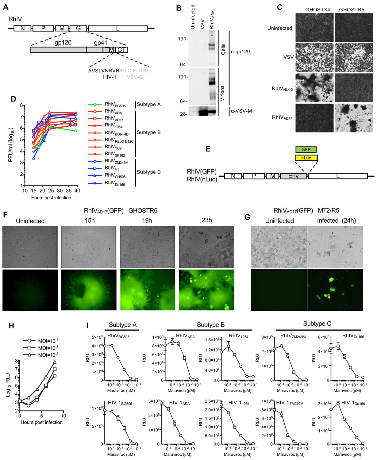
Characterization and in vitro replication properties of RhIV strains.
(A) Schematic representation of RhIV genomes in which VSV-G ectodomain and transmembrane sequences are replaced with HIV-1 Env counterparts. (B) Western blot analysis of HIV-1 gp160/120 and VSV-M protein levels in RhIV infected cells and extracellular virions. (C) Monolayers of GHOST-X4 or GHOST-R5 cells stained with crystal violet 24 hr after infection with VSV or RhIV strains. (D) Yield of various RhIV strains in plaque forming units/ml (PFU/ml) during replication in 293 T/CD4/CCR5 cells. (E) Schematic representation of RhIV genomes in which a GFP or nanoluciferase (nLuc) reporter is included. (F,G) Micrographs of GHOST-R5 (F) and MT4-R5 (G) at the indicated times after infection with RhIVAD17(GFP). (H) Luciferase expression in TZM-Bl cells over time following infection with RhIVAD17(nLuc) at the indicated MOIs. (I) Inhibition of RhIV(nLuc) and corresponsing HIV-1(nLuc) strains by the CCR5 inhibitor, Maraviroc.
Figure 2
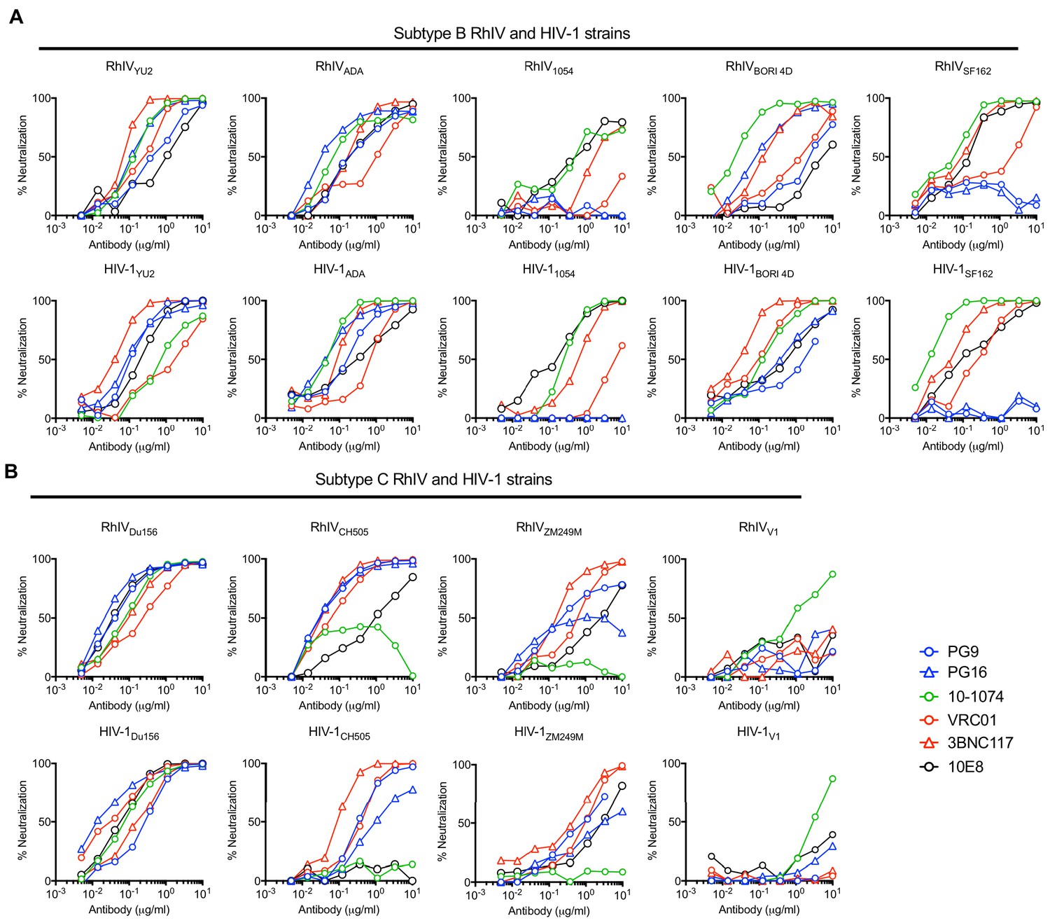
Neutralization properties of matched RhIV and HIV-1 strains.
(A, B) The indicated RhIV(nLuc) and HIV-1(nLuc) strains bearing subtype B (A) and subtype C (B) envelope proteins were incubated with broadly neutralizing antibodies targeting a V2 quaternary epitope (PG9,PG16), the V3 loop (10–1074), the CD4 binding site (VRC01, 3BNC17) or the MPER (10E8), prior to infection of TZM-Bl cells.
Figure 3
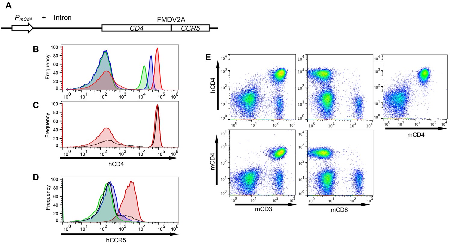
Transgenic mice with CD4+ T-cells that express human CD4 and CCR5.
(A) Schematic representation of the transgene construct that contains a murine Cd4 promoter and intron 1, linked to human CD4 and CCR5 cDNAs separated by sequences encoding an FMDV 2A termination/reinitiation site. (B) FACS analysis of hCD4 expression on unfractionated PBMC from three CD4+/CCR5+ transgenic mouse lines: A1 (red histogram) C18 (blue histogram) and B4 (green histogram). (C) FACS analysis of hCD4 expression on unfractionated PBMC from transgenic mouse line A1 (red histogram) and a human PBMC donor (black line). (D) FACS analysis of CCR5 expression on hCD4+ cells from A1 (red histogram) C18 (blue histogram) and B4 (green histogram) mouse lines and a human PBMC donor (black line). (E) FACS analysis of hCD4 expression in combination with mCD3, mCD8 or mCD4.
Figure 4 with 1 supplement
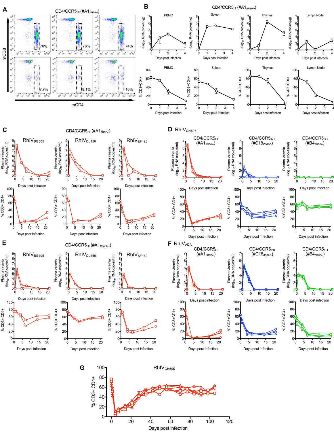
RhIV replication and pathology following infection of hCD4/hCCR5 transgenic mice.
(A) FACS analysis of CD4 and CD8 expression on T-cells (gated on CD3+ cells) in three A1Ifnar-/- mice prior to RhIV infection (upper row) and 3 days after RhIVCH505 infection (lower row). The % of CD3+ cells that were CD4+ is indicated. (B) RhIV RNA levels (log10 copies /µg total RNA, upper row) and CD4+ T-cell numbers (% of CD3+ cells, lower row) in A1Ifnar-/- mouse tissues following infection with RhIVCH505. Values are the mean ± sd of three mice at each time point. (C–F) RhIV viremia (log10 RNA copies/ml of plasma, upper rows) and blood CD4+ T-cell proportion (% of CD3+ cells, lower rows) in A1Ifnar-/- mice (C), A1Ifnar-/-, C18Ifnar-/-, and B4Ifnar-/- mice (D, F) or A1Ifnar+/+ mice (E) at the indicated times following infection with the indicated RhIV strains. Each symbol type on each chart represents an individual mouse (n = 2 to 4 for each virus/mouse strain combination) (G) Blood CD4+ T-cell proportion (% of CD3+ cells) in A1Ifnar-/- mice at the indicated times following infection with RhIVCH505. Each symbol type represents an individual mouse (n = 4).
Figure 4—figure supplement 1
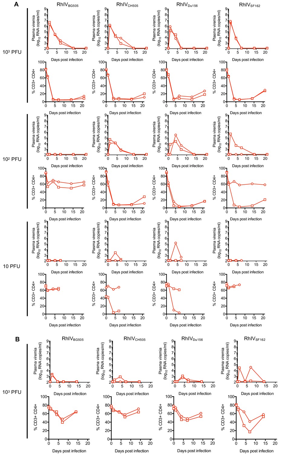
In vivo RhIV titrations.
(A, B) RhIV viremia (log10 RNA copies/ml of plasma, upper rows) and blood CD4+ T-cell proportion (% of CD3+ cells, lower rows) in A1Ifnar-/- mice (A) or A1Ifnar+/+ (B) at the indicated times following infection with 103 PFU, 102 PFU, or 10 PFU (as indicated), of RhiVBG505, RhiVCH505, RhiVDu156, or RhiVSF162. Each symbol type represents an individual mouse (n = 2 for each virus/dose combination).
Figure 5

Protection against RhIV infection by bNAbs.
(A, B) RhIV viremia (log10 RNA copies/ml of plasma, upper rows) and blood CD4+ T-cell proportion (% of CD3+ cells, lower rows) in A1Ifnar+/+ mice (A) or A1Ifnar-/- (B) at the indicated times following infection with RhIVBG505 (A) or RhIVCH505 (B). At 24 hr prior to infection mice were injected (s.c.) with PBS (control) or 1 mg of PG16 or 3BNC117 antibodies (A) or the indicated dose of 3BNC117 antibody (B). Each symbol type represents an individual mouse (n = 2 (A) or n = 3 (B) for each virus/antibody combination).
Figure 6 with 6 supplements

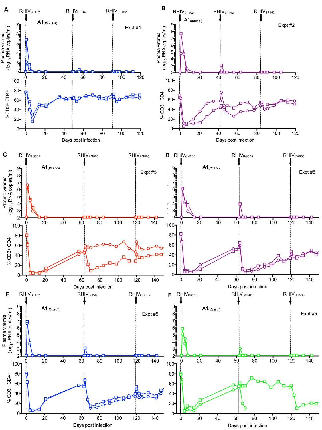
Protection against RhIV reinfection.
(A) RhIV viremia (log10 RNA copies/ml of plasma, upper rows) and blood CD4+ T-cell proportion (% of CD3+ cells, lower rows) in A1Ifnar-/- mice following infection with RhIVSF162 on days 0, 49 and 91. Each symbol type represents an individual mouse (n = 4). (B, C) RhIV viremia (log10 RNA copies/ml of plasma, upper rows) and blood CD4+ T-cell proportion (% of CD3+ cells, lower rows) in A1Ifnar+/+ mice (B) and A1Ifnar-/- mice (C) following infection with RhIVDu156, RhIVBG505, and RhIVSF162 on days, 0, 42 and 91, respectively. Each symbol type represents an individual mouse (n = 4). (D) RhIVBG505 and VSVMLV-E viremia (log10 RNA copies/ml of plasma, upper rows) and blood CD4+ T-cell proportion (% of CD3+ cells, lower rows) in A1Ifnar-/- mice following infection with RhIVBG505 on day 0 and day 28 (left panels), VSVMLV-E on day 0 and RhIVBG505 day 28 (center panels) or RhIVBG505 on day 0 and VSVMLV-E day 28 (right panels).
Figure 6—figure supplement 1
Repeat infections of mice with homologous or heterologous RhIV strains.
(A) RhiV viremia (log10 RNA copies/ml of plasma, upper rows) and blood CD4+ T-cell proportion (% of CD3+ cells, lower rows) in #A1Ifnar+/+ mice following infection with RhiVSF162 on day 0, day 48, and day 91. Each symbol type represents an individual mouse (n = 2). (B) RhiV viremia (log10 RNA copies/ml of plasma, upper rows) and blood CD4+ T-cell proportion (% of CD3+ cells, lower rows) in #A1Ifnar-/- mice following infection with RhiVSF162 on day 0, day 42, and day 84. Each symbol type represents an individual mouse (n = 2). (C) RhiV viremia (log10 RNA copies/ml of plasma, upper rows) and blood CD4+ T-cell proportion (% of CD3+ cells, lower rows) in #A1Ifnar-/- mice following infection with RhiVBG505 on day 0, day 63, and day 119. Each symbol type represents an individual mouse (n = 2). (D) RhiV viremia (log10 RNA copies/ml of plasma, upper rows) and blood CD4+ T-cell proportion (% of CD3+ cells, lower rows) in #A1Ifnar-/- mice following infection with RhiVCH505 on day 0, RhiVBG505 on day 63, and RhiVCH505 on day 119. Each symbol type represents an individual mouse (n = 2). (E) RhiV viremia (log10 RNA copies/ml of plasma, upper rows) and blood CD4+ T-cell proportion (% of CD3+ cells, lower rows) in #A1Ifnar-/- mice following infection with RhiVSF162 on day 0, RhiVBG505 on day 63, and RhiVCH505 on day 119. Each symbol type represents an individual mouse (n = 2). (F) RhiV viremia (log10 RNA copies/ml of plasma, upper rows) and blood CD4+ T-cell proportion (% of CD3+ cells, lower rows) in #A1Ifnar-/- mice following infection with RhiVDu156 on day 0, RhiVBG505 on day 63, and RhiVCH505 on day 119. Each symbol type represents an individual mouse (n = 2, one mouse died on day 68).
Figure 6—figure supplement 2
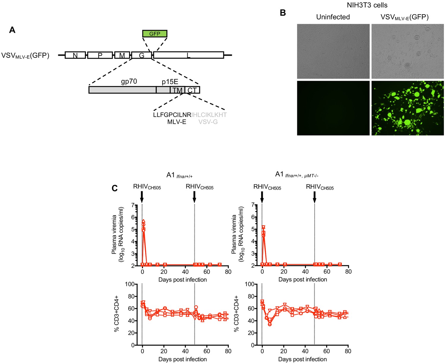
Construction of VSVMLV-E and Vaccine effect of RhIV infection does not require B-cells.
(A) Schematic representation of the VSVMLV-E genome in which the VSV-G ectodomain and transmembrane sequences are replaced with MLV-E Env counterparts and a GFP reporter is placed 3’ to Env coding sequences (B) Micrograph of NIH3T3 cells 24 hr after infection with VSVMLV-E(GFP). (C) RhiV viremia (log10 RNA copies/ml of plasma, upper rows) and blood CD4+ T-cell proportion (% of CD3+ cells, lower rows) in #A1Ifnar+/+ mice (left panels) and #A1Ifnar+/+, µMT-/- mice (right panels) following infection with RhiVCH505 on day 0 and day 49. Each symbol type represents an individual mouse (n = 4).
Figure 6—figure supplement 3
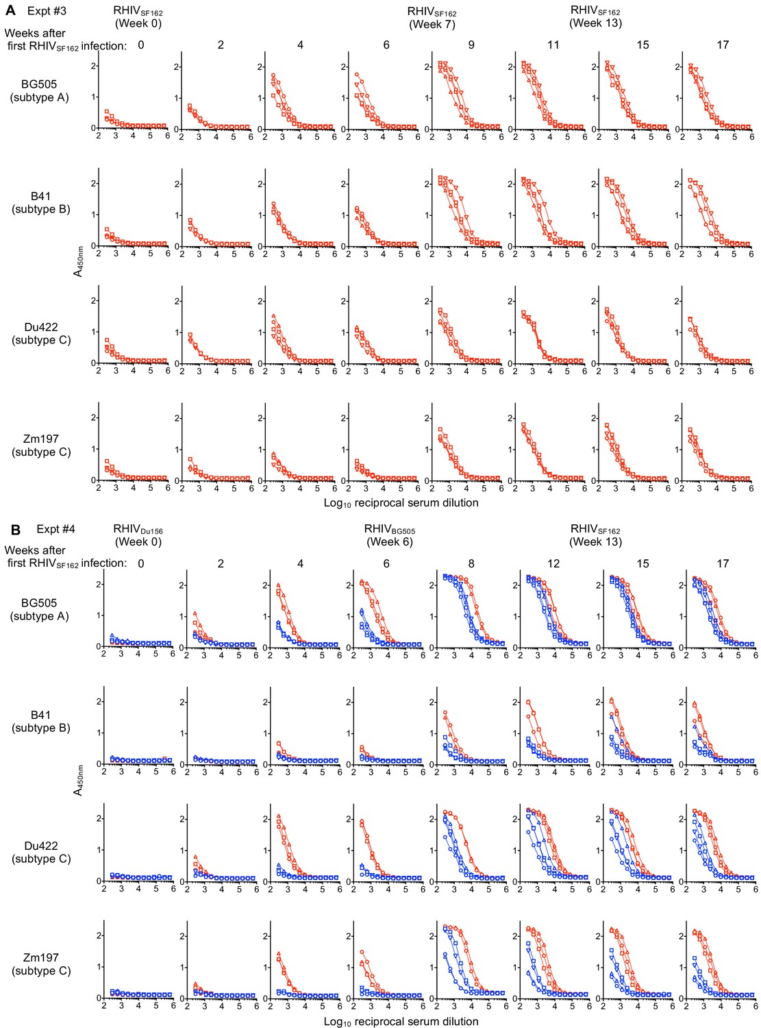
Gp160 (SOSIP) binding antibodies in RhIV infected mice (Expt #3 and #4).
(A,B) ELISA measurement of antibodies that bind to plates coated with gp160 (SOSIP) proteins from BG505 (subtype A), B41 (subtype B), Du422 (subtype C) and Zm197 (subtype C) HIV-1 strains. Absorbance at 450 nm (A450nm) plotted against Log10 reciprocal serum dilution (A) Serum from Expt #3 A1Ifnar-/- mice infected with RhIVSF162 on day 0, day 49 (week 7) and 91 (week 13). Each symbol type represents an individual mouse (n = 4) and corresponds to the mice depicted in Figure 6A. (B) Serum from Expt #4a A1Ifnar+/+ mice (blue symbols/lines) and Expt #4b A1Ifnar-/- mice (red symbols and lines) infected with RhiVDu156 on day 0, RhiVBG505 on day 42 (week 6), and RhiVSF162 on day 91 (week13). Each symbol type represents an individual mouse (n = 4) and corresponds to the mice depicted in Figure 6B and C.
Figure 6—figure supplement 4
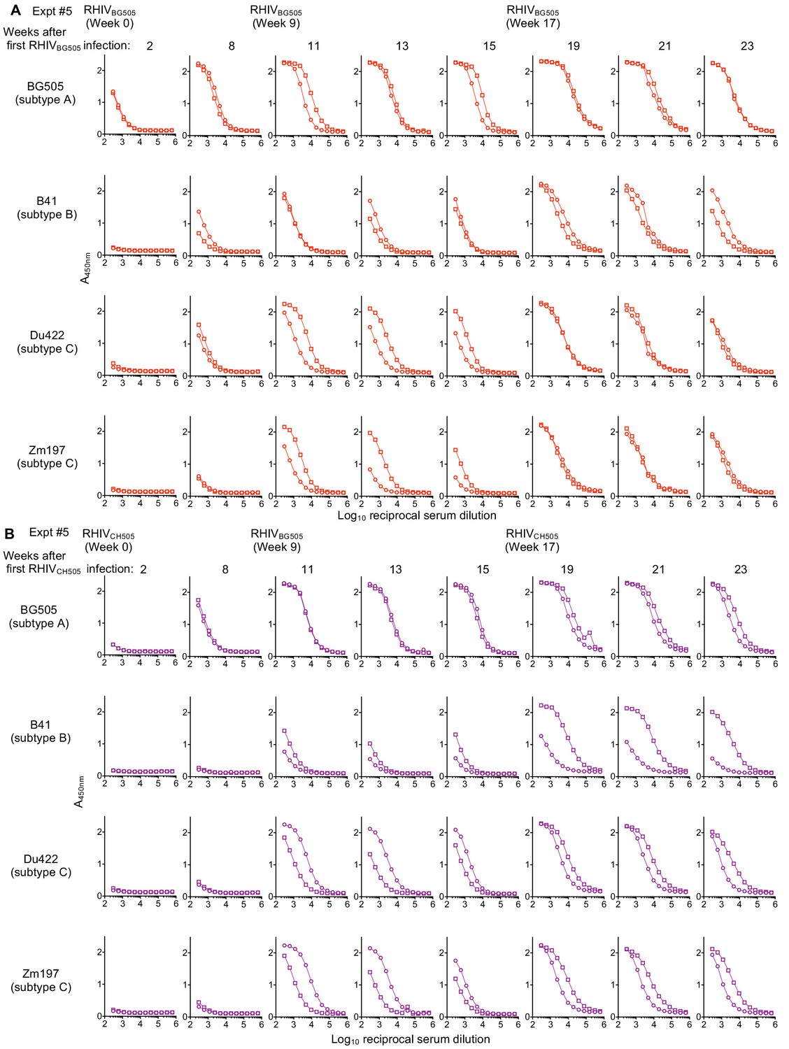
Gp160 (SOSIP) binding antibodies in RhIV infected mice (Expt #5).
(A,B) ELISA measurement of antibodies that bind to plates coated with gp160 (SOSIP) proteins from BG505 (subtype A), B41 (subtype B), Du422 (subtype C) and Zm197 (subtype C) HIV-1 strains. Absorbance at 450 nm (A450nm) plotted against Log10 reciprocal serum dilution (A) Serum from Expt #5 A1Ifnar-/- mice infected with RhIVBG505 on day 0, day 63 (week 9) and day 119 (week 17). Each symbol type represents an individual mouse (n = 2) Symbols and line colors corresponds to the mice depicted in Figure 6—figure supplement 1C. (B) Serum from Expt #5 A1Ifnar-/- mice infected with RhiVCH505 on day 0, RhiVBG505 on day 63 (week 9), and RhiVCH505 on day 119 (week 17). Each symbol type represents an individual mouse (n = 2). Symbols and line colors corresponds to the mice depicted in Figure 6—figure supplement 1D.
Figure 6—figure supplement 5
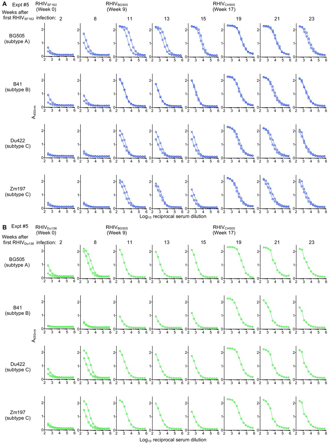
Gp160 (SOSIP) binding antibodies in RhIV infected mice (Expt #5).
(A,B) ELISA measurement of antibodies that bind to plates coated with gp160 (SOSIP) proteins from BG505 (subtype A), B41 (subtype B), Du422 (subtype C) and Zm197 (subtype C) HIV-1 strains. Absorbance at 450 nm (A450nm) plotted against Log10 reciprocal serum dilution. (A) Serum from Expt #5 A1Ifnar-/- mice infected with RhIVSF162 on day 0, RhIVBG505 on day 63 (week 9) and RhIVCH505 on day 119 (week 17). Each symbol type represents an individual mouse (n = 2) Symbols and line colors corresponds to the mice depicted in Figure 6—figure supplement 1E. (B) Serum from Expt #5 A1Ifnar-/- mice infected with RhiVDu156 on day 0, RhiVBG505 on day 63 (week 9), and RhiVCH505 on day 119 (week 17). Each symbol type represents an individual mouse (n = 2). Symbols and line colors corresponds to the mice depicted in Figure 6—figure supplement 1F.
Figure 6—figure supplement 6
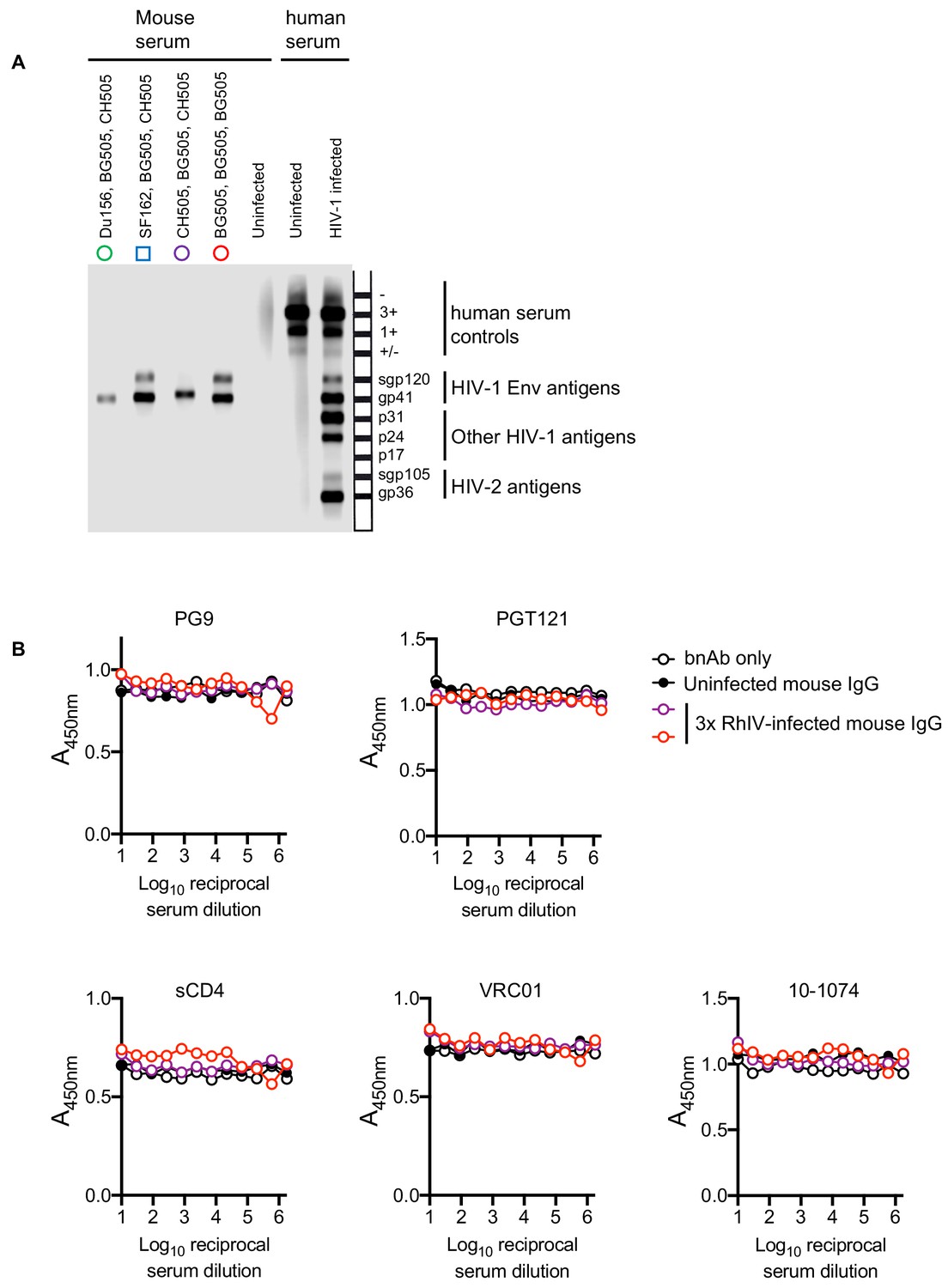
Characterization of antibodies in RhIV infected mice.
(A) INNO-LIA HIV I/II Score assay using sera from mice (Exp #5) Schematic on the right shows the position of the antigens deposited on each strip. Bands in the upper portion of the strips are controls for the presence of human serum and are not expected to react with mouse serum. Symbols and their colors above each lane correspond to the individual mice depicted in Figure 6—figure supplement 1. (B) Competition ELISA in which the indicated dilutions of IgG, purified from mouse sera, were tested for ability to block the binding of the indicated bnAbs to BG505 SOSIP.664 coated ELISA plates. Symbols and their colors correspond to the individual mice depicted in Figure 6—figure supplement 1.
Figure 7 with 1 supplement
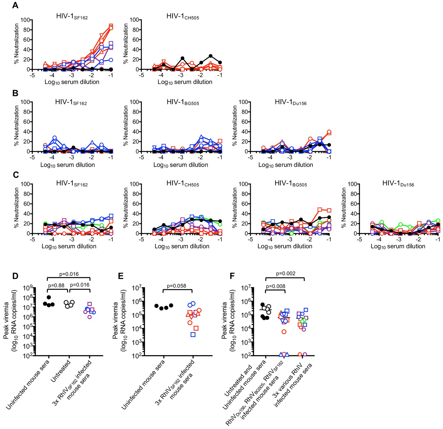
Neutralization and passive protection activity in sera from RhIV infected mice.
(A) Neutralization (using TZM-Bl target cells) of HIV-1(nLuc) strains by immunoglobulins purified from convalescent mouse sera after three infections with RhIVSF162. Symbol types and line colors correspond to individual mice in Expts #1 to #3 (depicted in Figure 6A, Figure 6—figure supplement 1A and B). Black symbols indicate sera from uninfected mice (B) Neutralization of HIV-1(nLuc) strains by immunoglobulins purified from convalescent mouse sera after infection with RhIVDu156, RhIVBG505 and RhIVSF162 in Expt #4. Symbol types and line colors correspond to individual mice (depicted in Figure 6B and C). (C) Neutralization (using TZM-Bl target cells) of HIV-1(nLuc) strains by immunoglobulins purified from convalescent mouse sera after three infections with various RhIV strains in Expt #5. Symbol types and line colors correspond to individual mice depicted in Figure 6—figure supplement 1C,D,E and F. (D) Peak viremia (day one post infection with 105 PFU RhIVSF162) following no treatment or passive administration of sera from uninfected mice, or mice that had previously been infected three times with RhIVSF162. Colored symbols indicate different donor mice, matched to correspond to donor mice from Expt #1 and #2 (depicted in Figure 6—figure supplement 1A and B). Black closed circles indicate sera from uninfected mice, Black open circles indicate no serum treatment. (E) Peak viremia (day one post infection with 103 PFU RhIVSF162) following passive administration of sera from uninfected mice, or mice that had previously been infected three times with RhIVSF162. Colored symbols indicate different donor mice, matched to correspond to donor mice from Expt #1 - #3 (depicted in Figure 6A, Figure 6—figure supplement 1A and B). (F) Peak viremia (day one post infection with 103 PFU RhIVSF162) following no treatment, passive administration of sera from uninfected mice, or mice that had previously been infected with RhIVDu156, RhIVBG505 and RhIVSF162 (Expt #4). Alternatively, serum from mice sequentially infected with various combinations of three RhIV strains (Expt #5) were used. Colored symbols indicate different donor mice, matched to correspond to donor mice from Expt #4 or Expt #5 (depicted in Figure 6B and C, or Figure 6—figure supplement 1C,D,E and F).
Figure 7—figure supplement 1
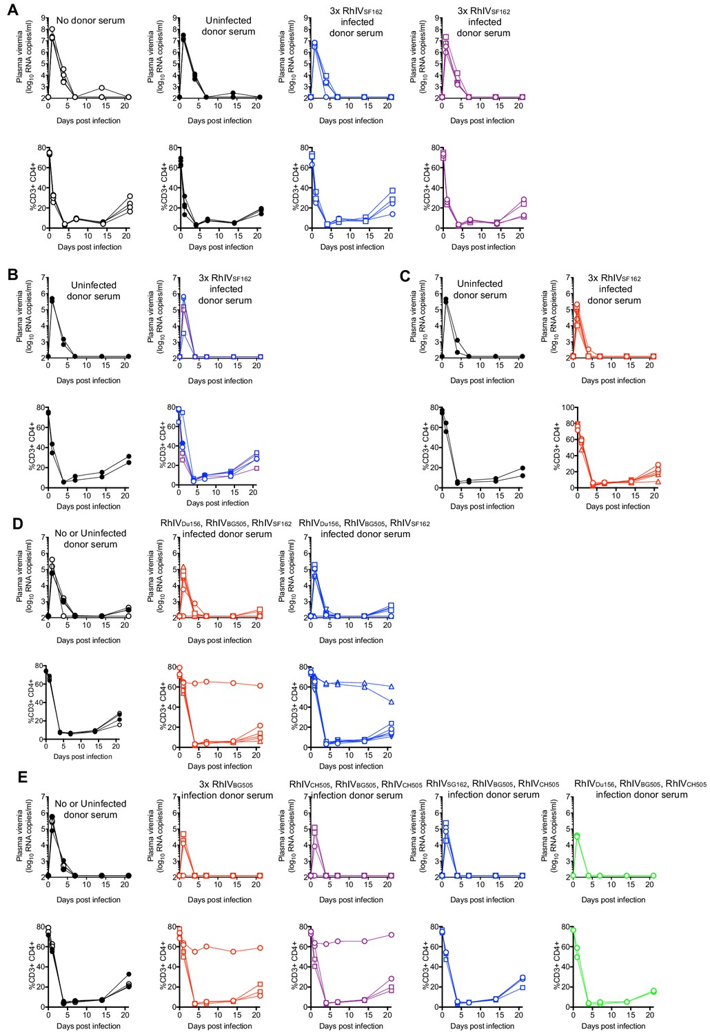
Passive serum transfer/protection experiments using donor serum from RhIV infected mice.
(A–E) RhiV viremia (log10 RNA copies/ml of plasma, upper rows) and blood CD4+ T-cell proportion (% of CD3+ cells, lower rows) in recipient #A1Ifnar-/- mice following infection with 105 PFU of RhiVSF162 (A) or 103 PFU of RhiVSF162 (B–E). One day prior to infection, recipient mice received either no treatment or passive administration of sera from uninfected mice, or mice that had previously been infected three times with RhiV strains in Expt #1 -#5 as detailed below. Serum was administered subcutaneously one day before intraperitoneal RhIV challenge. Each colored symbol type indicates a different donor mouse, and is matched to correspond to donor mice from experiments depicted in Figure 6 and Figure 6—figure supplement 1. (A) Donor sera were from Expt #1 and #2 mice that had been infected three times with RhiVSF162 and are depicted in Figure 6—figure supplement 1A and B (B) Donor sera were from Expt #1 and #2 mice that had been infected three times with RhiVSF162 and are described in Figure 6—figure supplement 1A and B (C) Donor sera were from Expt #3 mice that had been infected three times with RhiVSF162 and are described in Figure 6A (D) Donor sera were from Expt #4 mice that had been infected once each with RhiVDu156, RhiVBG505, and RhiVSF162 and are described in Figure 6B and 6C (E) Donor sera were from Expt #5 mice that had been infected with various RhIV strains and are described in Figure 6—figure supplement 1C,D,E and F.
Videos
Video 1
Spreading replication of RhIVAD17(GFP).
Cells (293 T/CD4/CCR5) were infected with RhIVAD17(GFP) at low MOI (0.0001) and placed in VivaView FL incubator fluorescence microscope imaging system (Olympus). At 6 hr after infection, individual GFP positive cells were identified and centered in a field of observation and images acquired every 5 min thereafter. The movie represents 24 hr of observation (from 6 hr to 30 hr after infection).
Tables
Key resources table
| Reagent type (species) or resource | Designation | Source or reference | Identifiers | Additional information |
|---|---|---|---|---|
| Strain, strain background (Vesicular Stomatitis Virus) | pVSV-FL+(2) Plasmid Expression Vector System | Kerafast | Cat#EH1002 | Anti-genomic sense plasmid with helper plasmids N, P, G and L |
| Genetic reagent (Mus musculus) | C57BL/6J-Tg(Cd4-CD4,CCR5)A1Bsz; C57BL/6J-Tg(Cd4-CD4,CCR5)C18Bsz; C57BL/6J-Tg(Cd4-CD4,CCR5)B4Bsz | This paper | Mouse lines with CD4 cell-specific expression of human CD4 and CCR5 | |
| Cell line (H. sapiens) | 293T | ATCC | CRL-3216 | |
| Cell line (H. sapiens) | GHOSTX4; GHOSTR5 | NIH AIDS Reagent Repository | Cat#3685;3944 | |
| Cell line (H. sapiens) | MT2 | NIH AIDS Reagent Repository | Cat#237 | |
| Antibody | Anti-mouse CD16/CD32 (purified rat monoclonal) | BD Pharmingen | Cat#553142 | FACS (2 uL per test) |
| Antibody | FITC Anti-mouse CD3 (rat monoclonal) | BD Pharmingen | Cat#555274 | FACS (2 uL per test) |
| Antibody | PerCP-Cy5.5 Anti-mouse CD4(rat monoclonal) | BD Pharmingen | Cat#550954 | FACS (2 uL per test) |
| Antibody | APC Anti-mouse CD8a (rat monoclonal) | Biolegend | Cat#100712 | FACS (1 uL per test) |
| Antibody | APC-Cy7 Anti-human CD4(mouse monoclonal) | Biolegend | Cat#317418 | FACS (2 uL per test) |
| Antibody | PE Anti-mouse CD19 (rat monoclonal) | BD Pharmingen | Cat#553786 | FACS (1 uL per test) |
| Antibody | PE Anti-human CD195/CCR5 (mouse monoclonal) | BD Pharmingen | Cat#560935 | FACS (2.5 uL per test) |
| Antibody | PE Anti-human CD195 (mouse monoclonal) | BD Pharmingen | Cat#550632 | FACS (2.5 uL per test) |
| Antibody | AlexaFluor 647 Anti-human CD4 (mouse monoclonal) | Biolegend | Cat#300520 | FACS (3 uL per test) |
| Antibody | Anti-HIV-1 gp120 (goat polyclonal) | American Research Products | Cat#12-6205-1 | WB (1:1000) |
| Antibody | Anti-VSV M (mouse monoclonal) | Kerafast | Cat#EB0011 | WB (1:2000) |
| Antibody | His-Tag Antibody (pAb, Rabbit) | GenScript | A00174-40 | ELISA coating at 0.5 mg/ml |
| Antibody | Goat anti-mouse IgG H and L (HRP) preadsorbed | Abcam | Ab97040 | ELISA (1:20000) |
| Antibody | Goat anti-human IgG H and L (HRP) preadsorbed | Abcam | Ab97175 | ELISA (1:20000) |
| Recombinant DNA reagent | pLHCX hCD4 2A CCR5 (plasmid) | This paper | Retroviral vector with human CD4/CCR5 | |
| Recombinant DNA reagent | pNL1.1 (plasmid) | Promega | #N1001; GenB:JQ437370 | Nanoluciferase cDNA |
| Recombinant DNA reagent | pAAVCMV_BG505-His | This paper | plasmid expressing his-tagged BG505 SOSIP | |
| Recombinant DNA reagent | pAAVCMV_B41-His | This paper | plasmid expressing his-tagged B41 SOSIP | |
| Recombinant DNA reagent | pAAVCMV_Du422-His | This paper | plasmid expressing his-tagged Du422 SOSIP | |
| Recombinant DNA reagent | pAAVCMV_Zm197-His | This paper | plasmid expressing his-tagged Zm197 SOSIP | |
| Sequence-based reagent | RL413 | This paper | Genotyping PCR primer | GAACCTGGTGGTGATGAGAGCCACTCA |
| Sequence-based reagent | RL425 | This paper | Genotyping PCR primer | TGCTTGCTTTAACAGAGAGAAGTTCGT |
| Sequence-based reagent | RL509 | PMID: 16617693 | RT-qPCR primer | TGATACAGTACAATTATTTTGGGAC |
| Sequence- based reagent | RL510 | PMID: 16617693 | RT-qPCR primer | GAGACTTTCTGTTACGGGATCTGG |
| Chemical compound, drug | Maraviroc | NIH AIDS Reagent Repository | Cat#11580 |
Additional files
Download links
A two-part list of links to download the article, or parts of the article, in various formats.
Downloads (link to download the article as PDF)
Open citations (links to open the citations from this article in various online reference manager services)
Cite this article (links to download the citations from this article in formats compatible with various reference manager tools)
Rhabdo-immunodeficiency virus, a murine model of acute HIV-1 infection
eLife 8:e49875.
https://doi.org/10.7554/eLife.49875


