Mutation of vsx genes in zebrafish highlights the robustness of the retinal specification network

  1. Joaquín Letelier  Is a corresponding author
  2. Lorena Buono
  3. María Almuedo-Castillo
  4. Jingjing Zang
  5. Constanza Mounieres
  6. Sergio González-Díaz
  7. Rocío Polvillo
  8. Estefanía Sanabria-Reinoso
  9. Jorge Corbacho
  10. Ana Sousa-Ortega
  11. Ruth Diez del Corral
  12. Stephan CF Neuhauss
  13. Juan R Martínez-Morales  Is a corresponding author
  1. Centro Andaluz de Biología del Desarrollo (CSIC/UPO/JA), Spain
  2. Centre for Integrative Biology, Facultad de Ciencias, Universidad Mayor, Chile
  3. IRCCS SYNLAB SDN, Via E. Gianturco, Italy
  4. Department of Molecular Life Sciences, University of Zürich, Switzerland
  5. Champalimaud Research, Champalimaud Centre for the Unknown, Portugal

Abstract

Genetic studies in human and mice have established a dual role for Vsx genes in retina development: an early function in progenitors’ specification, and a later requirement for bipolar-cells fate determination. Despite their conserved expression patterns, it is currently unclear to which extent Vsx functions are also conserved across vertebrates, as mutant models are available only in mammals. To gain insight into vsx function in teleosts, we have generated vsx1 and vsx2 CRISPR/Cas9 double knockouts (vsxKO) in zebrafish. Our electrophysiological and histological analyses indicate severe visual impairment and bipolar cells depletion in vsxKO larvae, with retinal precursors being rerouted toward photoreceptor or Müller glia fates. Surprisingly, neural retina is properly specified and maintained in mutant embryos, which do not display microphthalmia. We show that although important cis-regulatory remodelling occurs in vsxKO retinas during early specification, this has little impact at a transcriptomic level. Our observations point to genetic redundancy as an important mechanism sustaining the integrity of the retinal specification network, and to Vsx genes regulatory weight varying substantially among vertebrate species.

Editor's evaluation

This study provides important insights into how tissue specification networks, while often employing conserved genes across species, can differ in their network architecture, resulting in differences in how they buffer perturbations. This is shown for the Visual System Homeobox genes (VSX) in the zebrafish retinal specification pathway, where yet-to-be-defined compensatory mechanisms prevent microphthalmia in the absence of VSX function, something not observed in humans or mice. The evidence supporting the conclusions of the study is solid and provides a foundation for further molecular and genetic analysis of retinal specification. This work is relevant to developmental biologists with interests in tissue specification and gene regulatory networks.

https://doi.org/10.7554/eLife.85594.sa0

Introduction

The organogenesis of the vertebrate eye is a complex multistep process entailing the sequential activation of genetic programs responsible for the initial specification of the eye field, the patterning of the eye primordium into sub-domains, and the determination of the different neuronal types. Although we are far from understanding the precise architecture of the gene regulatory networks (GRNs) controlling eye formation, many of their central nodes have been already identified (Buono and Martinez-Morales, 2020; Fuhrmann, 2010; Heavner and Pevny, 2012; Martinez-Morales, 2016). They comprise transcriptional regulators recruited repeatedly for key developmental decisions at different stages of eye formation, and which mutation in humans is often associated to severe ocular malformations: that is, microphthalmia, anophthalmia, and coloboma. This is the case for SIX3, PAX6, RAX, SOX2, VSX2, or OTX2 (Gregory-Evans et al., 2004; Gregory-Evans et al., 2013).

Among the main regulators, the visual system homeobox transcription factors, Vsx1 and Vsx2, have been shown to control the development of visual circuits in vertebrate and invertebrate species (Burmeister et al., 1996; Erclik et al., 2008; Focareta et al., 2014). Vsx2, initially termed as Chx10, was the first gene of the family characterized in vertebrates (Liu et al., 1994). Vsx2/Chx10 shows a conserved expression pattern across vertebrate species, both in the retina (i.e. early in all optic cup precursors, and later in retinal bipolar cells), as well as in hindbrain and spinal cord interneurons (Ferda Percin et al., 2000; Kimura et al., 2013; Liu et al., 1994; Passini et al., 1997). A nonsense mutation in Vsx2 (Y176stop) turned to be the molecular cause of the phenotype exhibited by the classical mutant mice ocular retardation (or), which displays microphthalmia and optic nerve aplasia (Burmeister et al., 1996; Truslove, 1962). The phenotypic analysis of or mutants, as well as the examination of human patients with hereditary microphthalmia, revealed an essential role for Vsx2 in neuro-epithelial proliferation and bipolar cells differentiation (BarYosef et al., 2004; Burmeister et al., 1996; Ferda Percin et al., 2000). Subsequent studies indicated that, during optic cup formation, Vsx2 is a key factor in the binary decision between neural retina and retinal-pigmented epithelium (RPE) lineages. Genetic studies in mice and chick revealed that Vsx2 acts, downstream of the neural retina inducing ligands (i.e. FGFs), as a repressor of Mitf and Tfec genes and hence of the RPE identity (Horsford et al., 2005; Nguyen and Arnheiter, 2000; Rowan et al., 2004).

A few years after Vsx2 identification, a closely related paralog, Vsx1, was reported in several vertebrate species (Chen and Cepko, 2000; Chow et al., 2001; Levine et al., 1994; Passini et al., 1997). The proteins encoded by these paralogous genes have similar domains’ architecture, including well-conserved paired-like homeodomain and CVC (Chx10/Vsx-1 and ceh-10) regulatory modules, and share biochemical properties, binding with high affinity to the same DNA sequence motif ‘TAATTAGC’ (Capowski et al., 2016; Dorval et al., 2005; Ferda Percin et al., 2000; Heon, 2002). Although both genes display partially overlapping expression patterns in the retina, Vsx2 precedes Vsx1 expression in undifferentiated progenitors in all vertebrate models analysed. Furthermore, once retinal precursors exit the cell cycle, they are expressed in complementary sets of differentiated bipolar cells. Thus, Vsx1 is restricted to different types of ON and OFF cone bipolar cells in mice, and Vsx2 to S4 bipolar and Müller cells in zebrafish (Ohtoshi et al., 2004; Shi et al., 2011; Vitorino et al., 2009). In contrast to Vsx2, Vsx1 seems to have a minor contribution to retinal specification in mammals. A single case of sporadic microphthalmia has been associated to Vsx1 mutation in humans (Matías-Pérez et al., 2018), and its mutation in mice does not affect early retinal development even in a Vsx2 mutant background (Chow et al., 2004; Clark et al., 2008). However, Vsx1 mutation has been linked to inherited corneal dystrophies in humans, and is associated to abnormal electroretinogram (ERGs) recordings either in mice or in patients (Chow et al., 2004; Heon, 2002; Mintz-Hittner et al., 2004).

Despite all these advances on the developmental role of Vsx genes, many questions remain open. A fundamental issue is to understand to which extent Vsx gene functions are conserved across vertebrates. Previous antisense oligonucleotides or morpholino studies in zebrafish have shown that vsx2 knockdown results in microphthalmia and optic cup folding defects (Barabino et al., 1997; Clark et al., 2008; Gago-Rodrigues et al., 2015; Vitorino et al., 2009). However, these findings have not been validated using knockout lines, neither the role of vsx1 and vsx2 in fate determination and bipolar cells differentiation has been sufficiently explored in teleost fish.

To gain insight into the universality and diversity of Vsx functions, we have generated zebrafish mutants for vsx1 and vsx2 harboring deletions within the homeodomain-encoding exons. Surprisingly, eye morphology and size appear normal either in the individual or in the double vsx1/vsx2 mutants, thus indicating that vsx genes are not essential to initiate retinal development in zebrafish. The absence of early retinal malformations facilitates the phenotypic analysis of the mutants at later embryonic and larval stages. Defects in the visual background adaptation (VBA) reflex are observed in vsx1 mutant, and appear enhanced in double mutant larvae, suggesting partial or complete blindness. Analysis of ERG responses confirms vision loss, showing that the amplitude of the b-wave recordings is reduced in vsx1 mutants, and absent in double mutants. Interestingly, a single wild type copy of vsx1 is sufficient to prevent VBA and ERG defects, indicating that vsx2 loss of function can be compensated by vsx1. The analysis of neuronal-specific markers confirmed that retinal progenitors fail to differentiate into bipolar cells in double mutant embryos. Instead, we show that precursors at the inner nuclear layer (INL) can remain proliferative, undergo apoptosis, or be rerouted toward other retinal lineages, particularly differentiating as Müller glial cells. Finally, we investigate whether transcriptional adaptation (El-Brolosy et al., 2019) may compensate for vsx1/vsx2 loss-of-function during retinal specification. The transcriptomic analysis of core components of the retinal specification GRN do not support a transcriptional adaptation mechanism in vsx1/vsx2 double mutants, rather suggesting that the network robustness is by itself sufficient to sustain early eye development even in the absence of vsx1 and vsx2 function. In summary, whereas our work shows a conserved role for Vsx genes during bipolar cell differentiation, also indicates that their hierarchic weight within the eye GRNs varies considerably across vertebrate species.

Results

Zebrafish vsx double mutants show normal eye size but affected lamination of the retina

Despite the additional round of genome duplication occurring in the teleost lineage after the split with sarcopterygians (Meyer and Schartl, 1999), a single copy of both vsx1 and vsx2 was retained in zebrafish. In order to investigate the role of Vsx transcription factors during visual system formation in zebrafish, we generated mutants for both paralogs using CRISPR/Cas9. To optimize the generation of null animals, we targeted conserved regions encoding for the DNA binding domain of the proteins in their corresponding loci at chromosome 17 (Figure 1a). We generated a 245 bp deletion in vsx1 encompassing exon3, intron3, and exon4 of the gene (vsx1∆245). This mutation results in an in-frame deletion of 53 amino acids by the removal of 159 bp from exon3 (54 bp) and exon4 (105 bp; Figure 1—figure supplement 1a). In the case of vsx2, a 73 bp deletion was generated in exon 3 (vsx2∆73). This mutation deletes 24 amino acids of the core DBD of the protein and generates a premature stop codon in that domain (Figure 1—figure supplement 1b). Both deletions can be easily screened by PCR with primers flanking the mutation sites. Using Vsx1- and Vsx2-specific antibodies, we found that no Vsx2 or Vsx1 proteins could be detected by western blot in 24hpf vsxKO samples (Figure 1—figure supplement 1c, d). In addition, no maternal Vsx1 protein was detected in early 1.5hpf wildtype embryos (Figure 1—figure supplement 1c).

Figure 1 with 3 supplements see all
DNA-binding domain deletion of vsx genes affect neural retina formation and disrupt VBA reflex.

(a) CRISPR/Cas9 DNA editing tool was used to generate deletions (green box) in the highly conserved DBD from vsx1 (top) and vsx2 (bottom) TFs. Blue boxes represent gene exons, black boxes the location of sgRNAs used to guide Cas9 endonuclease and primers for screening are depicted as opposing arrowheads. b-d and f-h. Histological sections stained with nuclear marker DAPI and phalloidin-Alexa488 for actin filaments from WT (b-d, n≥8) and vsxKO central retinas (f-h, n≥10) at 48hpf (b, f), 72hpf (c, g) and 6dpf (d, h). (e, i). Head dorsal view from 6dpf WT (e) and vsxKO (i) larvae with insets showing their pigmentation pattern (white arrowhead). ONL: outer nuclear layer, INL: inner nuclear layer, GCL: ganglion cell layer, hpf: hours post-fertilization, dpf: days post-fertilization. Scale bar in (b-d) and (f-h): 50 µm, scale bar in e and i: 500 µm.

At 2-week post fertilization, no obvious macroscopic defects were observed in the visual system of either homozygous single mutants (i.e. vsx1∆245 or vsx2∆73) or homozygous double mutants vsx1∆245; vsx2∆73 (here termed vsxKO), which appeared normal in shape and size (Figure 1—figure supplement 2; Figure 1—figure supplement 3a–d). Homozygous single mutants, and even animals harboring a single wild type copy either of vsx1 (vsx1∆245+/-, vsx2∆73-/-) or vsx2 (vsx1∆245-/-; vsx2∆73+/-) reached adulthood and were fertile. However, double mutant larvae (vsx1∆245 -/-; vsx2∆73 -/-) died at around 3-week post fertilization, with the exception of a single unfertile escaper reaching adulthood (1 out of 152 larvae raised). For further analyses, double mutant embryos and larvae were obtained each generation by in-crossing of vsx1∆245+/-; vsx2∆73-/- or vsx1∆245-/-; vsx2∆73+/-animals. Once the proper recombinants were obtained, heterozygous lines maintenance was facilitated by the linkage between vsx1 and vsx2 mutant alleles, which tend to segregate together due to their proximity (10.6 Mb) in chromosome 17.

Histological sectioning of mutant retinas at 48hpf showed a small delay in the formation of the inner plexiform layer (IPL), but no obvious macroscopic optic cup malformations when compared to WT (Figure 1b and f). At 72hpf, both the outer plexiform layer (OPL) and the IPL appeared less organized in the double mutant retinas, which showed discontinuities/fenestrae (Figure 1c and g). At 6dpf, double mutant larvae showed all the layers of a normal retina, but the thickness of the outer (ONL) and inner (INL) nuclear layers was significantly increased and reduced respectively, when compared to siblings (Figure 1d and h; Figure 1—figure supplement 3e, h, i). In addition to retinal layer formation defects, vsxKO fish presented expanded pigmentation in skin melanocytes even when exposed to bright light for 20 min (Figure 1e and i; Figure 1—figure supplement 3a, d). This phenomenon is indicative of an impaired visual background adaptation (VBA) reflex, and is often associated with blindness in zebrafish (Fleisch and Neuhauss, 2006).

Visual function is impaired in single vsx1and vsxKO double mutants

To test the visual performance of the vsx mutants; ERG recordings were obtained from WT and mutants at 5 dpf (Figure 2). Zebrafish retina becomes fully functional at 5 dpf with the exception of late maturing rods (Bilotta et al., 2001) and thus, the recorded field potentials were mainly contributed by cones. Wild type larvae show a standard ERG response to light flash, characterized by a large positive b-wave representing the depolarization of ON bipolar cells (Figure 2a), which also masks the initial a-wave generated by photoreceptor (PR) hyperpolarization. Representative recordings from larvae harboring different vsx genotypes are shown in Figure 2a. We found that vsx2∆73 ERG response (green curve) was similar to the WT recording (blue curve). However, recordings in vsx1∆245 larvae showed a reduced b-wave compared to WT or vsx2∆73 larvae. From the 36 double mutant larvae recorded in total, 10 of them still showed a b-wave, though reduced in comparison to vsx1∆245 mutants, and much smaller than WT recordings. Moreover, in the remaining 26 double mutants recorded only the negative a-wave but not the b-wave (gray curve) was detected, suggesting that bipolar cells differentiation and/or function might be compromised. Statistical analysis of the average amplitude showed that the b-wave is significantly decreased in both vsx1∆245 single and vsxKO double mutants in comparison to WT at all tested light intensities (Figure 2b). In addition, the b-wave response amplitude in the double mutant was significantly reduced compared to vsx1 single mutants (Figure 2b). These measurements are in line with our previous observation indicating that double mutant retinas are more affected at the cellular level than single vsx1∆245 animals (Figure 1—figure supplement 3).

Figure 2 with 1 supplement see all
ERG response is reduced in vsxKO larvae.

(a) Representative ERG tracks at maximum light intensity from WT (blue), vsx1∆245 (red), vsx2∆73 (green) and vsxKO double mutants (grey and yellow) at 5dpf. For vsxKO larvae, two typical recordings are shown (grey and yellow tracks). (b). Averaged ERG b-wave amplitudes from WT (blue), vsx1∆245 (red), vsx2∆73 (green) and vsxKO (yellow) larvae. No significant differences were observed between WT and vsx2∆73 samples. vsx1∆245 and vsxKO mutants produce a significant reduction of the ERG b-wave amplitude compared with both WT and vsx2∆73 larvae throughout all light intensities tested (***p<0.0001, ****p<0.00001). Data are shown as mean ± SEM. In (a) and (b), vsx1∆245 (red tracks) represents both vsx1∆245-/- and vsx1∆245-/-; vsx2∆73+/-genotypes, while vsx2∆73 (green tracks) represents both vsx2∆73-/- and vsx1∆245+/-; vsx2∆73-/- genotypes. Data were collected from five independent experiments. For statistical comparison, one way ANOVA test was used. ms: milliseconds, mV: millivolts.

To quantitatively characterize eye performance, optokinetic response (OKR) recordings (Rinner et al., 2005) were obtained for WT and vsx mutant fish (Figure 2—figure supplement 1). To investigate the role of Vsx transcription factors at the behavioral level, eye movement velocity was recorded at 5dpf in WT and vsxKO mutant fish. We measured eye velocity varying different parameters of the moving stimuli, such as contrast (contrast sensitivity; Figure 2—figure supplement 1a), frequency (spatial resolution; Figure 2—figure supplement 1b) and angular velocity (temporal resolution; Figure 2—figure supplement 1c). In all conditions tested, we observed a significant reduction in eye velocity for vsx1 single and vsxKO double mutants when compared with vsx2∆73 larvae and WT controls (repeated measurement, ANOVA p<0.001). Taken together these physiological recordings confirmed significant sight impairment in vsx1 mutants, a phenotype that is further aggravated by vsx2 loss in vsxKO double mutants.

Extended proliferation wave and INL cell death in vsxKO double mutant retinas

As vsxKO double mutants showed stronger retinal architecture and visual defects than other vsx mutant combinations, we decided to focus further phenotypic analyses on them. To assess whether our observations on the increased thickness of the ONL and the decreased width of the INL (Figure 1—figure supplement 3) correlate with a proliferation and/or cell death unbalance, we examined both parameters in vsxKO fish. To investigate proliferation defects, we quantified the number of phosphohistone H3 positive (PH3+) cells in the retina of wild type and vsxKO animals throughout the lamination process: that is, at 24, 48, 60, and 72hpf (Figure 3a–f and m; Figure 3—figure supplement 1). At 24 and 48hpf, no difference in the number of PH3 + cells were observed between WT and vsxKO retinas (Figure 3a, d and m; Figure 3—figure supplement 1). However, at 60hpf, when the proliferation wave has largely finished in WT eyes, double mutant retinas continued to divide and showed a significant increase in PH3 + cells, particularly in the outer and peripheral regions (Figure 3b, e and m). Later on, at 72hpf, PH3 + cells were only detected in the CMZ and no significant difference in the number of proliferative cells was detected between WT and vsxKO retinas (Figure 3c, f and m). To test if cell death may account for the reduced INL width observed in double mutants (Figure 1—figure supplement 3h, i), we stained retinal cryosections at different time points with anti-cleaved caspase3 (C3) antibodies to detect cells that undergo apoptosis (Figure 3g–l and n). At 60hpf, C3-positive cells (C3+) could be observed rarely in WT or vsxKO retinas (Figure 3g, j and n). However, at both 72 and 96hpf, a significant increase in the number of apoptotic C3 + cells were detected in double mutants compared to WT (Figure 3h, i, k and l). Apoptotic cells concentrated mainly in the INL layer of the retina (Figure 3k, l and n), suggesting that cell death within this layer may contribute to the decreased thickness observed in vsxKO retinas. We also observed a few apoptotic C3 + cells in the ganglion cell layer (GCL) in vsxKO embryos (Figure 3k and l) suggesting than the survival of these cells may be compromised. To investigate this point, we decided to analyze the integrity of the retinal ganglion cells’ (RGCs) projections to the optic tectum by injecting DiI and DiO tracers in WT and vsxKO double mutant eyes at 6dpf (Video 1). No obvious differences in retino-tectal projections were detected between WT and double mutant larvae, indicating that the RGCs are not affected in vsxKO retinas compared to control animals.

Figure 3 with 1 supplement see all
Mitosis and apoptosis markers expression are increased in vsxKO retinas.

(a-f). Phospho-histone H3 (PH3) antibody staining reveals cell divisions in central retina cryosections from WT (a-c) and vsxKO (d-f) samples at three different developmental stages (48, 60, and 72hpf). Increased PH3 staining was observed in vsxKO retinas at 60hpf (white arrowheads in e) compared to WT samples (white arrowheads in b). (g-l). Caspase-3 (C3) antibody staining was used to evaluate cell death in central retina cryosections from WT (g-i) and vsxKO (j-l) samples at three different developmental stages (60, 72, and 96hpf). Aberrant C3 staining was observed in vsxKO retinas at 72 and 96hpf (white arrowheads in k and l) compared to WT samples (h and i). m. Quantification of PH3 positive cells in WT and vsxKO retinas at different stages. Using an unpaired t-test, a significant increase in PH3 positive cells was observed in vsxKO samples at 60hpf compared to WT (***p<0.0001) but no significant changes were observed at other stages analysed (48 and 72hpf). n. Quantification of C3 positive cells in WT and vsxKO retinas at different stages. Significant increase in C3-positive cells was observed in vsxKO samples at 72 and 96hpf compared to WT (***p<0.0001), but no change was observed at 60hpf using an unpaired t-test. Data is shown as mean ± SD. ONL: outer nuclear layer, INL: inner nuclear layer, GCL: ganglion cell layer, hpf: hours post-fertilization. Scale bar in (a-l): 50 µm.

Video 1
vsxKO larvae show normal GCL retinotectal projections.

(a, b). 3-D reconstructions of confocal stacks from zebrafish larval eyes injected with either DiO (green) or DiI (red) to label retinal ganglion cells and their projections to the optic tectum in wildtype (a, n=6) and vsxKO (b, n=8) at 6dpf. Note that vsxKO larvae show apparently normal retinotectal projections.

Abnormal cell fate specification in the retina in vsxKO

Our results indicated that, in contrast to Vsx2 early requirement in the mouse (Burmeister et al., 1996), vsx genes are not essential for the early specification of the neural retina in zebrafish (Figure 1; Figure 1—figure supplement 2). This fact facilitated the analysis of cell fate choices in vsxKO embryos. Although all retinal layers are present in double mutant animals (Figure 1—figure supplement 3), the identity of the cells within these layers required further investigation. To examine cell fate acquisition in the INL and ONL of mutant retinas, fluorescent antisense probes or antibodies for specific markers of PRs (prdm1a), bipolar (prox1, prkcbb), amacrine (ptf1a, pax6), and Müller glia cells (gfap) were examined at 48-72hpf (Figure 4; Figure 4—figure supplement 2).

Figure 4 with 3 supplements see all
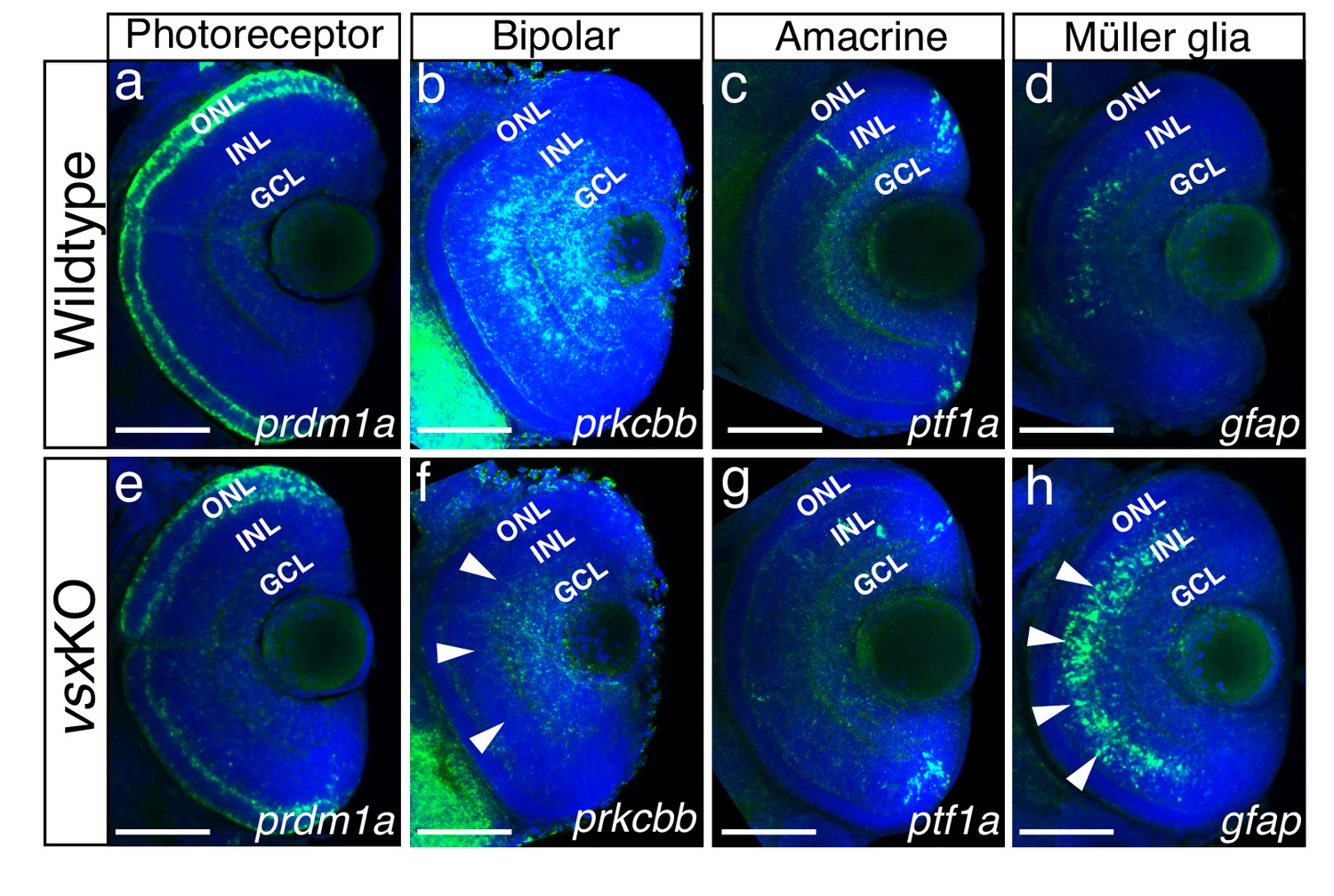
Altered expression of Bipolar and Müller glia cell markers in 3dpf vsx mutant fish.

(a-h). Confocal sections from in toto in situ hybridization experiments using specific fluorescent probes to label different cell types in wildtype and vsxKO retinas at 72hpf. No clear differences in the expression of the photoreceptor marker prdm1a were observed in ONL of wildtype (a) and mutant samples (e). Bipolar cell marker prkcbb expression (b, f) is considerably reduced in the INL of vsxKO mutant retinas (f, white arrowheads) compared to wildtype (b). Similar expression of the amacrine cell marker ptf1a is observed in the INL from wildtype (c) and vsxKO (g) retinas. Increased expression of the Müller glia cell marker gfap (d, h) is observed in the INL of vsxKO samples (h, white arrowheads) compared to wildtype (d) retinas. ONL: outer nuclear layer, INL: inner nuclear layer, GCL: ganglion cell layer. Scale bar in (a-h): 50 µm.

ONL/photoreceptors specification

Prdm1a (or Blimp1) is a transcription factor that has been shown to play an early role in the specification of PR identity, mainly by the suppression of bipolar cell fate genes, including vsx2 (Brzezinski et al., 2010; Katoh et al., 2010). Conversely, vsx2 acute knockdown by electroporation in the postnatal mouse retina triggers a bipolar to rod fate shift (Goodson et al., 2020; Livne-Bar et al., 2006). In this study, the comparative analysis of the transient marker prdm1a (Wilm and Solnica-Krezel, 2005) between wild type and vsxKO embryos revealed a mild downregulation in the mutants at 72 hpf (n=6) (Figure 4a and e), which is in agreement with the delayed differentiation of the photoreceptors we observed in vsxKO animals (Figure 4—figure supplement 1). However, when we examined terminal differentiation markers for cones (Ab Zpr-1) and rods (Ab Zpr-3) at 72 and 96hpf, a delayed differentiation of both cell types was observed in double mutant embryos (Figure 4—figure supplement 1). Whereas Zpr-1 and Zpr-3 staining could be detected in the entire ONL in wild type fish from 72hpf on (Figure 4—figure supplement 1a–c, h-j), in 72 hpf vsxKO embryos the staining was restricted to a few cells in the ventral retina (Figure 4—figure supplement 1d, k) and was only extended to the entire ONL at 96 hpf (Figure 4—figure supplement 1e, l). At 6dpf, there is a significant increase of Zpr1 fluorescent intensity in vsxKO compared to WT retinas (Figure 4—figure supplement 1c, f, g), but no major differences were observed in rod stain intensity (Figure 4—figure supplement 1j, m, n). This result suggests that PRs’ differentiation program is delayed in the absence of vsx function and that cone cells are overrepresented in the ONL of the double mutants. A prolonged period of precursors’ proliferation and/or competence could account for an increased number of PRs at larval stages, and thus for an expanded thickness of the ONL layer, as observed in double mutants at 6 dpf (Figure 1; Figure 1—figure supplement 3).

INL/bipolar cells specification

In the zebrafish retina, vsx1 and vsx2 expression has been reported in complementary subsets of bipolar cells, with vsx1 having a broader distribution and vsx2 being restricted to a few bipolar subtypes (Vitorino et al., 2009). To analyse bipolar cell integrity in vsxKO embryos, we first performed immunohistochemistry for the general INL marker prox1 (Figure 4—figure supplement 2; Dyer, 2003) and then fluorescent in situ hybridizations for the bipolar cell marker protein kinase Cb1 (prkcbb) (Figure 4). At 48hpf, no changes in the expression of prox1 was detected between WT and vsxKO retinas (Figure 4—figure supplement 2a, e). However, at 72hpf the nuclear distribution of prox1 in the INL is affected in vsxKO samples compared to WT retinas (Figure 4—figure supplement 2b, b’, f, f’) suggesting a lack of bipolar cells in vsxKO retinas. This observation was further confirmed by the fact that at 72hpf prkcbb expression is very reduced, if not absent, in the INL of double mutant retinas compared to WT (n=5) (Figure 4b and f). These results are in agreement with our previous histological (i.e., reduced INL thickness; Figure 1—figure supplement 3) and electrophysiological (i.e. reduced b-wave, Figure 2) observations in vsxKO larvae, and confirms that vsx genes are essential for bipolar cells specification in zebrafish. Although we can also detect a mild reduction of prkcbb at the GCL, and we cannot rule out transient defects in a particular RGCs subpopulation at 72 hpf (Figure 4f), the final RGC numbers seem normal in the vsxKO retinas as determined by DiI and DiO tracers (Video 1) as well as retinal histology at 6dpf (Figure 1).

INL/amacrine cells (AC) specification

A detailed histological analysis of the INL architecture in wild type and vsxKO embryos suggested that ACs specification was not severely affected in the double mutant (Figure 1d and h). To confirm this point, we followed the expression of ptf1a, a transcription factor encoding gene that is expressed in horizontal and AC and has been shown to play an essential role in their specification in the mouse retina (Fujitani et al., 2006). Using a fluorescent probe against ptf1a, which is expressed transiently in all types of amacrine cells in the embryonic zebrafish retina (Jusuf and Harris, 2009), we could determine that the ACs differentiation wave progresses normally through the central retina in wild type and vsxKO embryos at 48 hpf (n=5) (Figure 4—figure supplement 2c, g). Later in development, at 72 hpf, pft1a expression was no longer detected in the central retina and appeared restricted to the most peripheral region, being expressed at similar levels in both wild type and vsxKO retinas (n=10) (Figure 4c and g). In addition, the expression of the differentiated amacrine cell marker pax6 (Hitchcock et al., 1996) is not affected in vsxKO retinas compared to WT (Figure 4—figure supplement 2d, h). These observations suggest that vsx genes in zebrafish do not play a major role for amacrine cells specification.

INL/Müller glia cell specification

The abnormal expression of prox1 in the vsxKO fish INL (Figure 4—figure supplement 2b’, f’) suggests an unbalance in the contribution of the different cell types present in that retinal layer. Müller glia cell bodies are located in the INL where they provide structural and functional support to the retinal neurons (Goldman, 2014). To investigate if their differentiation occurs normally in vsx double mutants, we used a gfap antisense probe as glial marker (Bernardos and Raymond, 2006). We found a clear increase in the expression of gfap in vsxKO retinas compared to WT (n=5) (Figure 4d and h), suggesting that this cell type is overrepresented in vsxKO retinas, which may compensate for the reduction in bipolar cells observed in these animals.

In addition to their expression in the retina, Vsx transcription factors are also expressed in spinal cord interneurons (V2a and V2b cells), which are important to coordinate motor neuron activity and locomotion (Crone et al., 2008; Kimura et al., 2008). As reported here, vsx double mutants die around 2 weeks post-fertilization. This lethality could be due by spinal cord interneuron specification defects that may restrict the movement of the animals. To examine the integrity of V2a and V2b interneurons, we label both cell types with vsx1 and tal1 fluorescent antisense probes, that are expressed in V2a and V2b neurons, respectively (Figure 4—figure supplement 3). No significant differences in V2a or V2b spinal cord interneurons density was observed between WT (n=6) and vsx1∆245-/-, vsx2∆73-/- mutant fish (n=8) at 24hpf (Figure 4—figure supplement 3e). These results suggest that V2 motoneurons are properly specified in vsxKO animals. In agreement with this observation, obvious swimming defects were not observed in vsxKO larvae.

RNA-seq and ATAC-seq analyses of vsxKO reveal eye GRN robustness

The strong microphthalmia and abnormal specification of the neural retina reported in vsx2 mutant mice (Burmeister et al., 1996; Horsford et al., 2005; Rowan et al., 2004) are in contrast to our observation that in vsxKO embryos/larvae optic cup identity is normally established and maintained. In vsxKO mutants, the morphology and size of the optic cup, the precursors’ proliferation rate, as well as the distribution and expression of RPE specification markers (i.e. tfec and bhlhe40) appeared normal at 24hpf, as determined by PH3 staining and ISH, respectively (Figure 3—figure supplement 1a‐g). The only parameter altered in vsxKO later in development is the onset of retinal differentiation, which appeared slightly delayed as determined by atoh7 ISH at 26 hpf (Figure 3—figure supplement 1h, i). All these data point to a correct specification of the optic cup domains in the double mutants.

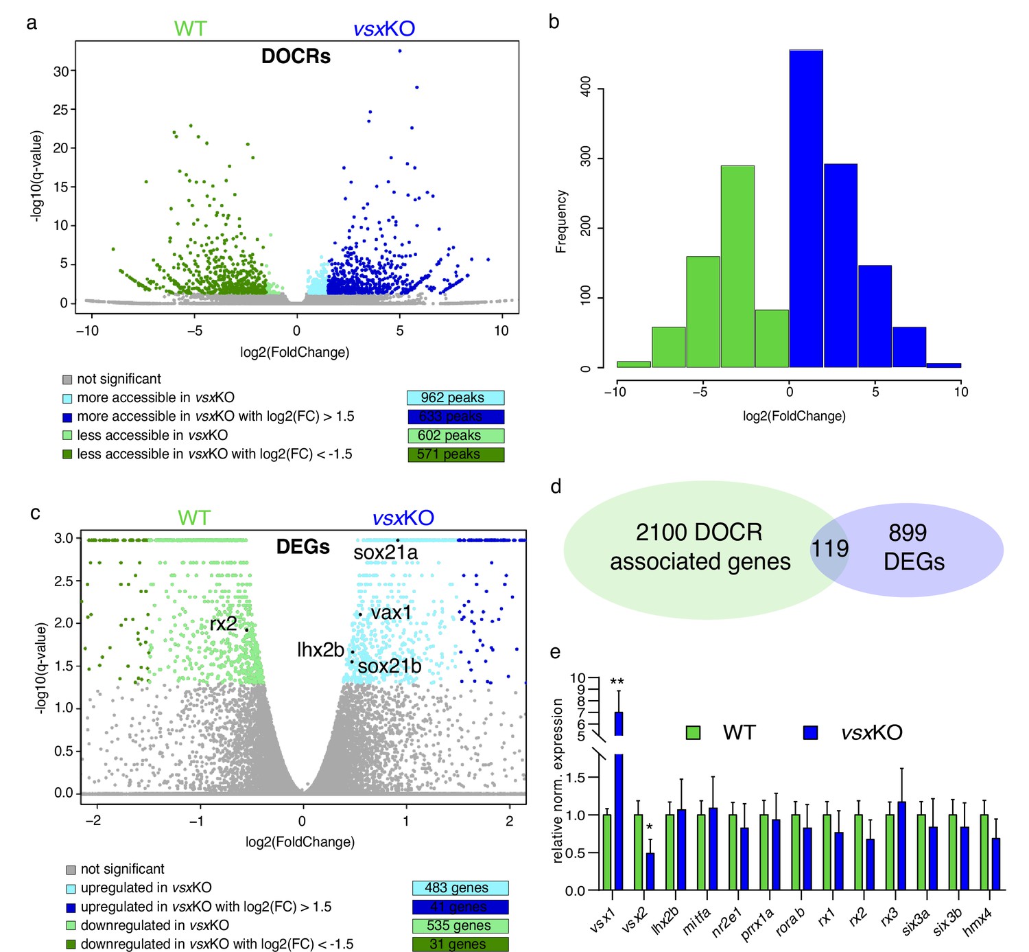
To gain insight into the molecular causes behind the discrepancy between mice and zebrafish mutants, we sought to investigate transcriptional and chromatin accessibility changes in mutant embryos during the specification of the neural retina (Figure 5). To this end, 18hpf embryo heads were collected from vsxKO and their wild type siblings and the rest of the tissue was used for PCR genotyping. We focused in this particular stage as it corresponds to the early bifurcation of the neural retina and RPE GRNs in zebrafish (Buono et al., 2021). To first identify changes in the cis-regulatory landscape associated to vsx loss of function, we examined wild type and mutant samples using ATAC-seq. This approach identified 1564 DNA regions with differential accessibility, most of them (1204) with a high fold change (i.e. log2 fold change > |1.5|). They include 633 regions more accessible in the mutant with a log2 fold change >1.5; and 571 less accessible with a log2 fold change <–1.5 (Figure 5a and b; Figure 5—source data 1). An analysis of enriched gene ontology terms for those genes (2219) neighboring the differentially opened regions revealed entries related to neuronal differentiation and eye development (Figure 5—source data 2). This observation suggests that vsx genes mutation results in the deregulation of hundreds of cis-regulatory elements mainly associated to retinal genes. In contrast, at a transcriptional level the comparative analysis of mutant and wild type samples by RNA-seq revealed expression changes only in a relatively small gene set (1018) (Figure 5c; Figure 5—source data 3). This collection comprised 41 up-regulated (log2 fold change >1.5) and 31 down-regulated (log2 Fold change <–1.5) genes, with only 3 up-regulated (vsx1, znf1109, and znf1102) and one down-regulated transcription factor (znf1091) above the threshold (log2 fold change > |1.5|) (Figure 5c). This observation indicated that the identified cis-regulatory changes are only translated in subtle changes at the transcriptional level. In fact, among the 2219 genes neighboring differentially open chromatin regions, only 5% (119) were associated to differentially expressed genes (Figure 5d). To further confirm the impact of vsx loss of function on the expression of core components of the neural retina GRN, we examined their levels by qPCR at 19 hpf (Figure 5e). Interestingly, in vsxKO embryos significant expression changes could be detected only for rx2 and lhx2b (although below the threshold log2 fold change > |1.5|). In addition, the transcripts of the mutated genes vsx1 and vsx2 were significantly upregulated and downregulated respectively in mutant embryos at optic cup stages, as determined by qPCR at 19hpf (Figure 5e) and confirmed by ISH at 24hpf (Figure 5—figure supplement 1). Taken together, these analyses suggest that the general architecture of the retinal GRN was not significantly altered upon vsx genes mutation.

Figure 5 with 4 supplements see all
Lack of Vsx TFs in the forming retina is buffered by genetic redundancy.

(a) Volcano plots illustrating chromatin accessibility variations upon vsx1 and vsx2 mutation in zebrafish retina at 18hpf. Each dot corresponds to an ATAC-seq peak, that is an open chromatin region. Grey dots indicate not significant variations, whereas colored dots point out significant differentially open chromatin regions. (b). Frequency of DOCRs’ fold change values. (c). Transcriptome variations in vsxKO retina at 18hpf. The genes reported in the plot are the only known retinal regulators whose transcriptional levels are affected by the loss of Vsx factors, with a very modest fold change. Essentially, RNA-seq experiments did not highlight a remarkable change of the levels of the main TFs governing the retinal GRN. (d). Correspondence between genes associated with DOCRs from ATAC-seq and DEGs from RNA-seq. (e). qPCR of the main retinal TFs confirming the stability of the eye gene network expression after vsx1 and vsx2 loss (n=3). **p<0.001, *p<0.01 using one-way ANOVA. Data is shown as mean ± SD. DOCR: differentially open chromatin regions, DEG: differentially expressed genes.

Figure 5—source data 1

List of all ATAC-seq peaks with differential accessibility in vsxKO vs WT.

Worksheet #1. Table of contents. Worksheet #2. List of all ATAC-seq peaks upregulated in the vsxKO with adjusted p-value <0.05. Genes associated with the peak are reported in the last column. Worksheet #3. List of all ATAC-seq peaks downregulated in the vsxKO with adjusted <i>P-value <0.05. Genes associated with the peak are reported in the last column.

https://cdn.elifesciences.org/articles/85594/elife-85594-fig5-data1-v1.xlsx
Figure 5—source data 2

Analysis of gene ontology terms for genes neighboring differentially opened regions in vsxKO.

Worksheet #1. Table of contents. Worksheet #2. Gene ontology terms enriched using as input the list of genes associated with differentially open chromatin region from ATAC-seq.

https://cdn.elifesciences.org/articles/85594/elife-85594-fig5-data2-v1.xlsx
Figure 5—source data 3

List of differentially expressed genes between WT and vsxKO embryos and cross-listing between DEGs and DOCRs from WT and vsxKO.

Worksheet #1. Table of contents. Worksheet #2. List of differentially expressed genes (DEGs) with q-value <0.05 between WT and vsxKO embryos Worksheet #3. Cross-list between DEGs from worksheet #2 and differentially open chromatin regions (DOCRs) more accessible in the vsxKO. Every DEG may be associated with more than one DOCR (column M). Worksheet #4. Cross-list between DEGs from worksheet #2 and differentially open chromatin regions (DOCRs) less accessible in the vsxKO. Every DEG may be associated with more than one DOCR (column M).

https://cdn.elifesciences.org/articles/85594/elife-85594-fig5-data3-v1.xlsx
Figure 5—source data 4

List of differentially expressed genes between WT vs vsx2MO and between vsxKO vs vsx2MO.

Worksheet #1. Table of contents. Worksheet #2. List of differentially expressed genes (DEGs) with q-value <0.05 between WT (uninjected) and vsx2-morpholino-injected embryos. Worksheet #3. List of differentially expressed genes (DEGs) with q-value <0.05 between vsx2-morpholino-injected and vsxKO embryos.

https://cdn.elifesciences.org/articles/85594/elife-85594-fig5-data4-v1.xlsx

To further understand the phenotypic discrepancy observed between vsx2 morphants and vsxKO mutants, we revisited previous splicing morpholino experiments (Gago-Rodrigues et al., 2015) confirming the reported microphthalmic phenotypes (Barabino et al., 1997; Clark et al., 2008; Gago-Rodrigues et al., 2015; Vitorino et al., 2009). Then we performed a full transcriptome analysis of vsx2 morphants by RNA-seq at 18 hpf using embryo heads (Figure 5—figure supplement 2a). Principal components analysis (PCA) of wild type, morphant and double mutant datasets revealed a very different regulatory response between mutant and morphant samples (Figure 5—figure supplement 2b; Figure 5—source data 4). This differential behavior was evident when core components of the eye specification gene regulatory networks were examined: whereas mild transcriptional differences were detected for rx2 and lhx2b in the mutants (Figure 5; Figure 5—figure supplement 2c), core components of the retinal network such as rx3, rx1, six3b, vax2, and lhx2b appeared upregulated in the morphants (Figure 5—figure supplement 2c). Particularly different was the expression of vsx2 itself, which appeared strongly downregulated in vsxKO mutants, but strongly upregulated in vsx2 morphants (Figure 5—figure supplement 2d, f). These results suggest that the dysregulation of the retinal network induced by the morpholinos (i.e. may be through compensatory mechanisms operating at the RNA level) is behind the early microphthalmia observed in the vsx2 morphants.

Our results point at vsx genes having a different regulatory weight for neural retina specification/maintenance in different species. To gain insight into this hypothesis, we mutated two of the three paralogs (i.e. vsx1 and vsx2.2) present in the genome of the far-related teleost medaka by CRISPR/Cas9 (Figure 5—figure supplement 3). We generated a 148 bp deletion in medaka vsx1 deleting 29 bp of intron 2 and 119 bp of exon3 (vsx1∆148). This frame shift mutation results in a deletion of 39 amino acids in the highly conserved DBD and the generation of a premature stop shortly after the deletion. In vsx2.1, a 319 bp deletion (vsx2.1∆319) was generated encompassing intron2 (47 bp), exon3 (124 bp), and intron3 (147 bp). This frame shift mutation deletes 41 amino acids of the core DBD of the protein, mutate critical Arginines and generates a premature stop codon 66 amino acids after the deletion. Interestingly, although the initial specification of the organ appeared normal also in medaka double mutant embryos at 4dpf, INL differentiation and eye growth was impaired at later stages in those animals (Figure 5—figure supplement 3b–e). This is in contrast to the normal eye size observed in vsxKO zebrafish larvae at 19dpf (Figure 5—figure supplement 4) and confirmed the assumption of differential regulatory weight among vertebrate species for vsx genes.

Discussion

In this study, we explore the universality of Vsx functions in the development of the vertebrate eye, by generating CRISPR/Cas9 mutations of the ‘visual system homeobox’ genes vsx1 and vsx2 in the far related teleost models, zebrafish and medaka. Genetic analyses in the mouse, as well as the chick, had revealed two distinct functions for Vsx genes during eye development: an early requirement for proliferation and specification of the neural retina precursors, and a later role in the differentiation of bipolar neurons (Burmeister et al., 1996; Horsford et al., 2005; Rowan et al., 2004). These two developmental roles depend on consecutive waves of gene expression and thus can be uncoupled by genetic interference within specific developmental windows (Goodson et al., 2020; Livne-Bar et al., 2006). Moreover, in mice, Vsx biphasic activity follows a partially independent cis-regulatory control by enhancers active either in precursors, bipolar cells, or both (Kim et al., 2008; Norrie et al., 2019; Rowan and Cepko, 2005). Accordingly, CRISPR-mediated ablation of a distal bipolar enhancer results in the specific depletion of these cells, without leading to microphthalmia or compromising the early specification of the mouse retina (Goodson et al., 2020; Norrie et al., 2019).

Here, we show that Vsx activity is essential for bipolar cells differentiation in teleost fish, indicating a broadly conserved role for these genes across vertebrates. This observation suggests that the genetic program controlling bipolars specification was inherited from a common vertebrate ancestor. The fact that Vsx homologous genes are also expressed in the visual-system of the invertebrates Drosophila and Cuttlefish (Sepia officinalis) further suggests that the function for these homeobox genes in the specification of visual interneurons may be a common theme in all metazoans (Erclik et al., 2008; Focareta et al., 2014). The absence of an earlier eye phenotype in zebrafish vsxKO embryos allowed us examining in detail the consequences of vsx loss on cell fate determination and sight physiology. Both our histological and electrophysiological analyses confirmed bipolar cells depletion in vsxKO retinas. We show that, unable to acquire the bipolar fate, retinal precursors follow alternative differentiation trajectories such as undergoing apoptosis, extending their proliferative phase, or differentiating as photoreceptors or Müller glia cells. A detour toward photoreceptors fate in zebrafish is in agreement with previous studies in mice showing that the Blimp1/Vsx2 antagonism controls the balance between rods and bipolar cells (Brzezinski et al., 2010; Goodson et al., 2020; Katoh et al., 2010; Wang et al., 2014). Interestingly, in vsxKO retinas we observed a noticeable delay in the onset of cones and rods terminal differentiation markers, zpr-1 and zpr-3 respectively, indicating that Vsx activity is not only required for correct fate specification, but also to determine the timing of the differentiation sequence, in agreement with previous data in mice (Rutherford et al., 2004). Arguably, more intriguing was our observation of an increased number of Müller glia cells in vsxKO retinas. Both glial and bipolars cells are late-born retinal types deriving from a common pool of precursors with restricted developmental potential (Bassett and Wallace, 2012; Hatakeyama et al., 2001; Rowan and Cepko, 2004; Satow et al., 2001). In mice, however, a significant increase in Müller glia cells has not been reported in experiments genetically interfering with vsx either postnatally (Goodson et al., 2020; Livne-Bar et al., 2006) or specifically in bipolar cells (Norrie et al., 2019). This apparent discrepancy might indicate some variations in the cell fate specification mechanisms among vertebrate species. Alternatively, the increase may have been overlooked in previous studies due to the small size of the Müller glia cell population. The fact that a trend toward an increase in Müller glia has been reported (Livne-Bar et al., 2006) may support this second possibility.

Despite the severe visual impairment and retinal lamination defects we observed in vsxKO larvae, their eyes appear normal in shape and size and no early morphological defects are observed in the optic cup. More importantly, neuro-retinal identity seemed perfectly maintained in double mutant animals, and we did not detect any trans-differentiation of the retina into pigmented cells. This finding, which is in contrast with the microphthalmia and the neural retina specification defects observed in mice (Burmeister et al., 1996; Horsford et al., 2005; Rowan et al., 2004), may indicate that Vsx genes do not play an early role in the establishment and maintenance of the neural retina identity in zebrafish. Although a potential rescue by maternally provided vsx genes could be hypothesized as an explanation for normal specification of the retina, this is an unlikely possibility as both transcripts are not detectable before zygotic genome activation (White et al., 2017). Our observations are also in contrast to previous reports in zebrafish using antisense oligonucleotides or morpholinos against vsx2, which show microphthalmia and optic cup malformations (Barabino et al., 1997; Clark et al., 2008; Gago-Rodrigues et al., 2015; Vitorino et al., 2009). A poor resemblance between morpholino-induced and mutant phenotypes has been previously described in zebrafish, with many mutations lacking observable phenotypes (Kok et al., 2015). Genetic compensation and, in particular, transcriptional adaptation (i.e. up-regulation of genes displaying sequence similarity) has been identified as the molecular mechanism accounting for genetic robustness in a number of these mutations (El-Brolosy et al., 2019; Rossi et al., 2015). However, our comparative transcriptomic analysis of vsxKO vs WT embryos does not support genetic compensation acting as a relevant mechanism at optic cup stages. We show that, despite that vsx loss of function results in the deregulation of hundreds of cis-regulatory regions associated to retinal genes, this has little impact on the expression of core components of the neural retina specification network.

Our previous analysis of transcriptome dynamics and chromatin accessibility in segregating NR/RPE populations indicated that the regulatory networks involved in the specification of the zebrafish eye are remarkably robust (Buono et al., 2021). In that study, we showed that the consensus motif 5’-TAATT-3’, which is central to the neural retina cis-regulatory logic, is shared by many homeodomain TFs co-expressed during retinal specification; including not only vsx1 and vsx2, but also rx1, rx2, rx3, lhx2b, lhx9, hmx1, and hmx4. Moreover, we show evidence that these TFs may co-regulate the same genes and cooperate within the same cis-regulatory modules (Buono et al., 2021). According to these observations, gene redundancy appears as a more parsimonious explanation for the absence of an early phenotype in vsxKO embryos. This would suggest that the regulatory weight of vsx genes within the retina network varies across vertebrate species. Several lines of evidence support this view. (i) Other mutations in genes encoding for TFs targeting the motif 5’-TAATT-3’, such as rx2 in medaka (Reinhardt et al., 2015) or lhx2 in zebrafish (Seth et al., 2006) do not compromise the identity of the neural retina tissue either. (ii) Even in vsx1/vsx2 double mutant mice, the central retina keeps the potential for differentiation into several neuronal types, indicating that other genes must cooperate in the specification of this tissue (Clark et al., 2008). In such scenario of complex epistatic interactions, it is not surprising the intrinsic variability in expressivity traditionally observed in ocular retardation mutants (Osipov and Vakhrusheva, 1983). In line with this, in Vsx2 mutant mice has been shown that neural retinal identity defects and microphthalmia (but not bipolar cells differentiation) can be partially restored by simply deleting a cell cycle gene (Green et al., 2003). (iii) Finally, here we show that the mutation of the paralogs vsx1 and vsx2.2 results in severe microphthalmia in medaka larvae. This finding confirms a variable role across species for Vsx genes in the specification and maintenance of the neural retina domain in vertebrates.

Methods

Animal experimentation and strains

All experiments performed in this work comply European Community standards for the use of animals in experimentation and were approved by ethical committees from Universidad Pablo de Olavide (#02/04/2018/041), Consejo Superior de Investigaciones Científicas (CSIC), the Andalusian government and Universidad Mayor (#25/2018). Zebrafish AB/Tübingen (AB/TU) and medaka iCab wild-type strains were staged, maintained and bred under standard conditions (Iwamatsu, 2004; Kimmel et al., 1995). Zebrafish Vsx mutants were maintained harboring a single copy of vsx1 (vsx1∆245+/-, vsx2∆73-/-) or vsx2 (vsx1∆245-/-; vsx2∆73+/-), while medaka Vsx mutants were maintained in heterozygosis (vsx1∆148+/-, vsx2.1∆319+/-).

Gene editing

Single guide RNAs (sgRNAs) targeting the DNA binding domains of vsx1 and vsx2 from zebrafish and vsx1 and vsx2.1 from medaka were designed using the CRISPRscan (Moreno-Mateos et al., 2015) and CCTop (Stemmer et al., 2015) design tools. Primers for sgRNA generation (see Table 1), were aligned by PCR to a universal CRISPR primer and the PCR product was further purified and used as template to sgRNA synthesis (Vejnar et al., 2016). To target individual vsx genes, a solution containing two sgRNAs (40 ng/μL each) and Cas9 protein (250 ng/μL) (Addgene; 47327) (Gagnon et al., 2014) were injected into one-cell-stage zebrafish and medaka embryos. Oligos used for screening of genomic DNA deletions flanking CRISPR target sites are detailed in Table 1. Wild-type and mutant PCR products from F1 embryos were further analysed by standard sequencing to determine germline mutations (Stab Vida).

Table 1
Nucleotide sequence of oligos used in this work.

Organism, gene of interest, application and nucleotide sequence is described in each column. Note that the target site is bolded in CRISPR/Cas9 primers used for vsx disruption.

OrganismGeneApplicationOligo sequence (5’–3’)
Danio reriovsx1CRISPR/Cas9TAATACGACTCACTATAGGGTTCCTCAAGTTGATGGGGTTTTAGAGCTAGAA
Danio reriovsx1CRISPR/Cas9TAATACGACTCACTATAGGTTTACGCGAGAGAAATGCGTTTTAGAGCTAGAA
Danio reriovsx2CRISPR/Cas9TAATACGACTCACTATAGGTGCCGGAGGACAGAATACGTTTTAGAGCTAGAA
Danio reriovsx2CRISPR/Cas9TAATACGACTCACTATAGGTGGAGAAAGCTTTTAACGGTTTTAGAGCTAGAA
Danio reriovsx1Genotyping FwATGACTGCCTTTCCGGTGAT
Danio reriovsx1Genotyping RvCTGCTGGCTCACCTAGAAGC
Danio reriovsx2Genotyping FwTCGTAATCTTTCCACTGATTCTGAT
Danio reriovsx2Genotyping RvTGTTCTAGAGCATATTGTCTGTTCC
Danio reriovsx1Cloning FwCGGGAAGAGAAGAAGCTACAGAT
Danio reriovsx1Cloning RvGCCTTCTCTTTTTCCTCTTTTGA
Danio reriovsx2Cloning FwCTGTTTTGTCGGAAAGTTTGAA
Danio reriovsx2Cloning RvCCAGCTGGTAAGATGTAAATATTGTT
Danio rerioptf1aCloning FwGGCTTAGACTCTTTCTCCTCCTC
Danio rerioptf1aCloning RvCGTAGTCTGGGTCATTTGGAGAT
Danio reriogfapCloning FwGTTCCTTCTCATCCTACCGAAAG
Danio reriogfapCloning RvGATCAGCAAACTTTGAGCGATAC
Danio reriopkcb1Cloning FwGCGCAGTAAGCACAAGTTCAAGG
Danio reriopkcb1Cloning RvCCCAGCCAGCATCTCATATAGC
Danio rerioprdm1aCloning FwTCAAAACGGCATGAACATCTATT
Danio rerioprdm1aCloning RvAGGGGTTTGTCTTTCAGAGAAGT
Danio reriotal1Cloning FwAGTATGATTTGCTCATCCTCCAA
Danio reriotal1Cloning RvTTTGTTTGTTTGCGCATTTAATA
Danio reriotfecCloning FwTATAAAGACCGGACGGGGACAAC
Danio reriotfecCloning RvCAGCTCCTGGATTCGTAGCTGGA
Danio reriobhlhe40Cloning FwTTGCAAATCGGCGAACAGGG
Danio reriobhlhe40Cloning RvGGAAACGTGCACGCAGTCG
Danio rerioeef1a1l1qPCR FwTCCACCGGTCACCTGATCTAC
Danio rerioeef1a1l1qPCR RvCAACACCCAGGCGTACTTGA
Danio reriovsx1qPCR FwTCTAGGTGAGCCAGCAGGAAT
Danio reriovsx1qPCR RvCCATGTCGTGTCGCTGTCTT
Danio reriovsx2qPCR FwGGGATTAATTGGGCCTGGAGG
Danio reriovsx2qPCR RvGCTGGCAGACTGGTTATGTTCC
Danio reriosix3aqPCR FwAAAAACAGGCTCCAGCATCAA
Danio reriosix3aqPCR RvAAGAATTGACGTGCCCGTGT
Danio reriosix3bqPCR FwTCCCCGTCGTTTTGTCTCTG
Danio reriosix3bqPCR RvAGAAGTTTAGGGTGGGCAGC
Danio reriolhx2bqPCR FwAGGCAAGATTTCGGATCGCT
Danio reriolhx2bqPCR RvTCTCTGCACCGAAAACCTGTA
Danio reriomitfaqPCR FwCTGATGGCTTTCCAGTAGCAGA
Danio reriomitfaqPCR RvGCTTTCAGGATGGTGCCTTT
Danio rerionr2e1qPCR FwCAAATCTGGCACACAGGGCG
Danio rerionr2e1qPCR RvCGACGAACCGTTCACCTCTT
Danio rerioprrx1aqPCR FwCTCACCGTCATACAGTGCCA
Danio rerioprrx1aqPCR RvAGAGTCTTTGACAGCCCAGC
Danio reriororabqPCR FwACAAACCAGCACCAGTTCGG
Danio reriororabqPCR RvCCTCCTGAAGAAACCCTTGCAT
Danio reriorx1qPCR FwAAGAACTTGCATCGGACGGT
Danio reriorx1qPCR RvTCGGAAGCTTGCATCCAGTT
Danio reriorx2qPCR FwTCGGGACGCATAAAGTGGAC
Danio reriorx2qPCR RvCGGGTCTCCCAAATCTGCAT
Danio reriorx3qPCR FwCCGAGTACAGGTGTGGTTCC
Danio reriorx3qPCR RvGTCAACCAGGGCTCTAACGG
Danio reriohmx4qPCR FwTGTCGACCCGCTTCTTTGAA
Danio reriohmx4qPCR RvTGATGAAGACAGCCATCCCG
Oryzias latipesvsx1CRISPR/Cas9TAATACGACTCACTATAGGCAGAGTGAGGTTCAGTGGGTTTTAGAGCTAGAA
Oryzias latipesvsx1CRISPR/Cas9TAATACGACTCACTATAGGTAGGGCCTGACCTGGATTGTTTTAGAGCTAGAA
Oryzias latipesvsx2.1CRISPR/Cas9TAATACGACTCACTATAGGGGATGATGAGAGTCAAGGGTTTTAGAGCTAGAA
Oryzias latipesvsx2.1CRISPR/Cas9TAATACGACTCACTATAGGAAAAAATAACAGAATTGAGTTTTAGAGCTAGAA
Oryzias latipesvsx1Genotyping FwAACAATAATTTAAAATGCGGAAAAA
Oryzias latipesvsx1Genotyping RvGAAACTAAAATCCCATTCAGTGCT
Oryzias latipesvsx2.1Genotyping FwATATCACGGGAAATTAAAATGCTC
Oryzias latipesvsx2.1Genotyping RvAAGTCAAATGTGCCATTGTTAGTC

Histology

Zebrafish and medaka samples from different developmental stages harboring mutations in vsx genes were deeply anesthetized for 5–10 min with 160 mg/L of tricaine (ethyl 3-aminobenzoate methanesulfonate salt; MS-222; Merck) before dissecting their heads. Heads including both eyes were fixed in 4% w/v paraformaldehyde (PFA, Merck) in 0.1 M phosphate buffer overnight at 4 °C and the remaining tissue were kept for genotyping by conventional PCR. Wild-type and vsxKO sorted heads were then washed several times in 1 X PBS, incubated in 30% sucrose-PBS overnight at 4 °C, embedded in OCT (Tissue Tek) using cryomolds (Tissue Tek) and frozen in liquid nitrogen for short term storage at –80 °C. Cryosectioning of samples was performed using a Leica CM1850 cryostat and 20-μm-thick transverse sections were collected in glass slides (Super Frost Ultra Plus, #11976299, Thermo Fisher Scientific) for Phalloidin (#A12379, Alexa fluor 488, Thermo Fisher Scientific) and 4′,6-Diamidine-2′-phenylindole dihydrochloride (DAPI, #10236276001, Merck) staining. Briefly, zebrafish and medaka eye transverse cryosections were dried at room temperature for ≥3 hr and washed with filtered PBST (0.1% Triton in 1 X PBS) five times for 5 min each wash. Then, slides were incubated with a solution containing 1/50 phalloidin Alexa fluor 488 in PBST supplemented with 5% DMSO (Merck) and covered with parafilm (Bemis) in a dark humid chamber overnight at 4 °C. After 30–60 min at room temperature, sections were incubated in a DAPI solution (1:1000 in PBST) and then washed five times for 5 min each wash with PBST. Slides were mounted with a drop of 15% glycerol in PBS and covered with 22x60 mm coverslips. Mounted slides were kept in the dark and confocal images were captured immediately (≤24 hr) using a Leica SPE microscope to detect Alexa 488 and DAPI signals from retina samples.

Eye size and retina layer width measurements

Zebrafish embryos obtained from in-crosses of either vsx1∆245+/-, vsx2∆73-/- or vsx1∆245-/-; vsx2∆73+/-fish, were raised for 2 weeks under standard conditions (Kimmel et al., 1995). At this stage larvae were anesthetized, the antero-posterior length was measured (in millimetres) and a lateral image of the head region was obtained (Olympus SZX16 binocular scope connected to an Olympus DP71 camera). In parallel, a tip of the tail was collected using a scalpel to extract genomic DNA using Chelex resin (C7901, Sigma) for PCR screening. Head images (all taken at the same magnification) sorted by their genotype (either wild-type or vsxKO) were analysed using Fiji software to measure eye surface. Total eye area was divided by fish antero-posterior length for each animal to normalize eye size. To measure retina INL and ONL layers width in zebrafish larvae, confocal images of eye cryostat sections from previously genotyped wild-type and vsxKO animals were taken using an immersion oil ×40 objective (SPE, Leica). These images were then analysed using Fiji software to measure INL and ONL width (μm).

Electroretinography (ERG)

ERG was recorded on 5 dpf larvae as previously described (Zang et al., 2015). 100ms light stimuli delivered by HPX-2000 (Ocean Optics) were attenuated (log-4 to log0) by neutral density filters and given with an interval of 15 s. Full light intensity was measured by spectrometer (Ocean Optics, USB2000+) with spectrum shown in S1 (SpectraSuite, Ocean Optics). Electronic signals were amplified 1000 times by a pre-amplifier (P55 A.C. Preamplifier, Astro-Med. Inc, Grass Technology), digitized by DAQ Board (SCC-68, National Instruments) and recorded by self-written Labview program (National Instruments). Figures were prepared using Microsoft Excel 2016.

Optokinetic response (OKR)

The OKR was recorded by the experiment setup as previously described (Mueller and Neuhauss, 2010). Briefly, 5dpf larvae were stimulated binocularly with sinusoidal gratings. To determine the contrast sensitivity, a spatial frequency of 20 cycles/360° and an angular velocity of 7.5 deg/s were used with varying contrast (5, 10, 20, 40, 70, and 100%). To study the spatial sensitivity, an angular velocity of 7.5 /s and 70% of the maximum contrast was used with different spatial frequency (7, 14, 21, 28, 42, 56 cycles/360°). To analyse the temporal sensitivity, maximum contrast and a spatial frequency of 20 cycles/360° were applied with increasing temporal frequency (5, 10, 15, 20, 25, 30 deg/s). Figures were presented by SPSS (Version 23.0. Armonk, NY: IBM Corp).

Immunohistochemistry in sections

Zebrafish wild-type and vsxKO retina sections from different developmental stages were analysed for the detection of apoptotic and mitotic cells using rabbit anti-active caspase-3 antibodies (BD Biosciences, 559565) and rabbit anti-phospho-histone H3 antibodies (Merck Millipore, 06–570), respectively. For the detection of cone and rod photoreceptors, zpr1 (ZIRC) and zpr3 (ZIRC) antibodies were used, respectively. Briefly, eye transverse cryosections were dried at room temperature for ≥3 hr, washed five times for 5 min each with PBST, blocked for ≥1 hr with 10% fetal bovine serum in PBST and incubated overnight in a humid chamber at 4 °C with the corresponding primary antibody. All primary antibodies were diluted 1:500 in blocking solution. After several washes with PBST, a 1:500 dilution of the secondary antibody (Alexa Fluor 555 goat anti-rabbit or goat anti-mouse antibodies, Thermo Fisher, #A-21429 and #A-21422, respectively) was added for 2 hr at room temperature. Following extensive washes with PBST, slides were mounted in 15% glycerol/PBS solution and sealed with 22x60 mm coverslips. Immunofluorescence confocal images were taken using a Leica SPE confocal microscope.

Whole-mount embryo immunofluorescence

Embryos collected from in-crossed vsx1+/-; vsx2-/- adult fish were dechorionated and fixed at 72 hpf with 4% Formaldehyde in PBS (FA). Fixed embryos were washed with PBS-Tween 0.5%-Triton 0.5% (PBST), treated with Proteinase K (10 µg/mL in PBST) for 30 min at 37 °C followed by PBST washes and a post-fixation step in FA for 30 min at room temperature (RT). After PBST washes, embryos were treated with cold acetone at –20 °C for 20 min, then washed again with PBST and incubated with freshly prepared blocking solution (5% normal goat serum, 1% BSA, 1% DMSO in PBST) at RT for 2 hr. Primary antibody specific for zebrafish Prox1 (GeneTex, GTX128354) and Pax6 (GeneTex, GTX128843) were diluted 1:100 in blocking solution and embryos were incubated overnight (ON) at 4 °C. Embryos were subsequently washed with PBST and incubated ON at 4 °C in the dark with the Alexa FluorTM 488 Goat anti-rabbit antibody (Invitrogen), diluted 1:500. Finally, embryos were washed with PBST and incubated ON at 4 °C with DAPI (Sigma) diluted 1:5000 in PBST. For imaging, embryos were embedded in 1% low-melting point agarose, transferred to glass-bottom culture dishes (MatTek corporation) and manually oriented. Confocal laser scanning microscopy was performed using an LSM 880 microscope (Zeiss). Images were processed using Fiji. After imaging, embryos were genotyped by PCR to identify vsx1-/-; vsx2-/- double mutant embryos.

RNA in situ hybridization

Fluorescence in situ hybridization experiments were performed as previously described (Bogdanović et al., 2012). Fragments of the vsx1, vsx2, ptf1a, prdm1a, gfap, prkcbb, tfec, bhlhe40 and tal1 genes were PCR amplified from zebrafish cDNA (SuperScript IV VILO Master Mix ThermoFisher Scientific, #11756050) using specific primers (Table 1). For vsx1 and vsx2 genes, the deleted region of the coding sequence in vsxKO mutants was excluded from the amplified fragment. PCR products were cloned into StrataClone PCR Cloning vector (Agilent, #240205), linearized with XbaI restriction enzyme (Takara, #1093B) and transcribed with a DIG-labeling Kit (Roche, #11277073910) using T3 polymerase (Roche, #11031163001) to obtain digoxigenin-labeled antisense probes. Probes were used at a final concentration of 2 ng/µl diluted in hybridization buffer (Thisse and Thisse, 2008). For atoh7, a colorimetric antisense digoxigenin-labeled RNA probe was prepared as reported elsewhere (Masai et al., 2000).

Morpholino injections

The vsx2E2I2 splicing morpholino was obtained from Gene Tools and injected as reported before (Gago-Rodrigues et al., 2015). For RNA-seq experiments, vsx2 morphants where co-injected with lyn-Td-tomato mRNA at a concentration of 50 ng/µL. At 16 hpf, red fluorescent embryos were pooled under the stereoscope and heads were dissected at 18 hpf for total RNA extraction.

RNA-seq

Total RNA was extracted from 18 hpf zebrafish embryos’ heads using 1 ml TRIzol (Invitrogen, #15596026) following the manufacturer’s protocol. The trunk and tail of the embryos was used to extract genomic DNA using Chelex resin (C7901, Sigma) for PCR screening. Potential DNA contamination was eliminated by treating RNA samples with TURBO DNAse-free kit (Ambion, #AM1907). The concentration of the RNA samples was evaluated by Qubit spectrophotometer (Thermo Fisher). Libraries were prepared with TruSeq stranded mRNA kit (Illumina) and sequenced 2x125 bp on an Illumina Nextseq platform. We obtain at least 33 million reads per sample. Three biological replicates were used for each analysed condition. Reads were aligned to the danRer10 zebrafish genome assembly using hisat2 (Kim et al., 2015). Transcript abundance was estimated with Cufflinks v2.2.1. Differential gene expression analysis was performed using Cuffdiff v2.2.1, setting an adjusted <i>P-value  <0.05. PCA analysis were done using CummeRbund, R package version 2.40.0 (Goff and Trapnell, 2022). Heatmap visualization was obtained with Clustvis (Metsalu and Vilo, 2015) using the FPKMs normalized by row as input.

qPCR

cDNA retrotranscription and qPCR were performed as previously described (Vázquez-Marín et al., 2019). eef1a1l1 gene was used as housekeeping normalization control. Primer sequences for amplified genes are detailed in Table 1.

ATAC-seq

Each ATAC-seq sample was obtained starting from a single 18 hpf zebrafish embryo’s head manually dissected, while the trunk and tail was kept for conventional PCR genotyping. Tagmentation and library amplification were performed using the FAST-ATAC protocol previously described (Corces et al., 2016). We obtained at least 70  M reads from the sequencing of each library. For data comparison, we used two biological replicates for each condition. Reads were aligned to the danRer10 zebrafish genome using Bowtie2 (Langmead and Salzberg, 2012) with -X 2000—no-mixed—no-unal parameters. PCR artifacts and duplicates were removed with the tool rmdup, available in the Samtools toolkit (Li et al., 2009). In order to detect the exact position where the transposase binds to the DNA, read start sites were offset by +4/–5 bp in the plus and minus strands. Read pairs that have an insert  < 130  bp were selected as nucleosome-free reads. Differential chromatin accessibility was calculated as reported (Magri et al., 2019). All chromatin regions reporting differential accessibility with an adjusted p-value  < 0.05 were considered as DOCRs. All the DOCRS have been associated with genes using the online tool GREAT (McLean et al., 2010) with the option ‘basal plus extension’. Gene ontology enrichment analysis of the genes associated with DOCRs was performed with PANTHER (Mi et al., 2021).

Labeling of retinotectal projections (DiI/DiO injections)

Following PCR genotyping, 6 dpf wild-type and vsx mutant larvae were fixed in 4% PFA overnight, washed in PBS and embedded in 1% low melting agarose (Sigma, #A9414) in PBS on an agarose filled Petri dish for injection. Each eye (between the lens and the retina) of the fish was injected either with 1% DiI (Invitrogen, #D275) or 1% DiO (Invitrogen, #D282) solutions in Chloroform (or dimethylformamide) with a pulled capillary glass mounted on a micromanipulator and under a stereomicroscope. A PV820 Pneumatic PicoPump (WPI) with the appropriate setting to deliver pressure to label the whole retina was used. Injected simples were washed in PBS, maintained overnight at 4 °C and mounted on low melting agarose to image on a Zeiss LSM 710 confocal microscope. Z-stacks (0.5 µm x 0.5 µm x 1 µm) were collected to visualize the optic nerve reaching the tectum and 3D reconstructions were generated using Zen blue edition software (Zeiss).

Total protein extraction and western blotting analysis

Vsx1 and Vsx2 protein presence was analysed by Western blotting. To accomplish this, three different samples were prepared: the first two contained 20 heads of wildtype or vsxKO embryos at 24 hpf stage and the third sample comprised 20 wildtype embryos at 1.5 hpf. Each set of embryos were shaken for 5 min at 1100 rpm in deyolking buffer (55 mM NaCl, 1.8 mM KCl and 1.25 mM NaHCO3). Tubes were then centrifuged at 300 g for 30 s, and subsequently the pellets were rinsed with wash buffer (10 mM Tris-HCl pH8.0, 110 mM NaCl, 3.5 mM KCl and 2.7 mM CaCl2). Then, each pellet was resuspended in 25 µL SDS buffer (100 mM TrisHCl pH 6.8, 4% SDS, 20% glycerol and 200 mM DTT) and heated at 95 °C for 5 min. After that, samples were centrifuged at 16,000 g for 20 min at 4 °C and supernatants were collected. Protein extracts were loaded in 10% TGX Stain-FreeTM FastCastTM Acrylamide (BioRad) and blotted onto nitrocellulose membranes. Western blot normalization was performed using total protein load following manufacturer protocol for Stain Free gels (Bio Rad). Vsx1 (A10801, https://www.antibodies.com/) and Vsx2 (X1180P, Abintek) antibodies were used at a 1:500 dilution, followed by incubation with anti-Rabbit IgG-HRP secondary antibody (AP160P, Chemicon), diluted to 1:10000 for Vsx1 detection and Rabbit anti-Sheep IgG-HRP secondary antibody (402100, Calbiochem) diluted to 1:2000 for Vsx2. Chemiluminescent signals were detected with SuperSignal West Femto Substrate (Thermo Scientific) in a ChemiDoc MP Imaging System (BioRad).

Statistical analysis

Quantitative data were evaluated using Prism 9.0 GraphPad software. Two-way ANOVA and a Tukey post hoc test was used to analyse ERG data, one-way ANOVA for OKR recordings and qPCR. Unpaired t test were used for PH3+ cell counts, C3+ cell counts, total eye area, retina layers’ width, trunk V2 neuron comparisons and Zpr1/3 fluorescent intensity labeling. n values and significance levels are indicated in figure legends.

Data availability

Sequencing data have been deposited in GEO under accession code GSE189739.

The following data sets were generated
    1. Letelier J
    2. Buono L
    (2022) NCBI Gene Expression Omnibus
    ID GSE189739. Mutation of Vsx genes in zebrafish highlights the robustness of the retinal specification network.

References

    1. Martinez-Morales JR
    (2016) Vertebrate eye gene regulatory networks
    Organogenetic Gene Networks: Genetic Control of Organ Formation 1:259–274.
    https://doi.org/10.1007/978-3-319-42767-6
    1. Osipov VV
    2. Vakhrusheva MP
    (1983)
    Variation in the expressivity of the ocular retardation gene in mice
    TSitologiia i Genetika 17:39–43.
    1. Truslove GM
    (1962)
    A gene causing ocular retardation in the mouse
    Journal of Embryology and Experimental Morphology 10:652–660.

Decision letter

  1. Edward M Levine
    Reviewing Editor; Vanderbilt University, United States
  2. Didier YR Stainier
    Senior Editor; Max Planck Institute for Heart and Lung Research, Germany
  3. William A Harris
    Reviewer; University of Cambridge, United Kingdom

In the interests of transparency, eLife publishes the most substantive revision requests and the accompanying author responses.

Decision letter after peer review:

[Editors’ note: the authors submitted for reconsideration following the decision after peer review. What follows is the decision letter after the first round of review.]

Thank you for submitting the paper "Mutation of Vsx genes in zebrafish highlights the robustness of the retinal specification network." for consideration by eLife. Your article has been reviewed by 3 peer reviewers, and the evaluation has been overseen by a Reviewing Editor and a Senior Editor. The following individual involved in review of your submission has agreed to reveal their identity: William A Harris (Reviewer #1).

Comments to the Authors:

While you can see that there was some enthusiasm for your work and all three reviewers agreed that this is an interesting study, too many questions remained open at this point to move forward with the current submission. I and all reviewers feel that these revisions would be a substantial amount of work and you might want to consider another journal. If however, all experimental needs could be met, a submission of a reworked submission might be considered.

Reviewer #1 (Recommendations for the authors):

This is a solid paper that makes a contribution to understanding the roles of Vsx genes in retinal development in vertebrates. There are often 2 or more Vsx genes in different vertebrates so the effects of knocking them out singly can only partially reveal the function of a full Vsx knockout. This manuscript shows that a CRISPR KO of the both Vsx genes in zebrafish leads to retinal phenotypes that are similar in several ways to that of Vsx2 mutant (ocular retardation or) mice, i.e. lack of bipolar cells accompanied fate switching, altered proliferation, and increased cell death.

There is also a significant difference between VsxKO zebrafish and or mutant mice. The KO fish do not appear to show microphthalmia while or mutant mice do. To begin to explore this, the authors looked at how Vsx2KO affects other genes in involved in retinal development in zebrafish using sequence analyses and gene ontologies, and they found that although the VsxKO impinges on the regulation of thousands of genes, very few actually showed significant changes at the transcriptome level. Most dysregulated, as would have been expected, were the mutant transcripts coming off the CRISPRed Vsx genes themselves. Although there were also changes in the level of Rx (as has previously reported in Vsx2 MO fish). Interestingly Rx mutants and Vsx2MO fish show microphthalmia.

It would have been interesting to compare how the GRN in zebrafish in affected by Vsx2KO to that of the or mutant mice, or even Vsx2 morphants. A difference might reveal why the one shows microphthalmia and the other not.

Is it possible to get transcriptomic data from or mutant mice or Vsx2 morphant fish for comparison of the genetic robustness and clues to the or phenotype and why it isn't seen in Vsx2KO?

I would have liked to have seen the INL in better detail, for example using Vsx reporter lines. So one could see what happens to the fate switched bipolar cells.

Reviewer #2 (Recommendations for the authors):

In Letelier et al. the authors investigate the functional conservation of the visual system homeobox transcription factors vsx1 and vsx2 using zebrafish as a model for the teleost species. Genetic mutations were generated for each gene through CRISPR-Cas9 targeting of the conserved DNA binding domain, which resulted in a vsx1 frameshift mutation and vsx2 early stop codon. Individual vsx1 or vsx2 homozygous mutants are viable and only vsx1 -/- animals present ocular phenotypes, specifically a decrease in visual function based on electroretinograms, the visual background adaptation reflex, and optokinetic repsonse. The lack of phenotypes, such as small eyes, in vsx2-/- animals is a departure from mammalian models of vsx2 loss of function. The vsx1-/-;vsx2-/- animals also do not feature small eyes but do present other retinal developmental phenotypes. These include increased outer nuclear layer thickness, decreased inner nuclear layer thickness, visual defects, increased cell death, prolonged proliferation, delayed photoreceptor differentiation, and changes to various cell fates. Finally, ATACseq analysis of open chromatin in vsx1-/-;vsx2-/- animals revealed significant changes to open chromatin while RNAseq only resulted in minor changes to RNA transcripts. Based on the collection of results, the authors conclude early zebrafish visual system development differs from mammals due to compensatory mechanisms built into the eye field gene regulatory network.

The strengths of this manuscript include the high quality data presented and the variety of techniques used to investigate the changes to vision and retinal histogenesis in vsx1;vsx2 double homozygous mutants. Despite these strengths there are other mechanistic explanations for the differences between zebrafish and mammalian vsx1;vsx2 function, which can be addressed with additional analysis.

1. Due to the lethality of vsx1-/-;vsx2-/- animals, the only way to produce double homozygous offspring is to use parents with a wild type allele of either gene. This leads to the potential of maternal contributions of the wild type allele during early development, including the time window of optic cup formation. A maternal contribution of either vsx1 or vsx2 could contribute to the lack of early phenotypes, such as small eyes. The lack of microphthalmia in the mutants studied may be due to the maternal contribution of functional vsx1 or vsx2 genes depending on the breeding scheme.

2. The vsx1 mutation is an in frame deletion and despite the loss of the DNA binding domain, several other regions of conserved sequence are retained. In addition, vsx1 is one of three genes significantly upregulated in the RNAseq analysis with a log2 fold change > |1.5|in vsx1-/-;vsx2-/- animals. The qPCR analysis of eye related genes resulted in a 7 fold increase in vsx1. This large increase in the mutant form of vsx1 may be masking phenotypes or inducing different phenotypes than true loss of function mutations.

3. The authors show the ONL of vsx1-/-;vsx2-/- retinas are larger compared to controls, yet there are no clear changes to the photoreceptor precursor marker prdm1a (Figure 4). Further, even though there is a delay in photoreceptor differentiation there are no visible differences in expression of Zpr1+ cones and Zpr3+ rods (Figure S4). It is unclear based on these set of results to what is causing the increased size of the ONL in vsx1-/-;vsx2-/- retinas.

4. The authors should address the issue of maternal contribution of wild type vsx1 and vsx2. This can be difficult to do experimentally, especially due to the mutations not completely eliminating vsx1 and vsx2 through nonsense mediated decay. If it is not possible to assess maternal wild type mRNA or protein the authors should comment on this issue within the manuscript text.

5. In relation to point 1, while there are minimal changes to the transcriptome at 18/19hpf there are changes to bipolar and MG cells, suggesting late transcriptional changes in the mutants. Does this provide further evidence for maternal contribution? Why does genetic redundancy buffer the early but not late phenotypes? Is otx2a or otx2b expression changed in the mutants?

5. Overexpression experiments with the mutant form of vsx1 should be performed to address whether or not it produces a retinal phenotype similar to what is observed in the mutants. A large presence of the Vsx1 mutant protein may produce ectopic interactions otherwise not observed.

6. Another interesting observation from the study is the increase in MG cells based on GFAP staining. A previous study from Hatakeyama et al. (2001 Development 128 (8): 1313-1322.) found misexpression of Vsx2 (Chx10) in explant cultures increased the numbers of MG cells. Livine-Bar et al. (2006. PNAS 103 (13) 4988-4993) performed a similar experiment and although they didn't report a significant increase in MG cells, the bar graph containing cell counts does show a trend towards increased MG (Figure 1D). Is there a role for the truncated version of the mutant Vsx2 generated in this study? While vsx2 levels are decreased via qPCR in the double mutants, the in situ in Figure S8 shows a significant increase in vsx2 expression at 72 hpf.

7. Cell counts of rods and cones will help clarify the disparity in staining abundance and ONL thickness changes. There is precedent for an increase in rods with late knockdown of Vsx2 expression (Livine-Bar et al. 2006).

Reviewer #3 (Recommendations for the authors):

Letelier and colleagues examined the genetic requirements of the two paralogous VSX genes, vsx1 and 2, in zebrafish retinal development by generating Crispr deletions that target each gene. A single copy of either gene is sufficient to prevent macroscopic changes or lethality, but double mutants (vsxKO) die at approximately two weeks. Most of the study then centers on the retinal phenotypes of the single and vsxKO phenotypes, largely focusing on the known phenotypic characteristics of vsx1 and vsx2 mutants or knockdowns in other species such as mouse, medaka, and chick. Notable differences in zebrafish versus the other species were the lack of microphthalmia, retinal hypocellularity in the early retina, and ectopic pigmentation. These phenotypes are typically due to vsx2 loss of function, and the authors show that genetic compensation or redundancy between vsx1 and vsx2 is not the reason. A similar lack of compensation/redundancy between vsx1 and vsx2 for these early retinal phenotypes was previously shown in mouse and was suggested in zebrafish. Notable similarities were the reduction in bipolar cell function and the resulting defects in visual signaling as noted by ERG and the visual background adaptation (VBA) reflex. Other cross-species similarities were delayed differentiation of photoreceptors, and a skew in the proportions of late cell types, notably rods and Muller glia. Bulk RNA sequencing and ATAC seq in 18 hpf wild type and mutant retina demonstrated a limited degree of overlap with respect to differentially expressed genes and nearby differentially accessible chromatin regions. These last findings combined with the lack of microphthalmia and apparent lack of changes in early retinal development led the authors to suggest that these differences highlight the robustness of the retinal specification network.

With a couple of exceptions, the phenotypic characterizations and interpretations are supported with data, but the depth of analysis is generally not sufficient to reveal mechanistic insights or push the field beyond what has already been characterized. The main conclusion that the work provides insight into the robustness of the retinal specification network is not supported by data. The lack of an early retinal phenotype reported here is not consistent with other reports of vsx knockdown in zebrafish. The speculation that the differences are due to morpholinos versus genetic manipulation is concerning because no proof of this is provided. In that this outcome calls into question prior research by others and one of the senior authors of this study, it is imperative to provide an explanation supported by data.

1. The generation of the Crispr deletions is an important step for better understanding the genetic requirements of vsx1 and vsx2 in zebrafish. More information is needed, however, to understand the true nature of the mutations, This should include a graphic showing how the mutant alleles are altered with respect to open reading frames, domains, and structural motifs. Data should also be provided demonstrating the degree of mutant protein expression, preferably by western blot. This could then allow the authors to determine whether these alleles are nulls, hypomorphs, neomorphs, etc. This issue could also be relevant to understanding why the vsx2 mutant differs from morpholino knockdown

2. The lack of microphthalmia is an unexpected outcome. The apparent lack of a reduced proliferation phenotype at 48 hpf and a lack of overt pigmentation in the retina suggest that the early retinal phenotypes observed in vsx2 mutant mice, medaka, and vsx2(R200Q) mutant human organoids are not occurring in zebrafish. But the earliest stages of retinal development were not presented. It is well established that the proliferation and identity defects in vsx2 mutant mice are revealed very soon after vsx2 onset. In addition, the initiation of neurogenesis is delayed in mouse vsx2 mutants. If the authors performed an earlier phenotypic analysis and their interpretation holds, this would be one of the more novel aspects of the study. And the study would be greatly strengthened by data that provide new mechanistic insights and/or future directions for the field.

3. A lack of pigmentation does not rule out changes in gene expression that would indicate problems with retinal identity. Pigmentation is the outcome of a differentiation pathway, whereas identity issues could be indicated by the ectopic expression of genes related to other tissues such as RPE, ciliary epithelium, etc.

4. Issues 2 and 3 pertain to the early retinal phenotypes observed with multiple alleles in mice, medaka and to a certain extent with results in zebrafish vsx2/chx10 morphants. The apparent differences across species could very well be interesting, but the differences between prior morpholino data versus the mutant data here is troubling. First, Clark et al. (PMCID: PMC3315787) showed obvious changes in cyclind1 and vsx1 in chx10 morphants at 24 hpf, a finding very consistent across species. Second, this is concerning in its implication for one of the senior author's prior work. Gago-Rodrigues et al. (DOI: 10.1038/ncomms8054) performed well controlled experiments showing that their vsx2 knockdown was specific and used this approach to demonstrate microphthalmia and defective optic cup morphogenesis through a mechanism positing that vsx2 promotes the expression of ojoplano (opo). It was a rigorous study and very consistent with what the corresponding author's group reported in medaka. The lack of data to support a suitable explanation diminishes the findings presented here. If the authors believe it is due to redundancy by a non-vsx gene, then that should be shown.

5. The data for visual impairment and reduced bipolar function is strong. However, evidence of the fate of the bipolar cells is lacking. In germline vsx2 mouse mutants and early postnatal Crispr inactivations, the bipolar cells fail to form or rapidly die. In late postnatal Crispr inactivation and in vsx1 mutants, bipolar cells form but don't differentiate properly and their function is impaired. More direct evidence of how bipolar cell formation or function is affected should be provided.

6. Related to Point 2 above:

a. At the minimum, a 24 hpf timepoint should be analyzed with proliferation and cell cycle markers (e.g. pHH3, cyclind1 BrdU, pcna) and expression for RPE/pigmentation genes (e.g. mitf, otx1, dct, to name a few) should be done.

b. The delay of neurogenesis in mouse vsx2 mutants is clearly indicated by markers of retinal ganglion cells, Tuj1 staining, and markers neurogenic progenitors (e.g. atoh7, neurog2, ascl1, otx2 (also marks photoreceptor precursors)). These or equivalent zebrafish markers should be assessed at the appropriate timepoint (soon after retinal neurogenesis normally initiates).

c. Related to the neurogenesis question, the optic nerve is absent in the panel in Figure 1f. Is this because it is not yet apparent (consistent with delay) or is it on another section? If the latter, then the panel should be replaced so as not to give the impression that its present in the wild type at 48hpf but not in the vsxKO.

7. Related to Point 3 above:

a. A more detailed analysis of the RNA seq data would begin to address this and a good reference for nonretinal gene expression is provided in Rowan et al. (2004). Evidence of ectopic gene expression should be followed by qPCR and in situ hybridization or immunohistochemistry. A later timepoint should also be used, for example at the start of retinal neurogenesis.

8. Related to Point 5 above:

a. This can be addressed in several ways including more markers, birthdating, morphological analysis by EM.

b. It seems that the enhanced apoptosis in the INL at the 72 and 96 hpf timepoints could be indicating that bipolar fated precursors are dying. An early bipolar marker costained with aC3 could reveal this.

c. This is perhaps a long shot but could the apoptosis at 72 and 96 hpf be related to the increased proliferation at 60 and 72 hpf? A pulse of Brdu at ~60 hpf followed by staining at 72 hpf for BrdU and aC3 and showing colocalization could link them. And if this is when bipolar cells are being born, an interesting picture begins to emerge, especially if the three processes can be tied together.

9. The characterization of some cell types was rather cursory in detail. More cell type markers should be presented.

10. The observations made for the ptf1a and prdm1a were not in agreement with the images shown.

11. Related to the RNA seq and ATAC seq data:

a. The choice of 18 hpf for these analyses seems quite early. At what stage is vsx2 expression activated?

b. The decision to use heads for the RNA seq and ATAC seq data could result in a significant loss of resolution for retinal gene expression and chromatin accessibility. A better approach would have been to use a later timepoint when retinas could be dissected away from the RPE and other tissues. Several studies for vsx2 in zebrafish have reported gene expression at 24 hpf. A similar issue exists for the qPCR. Many of the genes analyzed are not necessarily retina specific and changes in expression could be obscured by the presence of other tissues in addition to the very early stage that was used.

c. A table of the differential gene expression analysis from the RNA-seq and a table for the DOCR analysis from the ATAC seq should be provided, preferably the whole datasets.

d. A table showing the 119 genes that are predicted to overlap by DEG and DOCR analysis should be provided.

e. What are the criteria for associating a DOCR with a DEG?

f. Gene enrichment analyses (KEGG, GO, etc) should be done on these different gene cohorts.

12. There are a few instances of omitted or inaccurate reporting of previous findings.

a. The delay in photoreceptor gene expression was described here as a novel finding, but Rutherford et al. (DOI: 10.1167/iovs.03-0332) reported on a similar phenotype in the vsx2 mutant mouse.

b. Lines 444-447: the statement that 'Our observations are also in contrast to previous reports in zebrafish using morpholinos against vsx2, which show microphthalmia and ocular malformations (Barabino et al., 1997; Gago-Rodrigues et al., 2015; Vitorino et al., 2009) is not accurate and incomplete. Barabino et al. used antisense oligonucleotides, not morpholinos, and Clark et al. (PMCID: PMC3315787) was not cited here even though vsx2 and vsx1 morpholinos were used in that study.

c. The vsx1 gene was first reported in 1994, not in 1997 as suggested (https://doi.org/10.1002/cne.903480409).

13. The lethality phenotype of the vsxKO fish is not consistent with any other study on vsx mutants. If the authors can identify the genetic cause, it could be quite informative. Have different genetic backgrounds been tested? The severity of the Vsx2 mutant phenotypes in mice are background dependent. Perhaps this could also explain the lack of microphthalmia in vsxKO fish.

[Editors’ note: further revisions were suggested prior to acceptance, as described below.]

Thank you for resubmitting your work entitled "Mutation of vsx genes in zebrafish highlights the robustness of the retinal specification network" for further consideration by eLife. Your revised article has been evaluated by Didier Stainier (Senior Editor) and a Reviewing Editor.

The manuscript has been improved but there are some remaining issues that need to be addressed, as outlined below:

Essential revisions:

1) All reviewers were in consensus that additional experiments were not necessary. Reviewers 1 and 2 listed several points that seek clarification, especially on new and supplemental datasets. These points should be addressed in the revised manuscript and in a brief response to the reviewers.

2) The broad questions asked by reviewer 2 will be left to the authors' discretion to include in the revised manuscript.

Reviewer #1 (Recommendations for the authors):

The authors have addressed my prior concerns and the inclusion of the additional data has strengthened the study considerably. The study succeeds in revealing important distinctions in early vertebrate retinal development that raises interesting questions about how the retinal GRN functions as a network in different species. While the evidence is now strong for the differences between the phenotypes generated by the Crispr mutants compared to the morpholinos, it still is not clear why this is happening. At this point, however, I believe this issue goes beyond the scope of the present study, especially since the authors provided important controls and data. My comments below are primarily about data accessibility and clarifications of the supplemental datasets.

I will leave this to the authors' discretion, but some of the data in the supplemental figures could fit into the main figures which could help the manuscript flow better. Figure S4 – S6, S9, and S10 come to mind.

I'm not familiar with the PCA plot in Figure S10b. It appears to be a combination of a MA plot and a PCA plot and it's unclear what the vectors for the genotypes are originating from. Some clarification of this type of plot would help, especially given the importance of the point being made in this figure.

The supplemental datasets are hard to follow since they lack titles and descriptions. Detailed descriptions could be provided that are comparable to a figure legend. It could also help if the first worksheet for each dataset file has a key with notes.

With respect to datasets 3 and 4:

1. Dataset 3 appears to be the intersections of differentially expressed genes in the vsxKO with the ATAC data. Is the data referring to gene expression?

2. Dataset 4 appears to contain differential gene expression analysis between the morphant and wild type (worksheet 1) and mutant and morphant (worksheet 2). Is the wild type in worksheet 1 uninjected or control MO-injected?

3. Related to this, were all libraries prepared and sequenced together? If not, direct comparisons can be fraught with batch effect issues that are not easily corrected. If prepared and sequenced at different times, provide documentation and evidence for successful batch effect correction.

4. On this same point, there are less direct, qualitative ways to compare differential gene expression between sample groups that avoid issues with batch effect corrections such as Venn diagrams or other intersectional analyses. This might be sufficient for the point being made if the samples are from different experiments.

5. What is the significance of filtering dataset 4 with V1+V2 values greater than 10?

6. The p and q values in both analyses in dataset 4 are highly repetitive within their respective columns. I'm not used to seeing this, so if it is normal and doesn't need fixing, please provide a statistical explanation for this.

It would be useful if the authors could provide the complete results of the differential expression analysis for vsxKO compared to its respective control condition (wild type or one of the partial combinatorial mutants).

In lines 465-474, the discussion highlighting the differences in Muller glia in the vsxKO compared to the mouse orJ mutant presents reasonable speculation as to the ultimate fate of the missing bipolar cells. A point for the authors to consider is that Rowan et al. (doi: 10.1016/j.ydbio.2004.03.039) noted that their Chx10 BAC reporter was primarily expressed in Muller glia when crossed into the orJ mutant. While they didn't quantify the proportion of Muller glia in the mutant, they did suggest that Vsx2 was preferentially expressed in Muller glia rather than rods.

In Figure 4 legend towards the end, the term "double mutant samples" is used. In keeping with the naming established earlier in the paper, it would be more consistent to refer to them as "vsxKO samples"

Reviewer #2 (Recommendations for the authors):

In the revised version of the manuscript "Mutation of Vsx genes in zebrafish highlights the robustness of the retinal specification network" the authors included new time points of their descriptive data, provided protein analysis showing loss of vsx1/vsx2 protein in the mutants and provided RNAseq analysis comparing the transcriptional differences associated with the inconsistent phenotypes between previously published morphants and the newly generated mutant lines. The morphant data helps solidify the differences in early phenotypes and is applicable to all researchers using zebrafish as a model system by further highlighting the importance to be vigilant of the zebrafish's ability to compensate for gene redundancy. Together the new data has strengthened the authors' conclusions and nicely addressed many of the previous reviewer's questions.

https://doi.org/10.7554/eLife.85594.sa1

Author response

[Editors’ note: the authors resubmitted a revised version of the paper for consideration. What follows is the authors’ response to the first round of review.]

Reviewer #1 (Recommendations for the authors):

This is a solid paper that makes a contribution to understanding the roles of Vsx genes in retinal development in vertebrates. There are often 2 or more Vsx genes in different vertebrates so the effects of knocking them out singly can only partially reveal the function of a full Vsx knockout. This manuscript shows that a CRISPR KO of the both Vsx genes in zebrafish leads to retinal phenotypes that are similar in several ways to that of Vsx2 mutant (ocular retardation or) mice, i.e. lack of bipolar cells accompanied fate switching, altered proliferation, and increased cell death.

There is also a significant difference between VsxKO zebrafish and or mutant mice. The KO fish do not appear to show microphthalmia while or mutant mice do. To begin to explore this, the authors looked at how Vsx2KO affects other genes in involved in retinal development in zebrafish using sequence analyses and gene ontologies, and they found that although the VsxKO impinges on the regulation of thousands of genes, very few actually showed significant changes at the transcriptome level. Most dysregulated, as would have been expected, were the mutant transcripts coming off the CRISPRed Vsx genes themselves. Although there were also changes in the level of Rx (as has previously reported in Vsx2 MO fish). Interestingly Rx mutants and Vsx2MO fish show microphthalmia.

It would have been interesting to compare how the GRN in zebrafish in affected by Vsx2KO to that of the or mutant mice, or even Vsx2 morphants. A difference might reveal why the one shows microphthalmia and the other not.

We thank this reviewer for the positive comments on our work. Regarding the issue of the phenotypic discrepancies observed between vsx2 morphants and vsxKO mutants, in the revised version we are now including the requested RNA-seq data in the new Supplementary Figure S10. (see note 1)

We have confirmed our previous vsx2 morpholino injection results (see Gago-Rodrigues et al., 2015 Nat Comm, with additional controls; see Supplementary Figure S10), retrieving the microphthalmic phenotypes described by us and other groups (Vitorino et al., 2009; Clark et al., 2008; Barabino et al., 1997). We then performed a comparative RNA-seq analysis of the transcriptional changes in mutants and morphants. Surprisingly, the results clearly showed that, in contrast to the very mild transcriptional changes observed in the double mutants, the neural retina specification network appears strongly upregulated in the morphants; which also display a general downregulation of RPE markers. These results suggest that the dysregulation of the retinal network induced by the morpholinos (likely through compensatory mechanisms that operate at the RNA level) are the molecular cause of the early microphthalmia observed in the vsx2 morphants.

Unfortunately, we did not examine the expression levels of NR and RPE specifiers in the vsx2 morphants in our previous work (Gago-Rodrigues et al. 2015 Nat comm). We assumed a downregulation of the neural retina GRN, thus misinterpreting the molecular cause of the phenotype observed in the morphants. Previously, Vitorino et al. had performed expression analyses (by RT-PCR) in vsx2 morphants for a few NR and RPE markers, such as foxn4, mitf, rx3 or crx, (Vitorino et al., 2009). However, these analyses are less informative, as were carried out from 55 hpf on, long after the specification of the neural retina and RPE domains, which occurs in the 15-18 hpf window (Buono et al., 2021 Nat comm PMID: 34162866). (see note 2)

Note1: We already had these data before submitting our work to eLife. At the time, it was unclear to us to which extent adding these data to the manuscript, which did not alter any of the main conclusions of our work, may deviate the attention from our main findings setting the focus on previous work rather than in the current findings. In retrospect, in the light of the referees’ comments, we realized that not including these experiments in the initial submission was a mistake.

Note 2: I would like to emphasize that by any means our observations attempt to disregard previous vsx2 morpholino studies (among others by us) that consistently reported microphthalmia in zebrafish morphants. These were well controlled and reproducible studies in which the molecular causes of the observed eye-specific phenotype were simply misinterpreted (likely due to the phenotypic descriptions in mutant mice).

Is it possible to get transcriptomic data from or mutant mice or Vsx2 morphant fish for comparison of the genetic robustness and clues to the or phenotype and why it isn't seen in Vsx2KO?

We are including the vsx2 morphant RNA-seq data requested by the referee in the new Supplementary Figure S10.

I would have liked to have seen the INL in better detail, for example using Vsx reporter lines. So one could see what happens to the fate switched bipolar cells.

We agree in that following the precursors by live imaging would have help to visualize the differentiation trajectories. Unfortunately, the time required to set the crosses of reporter lines in the double mutant background to obtain homozygous vsxKO goes beyond the re-submission window. Nevertheless, we have added new molecular markers to our analyses and examined additional developmental stages to strengthen our conclusions on the fate of the precursors failing to differentiate as bipolar cells (see Supplementary Figures S6 and Figure S7).

Reviewer #2 (Recommendations for the authors):

In Letelier et al. the authors investigate the functional conservation of the visual system homeobox transcription factors vsx1 and vsx2 using zebrafish as a model for the teleost species. Genetic mutations were generated for each gene through CRISPR-Cas9 targeting of the conserved DNA binding domain, which resulted in a vsx1 frameshift mutation and vsx2 early stop codon. Individual vsx1 or vsx2 homozygous mutants are viable and only vsx1 -/- animals present ocular phenotypes, specifically a decrease in visual function based on electroretinograms, the visual background adaptation reflex, and optokinetic response. The lack of phenotypes, such as small eyes, in vsx2-/- animals is a departure from mammalian models of vsx2 loss of function. The vsx1-/-;vsx2-/- animals also do not feature small eyes but do present other retinal developmental phenotypes. These include increased outer nuclear layer thickness, decreased inner nuclear layer thickness, visual defects, increased cell death, prolonged proliferation, delayed photoreceptor differentiation, and changes to various cell fates. Finally, ATACseq analysis of open chromatin in vsx1-/-;vsx2-/- animals revealed significant changes to open chromatin while RNAseq only resulted in minor changes to RNA transcripts. Based on the collection of results, the authors conclude early zebrafish visual system development differs from mammals due to compensatory mechanisms built into the eye field gene regulatory network.

The strengths of this manuscript include the high quality data presented and the variety of techniques used to investigate the changes to vision and retinal histogenesis in vsx1;vsx2 double homozygous mutants. Despite these strengths there are other mechanistic explanations for the differences between zebrafish and mammalian vsx1;vsx2 function, which can be addressed with additional analysis.

We have carefully considered all the comments raised and we have made an effort to address all of them in detail.

1. Due to the lethality of vsx1-/-;vsx2-/- animals, the only way to produce double homozygous offspring is to use parents with a wild type allele of either gene. This leads to the potential of maternal contributions of the wild type allele during early development, including the time window of optic cup formation. A maternal contribution of either vsx1 or vsx2 could contribute to the lack of early phenotypes, such as small eyes. The lack of microphthalmia in the mutants studied may be due to the maternal contribution of functional vsx1 or vsx2 genes depending on the breeding scheme.

A potential maternal contribution by vsx genes (i.e., that could rescue the early eye phenotype) is a very unlikely hypothesis. Despite a previous report claiming vsx genes having such a maternal contribution (Xu et al., Dev Biol 2014), recent data from several independent groups using RNA-seq profiling throughout early zebrafish development clearly demonstrates that vsx1 and vsx2 are not maternally contributed. See information from the Zebrafish Expression atlas (https://www.ebi.ac.uk/gxa/home) Author response image 1: White et al., 2017, eLife PMID: 29144233. that was also confirmed in the datasets provided in Vejnar et al., 2019, PMID: 31227602, Genome Res; and Marletaz et al., 2018 Nature; (PMID: 30464347). Furthermore, we could confirm by western blot that Vsx1 is not maternally contributed as a protein (Supplementary Figure S1). Finally, the fact that double mutant embryos were obtained by in-crossing of either vsx1∆245+/-; vsx2∆73-/- or vsx1∆245-/-; vsx2∆73+/- animals, rules out any possibility for a maternal rescue of Vsx function.

Author response image 1

2. The vsx1 mutation is an in frame deletion and despite the loss of the DNA binding domain, several other regions of conserved sequence are retained. In addition, vsx1 is one of three genes significantly upregulated in the RNAseq analysis with a log2 fold change > |1.5|in vsx1-/-;vsx2-/- animals. The qPCR analysis of eye related genes resulted in a 7 fold increase in vsx1. This large increase in the mutant form of vsx1 may be masking phenotypes or inducing different phenotypes than true loss of function mutations.

The hypothesis of vsx1 retaining some function after the deletion of critical residues of the DNA binding domain and part of the CVC domain (see Supplementary Figure S1) seems also quite unlikely, as point mutations compromising the homeodomain DNA binding affinity have been proved essential for Vsx function. Mutation of critical Arginine residues in the Vsx2 homeodomain behave phenotypically as a null allele in mice (Zou and Levin 2012, PMID: 23028343). Thus, considering the high sequence conservation at the homeodomain in Vsx proteins, and the large deletion generated in the vsx1 allele here described it is logic to assume that our allele behaves also as a null.

Additionally, although vsx1 mRNA is upregulated in vsxKO mutants, the levels of the predicted in-frame Vsx1 truncated protein are undetectable, as we have determined by western blot (Supplementary Figure S1). This, together with the recessive nature of the vsx1 mutation, argue against any kind of neomorphic effect in our Vsx double mutants.

3. The authors show the ONL of vsx1-/-;vsx2-/- retinas are larger compared to controls, yet there are no clear changes to the photoreceptor precursor marker prdm1a (Figure 4). Further, even though there is a delay in photoreceptor differentiation there are no visible differences in expression of Zpr1+ cones and Zpr3+ rods (Figure S4). It is unclear based on these set of results to what is causing the increased size of the ONL in vsx1-/-;vsx2-/- retinas.

We agree with the referee’s comment. In the previous version, we quantify the increased thickness of the ONL at 6 dpf, whereas Zpr1 and Zpr3 stainings were examined only at 72 and 96 hpf. To address this point, we extended our observations to quantify Zpr1 and Zpr3 staining also at 6 dpf. The new data, showing a significant increase of photoreceptors’ associated staining in vsxKO retinas, are now included in Supplementary Figure S6.

4. The authors should address the issue of maternal contribution of wild type vsx1 and vsx2. This can be difficult to do experimentally, especially due to the mutations not completely eliminating vsx1 and vsx2 through nonsense mediated decay. If it is not possible to assess maternal wild type mRNA or protein the authors should comment on this issue within the manuscript text.

As mentioned before, a potential maternal contribution by vsx genes is a very unlikely hypothesis as RNA-seq data convincingly show these genes are not maternally provided. Please see full comments above. We have included the following sentence in the discussion to refer to this possibility. “…retina identity in zebrafish. Although a potential rescue by maternally provided vsx genes could be hypothesized as an explanation for normal specification of the retina, this is an unlikely possibility as both transcripts are not detectable before zygotic genome activation (White et al., 2017, eLife PMID: 29144233”).

5. In relation to point 1, while there are minimal changes to the transcriptome at 18/19hpf there are changes to bipolar and MG cells, suggesting late transcriptional changes in the mutants. Does this provide further evidence for maternal contribution? Why does genetic redundancy buffer the early but not late phenotypes? Is otx2a or otx2b expression changed in the mutants?

A fundamental difference between the NR specification GRN and the bipolar cells network is that, during the early specification of the tissue, numerous homeodomain TFs (i.e., including not only vsx1 and vsx2, but also rx1, rx2, rx3, lhx2b, lhx9, hmx1, and hmx4) converge on the same cis-regulatory modules through the 5’-TAATT-3’ motif, increasing the robustness of the network (Buono et al., 2021, Nat comm PMID: 34162866). We already commented on this important aspect in the Discussion section. Regulatory redundancy is even more pronounced in teleost species, due to the existence of additional paralogs after the extra round of genome duplication specific of this clade. The scenario is quite different for the bipolar cells’ specification network, as many of these factors are no longer expressed in precursors committed to the bipolar lineage but acquire specialized functions in the differentiation of other neuronal types. This segregation of the eye specifiers into cell-specific networks, makes bipolar specification critically dependent on vsx genes’ function.

Answering the second question: according to our RNA-seq data set at stage 18hpf, the expression of otx genes is not significantly different in vsxKO mutants when compared to wild type animals (See supplementary dataset 3).

5. Overexpression experiments with the mutant form of vsx1 should be performed to address whether or not it produces a retinal phenotype similar to what is observed in the mutants. A large presence of the Vsx1 mutant protein may produce ectopic interactions otherwise not observed.

As already mentioned, it is very unlikely that vsx1 retains some function after the deletion of critical residues of its DNA binding domain and part of the CVC domain (see Supplementary Figure S1), for point mutations in critical Arginines are sufficient to block Vsx function (Zou and Levin 2012, PMID: 23028343). On the other hand, we find challenging the interpretation of an overexpression experiment as suggested by the referee (e.g., by injecting mRNA at one-cell stage). It is important to consider that vsxKO mutants do not display an early phenotype in retinal specification, and that the late phenotypes on bipolar differentiation are not apparent before 72 hpf, when the injected mRNA would no longer be present in the embryo. In addition, although vsx1 expression is elevated in the mutants, the new Western blot data show similar if not lower Vsx1 protein levels (Supplementary Figure S1). These considerations, together with the fact that toxicity and artifacts are also a possibility in overexpression experiments, discourage us to attempt the suggested experiments.

6. Another interesting observation from the study is the increase in MG cells based on GFAP staining. A previous study from Hatakeyama et al. (2001 Development 128 (8): 1313-1322.) found misexpression of Vsx2 (Chx10) in explant cultures increased the numbers of MG cells. Livine-Bar et al. (2006. PNAS 103 (13) 4988-4993) performed a similar experiment and although they didn't report a significant increase in MG cells, the bar graph containing cell counts does show a trend towards increased MG (Figure 1D).

We thank this reviewer for pointing to us the article by Hatakeyama et al., 2001, which further supports the proximity of the Müller glia and bipolar cells differentiation trajectory. We have now included this reference in the discussion and changed the corresponding paragraph accordingly to also mention the observed trend:

“Both glial and bipolars cells are late-born retinal types deriving from a common pool of precursors with restricted developmental potential (Bassett and Wallace, 2012; Satow et al., 2001; Hatakeyama et al. 2001). In mice, however, a significant increase in Müller glia cells has not been reported in experiments genetically interfering with vsx either postnatally (Goodson et al., 2020; Livne-Bar et al., 2006) or specifically in bipolar cells (Norrie et al., 2019)”

“Alternatively, the increase may have been overlooked in previous studies due to the small size of the Müller glia cell population. The fact that a trend towards an increase in Müller glia has been reported (Livne-Bar et al., 2006) may support this second possibility”.

Is there a role for the truncated version of the mutant Vsx2 generated in this study? While vsx2 levels are decreased via qPCR in the double mutants, the in situ in Figure S8 shows a significant increase in vsx2 expression at 72 hpf.

It is extremely unlikely that the truncated Vsx2 protein is functional. As we argue previously for vsx1, the transcriptional properties of members of this family crucially depend on the homeodomain, a domain that is severely truncated in the mutants. In addition, mRNA levels are significantly reduced at 18hpf, and no protein could be detected by western blot in vsxKO mutants at 24hpf (Supplementary Figure S1). Furthermore, in our vsx2 mutant the CRISPR deletion comprised critical conserved Arginines (R200 and R227) causative of microphthalmia in mouse and human patients (see Supplementary Figure S1).

The fact that vsx2 mRNA levels appear upregulated at 72 hpf would argue for a dynamic regulatory logic during development. We have already commented on this in the following paragraph of the discussion: “Vsx biphasic activity follows a partially independent cis-regulatory control by enhancers active either in precursors, bipolar cells, or both (D. S. Kim et al., 2008; Norrie et al., 2019; Rowan and Cepko, 2005)”.

7. Cell counts of rods and cones will help clarify the disparity in staining abundance and ONL thickness changes. There is precedent for an increase in rods with late knockdown of Vsx2 expression (Livine-Bar et al. 2006).

We agree with the referee’s comment. We have addressed this point by quantifying Zpr1 and Zpr3 staining at 6 dpf, when the ONL was significantly wider. The new data, showing a significant increase of photoreceptors’ associated staining in vsxKO mutant retinas, are now included in Supplementary Figure S6.

Reviewer #3 (Recommendations for the authors):

Letelier and colleagues examined the genetic requirements of the two paralogous VSX genes, vsx1 and 2, in zebrafish retinal development by generating Crispr deletions that target each gene. A single copy of either gene is sufficient to prevent macroscopic changes or lethality, but double mutants (vsxKO) die at approximately two weeks. Most of the study then centers on the retinal phenotypes of the single and vsxKO phenotypes, largely focusing on the known phenotypic characteristics of vsx1 and vsx2 mutants or knockdowns in other species such as mouse, medaka, and chick. Notable differences in zebrafish versus the other species were the lack of microphthalmia, retinal hypocellularity in the early retina, and ectopic pigmentation. These phenotypes are typically due to vsx2 loss of function, and the authors show that genetic compensation or redundancy between vsx1 and vsx2 is not the reason. A similar lack of compensation/redundancy between vsx1 and vsx2 for these early retinal phenotypes was previously shown in mouse and was suggested in zebrafish. Notable similarities were the reduction in bipolar cell function and the resulting defects in visual signaling as noted by ERG and the visual background adaptation (VBA) reflex. Other cross-species similarities were delayed differentiation of photoreceptors, and a skew in the proportions of late cell types, notably rods and Muller glia. Bulk RNA sequencing and ATAC seq in 18 hpf wild type and mutant retina demonstrated a limited degree of overlap with respect to differentially expressed genes and nearby differentially accessible chromatin regions. These last findings combined with the lack of microphthalmia and apparent lack of changes in early retinal development led the authors to suggest that these differences highlight the robustness of the retinal specification network.

With a couple of exceptions, the phenotypic characterizations and interpretations are supported with data, but the depth of analysis is generally not sufficient to reveal mechanistic insights or push the field beyond what has already been characterized.

We thank this reviewer for the positive comments on our work. We have considered in detail all the comments and suggestions, and we feel confident that have address most if not all of them.

The main conclusion that the work provides insight into the robustness of the retinal specification network is not supported by data. The lack of an early retinal phenotype reported here is not consistent with other reports of vsx knockdown in zebrafish. The speculation that the differences are due to morpholinos versus genetic manipulation is concerning because no proof of this is provided. In that this outcome calls into question prior research by others and one of the senior authors of this study, it is imperative to provide an explanation supported by data.

We thank this reviewer for the positive comments on our work. Regarding the issue of the phenotypic discrepancies observed between vsx2 morphants and vsxKO mutants, in the revised version we are now including the requested RNA-seq data in the new Supplementary Figure S10. (see note 1)

We have confirmed our previous vsx2 morpholino injection results (see Gago-Rodrigues et al., 2015 Nat Comm, with additional controls; see Supplementary Figure S10), retrieving the microphthalmic phenotypes described by us and other groups (Vitorino et al., 2009; Clark et al., 2008; Barabino et al., 1997). We then performed a comparative RNA-seq analysis of the transcriptional changes in mutants and morphants. Surprisingly, the results clearly showed that, in contrast to the very mild transcriptional changes observed in the double mutants, the neural retina specification network appears strongly upregulated in the morphants; which also display a general downregulation of RPE markers. These results suggest that the dysregulation of the retinal network induced by the morpholinos (likely through compensatory mechanisms that operate at the RNA level) are the molecular cause of the early microphthalmia observed in the vsx2 morphants.

Unfortunately, we did not examine the expression levels of NR and RPE specifiers in the vsx2 morphants in our previous work (Gago-Rodrigues et al. 2015 Nat comm). We assumed a downregulation of the neural retina GRN, thus misinterpreting the molecular cause of the phenotype observed in the morphants. Previously, Vitorino et al. had performed expression analyses (by RT-PCR) in vsx2 morphants for a few NR and RPE markers, such as foxn4, mitf, rx3 or crx, (Vitorino et al., 2009). However, these analyses are less informative, as were carried out from 55 hpf on, long after the specification of the neural retina and RPE domains, which occurs in the 15-18 hpf window (Buono et al., 2021 Nat comm PMID: 34162866). (see note 2)

Note1: We already had these data before submitting our work to eLife. At the time, it was unclear to us to which extent adding these data to the manuscript, which did not alter any of the main conclusions of our work, may deviate the attention from our main findings setting the focus on previous work rather than in the current findings. In retrospect, in the light of the referees’ comments, we realized that not including these experiments in the initial submission was a mistake.

Note 2: I would like to emphasize that by any means our observations attempt to disregard previous vsx2 morpholino studies (among others by us) that consistently reported microphthalmia in zebrafish morphants. These were well controlled and reproducible studies in which the molecular causes of the observed eye-specific phenotype were simply misinterpreted (likely due to the phenotypic descriptions in mutant mice).

1. The generation of the Crispr deletions is an important step for better understanding the genetic requirements of vsx1 and vsx2 in zebrafish. More information is needed, however, to understand the true nature of the mutations, This should include a graphic showing how the mutant alleles are altered with respect to open reading frames, domains, and structural motifs. Data should also be provided demonstrating the degree of mutant protein expression, preferably by western blot. This could then allow the authors to determine whether these alleles are nulls, hypomorphs, neomorphs, etc. This issue could also be relevant to understanding why the vsx2 mutant differs from morpholino knockdown

We do agree with this referee in that including more information on the domains affected by the deletions will improve the clarity of the work. A new scheme has been generated and is now shown in Supplementary Figure S1.

In the new Supplementary Figure S1 we also show protein expression by western blot using Vsx1 and Vsx2 specific antibodies. We show that mutant Vsx2 and Vsx1 proteins cannot be detected by WB, suggesting reduced stability. As already pointed out to referee 2, it is very unlikely that this mutant Vsx1 protein retains some function: particularly after the deletion of its DNA binding domain and part of the CVC domain (see Supplementary Figure S1). It has been described that the mutation of critical Arginines in the homeodomain is sufficient to block Vsx function leading to a null allele (Zou and Levin 2012, PMID: 23028343). In our vsx2 mutant, the deletion comprised those conserved critical amino acids causative of microphthalmia in mouse and human patients (see Supplementary Figure S1).

2. The lack of microphthalmia is an unexpected outcome. The apparent lack of a reduced proliferation phenotype at 48 hpf and a lack of overt pigmentation in the retina suggest that the early retinal phenotypes observed in vsx2 mutant mice, medaka, and vsx2(R200Q) mutant human organoids are not occurring in zebrafish. But the earliest stages of retinal development were not presented. It is well established that the proliferation and identity defects in vsx2 mutant mice are revealed very soon after vsx2 onset. In addition, the initiation of neurogenesis is delayed in mouse vsx2 mutants. If the authors performed an earlier phenotypic analysis and their interpretation holds, this would be one of the more novel aspects of the study. And the study would be greatly strengthened by data that provide new mechanistic insights and/or future directions for the field.

In the revised version of the manuscript, we have followed all the reviewer suggestions and extended our observations to early developmental stages. All the data have been included in the new Supplementary Figure S5. We show that, in agreement with our previous observations at 48hpf, no significant differences in proliferation rates are detected at 24 hpf, as examined by PH3 staining. Interestingly, we found that the onset of atoh7 expression is slightly but consistently delayed in the double mutants; as it has been reported in mice. These data, together with the correct specification of the RPE domain (see point below and Supplementary Figure S5), support our hypothesis of a normal specification of the optic cup domains in vsxKO mutants. Thus, this information has now been included in the manuscript (Supplementary Figure S5). We thank the reviewer for pointing us in this direction.

3. A lack of pigmentation does not rule out changes in gene expression that would indicate problems with retinal identity. Pigmentation is the outcome of a differentiation pathway, whereas identity issues could be indicated by the ectopic expression of genes related to other tissues such as RPE, ciliary epithelium, etc.

This aspect was already addressed by our comparative RNAseq analysis of WT and vsxKO mutant embryos at 18 hpf. None of the RPE specifiers were significantly dysregulated in the vsxKO embryos during the specification of the optic cup domains (See supplementary dataset 3). To further confirm this, we examined the expression of two core components of the RPE GRN: bhlhe40 and tfec by fluorescent ISH. This analysis (now included in Supplementary Figure S5) shows normal expression of both markers in vsxKO mutants.

4. Issues 2 and 3 pertain to the early retinal phenotypes observed with multiple alleles in mice, medaka and to a certain extent with results in zebrafish vsx2/chx10 morphants. The apparent differences across species could very well be interesting, but the differences between prior morpholino data versus the mutant data here is troubling. First, Clark et al. (PMCID: PMC3315787) showed obvious changes in cyclind1 and vsx1 in chx10 morphants at 24 hpf, a finding very consistent across species. Second, this is concerning in its implication for one of the senior author's prior work. Gago-Rodrigues et al. (DOI: 10.1038/ncomms8054) performed well controlled experiments showing that their vsx2 knockdown was specific and used this approach to demonstrate microphthalmia and defective optic cup morphogenesis through a mechanism positing that vsx2 promotes the expression of ojoplano (opo). It was a rigorous study and very consistent with what the corresponding author's group reported in medaka. The lack of data to support a suitable explanation diminishes the findings presented here. If the authors believe it is due to redundancy by a non-vsx gene, then that should be shown.

We have addressed this point in detail in the reply to the general comments of this referee (see above), as well as in those to referee #1. In the revised version we are now including RNA-seq data also comparing the transcriptome of wild type and vsx2 morphants at 18 hpf. These data, which explain the phenotypic discrepancies between mutants and morphants, have been included in the Supplementary Figure S10.

5. The data for visual impairment and reduced bipolar function is strong. However, evidence of the fate of the bipolar cells is lacking. In germline vsx2 mouse mutants and early postnatal Crispr inactivations, the bipolar cells fail to form or rapidly die. In late postnatal Crispr inactivation and in vsx1 mutants, bipolar cells form but don't differentiate properly and their function is impaired. More direct evidence of how bipolar cell formation or function is affected should be provided.

Our data point to multiple fate alternatives for precursors that fail to differentiate as bipolar cells: such as undergoing apoptosis, extending their proliferative phase, or differentiating, as photoreceptors or Müller glia cells. Therefore, to address this issue properly would have required following individual precursors by live imaging to visualize their differentiation trajectories, which is beyond the initial objectives of this work.

A possibility could have been crossing the mutant line with different reporters and performing imaging analyses. Unfortunately, the time required to set the crosses in the double mutant background to obtain homozygous vsxKO mutants goes over the re-submission window. Nevertheless, following the reviewer comments, we have added new molecular markers to our analyses and examined additional developmental stages to strengthen our general conclusions on the fate of the precursors failing to differentiate as bipolar cells (see Supplementary Figures S6 and S7).

6. Related to Point 2 above:

a. At the minimum, a 24 hpf timepoint should be analyzed with proliferation and cell cycle markers (e.g. pHH3, cyclind1 BrdU, pcna) and expression for RPE/pigmentation genes (e.g. mitf, otx1, dct, to name a few) should be done.

b. The delay of neurogenesis in mouse vsx2 mutants is clearly indicated by markers of retinal ganglion cells, Tuj1 staining, and markers neurogenic progenitors (e.g. atoh7, neurog2, ascl1, otx2 (also marks photoreceptor precursors)). These or equivalent zebrafish markers should be assessed at the appropriate timepoint (soon after retinal neurogenesis normally initiates).

c. Related to the neurogenesis question, the optic nerve is absent in the panel in Figure 1f. Is this because it is not yet apparent (consistent with delay) or is it on another section? If the latter, then the panel should be replaced so as not to give the impression that its present in the wild type at 48hpf but not in the vsxKO.

All these aspects have been addressed in the revised version as discussed above. PH3 stainings, and ISH have been performed to examine proliferation, differentiation and RPE specification in the mutants. The information is now summarized in Supplementary Figure S5. Regarding the point 1c, we have substituted the panel in Figure 1f. The optic nerve develops normally in the vsxKO mutants, as clearly shown in Supplementary movie 1.

7. Related to Point 3 above:

a. A more detailed analysis of the RNA seq data would begin to address this and a good reference for nonretinal gene expression is provided in Rowan et al. (2004). Evidence of ectopic gene expression should be followed by qPCR and in situ hybridization or immunohistochemistry. A later timepoint should also be used, for example at the start of retinal neurogenesis.

As stated in point 3, our comparative RNAseq analysis of WT and vsxKO mutant embryos at 18 hpf is addressing this point specifically. We apologize for unintentionally did not upload Supplementary data sets 1, 2 and 3 with the first version of the manuscript. This important dataset will be now uploaded (together with a new Supplementary dataset 4 showing the vsx2 morphants transcriptomics). As pointed out, none of the RPE specifiers were significantly dysregulated in the vsxKO embryos during the specification of the optic cup domains. We have further confirmed this point by examining the expression of two core components of the RPE GRN: bhlhe40 and tfec by ISH. This analysis (now included in Supplementary Figure S5) shows normal expression of both markers in vsxKO mutants.

8. Related to Point 5 above:

a. This can be addressed in several ways including more markers, birthdating, morphological analysis by EM.

b. It seems that the enhanced apoptosis in the INL at the 72 and 96 hpf timepoints could be indicating that bipolar fated precursors are dying. An early bipolar marker costained with aC3 could reveal this.

c. This is perhaps a long shot but could the apoptosis at 72 and 96 hpf be related to the increased proliferation at 60 and 72 hpf? A pulse of Brdu at ~60 hpf followed by staining at 72 hpf for BrdU and aC3 and showing colocalization could link them. And if this is when bipolar cells are being born, an interesting picture begins to emerge, especially if the three processes can be tied together.

As we mentioned in the comments to the public review (point 5), our data point to multiple fate alternatives for precursors failing to differentiate as bipolar cells: apoptosis, extended proliferation, and alternative fate acquisition as photoreceptors or Müller glia cells. We think that to ultimately address the differentiation trajectories in the mutants would have required following individual precursors by live imaging; something that is out of the scope of our work; which already covers many aspects using a broad methodological approach. Nevertheless, to strengthen our general conclusions on the fate of the precursors we have added now a few more molecular markers to our analyses (pax6 and prox1) and examined additional developmental stages for Zpr1 and Zpr3 stainings (see Supplementary Figures S6 and S7).

9. The characterization of some cell types was rather cursory in detail. More cell type markers should be presented.

This relates to the previous point. We have added a few more markers to the analysis to strengthen our general conclusions on the fate of the precursors (see Supplementary Figures S6 and S7).

10. The observations made for the ptf1a and prdm1a were not in agreement with the images shown.

We partially disagree with the referee in this specific point. We think that images included in Figures 4 and S7 (Supplementary Figure S5 in the previous version) showing ptf1a expression at two different stages clearly indicate similar levels of expression in wild type and vsxKO embryos. However, we concede to the referee that prdm1a expression levels are slightly downregulated in the mutants (Figure 4). This downregulation will be in agreement with the delayed differentiation of the photoreceptors we observed in the mutants (Supplementary Figure S6). We have now changed the text to acknowledge this prdm1a mild downregulation in the mutants.

11. Related to the RNA seq and ATAC seq data:

a. The choice of 18 hpf for these analyses seems quite early. At what stage is vsx2 expression activated?

b. The decision to use heads for the RNA seq and ATAC seq data could result in a significant loss of resolution for retinal gene expression and chromatin accessibility. A better approach would have been to use a later timepoint when retinas could be dissected away from the RPE and other tissues. Several studies for vsx2 in zebrafish have reported gene expression at 24 hpf. A similar issue exists for the qPCR. Many of the genes analyzed are not necessarily retina specific and changes in expression could be obscured by the presence of other tissues in addition to the very early stage that was used.

In our previous work (Buono et al. 2021, Nat comm PMID: 34162866) we analyzed in detail the specification of the neural retina and RPE networks by examining both transcriptional dynamics and chromatin accessibility in each domain. From our data, as well as from anatomical observations (Li et al., 2000, PMID: 10822269; Kwan et al., 2012, PMID: 22186726), it is easy to conclude that it is precisely within the 15 to 18 hpf window that the two GRNs bifurcate and the cells acquire morphological features specific of each domain. We took all this into consideration to choose 18hpf as the optimal stage for RNAseq and ATACseq studies. The use of heads as starting material was also coherent. First the optic vesicles represent almost two thirds of the head volume at this particular stage. In addition, once the neural tube is excluded, the neural retina is the only domain in which vsx genes are expressed at these early stages. Thus, it is logical to assume that will be mostly the retinal tissues those affected by the mutation. Taking all together, we think that the presence of additional tissues in the samples will have a minimal masking effect when it comes to analyze the status of the neural retina and RPE networks: particularly because many of the key specifiers in each compartment are also eye-specific genes.

c. A table of the differential gene expression analysis from the RNA-seq and a table for the DOCR analysis from the ATAC seq should be provided, preferably the whole datasets.

d. A table showing the 119 genes that are predicted to overlap by DEG and DOCR analysis should be provided.

e. What are the criteria for associating a DOCR with a DEG?

f. Gene enrichment analyses (KEGG, GO, etc) should be done on these different gene cohorts.

We apologize to all the reviewers for, during the submission, we forgot uploading the necessary Supplementary dataset 1 (Differentially opened chromatin regions; DORCs), Supplementary dataset 2 (enriched GO terms in genes associated to DOCRs) and Supplementary dataset 3 (Differentially expressed genes, DEGs) as items attached to the manuscript. We are now providing these important datasets, together with a new one (Supplementary dataset 4), to include DEGs in vsx2 morphants. In the revised version of the manuscript, Supplementary Figure S7 has been removed from the manuscript as the information contained in that figure is redundant with Supplementary dataset 2.

Following the referee’s suggestion, a table with the overlap between DEGs and genes associated to DOCRs has also been added to the Supplementary dataset 3. The criteria for DORCs gene assignment was already included in the methods “All the DOCRS have been associated with genes using the online tool GREAT (McLean et al., 2010) with the option “basal plus extension”. Finally, we perform an analysis of GO enriched terms in the set of 119 overlapping genes. This analysis did not yield GO terms with an adjusted p value < 0.05 and thus the data has not been included in the manuscript.

12. There are a few instances of omitted or inaccurate reporting of previous findings.

a. The delay in photoreceptor gene expression was described here as a novel finding, but Rutherford et al. (DOI: 10.1167/iovs.03-0332) reported on a similar phenotype in the vsx2 mutant mouse.

We thank the reviewer for calling our attention on this article. We are now including a reference to it in the Discussion section.

b. Lines 444-447: the statement that 'Our observations are also in contrast to previous reports in zebrafish using morpholinos against vsx2, which show microphthalmia and ocular malformations (Barabino et al., 1997; Gago-Rodrigues et al., 2015; Vitorino et al., 2009) is not accurate and incomplete. Barabino et al. used antisense oligonucleotides, not morpholinos, and Clark et al. (PMCID: PMC3315787) was not cited here even though vsx2 and vsx1 morpholinos were used in that study.

c. The vsx1 gene was first reported in 1994, not in 1997 as suggested (https://doi.org/10.1002/cne.903480409).

We have now included in the text the missing reference and corrected the corresponding sentences.

13. The lethality phenotype of the vsxKO fish is not consistent with any other study on vsx mutants. If the authors can identify the genetic cause, it could be quite informative. Have different genetic backgrounds been tested? The severity of the Vsx2 mutant phenotypes in mice are background dependent. Perhaps this could also explain the lack of microphthalmia in vsxKO fish.

A majority of the published mutations severely compromising vision in zebrafish are lethal during larval stages and/or require extraordinary measures to be further raised to adulthood. This has been reported even if the mutations affect eye-specific genes, such as atho7 (Neuhaus et al., 1999, PMID: 10493760), suggesting that lethality is likely linked to a compromised feeding behavior. In our case, we have managed to raise only a single double mutant escaper after numerous attempts. In addition to our previous efforts, for this revision we have raised 90 larvae with a compromised visual background adaptation response (3 tanks with 30 larvae each from independent crosses, 30 larvae raised each week); and after 2 months, none of the survival fish were vsx double mutants.

[Editors’ note: what follows is the authors’ response to the second round of review.]

Reviewer #1 (Recommendations for the authors):

The authors have addressed my prior concerns and the inclusion of the additional data has strengthened the study considerably. The study succeeds in revealing important distinctions in early vertebrate retinal development that raises interesting questions about how the retinal GRN functions as a network in different species. While the evidence is now strong for the differences between the phenotypes generated by the Crispr mutants compared to the morpholinos, it still is not clear why this is happening. At this point, however, I believe this issue goes beyond the scope of the present study, especially since the authors provided important controls and data. My comments below are primarily about data accessibility and clarifications of the supplemental datasets.

We thank the reviewer for the positive comments on our work. In this version of the manuscript all the comments raised by the reviewer have been addressed.

I will leave this to the authors' discretion, but some of the data in the supplemental figures could fit into the main figures which could help the manuscript flow better. Figure S4 – S6, S9, and S10 come to mind.

As suggested by the reviewer, we were thinking extensively with all the authors to include some of the supplementary figures as main figures in the manuscript. However, as in the eLife online version of the article, supplementary figures are linked to main figures as figure supplements, we think this format will help substantially the manuscript to flow better.

I'm not familiar with the PCA plot in Figure S10b. It appears to be a combination of a MA plot and a PCA plot and it's unclear what the vectors for the genotypes are originating from. Some clarification of this type of plot would help, especially given the importance of the point being made in this figure.

The PCA plot presented in Figure 5—figure supplement 2b (former Figure S10b) is the default graphical output obtained by the PCA plot function of the R package Cummerbund (Goff L, Trapnell C, Kelley D, 2022. cummeRbund: Analysis, exploration, manipulation, and visualization of Cufflinks high-throughput sequencing data. R package) where the dots represent expression values transformed into PC and the vectors show the direction of the variation depending on the PC. However, we agree with the reviewer that this kind of plot could be misleading, and it does not add much to the understanding of the represented results. Hence, we decided to modify the Figure 5—figure supplement 2b (former Figure S10b) graph and represent the data as a standard PCA plot with no dots or vectors.

The supplemental datasets are hard to follow since they lack titles and descriptions. Detailed descriptions could be provided that are comparable to a figure legend. It could also help if the first worksheet for each dataset file has a key with notes.

As suggested by the reviewer, a detailed description has been added in the first worksheet of each Supplementary Dataset.

With respect to datasets 3 and 4:

1. Dataset 3 appears to be the intersections of differentially expressed genes in the vsxKO with the ATAC data. Is the data referring to gene expression?

Yes, the Figure 5-source data 3 (former Supplementary Dataset 3) is the intersection of differentially expressed genes (DEGs) in the vsxKO (vs WT) with the significantly differentially open chromatin regions (DOCRs) from ATAC-seq, that refers to gene expression. To complement that dataset, a complete list of DEGs (vsxKO vs WT) with relative expression data has been added as a new worksheet in the same Figure 5-source data 3.

2. Dataset 4 appears to contain differential gene expression analysis between the morphant and wild type (worksheet 1) and mutant and morphant (worksheet 2). Is the wild type in worksheet 1 uninjected or control MO-injected?

As stated in the legend of Figure 5—figure supplement 2 (former Figure S10), WT (uninjected) animals were used as controls in this experiment. To further clarify this point, we included that information in Figure 5-source data 4 legend (former Supplementary Dataset 4). We didn´t use control MOs for this experiment as the efficiency of the splicing morpholino was assessed directly in the RNA-seq experiments (see Figure 5—figure supplement 2f).

3. Related to this, were all libraries prepared and sequenced together? If not, direct comparisons can be fraught with batch effect issues that are not easily corrected. If prepared and sequenced at different times, provide documentation and evidence for successful batch effect correction.

We thank the reviewer to raise this important point. As stated in Note1 of the first rebuttal letter (after the initial round of revisions), we had the morpholino data before submitting our work to eLife, so no batch effect is observed as all RNA-seq and ATAC-seq libraries were prepared and sequenced together.

These following two paragraphs comes from the initial rebuttal letter:

“We thank this reviewer for the positive comments on our work. Regarding the issue of the phenotypic discrepancies observed between vsx2 morphants and vsxKO mutants, in the revised version we are now including the requested RNA-seq data in the new Supplementary Figure S10. (see note 1)”

Note1: We already had these data before submitting our work to eLife. At the time, it was unclear to us to which extent adding these data to the manuscript, which did not alter any of the main conclusions of our work, may deviate the attention from our main findings setting the focus on previous work rather than in the current findings. In retrospect, in the light of the referees’ comments, we realized that not including these experiments in the initial submission was a mistake.

4. On this same point, there are less direct, qualitative ways to compare differential gene expression between sample groups that avoid issues with batch effect corrections such as Venn diagrams or other intersectional analyses. This might be sufficient for the point being made if the samples are from different experiments.

Please refer to the previous reply (point 3). No batch effect is possible as all RNA-seq and ATAC-seq libraries were prepared and sequenced together.

5. What is the significance of filtering dataset 4 with V1+V2 values greater than 10?

Filtering out genes that have all zero expression values or very low expression values is a common practice during bulk RNA-seq analysis. This practice is meant to filter the data and eliminate background that could lead to statistical artifacts. We empirically set 10 as threshold value basing on both the distribution of our data and the background expression of negative control markers for our experimental setting.

Regarding the stringency of filtering parameters for RNA-seq analysis, we noticed an inconsistency of our data between the DEG list used for Figure 5c and 5d. We uniformed the results with a conservative strategy and fixed the main text accordingly.

6. The p and q values in both analyses in dataset 4 are highly repetitive within their respective columns. I'm not used to seeing this, so if it is normal and doesn't need fixing, please provide a statistical explanation for this.

This happens because the most recent versions of Cuffdiff (the software that we used for the differential expression analysis presented in this paper, see Methods section for further details) use a kind of permutation sampling procedure to assess the significance of differential expression. This is why the results present “bins" of significance values.

It would be useful if the authors could provide the complete results of the differential expression analysis for vsxKO compared to its respective control condition (wild type or one of the partial combinatorial mutants).

As mentioned in point 1, a complete list of DEGs for vsxKO compared to wild type has been added as a new worksheet in the Figure 5-source data 3 (former Supplementary Dataset 3).

In lines 465-474, the discussion highlighting the differences in Muller glia in the vsxKO compared to the mouse orJ mutant presents reasonable speculation as to the ultimate fate of the missing bipolar cells. A point for the authors to consider is that Rowan et al. (doi: 10.1016/j.ydbio.2004.03.039) noted that their Chx10 BAC reporter was primarily expressed in Muller glia when crossed into the orJ mutant. While they didn't quantify the proportion of Muller glia in the mutant, they did suggest that Vsx2 was preferentially expressed in Muller glia rather than rods.

We thank the reviewer for pointing to us the article by Rowan and Cepko, 2004 (PMID: 15223342); which further supports the proximity of the Müller glia and bipolar cells differentiation trajectory. We have now included this reference in the discussion: “Both glial and bipolars cells are late-born retinal types deriving from a common pool of precursors with restricted developmental potential (Bassett and Wallace, 2012; Hatakeyama et al. 2001; Rowan and Cepko, 2004; Satow et al., 2001).

In Figure 4 legend towards the end, the term "double mutant samples" is used. In keeping with the naming established earlier in the paper, it would be more consistent to refer to them as "vsxKO samples"

OK, done.

https://doi.org/10.7554/eLife.85594.sa2

Article and author information

Author details

  1. Joaquín Letelier

    1. Centro Andaluz de Biología del Desarrollo (CSIC/UPO/JA), Sevilla, Spain
    2. Centre for Integrative Biology, Facultad de Ciencias, Universidad Mayor, Santiago, Chile
    Contribution
    Conceptualization, Data curation, Formal analysis, Supervision, Funding acquisition, Investigation, Methodology, Writing – original draft, Project administration, Writing – review and editing
    Contributed equally with
    Lorena Buono
    For correspondence
    joaquin.letelier@umayor.cl
    Competing interests
    No competing interests declared
    ORCID icon "This ORCID iD identifies the author of this article:" 0000-0002-2406-0337
  2. Lorena Buono

    1. Centro Andaluz de Biología del Desarrollo (CSIC/UPO/JA), Sevilla, Spain
    2. IRCCS SYNLAB SDN, Via E. Gianturco, Naples, Italy
    Contribution
    Data curation, Formal analysis, Investigation, Methodology
    Contributed equally with
    Joaquín Letelier
    Competing interests
    No competing interests declared
    ORCID icon "This ORCID iD identifies the author of this article:" 0000-0002-5457-4515
  3. María Almuedo-Castillo

    Centro Andaluz de Biología del Desarrollo (CSIC/UPO/JA), Sevilla, Spain
    Contribution
    Data curation, Formal analysis, Investigation
    Competing interests
    No competing interests declared
  4. Jingjing Zang

    Department of Molecular Life Sciences, University of Zürich, Zürich, Switzerland
    Contribution
    Data curation, Investigation
    Competing interests
    No competing interests declared
  5. Constanza Mounieres

    Centre for Integrative Biology, Facultad de Ciencias, Universidad Mayor, Santiago, Chile
    Contribution
    Investigation
    Competing interests
    No competing interests declared
  6. Sergio González-Díaz

    Centro Andaluz de Biología del Desarrollo (CSIC/UPO/JA), Sevilla, Spain
    Contribution
    Investigation
    Competing interests
    No competing interests declared
  7. Rocío Polvillo

    Centro Andaluz de Biología del Desarrollo (CSIC/UPO/JA), Sevilla, Spain
    Contribution
    Funding acquisition, Investigation
    Competing interests
    No competing interests declared
  8. Estefanía Sanabria-Reinoso

    Centro Andaluz de Biología del Desarrollo (CSIC/UPO/JA), Sevilla, Spain
    Contribution
    Conceptualization, Resources, Formal analysis, Supervision, Funding acquisition, Investigation, Writing – original draft, Project administration, Writing – review and editing
    Competing interests
    No competing interests declared
  9. Jorge Corbacho

    Centro Andaluz de Biología del Desarrollo (CSIC/UPO/JA), Sevilla, Spain
    Contribution
    Data curation, Formal analysis, Investigation, Methodology
    Competing interests
    No competing interests declared
  10. Ana Sousa-Ortega

    Centro Andaluz de Biología del Desarrollo (CSIC/UPO/JA), Sevilla, Spain
    Contribution
    Investigation
    Competing interests
    No competing interests declared
  11. Ruth Diez del Corral

    Champalimaud Research, Champalimaud Centre for the Unknown, Lisbon, Portugal
    Contribution
    Investigation
    Competing interests
    No competing interests declared
    ORCID icon "This ORCID iD identifies the author of this article:" 0000-0003-2649-7214
  12. Stephan CF Neuhauss

    Department of Molecular Life Sciences, University of Zürich, Zürich, Switzerland
    Contribution
    Funding acquisition, Investigation
    Competing interests
    No competing interests declared
    ORCID icon "This ORCID iD identifies the author of this article:" 0000-0002-9615-480X
  13. Juan R Martínez-Morales

    Centro Andaluz de Biología del Desarrollo (CSIC/UPO/JA), Sevilla, Spain
    Contribution
    Conceptualization, Resources, Formal analysis, Supervision, Funding acquisition, Investigation, Writing – original draft, Project administration, Writing – review and editing
    For correspondence
    jrmarmor@upo.es
    Competing interests
    No competing interests declared
    ORCID icon "This ORCID iD identifies the author of this article:" 0000-0002-4650-4293

Funding

Fondo Nacional de Desarrollo Científico y Tecnológico (11180727)

  • Joaquín Letelier

Fondo Nacional de Desarrollo Científico y Tecnológico (1230903)

  • Joaquín Letelier

JUNTA DE ANDALUCIA (PY20_00006)

  • Juan R Martínez-Morales

Consejo Superior de Investigaciones Científicas (2020AEP014)

  • Juan R Martínez-Morales

Spanish Ministry of Science, Innovation and Universities (BFU2017-86339P)

  • Juan R Martínez-Morales

Spanish Ministry of Science, Innovation and Universities (CEX2020-001088-M)

  • Juan R Martínez-Morales

Spanish Ministry of Science, Innovation and Universities (PID2020-112566GB-I00)

  • Juan R Martínez-Morales

Spanish Ministry of Science, Innovation and Universities (RED2018-102553-T)

  • Juan R Martínez-Morales

The funders had no role in study design, data collection and interpretation, or the decision to submit the work for publication.

Acknowledgements

We thank Marta Magri for their scientific advice and the CABD Proteomics, Aquatic Vertebrates and Functional Genomics facilities for their excellent technical assistance. This work was supported by grants awarded to JL from ANID (FONDECYT Iniciación #11180727, FONDECYT Regular #1230903) and JRM-M from Junta de Andalucía (Reference PY20_00006), CSIC (Reference 2020AEP014), and Spanish Ministry of Science, Innovation and Universities: (References BFU2017-86339P, RED2018-102553-T, PID2020-112566GB-I00, and CEX2020-001088-M).

Ethics

All experiments performed in this work comply European Community standards for the use of animals in experimentation and were approved by ethical committees from Universidad Pablo de Olavide (#02/04/2018/041), Consejo Superior de Investigaciones Científicas (CSIC), the Andalusian government and Universidad Mayor (#25/2018). Zebrafish AB/Tübingen (AB/TU) and medaka iCab wild-type strains were staged, maintained and bred under standard conditions.

Senior Editor

  1. Didier YR Stainier, Max Planck Institute for Heart and Lung Research, Germany

Reviewing Editor

  1. Edward M Levine, Vanderbilt University, United States

Reviewer

  1. William A Harris, University of Cambridge, United Kingdom

Version history

  1. Preprint posted: January 21, 2022 (view preprint)
  2. Received: December 15, 2022
  3. Accepted: April 14, 2023
  4. Version of Record published: May 25, 2023 (version 1)

Copyright

© 2023, Letelier, Buono et al.

This article is distributed under the terms of the Creative Commons Attribution License, which permits unrestricted use and redistribution provided that the original author and source are credited.

Metrics

  • 772
    Page views
  • 75
    Downloads
  • 1
    Citations

Article citation count generated by polling the highest count across the following sources: PubMed Central, Crossref, Scopus.

Download links

A two-part list of links to download the article, or parts of the article, in various formats.

Downloads (link to download the article as PDF)

Open citations (links to open the citations from this article in various online reference manager services)

Cite this article (links to download the citations from this article in formats compatible with various reference manager tools)

  1. Joaquín Letelier
  2. Lorena Buono
  3. María Almuedo-Castillo
  4. Jingjing Zang
  5. Constanza Mounieres
  6. Sergio González-Díaz
  7. Rocío Polvillo
  8. Estefanía Sanabria-Reinoso
  9. Jorge Corbacho
  10. Ana Sousa-Ortega
  11. Ruth Diez del Corral
  12. Stephan CF Neuhauss
  13. Juan R Martínez-Morales
(2023)
Mutation of vsx genes in zebrafish highlights the robustness of the retinal specification network
eLife 12:e85594.
https://doi.org/10.7554/eLife.85594

Further reading

    1. Chromosomes and Gene Expression
    2. Developmental Biology
    Virginia L Pimmett, Mounia Lagha
    Insight

    Imaging experiments reveal the complex and dynamic nature of the transcriptional hubs associated with Notch signaling.

    1. Cell Biology
    2. Developmental Biology
    Simon Schneider, Andjela Kovacevic ... Hubert Schorle
    Research Article

    Cylicins are testis-specific proteins, which are exclusively expressed during spermiogenesis. In mice and humans, two Cylicins, the gonosomal X-linked Cylicin 1 (Cylc1/CYLC1) and the autosomal Cylicin 2 (Cylc2/CYLC2) genes, have been identified. Cylicins are cytoskeletal proteins with an overall positive charge due to lysine-rich repeats. While Cylicins have been localized in the acrosomal region of round spermatids, they resemble a major component of the calyx within the perinuclear theca at the posterior part of mature sperm nuclei. However, the role of Cylicins during spermiogenesis has not yet been investigated. Here, we applied CRISPR/Cas9-mediated gene editing in zygotes to establish Cylc1- and Cylc2-deficient mouse lines as a model to study the function of these proteins. Cylc1 deficiency resulted in male subfertility, whereas Cylc2-/-, Cylc1-/yCylc2+/-, and Cylc1-/yCylc2-/- males were infertile. Phenotypical characterization revealed that loss of Cylicins prevents proper calyx assembly during spermiogenesis. This results in decreased epididymal sperm counts, impaired shedding of excess cytoplasm, and severe structural malformations, ultimately resulting in impaired sperm motility. Furthermore, exome sequencing identified an infertile man with a hemizygous variant in CYLC1 and a heterozygous variant in CYLC2, displaying morphological abnormalities of the sperm including the absence of the acrosome. Thus, our study highlights the relevance and importance of Cylicins for spermiogenic remodeling and male fertility in human and mouse, and provides the basis for further studies on unraveling the complex molecular interactions between perinuclear theca proteins required during spermiogenesis.