The shape of the bacterial ribosome exit tunnel affects cotranslational protein folding

  1. Renuka Kudva
  2. Pengfei Tian
  3. Fátima Pardo-Avila
  4. Marta Carroni
  5. Robert B Best
  6. Harris D Bernstein
  7. Gunnar von Heijne  Is a corresponding author
  1. Stockholm University, Sweden
  2. National Institutes of Health, United States
  3. Stanford University, United States

Abstract

The E. coli ribosome exit tunnel can accommodate small folded proteins, while larger ones fold outside. It remains unclear, however, to what extent the geometry of the tunnel influences protein folding. Here, using E. coli ribosomes with deletions in loops in proteins uL23 and uL24 that protrude into the tunnel, we investigate how tunnel geometry determines where proteins of different sizes fold. We find that a 29-residue zinc-finger domain normally folding close to the uL23 loop folds deeper in the tunnel in uL23 Δloop ribosomes, while two ~ 100 residue proteins normally folding close to the uL24 loop near the tunnel exit port fold at deeper locations in uL24 Δloop ribosomes, in good agreement with results obtained by coarse-grained molecular dynamics simulations. This supports the idea that cotranslational folding commences once a protein domain reaches a location in the exit tunnel where there is sufficient space to house the folded structure.

https://doi.org/10.7554/eLife.36326.001

Introduction

A large fraction of cellular proteins likely start to fold cotranslationally in the ~100 Å long exit tunnel in the ribosomal large subunit (Milligan and Unwin, 1986; Ban et al., 2000; Nissen et al., 2000), before they emerge into the cytosolic environment. In E. coli ribosomes, portions of the 23S rRNA and a few universally conserved proteins line the exit tunnel, Figure 1A. The tunnel proteins uL4, uL22, and uL23 consist of globular domains that are buried within the rRNA, and β-hairpin loops that protrude into the tunnel (Klein et al., 2004). These loops help stabilize the tertiary structure of 23S rRNA (Lawrence et al., 2016) and contribute towards the unique geometry of the tunnel (Ban et al., 2000; Nissen et al., 2000; Harms et al., 2001). uL24 and uL29 are located near the end of the tunnel, and a hairpin loop in uL24 forms a finger-like structure that partially obstructs the tunnel exit port.

Figure 1 with 2 supplements see all
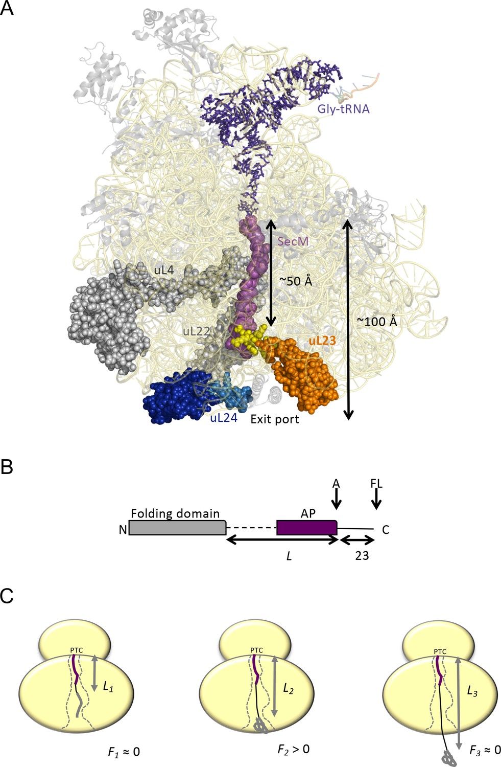
Cotranslational protein folding assay.

(A) Front view of the 50S subunit of the E.coli ribosome adapted from PDB 3JBU (Zhang et al., 2015), with tunnel proteins uL4 and uL22 indicated in gray. The globular domain of uL23 is indicated in orange with the β-hairpin loop depicted in yellow. uL24 is shown in dark blue, with the loop at the tunnel exit shown in light blue. The exit tunnel, outlined by a stalled SecM nascent chain (purple), is ~100 Å in length. (B) The arrest-peptide assay (Nilsson et al., 2015). The domain to be studied is placed L residues upstream of the critical proline at the C-terminal end of the 17-residue long arrest peptide (AP) from the E. coli SecM protein. A 23-residue long stretch of the E. coli LepB protein is attached downstream of the AP, allowing us to separate the arrested (A) and full-length (FL) products by SDS-PAGE after translation. Constructs are translated in the PURExpress in vitro translation system supplemented with WT, uL23 Δloop, or uL24 Δloop high-salt washed ribosomes for 20 min. The relative amounts of arrested and full-length protein were estimated by quantification of SDS-PAGE gels, and the fraction of full-length protein was calculated as fFL = IFL/(IA +IFL) where IA and IFL are the intensities of the bands corresponding to the A and FL products. (c) fFL is a proxy for the force F that cotranslational folding of a protein domain exerts on the AP. At short linker lengths, both F and fFL ≈ 0 because the domain is unable to fold due to lack of space in the exit tunnel. At intermediate linker lengths, F and fFL > 0 because the domain pulls on the nascent chain as it folds. At longer linker lengths, F and fFL ≈ 0 because the domain is already folded when the ribosome reaches the end of the AP.

https://doi.org/10.7554/eLife.36326.002

Inspired by observation that protein domains fold in different parts of the exit tunnel depending on their molecular weight (O'Brien et al., 2010; O'Brien et al., 2011; Trovato and O'Brien, 2016; Samelson et al., 2018; Farías-Rico et al., 2018), we now ask what role the geometry of the exit tunnel plays in determining where these domains fold. To explore this question, we employ the same arrest peptide-based approach (and coarse-grained MD simulations) used in our previous studies of cotranslational protein folding (Nilsson et al., 2015; Nilsson et al., 2017), but with ribosomes that carry deletions in either the uL23 or the uL24 hairpin loop. Our findings provide strong evidence that the tunnel geometry determines where in the tunnel a protein starts to fold.

Results and discussion

The folding assay

Our experimental set-up, Figure 1B, exploits the ability of the SecM translational arrest peptide (AP) (Nakatogawa and Ito, 2001) to act as a force sensor (Ismail et al., 2015; Ismail et al., 2012; Goldman et al., 2015), making it possible to detect the folding of protein domains in the exit tunnel (Nilsson et al., 2015; Goldman et al., 2015). In brief, the domain to be studied is cloned, via a linker, to the AP, L residues away from its C-terminal proline. The AP is followed by a C-terminal tail, to ensure that arrested (A) nascent chains can be cleanly separated from full-length (FL) chains by SDS-PAGE. Constructs with different L are translated in the PURE in vitro translation system (Shimizu et al., 2005), and the fraction full-length protein (fFL) is determined for each L. For linkers that, when stretched, are long enough to allow the protein to reach a part of the exit tunnel where it can fold, force will be exerted on the AP by the folding protein, reducing stalling and increasing fFL. (Tian et al., 2018), Figure 1C. A plot of fFL vs. L thus shows where in the exit tunnel a protein starts to fold and at which linker length folding no longer causes increased tension in the nascent chain.

A number of earlier studies have provided strong support for the notion that the dominant peak in a fFL profile corresponds to folding into the native state (as opposed to, e.g., non-specific compaction of the nascent chain): (i) folded proteins have been visualized in the exit tunnel by cryo-EM of ribosome-nascent chain complexes at L-values corresponding to the dominant fFL peak (Nilsson et al., 2015; Nilsson et al., 2017; Tian et al., 2018), (ii) the dominant fFL peak disappears when proteins that depend on metals or other ligands for folding are translated in the absence of the ligand (Farías-Rico et al., 2018; Nilsson et al., 2015), (iii) the dominant fFL peak corresponds closely to the tether length at which protein domains become resistant to on-ribosome pulse-proteolysis by thermolysin (Farías-Rico et al., 2018) or at which folding can be detected by other techniques such as NMR or FRET (Kemp et al., 2018), (iv) the amplitude of the fFL peak correlates with the folding free energy of a domain (Farías-Rico et al., 2018).

uL23 Δloop and uL24 Δloop ribosomes

The E. coli strains HDB143 (uL23 Δloop; uL23 residues 65–74 deleted) and HDB144 (uL24 Δloop; uL24 residues 43–57 deleted) have previously been shown to be viable (Peterson et al., 2010), as is a strain where uL23 has been replaced by a homologue from spinach chloroplast ribosomes that also lacks the β-hairpin loop (Bubunenko et al., 1994; Bieri et al., 2017). These strains were used to purify high-salt-washed ribosomes that were used to translate proteins in the commercially available PURExpress Δ-Ribosome kit. Analysis of the purified ribosomes by SDS-PAGE and western blotting demonstrated the expected size differences compared to wildtype for the uL23 Δloop and uL24 Δloop proteins, Figure 1—figure supplement 1.

Cryo-EM structure of uL23 Δloop ribosomes

The loop deleted in the uL24 Δloop ribosomes does not interact with neighboring parts of the ribosome, Figure 2A, and hence its removal would not be expected to alter the structure of other parts of the exit tunnel. In contrast, the loop deleted in uL23 Δloop ribosomes is located deep in the exit tunnel, Figure 2B, ~40–50 Å from the exit and it is not clear a priori whether its removal may cause rearrangements in other tunnel components. For this reason, we determined a cryoEM structure of the uL23 Δloop 70S ribosome at an average resolution of 3.3 Å, Figure 2C, Figure 2—figure supplement 1, and found that the shape of the tunnel remains unchanged in the uL23 Δloop ribosome when compared with wildtype (WT) E. coli ribosomes, except for an increase in volume resulting from the absence of the uL23 loop Figure 2C–D. We estimated this increase using the POVME algorithm (Durrant et al., 2011; Durrant et al., 2014). Compared to WT E. coli ribosomes, the tunnel volume increases by 2,064 Å3 in uL23 Δloop ribosomes, see Video 1, about 1/3 of the size of ADR1a (5,880 Å3) calculated by the same method.

Figure 2 with 1 supplement see all
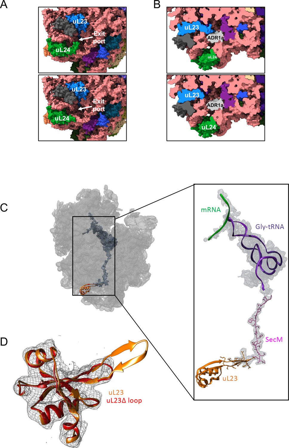
Structural consequences of removing the hairpin loops in uL24 and uL23 modeled after PDB 3JBU of the SecM stalled ribosome.

(A) In wildtype ribosomes, the loop in uL24 partially obstructs the exit tunnel (top panel). Its removal in uL24 Δloop ribosomes creates a wide opening into the tunnel (bottom panel). (B) In wildtype ribosomes, the loop in uL23 extends into the exit tunnel (top panel). Its removal in uL23 Δloop ribosomes creates an open space around the area where the ADR1a domain is known to fold (Nilsson et al., 2015). The ADR1a structure is from PDB 5A7U. (C) Cryo-EM structure of the uL23 Δloop 70 S ribosome (EMD-4319), fitted to PDB 3JBU (that includes a Gly-tRNA and a 26-residue long arrested SecM AP) to locate uL23 (orange) and the exit tunnel. The enlarged region shows a difference map (in mesh) obtained by subtracting the cryo-EM map of the uL23 Δloop 70 S ribosome from a map generated from 3JBU in Chimera. The difference map shows that the only difference in volume between the two maps is the tRNA (in magenta), the SecM AP (in pink), and the loop deleted from uL23. (D) Extracted cryo-EM density (in mesh) for uL23 in the uL23 Δloop ribosome EMD-4319. Wildtype uL23 (orange) and a de novo-built model for the mutant uL23 Δloop protein (PDB 6FU8; red) are shown in ribbon representation.

https://doi.org/10.7554/eLife.36326.005
Video 1
The ribosome exit tunnel (mesh), as calculated for PDB 3JBU, uL23 Δloop and uL24 Δloop ribosomes by POVME.

See Figure 1A for coloring scheme. The b-hairpin loops deleted in uL23 Δloop and uL24 Δloop ribosomes are shown in yellow and light blue, respectively. To facilitate the visualization of the exit tunnel, spheres left outside the exit tunnel after POVME processing were manually removed.

https://doi.org/10.7554/eLife.36326.007

ADR1a folds deeper in the exit tunnel in uL23 Δloop but not in uL24 Δloop ribosomes

ADR1a constructs of different linker lengths (L) were translated in the PURExpress Δ-Ribosome kit supplemented with purified WT, uL23 Δloop, or uL24 Δloop ribosomes, either in the presence of 50 μM Zn2+ (to promote folding of ADR1a) or in the presence of 50 μM of the zinc-specific chelating agent TPEN (to prevent folding of ADR1a; TPEN is required to remove residual amounts of Zn2+ from the PURE lysate) (Figure 3A, Figure 3—figure supplements 14). Translation rates in PURE are ~10 fold slower than in vivo (Capece et al., 2015), but since the proteins studied here fold on micro-to-millisecond time scales, that is considerably faster than the in vivo translation rate, it is safe to assume that the folding reaction has time to equilibrate between each translation step both in vivo and in the PURE system.

Figure 3 with 10 supplements see all
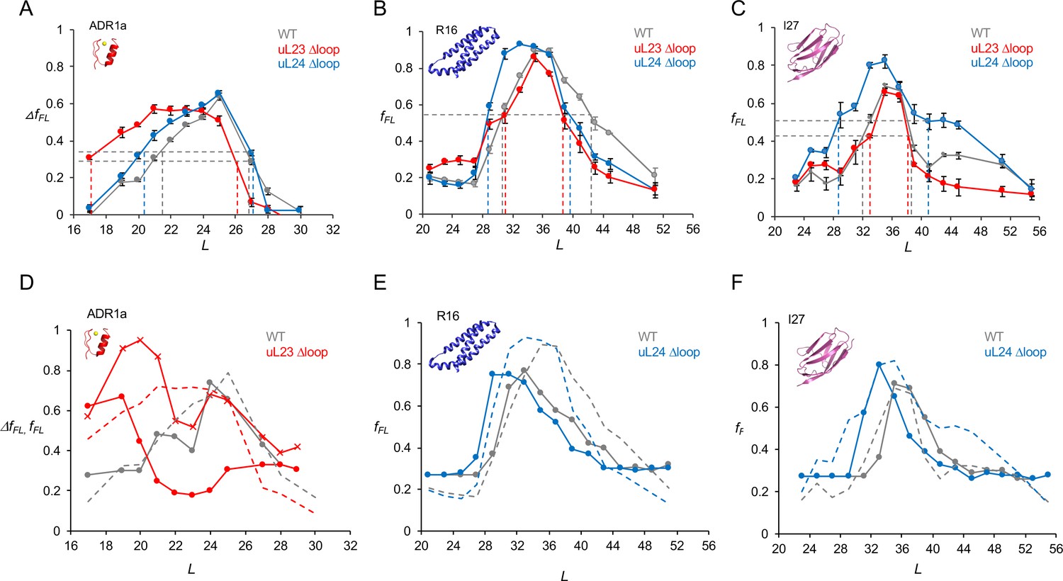
Cotranslational folding in WT, uL23 delta-loop, and uL24 delta-loop ribosomes.

(A) ΔfFL profiles (ΔfFL = fFL(50 μM Zn2+) – fFL(50 μM TPEN)) for ADR1a constructs translated in the PURE system supplemented with in-house purified WT (gray), uL23 Δloop (red), and uL24 Δloop (blue) E. coil ribosomes. (B) fFL profiles for spectrin R16 constructs translated in the PURE system supplemented with in-house purified WT (gray), uL23 Δloop (red), and uL24 Δloop (blue) E. coli ribosomes. (C) fFL profiles for titin I27 constructs translated in the PURE system supplemented with in-house purified WT (gray), uL23 Δloop (red), and uL24 Δloop (blue) E. coli ribosomes. Error bars in panels a-c show SEM values calculated from at least three independent experiments. Dashed lines indicate Lonset and Lend values, c.f., Table 1. fFL profiles for non-folding mutants of R16 and I27 are found in (Nilsson et al., 2017; Tian et al., 2018). (D) Simulated fFL profiles (full lines) for ADR1a, spectrin R16, and titin I27 obtained with WT (gray), uL23 Δloop (red), and uL24 Δloop (blue) ribosomes. The corresponding experimental fFL profiles from panels a-c are shown as dashed lines. The simulated ADR1a fFL profile marked by X’s was obtained with a uL23 Δloop(70-72) ribosome model. Simulated fFL profiles for ADR1a with uL24 Δloop ribosomes, and for R16 and I27 with uL23 Δloop ribosomes, are essentially identical to the corresponding profiles obtained with WT ribosomes, and are shown in Figure 3—figure supplement 10.

https://doi.org/10.7554/eLife.36326.008

Similar to previous results (Nilsson et al., 2015), we saw efficient stalling when ADR1a was translated in the presence of TPEN at L ≥ 19 residues, Figure 3—figure supplement 8. Further, there is a slight but significant increase in fFL at L = 17 residues in the presence of TPEN (hence not related to folding); this has been observed before (Nilsson et al., 2015) and we hypothesize that it is due to a weakening in the arrest potency of SecM by the ADR1a residues that abut the AP in this construct (see Figure 3—figure supplement 9 for sequences). To correct for this effect, we calculated ΔfFL = fFL(Zn2+) – fFL(TPEN), Figure 3A. In the presence of Zn2+, the ΔfFL profiles for WT and uL24 Δloop ribosomes are very similar: ΔfFL starts to increase around L = 20–21 residues and peaks at L = 25 residues (gray and blue curves). In contrast, for the uL23 Δloop ribosomes, ΔfFL starts to increase already at L = 17 residues and peaks at L = 21–24 residues (red curve). To quantify these differences, for each fFL curve we calculated the linker lengths characterizing the onset and end of the peak (Lonset and Lend; defined as the L-values for which the curve has half-maximal height, as indicated in Figure 3A), as well as the L-value corresponding to the peak of the curve (Lmax), Table 1.

Table 1
Lonset, Lmax, and Lend values calculated from the fFL profiles in Figure 3.
https://doi.org/10.7554/eLife.36326.020
ADR1aR16I27
WTuL23 ΔloopuL24 ΔloopWTuL23 ΔloopuL24 ΔloopWTuL23 ΔloopuL24 Δloop
Lonset211720313129323328
Lmax252225353533353535
Lend272627423940383841

A previous cryo-EM study demonstrated that the 29-residue ADR1a domain folds deep inside the ribosome exit tunnel in a location where it is in contact with the uL23 loop (Nilsson et al., 2015), Figure 2B. The additional space available in uL23 Δloop ribosomes makes it possible for ADR1a to start to fold at 3–4 residues shorter linker lengths (Lonset). Assuming an extended conformation of the linker segment (~3 Å per residue), ADR1a folds ~9–12 Å deeper in the exit tunnel in uL23 Δloop ribosomes than in WT ribosomes.

Spectrin and titin domains fold deeper in the exit tunnel in uL24 Δloop ribosomes

The 109-residue α-spectrin R16 domain has been shown to fold cotranslationally at L ≈ 35 residues, in close proximity to uL24 in the exit port region (Nilsson et al., 2017). As seen in Figure 3B and Table 1, with both WT and uL23 Δloop ribosomes, R16 has Lonset = 31 residues and Lmax = 35 residues (gray and red curves). For the uL24 Δloop ribosomes however, Lonset = 29 residues and Lmax = 33 residues (Table 1), suggesting that that spectrin R16 folds ~6–7 Å deeper in the exit tunnel when the uL24 loop does not obstruct the tunnel exit port.

Similar results were obtained for the 89-residue titin I27 domain, Figure 3C. Previous studies have shown that the I27 domain folds at linker lengths L = 35–39 residues and that it folds in about the same location as does spectrin R16, in close proximity to the uL24 loop (Tian et al., 2018). The fFL profile is not affected by the uL23 loop deletion, but folding commences at ~4 residues shorter linker lengths in uL24 Δloop ribosomes, similar to R16 (Table 1).

Coarse-grained molecular dynamics simulations

In order to provide a more detailed structural framework for interpreting the fFL profile results, we performed coarse-grained molecular dynamics simulations of the cotranslational folding of ADR1a, spectrin R16, and titin I27 in WT, uL23 Δloop, and uL24 Δloop ribosomes, using a recently described model that allows us to calculate fFL profiles from the simulations (Tian et al., 2018). The essence of the method is that the simulations are used to determine folded and unfolded populations at each linker length, and the forces associated with them. Combining this information with the experimentally determined force-dependent escape rate of the AP from the ribosome (Goldman et al., 2015) in a kinetic model allows fFL to be calculated. Simulated (full lines) and experimental (dashed lines) fFL profiles are shown in Figure 3D–F, and detailed simulation results, together with representative snapshots from the simulations of the folded domains at LLonset, are shown in Figure 3—figure supplement 10. For all three proteins, the Lonset values are well reproduced by the simulations, both in WT and Δloop ribosomes. Lmax values are also well captured by the simulations for ADR1a (WT ribosomes) and I27 (both WT and uL24 Δloop ribosomes), but are shifted to somewhat lower values in the R16 simulations.

The simulated ADR1a fFL profile for uL23 Δloop ribosomes, while showing an early onset of folding in agreement with the experimental profile, has a much smaller Lmax value. We also performed a simulation using a ribosome model with a smaller deletion in the uL23 loop (residues 70–72; red curve marked by X’s); in this case, the peak in the simulated profile extends between Lonset and Lend values that are more similar to the experimental profile for uL23 Δloop ribosomes. The shape of the fFL profile for ADR1a is clearly highly sensitive to fine structural details of the exit tunnel and therefore somewhat difficult to reproduce by coarse-grained simulations.

In summary, both the experimental and simulation results are consistent with the idea that proteins start to fold as soon as they reach a part of the exit tunnel that is large enough to hold the folded protein. Judging from the fFL profiles, the 29-residue ADR1a domain folds approximately ~9–12 Å deeper in the exit tunnel in uL23 Δloop ribosomes than in WT and uL24 Δloop ribosomes, while the 89- and 109-residue titin and spectrin domains fold ~6–10 Å deeper inside the tunnel in uL24 Δloop ribosomes than in WT and uL23 Δloop ribosomes; the corresponding values estimated from the simulations are ~6 Å for ADR1a and ~13–15 Å for I27 and R16 (Figure 3—figure supplement 10 panel B).

Both the uL23 and uL24 loops thus serve to reduce the space available for folding, but in different parts of the exit tunnel. The uL24 loop is particularly interesting in this regard. In bacterial ribosomes, it partially blocks the tunnel exit port, closing off what would otherwise be a wide, funnel-like opening, Figure 2A, and thereby prevents domains of Mw ≥10 kDa from folding inside the exit tunnel. It is conserved (in length, if not in sequence) in bacterial ribosomes, Figure 1—figure supplement 2, suggesting that this will be the case not only for E. coli ribosomes but for bacterial ribosomes in general. Eukaryotic ribosome tunnels have different geometries owing to expansion segments in their rRNA as well as an increased number of proteins and a wider exit port (Wilson and Doudna Cate, 2012; Filipovska and Rackham, 2013); uL24 is among the most divergent proteins compared to bacteria (Melnikov et al., 2015). We therefore expect the precise relation between the onset of folding and protein Mw to be somewhat different in eukaryotic ribosomes, as also suggested by a recent study (Schiller et al., 2017). At present, we do not know to what extent the shape of the ribosome exit tunnel has evolved to optimize the conditions for cotranslational protein folding in different organisms and organelles, but it is not unlikely that such a connection exists.

Materials and methods

Key resources table
Reagent type
(species) or
resource
DesignationSource or referenceIdentifiersAdditional information
Strain, strain
background
(Escherichia coli)
HDB140, HDB143,
HDB144, Strain
background N281
10.1111/j.1365–2958
.2010.07325.x
NAStrains used to isolate
high-salt washed
ribosomes in this study.
AntibodyuL23, uL2410.1111/j.1365–2958
.2010.07325.x
NA1:8000 dilution used
(incubated for one hour).
Secondary antibody:
Mouse (1:20,000 dilution
incubated for one hour).
Nitrocellulose membrane
blocked with 5% Milk in
TBS-T for an hour.
Peptide,
recombinant
protein
uL23 Δ loop,
uL24 Δ loop
10.1111/j.1365–2958
.2010.07325.x
UniProtKB- P0ADZ0 (rplW)
UniProtKB-P60624 (rplX)
Referred to as HDB 143
and HDB 144 in original
paper. Refers to genes
rplWΔ65–74 and rplX
Δ43–57 respectively.
Commercial
assay or kit
GeneJET Plasmid
miniprep kit
Thermo Fisher
Scientific
RRID:SCR_008452
Cat no. K0502Used to purify plasmids
Commercial
assay or kit
PURExpress Δ
Ribosome kit
New England BiolabsCat no. E3313SKit was supplemented
with ribosomes purified
in the lab. Translation
carried out at 37°C for 20 min.
Software,
algorithm
EasyQuantdoi: 10.1038/
nsmb.2376
NAUsed to quantify relative
fraction full-length of
translated protein from
SDS-PAGE
Software,
algorithm
cryoSPARC version v2Structura
Biotechnology Inc
NAUsed for ab-initio
reconstruction of uL23Δloop
ribosomes. The following
operations were carried
out as part of the
cryoSPARC workflow: 2D
classification, Ab initio
reconstruction, Homogeneous
refinement, Sharpening and
map flipping to correct for
handedness, local resolution.
Software,
algorithm
UCSF ChimeraXSCR_015872Used to make Figure 2A
Software,
algorithm
UCSF Chimera v. 1.12J Comput Chem.
2004 Oct;25
(Nilsson et al., 2015):
1605–12.
SCR_004097Used to visualise the cryoEM
map, fit PDB models 3JBU,
4YBB to check for differences
in maps. Used to make figures.
Software,
algorithm
Jalview v 2.10.4doi: 10.1093/
bioinformatics/btp033
SCR_006459Use for generating multiple
sequence alignments of
uL23 and uL24 in the
supplementary figures.
ChemicalPotassium acetateSigma-Aldrich
(SCR_008988)
Cat no. P1190Source of potassium
ions to stabilise ribosomes
ChemicalMagnesium acetateSigma-Aldrich
(SCR_008988)
Cat no. M5661Source of Magnesium
ions to stabilise ribosomes
ChemicalcOmplete protease
inhibitor cocktail
Sigma-Aldrich
(SCR_008988)
Cat no. 04693116001Used as a protease
inhibitor during cell
lysis to obtain ribosomes
ChemicalN,N,N′,N′-Tetrakis
(2-pyridylmethyl)
ethylenediamine
Sigma Aldrich
(SCR_008988)
Cat no. P4413Used to chelate Zn
for the -Zn reactions
in the ADR1 plot.
Chemicalthreo-1,4-Dimercapto-2,
3-butanediol
DL-Dithiothreitol
Sigma Aldrich
(SCR_008988)
Cat no. DTT-ROReducing agent added
to buffers during ribosome
purification and as a
reductant for SDS-PAGE
ChemicalTris BaseSigma-Aldrich (SCR_008988)Cat no. T1503Buffering agent during
ribosome preparation
Chemical35S MethioninePerkin-ElmerCat no.
NEG009T001MC
35S Methionine is
incorporated into the
protein during in vitro
translation and aids
detection by
phosphorimaging.

Enzymes and chemicals

Request a detailed protocol

The PURExpress Δ Ribosome kit was purchased from New England Biolabs (Cat no. E3313S). The components used to prepare Lysogeny Broth (LB Medium) for ribosome isolation were obtained from BD Biosciences and all other chemicals used were sourced from Merck Sigma Aldrich. (35S) Methionine was purchased from Perkin Elmer. Bis-Tris gels and plasmid isolation kits were obtained from Thermo Scientific.

Plasmids

All ADR1, spectrin and titin constructs fused to the E. coli SecM AP via a variable linker were expressed from the pET19b vector, as described previously (Nilsson et al., 2015; Nilsson et al., 2017; Tian et al., 2018). The spectrin constructs used in this study lacked the soluble domain of LepB at the N-terminus.

Strains and antisera

Request a detailed protocol

Strains HDB140 (referred to as WT), HDB143 (referred to as uL23 Δloop) and HDB144 (referred to as uL24 Δloop), as well as rabbit polyclonal antisera against uL23 and uL24, are described in (Peterson et al., 2010).

Isolation of ribosomes

Request a detailed protocol

Ribosomes were purified from the strains HDB140, HDB143, and HDB144. The strains were cultured in Lysogeny broth (LB) to an A600 of 1.0 at 37°C and chilled on ice for 15 min before they were harvested by centrifugation at 4000 g for 10 min. The cell pellet was washed twice with Buffer A at pH 7.5 (10 mM Tris-OAc, 14 mM Mg(OAc)2, 60 mM KOAc, 1 mM DTT, 0.1% Complete Protease Inhibitor) and lysed using the Emulsifex (Avestin) at a pressure of 8000 psi. The cell lysate was loaded on a sucrose cushion at pH 7.5 (50 mM Tris-OAc, 1 M KOAc, 15 mM Mg(OAc)2,1.44 M sucrose, 1 mM DTT, 0.1% Complete Protease Inhibitor) and centrifuged at 80,000xg in a Ti70 rotor (Beckman-Coulter) for 17 hr. The obtained ribosomal pellet was resuspended in Buffer B at pH 7.5 (50 mM Tris-OAc, 50 mM KOAc, 5 mM Mg(OAc)2, 1 mM DTT), flask frozen in liquid nitrogen and stored at −80°C. This suspension of ribosomes is presumed to consist of a pool of non-translating 30S, 50S and 70S particles due to the concentration of Mg2+ in the buffer they are in. Each batch of ribosomes that was prepared was tested for optimal translation by titrating different volumes in the PURExpress Δ-Ribosome kit.

In vitro transcription and translation

Request a detailed protocol

The generated constructs were translated for 20 min. in the PURExpress Δ-Ribosome kit supplemented with high-salt-washed ribosomes isolated from HDB140, HDB143 (uL23 Δloop), or HDB144 (uL24 Δloop). Plasmid DNA of each construct (300 ng) was used as a template for polypeptide synthesis, and translation was carried out in the presence of (35S) Methionine at 37°C for 20 min and shaking at 500 r.p.m. For ADR1a constructs, the translation reactions also included either 50 μM zinc acetate or 50 μM of the Zn2+ chelator TPEN. Translation was stopped by treating the sample with a final concentration of 5% trichloroacetic acid (TCA) and incubated on ice for 30 min. The TCA precipitated samples were subsequently centrifuged at 20,000 g for 10 min in a tabletop centrifuge (Eppendorf) and the pellet obtained was solubilized in sample buffer, supplemented with RNaseA (400 μg/ml), and incubated at 37°C for 15 min. The samples were resolved on 12% Bis-Tris gels (Thermo Scientific) in MOPS buffer for ADR1 and MES buffer for Spectrin and Titin. Gels were dried and subjected to autoradiography and scanned using the Fujifilm FLA-9000 phosphorimager for visualization of radioactively labeled translated proteins.

Quantification of radioactively labelled proteins

Request a detailed protocol

The protein bands on the gel were quantified using MultiGauge (Fujifilm) from which one-dimensional intensity profiles of each gel lane was extracted. This information was subsequently fit to a Gaussian distribution using EasyQuant (Rickard Hedman, Stockholm University). The sum of the arrested and full-length bands was calculated, and this was used to estimate the fraction full-length protein for each construct.

Cryo-EM sample preparation and data processing

Request a detailed protocol

The uL23 Δloop ribosomes (4 A260/ml) diluted in grid buffer (20 mM HEPES-KOH, 50 mM KOAc, 10 mM Mg(OAc)2, 125 mM sucrose, 2 mM Trp, 0.03% DDM) were loaded on Pelco TEM 400 mesh Cu grids pre-coated with 2 nm thick carbon and frozen using the Vitrobot Mark IV (FEI). Data were collected on the Titan Krios (FEI) microscope operated at 300 keV and equipped with a Falcon II direct electron detector. The camera was set to a nominal magnification of 75,000X, which resulted in a pixel size of 1.09 Å at the sample level and a defocus range of −1 to −3 μm.

The frame dose used was 1.17 e/Å2, and 20 frames were aligned using MotionCor2 (Li et al., 2013) within the Scipion software suite (de la Rosa-Trevín et al., 2016). The micrographs were visually inspected and those within a resolution threshold of 5 Å were selected, yielding 3522 micrographs.

471,272 particles were picked using Xmipp manual-pick followed by particle extraction within Scipion and further processing in CryoSPARC (Punjani et al., 2017). Two rounds of 2D classification were done, and particles resembling 30S and 50S subunits alone were discarded after visual inspection of the classes. The remaining 297,363 particles of the 70S ribosome were subjected to ab initio reconstruction into three classes to further sort out heterogeneity. A single homogeneous class consisting of 132,029 particles was used for final homogeneous refinement that resulted in a final map with an average FSC resolution at 0.143 of 3.28 Å. The obtained map of the 70S ribosome was sharpened and corrected for handedness in CryoSPARC fitted with PDBs 3JBU and 4YBB in Chimera (Pettersen et al., 2004). Local resolution and FSC at 0.143 was estimated in cryoSPARC. The electron microscopy map was deposited in the Electron Microscopy Data Bank.

The initial model for uL23 Δloop was built with Coot, and improved by energy minimization in a solvated dodecahedron box of explicit TIP3P waters, neutralized with chloride ions and using the Amber 99SB-ILDN force field (Lindorff-Larsen et al., 2010). The steepest descent minimization method implemented in GROMACS 2016.1 was used (Abraham et al., 2015; Pal et al., 2014). Even after minimization, the backbone of the new loop formed after the deletion of residues 65–74 still showed improper geometry and Ramachandran outliers, so we used kinematic sampling (Bhardwaj et al., 2016) to model alternative loop conformations, and then we selected the loop that could fit the electron density and had the best Ramachandran score.

Figures were prepared using MacPymol 1.8.6.2 (Schrödinger LLC), Chimera (Pettersen et al., 2004), and ChimeraX (Goddard et al., 2018).

Calculation of tunnel volume

View detailed protocol

The volume calculations were performed with POVME 2.0 (1). We used the E. coli SecM structure PDB 3JBU as a reference. To determine the inclusion region, we generated a series of overlapping spheres- eight with a 20 Å radius, and one with a 40 Å radius. In order to have a complete coverage of the exit tunnel, the centers of the spheres were chosen to match the coordinates corresponding to alternating Cα atoms of the amino acids of the SecM arrest peptide located within the exit tunnel (for the 20 Å radius spheres the residues use as centers were D11, F13, T15, V17, I19, Q21, Q23, I25, A27, G28 and for the 40 Å radius sphere the residue was E3). Grid Spacing was set to 2.0 Å, and the distance cut-off to 1.09 Å. For all three cases (WT, uL24 Δ loop, uL23 Δ loop), we used the same inclusion region. We also removed the SecM arrest peptide located within the exit tunnel. For uL23 Δloop ribosomes residues 65–75 were removed from uL23, and for uL24 Δloop ribosomes residues 42–57 were removed from uL24 (numbering based on PDB 3JBU) prior to the calculation.

Kinetic model to calculate fraction full length protein fFL(t)

Request a detailed protocol

The theoretical force profiles (Figure 3D–F) for ADR1a, I27, and R16 were calculated based on a kinetic model introduced in our previous study (Tian et al., 2018). Briefly, the rate, ke, of the arrest peptide sequence escape from the peptidyl transfer center with a force (F) exerted by the folding protein can be calculated using the Bell model:

ke(F)=k0eFΔx/kBT,

where Δx is the distance from the free energy minimum to the transition state, k0 the rupture rate when force equals to zero, kB is Boltzmann’s constant, and T the absolute temperature. In this study, k0 and Δx are set to be 3.4 ×10⁻⁴ s−1 and 4.5 Å, respectively, based on a previous experimental study (Goldman et al., 2015) in which k0 and Δx were estimated to be in the range of 0.5 ×10⁻⁴ to 20 ×10−4 s−1 and 1-8 Å, respectively.

We assume that the folding and unfolding of the protein is much faster than the escape from the ribosome. Then the time-dependent force profile fFLt can be obtained approximately by the mean pulling forces exerted when the protein is unfolded, Fu, or folded, Ff, and the unfolded and folded populations of Pu and Pf respectively,

fFL(t)1-exp-tPukeFu+PfkeFf.

Note that the values of Fu, Ff, Pu, Pf are dependent on the linker length L, and can be determined by molecular dynamics simulations.

Molecular dynamics simulations of ribosome-nascent chain complex

Request a detailed protocol

A coarse-grained model was employed to simulate folding of ADR1a, titin I27, and spectrin R16 on the ribosome. The ribosome was modelled on the 50S subunit of the E. coli ribosome (PDB 3OFR; Dunkle et al., 2010) Each amino acid in the nascent chain and ribosome was represented by one bead at the position of the α-carbon atom, each RNA residue was modelled by three beads located at the positions of phosphate P, sugar C4’, and base N3 atoms (Voss and Gerstein, 2005). The uL23 ∆loop ribosome was modelled by replacing the coordinates of the wildtype uL23 protein with the cryo-EM structure of the uL23 ∆loop protein from this study (PDB 6FU8), after being aligned to the wild type protein. The uL24 ∆loop ribosome was modelled by replacing the coordinates of the wildtype uL24 protein with the structure of the uL24 ∆loop protein built by homology modelling with Modeller (Eswar et al., 2006).

The interactions within the nascent chain were governed by a standard structure-based model (Karanicolas and Brooks, 2002), which allowed it to reversibly fold to the native state and unfold. Interactions between the protein and ribosome beads were purely repulsive. The pulling force (F) exerted on the arrest peptide by the folding of the protein (ADR1a, R16, or I27) was measured by the extension of the harmonic pseudobond potential between the last and the second last amino acid of the SecM arrest peptide. More details can be found in our previous study (Tian et al., 2018).

Data availability

The Cryo-EM map has been deposited under accession code EMD-4319, and the Atomic model for uL23 Dloop deposited under PDB accession number 6FU8.

The following data sets were generated
    1. Kudva R
    2. von Heijne G
    (2018) EMDataBank
    ID 4319. E.coli ribosome with Δ-hairpin of uL23 deleted.
    1. Kudva R
    2. von Heijne G
    (2018) Protein Data Bank
    ID 6FU8. Atomic model for uL23 delta-loop.

References

  1. Book
    1. Pal S
    2. Abraham MJ
    3. Kutzner C
    4. Hess B
    (2014)
    Solving Software Challenges for Exascale
    Cham, Switzerland: Springer.

Decision letter

  1. John Kuriyan
    Senior and Reviewing Editor; University of California, Berkeley, United States

In the interests of transparency, eLife includes the editorial decision letter and accompanying author responses. A lightly edited version of the letter sent to the authors after peer review is shown, indicating the most substantive concerns; minor comments are not usually included.

[Editors’ note: this article was originally rejected after discussions between the reviewers, but the authors were invited to resubmit after an appeal against the decision.]

Thank you for submitting your work entitled "The Shape of the Ribosome Exit Tunnel Affects Cotranslational Protein Folding" for consideration by eLife. Your article has been reviewed by three peer reviewers, and the evaluation has been overseen by John Kuriyan as the Senior and Reviewing Editor. The reviewers have opted to remain anonymous.

Based on discussions with the reviewers and the individual reviews below, we regret to inform you that your work will not be considered further for publication in eLife. The reviewers and the editors agree that this paper is an interesting and important study of the folding of nascent peptides, and how the shape of the ribosome exit tunnel governs that folding. Despite the interest in the work, after discussion with the reviewers, we have concluded that the paper is not quite definitive enough in its conclusions for further consideration at eLife. We remain interested in the work, and if you are able to provide additional data to support the conclusions and to address the reviewer comments satisfactorily, we would welcome a resubmission. We recognize, however, that you might wish to submit the manuscript elsewhere at this point.

The three individual reviews are appended below.

Reviewer 1:

This paper demonstrates the influence that changes in geometry of the peptide exit tunnel can have on the timing of co-translational protein folding. The authors conduct in vitro translation assays to translate folding domain fused to the SecM stalling sequence with varying linker length to measure at which linker lengths the nascent chain starts to fold on the ribosome, applying a tugging force to rescue the stall and translate the full protein. After using Cryo-EM to confirm that the loop deletion mutations on uL23 and uL24 doesn't change the architecture of the peptide exit tunnel, the authors indeed show that the loop deletions alter where the nascent chain folding occurs in the tunnel. Overall, the findings show a straightforward method to probe the location within the peptide exit tunnel where the protein folding occurs, and is indeed an advance in the field. The novelty here is in the manipulation of the tunnel itself, and the correlation to structural data. However, the question remains whether the science presented here rises to the level of publication in eLife. There are some critiques that the authors should address outlined below:

1) It is not explicitly mentioned whether the uL23Δloop ribosomes prepared for Cryo-EM is a 50S, empty 70S, or stalled translation complexes. This needs to be made in the Materials and methods section under the "Cryo-EM sample preparation and data processing" part.

2) Why is there an "increase in fFL seen at L = 17 residues in the presence of TPEN"? What does TPEN do to cause the increase in fFL? A brief statement in the text commenting on this effect would be helpful. If indeed this is an effect of TPEN, is there a way to do the same experiment in the absence of Zn2+ or TPEN to confirm this?

3) The authors commented on the universality of the uL24 loop in bacteria. It would be great to also comment on the non-universality of uL23 or uL24 loops in other domains of life (like the absence of uL23 loop in spinach chloroplast ribosomes mentioned in the paper) and how it may suggest that co-translational protein folding may work differently in another organism due to differences in geometry of its ribosomal peptide exit tunnel.

Reviewer 2:

Kudva et al. investigate how the geometry of the ribosomal tunnel affects folding. The main intent of this study is to understand how modifications on the ribosome influence the location of co-translational folding of proteins of different sizes. The authors first describe the folding assay; briefly, the force that protein folding close to the exit vestibule can exert on the SecM stalling sequence can consequently cause ribosome readthrough the mRNA and translate additional residues.

The authors explain the basis of the modified ribosomes HDB143 (ΔuL23) and HDB144 (ΔuL24) and solve the cryoEM structure of ΔL23 ribosomes. They further characterise the structural differences between wt and mutant by comparing the tunnel volumes. The authors then analyse the force profile of the ADR1, spectrin and titin domains on the wt, ΔL23 and ΔL24 ribosomes in an attempt to dissect the contribution of the two loops from these proteins in protein folding. They observe that ADR1 folds in the exit tunnel in the absence of loop in uL23 but not uL24, whereas the titin and spectrin (R15, R16) domains fold inside the tunnel in ΔL24 rather than the wt and ΔL23.

This study attempts to deconvolute the tunnel components that may be important for co-translational folding using the force assay which the authors are known for. This is a valuable, complex and rich experiment. It seems to me, however, not enough to interpret such complex events of translational and folding. This is my main concern: all conclusions are drawn based on the force experiments alone. Also, although some structural data are presented they are not used to rationalize the force results. It seems therefore the authors try to overstretch interpretation of their data.

Overall the manuscript contains some nice experiments and this is an interesting study. The study however, seems to need more substance to support some of its more far reaching statements. For example, the broader implications at the end would be excellent, had the paper have more depth. For me, this study in its current form leaves too many open questions.

1) Can the authors comment on the experimental conditions used for translation. Do the mutant ribosomes have the same translational kinetics as for wt? Are any differences likely to influence the force plots?

2) The cryoEM structure of the uL23 ribosomes is interesting, but why the authors did not investigate programmed ribosomes as they have previously? However, using the uL23 structure, can the authors comment further on the structural environment that the ADR1a would have in the uL23 ribosome compared to WT ribosomes? Does it have more space than in the original location? Given the rationale that the authors use to describe where folding begins to takes place (e.g. ca. 17aa in uL23 deletion). More information regarding the significance of structure would help.

3) It is not very clear to me how the authors define folding. Descriptions of the plots include words like "starting to increase, starting to fold". More information is needed on how these points are determined because to me it seems very subjective. In the absence of unfolded controls (e.g. in R15 and R16), the context for the "dips" and "rise/peaks" are difficult to rationalise.

4) On a similar point, overall, the analysis of Figure 3 appears ambiguous. For example, in Figure 3B red (uL23), data points from L=21-24 are effectively identical, all in error range, which casts potential doubt on the contention that there is a shift by 2-3 residues. The same types of difficulties arise in following analysis Figure 3D and 3E. One cannot clearly say there is 2-3 residue shifts. In order to focus on this small difference between the profiles, there should be more detailed analysis on other differences between the force profiles, for instance, why there are significant differences in fFL values between different ribosomes with the same length of nascent chains.

5) In ADR1a in the ΔuL23 ribosomes the first point (17aa) is non-zero even after subtracting the unfolded control. This suggests that the ADR1a is beginning to fold 51Ang (3ang per aa) from the PTC, approximately near uL22. If this is a folding, why is there never a clear peak? The authors say that the EM data tells us that the tunnel hasn't changed – even if the uL23 loop is gone and folding 'begins' early, (a) why isn't a complete fold reached (b) why would the ADR1a complete its fold in a narrow region of the tunnel rather than the vestibule exit? The "spring constant" idea is attractive – if the tunnel has opened up, then the NC can begin to sample conformations immediately, and in this case never reaches a maximum point of tension in the tunnel.

6) There are differences in Figure 1B observed between wt ribosomes in the PURE-kit or those prepared n-house and no explanation is given as to why? The WT and the WT(purified) ribosomes also show different patterns, even if the peak appears to be retained. Can the authors comment on the general reproducibility? Additionally, the PURE-derived force profile has no error bars – were replicates performed? Why is Δf used solely for this graph?

7) The use of unfolded controls for ADR1a shows clearly need to subtract data – why was this not done for R15, R16 and I27 plots?

8) Overall, the inclusion of both R15 and R16 is perhaps somewhat strange: on one hand, analysis of R15 is more convincing that other nascent chains are used to elaborate on the differences in force profiles. However, can the authors elaborate further on the significance of R15 and R16 and how these results relate to their previous work? For the R15 force profile, the peak of the wild type does not look the same as the previously reported, as it has a peak of 37 amino acids. Although the authors explain that this is because a folding intermediate is formed this is not clear or supported by other data. They also attribute the observed difference in force amplitude between wt and ΔuL23 to the lower "spring constant" due to the wider tunnel in the latter. The discussion of a 'spring tension' was interesting, but not explored and it seemed a pity in this context. Again, supporting data are needed.

9) The folding intermediate proposed in R15 – why would this intermediate only exist in uL23 deleted and WT ribosomes, but not in uL24 deleted ribosome? This seemed to be unclear. If the protein off the ribosome is renatured from an unfolded state does it form intermediates on its folding pathway?

10) It is not clear where in the ribosome the 6-9 Å measurement corresponds to in both the ΔuL23 ribosomes (subsection “ADR1a folds deeper in the exit tunnel in uL23 Δloop but not in uL24 Δloop ribosomes”, last paragraph) and in ΔuL24 ribosomes (subsection “Spectrin and titin domains fold deeper in the exit tunnel in uL24 Δloop ribosomes”, third paragraph). How does this correlate to the structure – i.e. is it consistent with the approximate location for the uL23 deletion?

11) The concluding statements appear to extrapolate from the data too much, and don't appear to be supported by the experimental data presented in the study – for example: "may prevent premature folding".…."that depend on interactions with TF…" Similarly, the uL24 loop statements about correlating length and sequence is not particularly well supported either – it seems to be a rather loose conclusion to an otherwise interesting paper.

Reviewer 3:

This manuscript reports a study of cotranslational folding, with the major conclusion that the shape of the exit tunnel modulates the cotranslational folding process. This conclusion is reached by studying two different proteins and two different forms of the ribosome, with the clever idea of truncating parts of L23 and L24.

My major concern with this manuscript is that the data presented do not adequately support the conclusions. The only experimental technique used is an assay in which of a SecM translational arrest peptide acts, broadly speaking, as a force sensor, whereby the measurements are performed by SDS-PAGE.

It is very difficult to assess the errors associated with this assay, in part because the primary data are not reported.

Most importantly, however, these conclusions should be validated using an independent technique. In the absence of this validation, one should put an excessive trust in the assay used, which would seem to be low resolution at best.

Overall, the publication of this work would be premature.

[Editors’ note: what now follows is the decision letter after the authors submitted for further consideration.]

Thank you for submitting your article "The Shape of the Ribosome Exit Tunnel Affects Cotranslational Protein Folding" for consideration by eLife. Your article has been reviewed by two peer reviewers, and the evaluation has been overseen by John Kuriyan as the Reviewing Editor and Senior Editor. The reviewers have opted to remain anonymous.

Your paper is now, in principle, acceptable for publication, but we are asking for some minor revisions (see below).

Your paper was seen by two of the original reviewers. One of the reviewers approves the revised manuscript. The other reviewer continues to object to the experimental basis for the paper, stating:

"The issue here is whether a single technique can be used to draw conclusions in a high impact paper. I am afraid that I believe that the answer is negative. I take the point made by the authors that this technique has been validated on other systems, but the fact remains that it is not as high resolution and clear cut as other techniques, such as cryo-EM or NMR.

Although I appreciate the quality and the scope of this manuscript, I do not think that the reliability of the conclusions meets the standard required by eLife."

We are making an editorial decision to proceed with publication of your paper at eLife. As someone outside the field, I find the entire body of work describing co-translational folding (either inside the ribosome or in its vicinity) to be extremely interesting, and yielding results that could not have been anticipated a few years ago. Although the results may now seem obvious to people in the field, I feel that the results are still very intriguing. I also note that there are controversies, but this reflects the complexity of the phenomena, and the limited view provided by different experimental techniques. Time will sort this out.

Regarding the objection that the paper relies on only one experimental technique, we accept your argument that this method (protease sensitivity with arrest peptide fusions) has been sufficiently validated in past work. While it would be nice to have the results of other experimental probes, we recognize that you have submitted this work as a short report. Also, I am not sure that NMR and cryoEM necessarily provide definitive answers in cases where a dynamic process is being observed. They too are subject to artifacts arising from restrictions on what can be observed.

Before publication, we encourage you to include in the paper more context for the importance and significance of the work, such as provided in your recent PNAS paper (Farias-Rico et al., 2018). We also suggest that you include some additional explanation for the validation of the method, as you suggest in your response to reviewers. You may do this as you think best suits your purposes, and we will approve your revised manuscript for publication without further delay.

https://doi.org/10.7554/eLife.36326.027

Author response

[Editors’ note: the author responses to the first round of peer review follow.]

Thanks for the thorough review of our manuscript. We take heart in the fact that you say that you’re still interested in the work, despite the rejection (which we understand, given some of the reviewer comments).

Our reading of the reviewer reports (especially reviewers 2 and 3) suggests that the main critique of the manuscript in its present form is that we use only the arrest-peptide based force-measurement assay in the paper, an assay that reviewer 3 in particular feels is not well validated against other techniques for studying cotranslational folding. As detailed in the response to reviewer 3 below, we (emphatically…) do not agree that the technique has not been properly validated, but realize that we should have been more explicit in summarizing previously published data to address this issue.

The second general issue we want to raise is that we wrote this paper in an attempt to make a single, major point based on just one experiment (well, one experiment for each protein domain and each type of mutant ribosome) and therefore, following the eLife Author Instructions, submitted it as a Short Report. Judging from their comments, we’re not sure that the reviewers appreciated that it wasn’t intended as a full paper.

Reviewer 1:

This paper demonstrates the influence that changes in geometry of the peptide exit tunnel can have on the timing of co-translational protein folding. The authors conduct in vitro translation assays to translate folding domain fused to the SecM stalling sequence with varying linker length to measure at which linker lengths the nascent chain starts to fold on the ribosome, applying a tugging force to rescue the stall and translate the full protein. After using Cryo-EM to confirm that the loop deletion mutations on uL23 and uL24 doesn't change the architecture of the peptide exit tunnel, the authors indeed show that the loop deletions alter where the nascent chain folding occurs in the tunnel. Overall, the findings show a straightforward method to probe the location within the peptide exit tunnel where the protein folding occurs, and is indeed an advance in the field. The novelty here is in the manipulation of the tunnel itself, and the correlation to structural data. However, the question remains whether the science presented here rises to the level of publication in eLife. There are some critiques that the authors should address outlined below:

1) It is not explicitly mentioned whether the uL23Δloop ribosomes prepared for Cryo-EM is a 50S, empty 70S, or stalled translation complexes. This needs to be made in the Materials and methods section under the "Cryo-EM sample preparation and data processing" part.

The cryo-EM was done with non-translating 70S ribosomes. This is now stated and highlighted in the Materials and methods section.

2) Why is there an "increase in fFL seen at L = 17 residues in the presence of TPEN"? What does TPEN do to cause the increase in fFL? A brief statement in the text commenting on this effect would be helpful. If indeed this is an effect of TPEN, is there a way to do the same experiment in the absence of Zn2+ or TPEN to confirm this?

In the L=17 construct, ADR1a is appended directly upstream of the SecM arrest peptide (Figure 3—figure supplement 9). Although we haven’t tested this, we believe that the slight increase in fFL seen in the presence of TPEN reflects a weakening of the arrest potency of the AP caused by the abutting residues. It is unlikely that TPEN is responsible for the effect, as this would require some kind of highly specific interaction of TPEN and this precise construct. Since PURE has some residual Zn2+, it is necessary to use TPEN for the no-Zn2+ controls. This is now stated in the text.

3) The authors commented on the universality of the uL24 loop in bacteria. It would be great to also comment on the non-universality of uL23 or uL24 loops in other domains of life (like the absence of uL23 loop in spinach chloroplast ribosomes mentioned in the paper) and how it may suggest that co-translational protein folding may work differently in another organism due to differences in geometry of its ribosomal peptide exit tunnel.

We now include multiple-sequence alignments of uL23 and uL24 homologs as Figure 1—figure supplement 2, showing the absence/presence of the loops in different phyla (as a case in point, both the uL23 and uL24 loops are absent in mammalian ribosomes). We have also added a comment that the uL24 homolog in mammalian ribosomes does not obstruct the exit tunnel to the extent that it does in bacterial ribosomes, leaving a wider exit area. Indeed, in a recent paper (available on BioRxiv https://doi.org/10.1101/201913), we show that ADR1a folds at a somewhat shorter tether length in mammalian ribosomes than in E. coli ribosomes.

Reviewer 2:

[…] This study attempts to deconvolute the tunnel components that may be important for co-translational folding using the force assay which the authors are known for. This is a valuable, complex and rich experiment. It seems to me, however, not enough to interpret such complex events of translational and folding. This is my main concern: all conclusions are drawn based on the force experiments alone. Also, although some structural data are presented they are not used to rationalize the force results. It seems therefore the authors try to overstretch interpretation of their data.

We address the criticism that the conclusions are based on the force experiments alone in our response to reviewer 3 below. The structural data is included to show that the tunnel is indeed larger in size in uL23 Δloop ribosomes and that no other structural alterations are induced by the deletion, beyond the removal of the loop itself.

Concerning the cryo-EM, we have of course considered obtaining cryo-EM structures for each of the mutant ribosomes with folded domains in the tunnel. However, based on our previous studies where we have complemented the force-measurement assay with cryo-EM maps, we can conclude that there is a very good correlation between where in the tunnel protein domains fold as determined by force-profiles and as deduced from cryo-EM maps. Obtaining cryo-EM maps for ADR1a, spectrin, and I27 for each ribosome mutant is a massive project in itself, that at best would make it possible to be a little more precise in determining the location in the tunnel where folding is initiated but would not alter the main conclusion about the effects of tunnel size on folding.

As an alternative approach to cryo-EM, we now include coarse-grained MD simulations of the cotranslational folding of the ADR1a, R16, and I27 domains in WT, uL23 Δloop, and uL24 Δloop ribosomes, Figure 3 and Figure 3—figure supplement 10. Lonset values (see below) are reproduced surprisingly well by these simulations, allowing us to visualize the location on the exit tunnel where these proteins start to fold, as shown in Figure 3—figure supplement 10B.

Overall the manuscript contains some nice experiments and this is an interesting study. The study however, seems to need more substance to support some of its more far reaching statements. For example, the broader implications at the end would be excellent, had the paper have more depth. For me, this study in its current form leaves too many open questions.

Indeed, as do most studies, the study raises new questions that will require substantial work to address; we nevertheless feel that it makes a clear and (hopefully) interesting point. This is why we submitted it as a Short Report.

1) Can the authors comment on the experimental conditions used for translation. Do the mutant ribosomes have the same translational kinetics as for wt? Are any differences likely to influence the force plots?

The ribosomes used have been tested for translational kinetics. Their translation rate in the PURE system used is between 0.4 – 0.5 amino acids/s, similar to published data (Capece et al., 2015). This is 10x slower than in vivo, but the proteins studied fold on micro-to-millisecond time scales, i.e., considerably faster than the in vivo translation rate. It thus seems safe to assume that the folding reaction equilibrates between each translation step both in vivo and in the PURE system. We have included a comment on this in the text.

2) The cryoEM structure of the uL23 ribosomes is interesting, but why the authors did not investigate programmed ribosomes as they have previously? However, using the uL23 structure, can the authors comment further on the structural environment that the ADR1a would have in the uL23 ribosome compared to WT ribosomes? Does it have more space than in the original location? Given the rationale that the authors use to describe where folding begins to takes place (e.g. ca. 17aa in uL23 deletion). More information regarding the significance of structure would help.

See comment above on the use of cryo-EM. Our data indicates that ADR1 starts to fold at L ≈ 17 residues in uL23 δ-loop ribosomes. Based on the MD simulations (above), we now provide more structural detail on how the deletion of the uL23 loop creates sufficient space for ADR1a to fold deeper in the tunnel.

3) It is not very clear to me how the authors define folding. Descriptions of the plots include words like "starting to increase, starting to fold". More information is needed on how these points are determined because to me it seems very subjective. In the absence of unfolded controls (e.g. in R15 and R16), the context for the "dips" and "rise/peaks" are difficult to rationalise.

We have clarified this point. To be precise, we extract three L-values from a given force profile: Lonset (the L-value for which fFL has risen to its half-maximal value), Lmax (the L-value where fFL is maximal), and Lend(the L-value where fFL has dropped to its half-maximal value). The interval Lonset to Lend corresponds to the tether lengths at which folding affects fFL. See comment below on validation of the force-measurement assay. Non-folding controls for both the spectrin domains and the I27 domain can be found in previous publications; we now refer to these publications in the legend of Figure 3.

4) On a similar point, overall, the analysis of Figure 3 appears ambiguous. For example, in Figure 3B red (uL23), data points from L=21-24 are effectively identical, all in error range, which casts potential doubt on the contention that there is a shift by 2-3 residues. The same types of difficulties arise in following analysis Figure 3D and 3E. One cannot clearly say there is 2-3 residue shifts. In order to focus on this small difference between the profiles, there should be more detailed analysis on other differences between the force profiles, for instance, why there are significant differences in fFL values between different ribosomes with the same length of nascent chains.

These shifts are now discussed based on the Lonset and Lmax values presented in Table 1. We hope that this helps clarify our reasoning.

5) In ADR1a in the ΔuL23 ribosomes the first point (17aa) is non-zero even after subtracting the unfolded control. This suggests that the ADR1a is beginning to fold 51Ang (3ang per aa) from the PTC, approximately near uL22. If this is a folding, why is there never a clear peak? The authors say that the EM data tells us that the tunnel hasn't changed – even if the uL23 loop is gone and folding 'begins' early, (a) why isn't a complete fold reached (b) why would the ADR1a complete its fold in a narrow region of the tunnel rather than the vestibule exit? The "spring constant" idea is attractive – if the tunnel has opened up, then the NC can begin to sample conformations immediately, and in this case never reaches a maximum point of tension in the tunnel.

Concerning the L=17 point, see the response to reviewer 1. Concerning the tunnel in uL23 Δloop ribosomes, what we say is that the shape of the tunnel doesn’t change in any appreciable way beyond what is expected from the absence of the loop. Our main point is that the wider tunnel in the area normally occupied by the uL23 loop allows ADR1a to complete folding in this (no longer “narrow”) part of the tunnel. We now further illustrate this point with the MD simulations, Figure 3 and Figure 3—figure supplement 10.

6) There are differences in Figure 1B observed between wt ribosomes in the PURE-kit or those prepared n-house and no explanation is given as to why? The WT and the WT(purified) ribosomes also show different patterns, even if the peak appears to be retained. Can the authors comment on the general reproducibility? Additionally, the PURE-derived force profile has no error bars – were replicates performed? Why is Δf used solely for this graph?

We did not do replicates for the PURE-kit ribosomes because we were only interested in checking whether there were any obvious differences between the two preparations (there aren’t), not in making a precise comparison. All other results reported in the paper were obtained with the in-house preparation, and hence can be directly compared. Not to unnecessarily confuse the reader, we have removed the PURE-kit-ribosome data from the manuscript.

7) The use of unfolded controls for ADR1a shows clearly need to subtract data – why was this not done for R15, R16 and I27 plots?

A force-profile for a non-folding point mutant of I27 has already been published (Nilsson et al., 2017); it’s at background levels throughout and subtraction would make little difference. For the spectrin domains, fFL was measured for non-folding mutants at a few L-values close to Lmax and also shown to reduce fFL to near background (ref 13). We now refer to these data explicitly in the legend to Figure 3.

8) Overall, the inclusion of both R15 and R16 is perhaps somewhat strange: on one hand, analysis of R15 is more convincing that other nascent chains are used to elaborate on the differences in force profiles. However, can the authors elaborate further on the significance of R15 and R16 and how these results relate to their previous work? For the R15 force profile, the peak of the wild type does not look the same as the previously reported, as it has a peak of 37 amino acids. Although the authors explain that this is because a folding intermediate is formed this is not clear or supported by other data. They also attribute the observed difference in force amplitude between wt and ΔuL23 to the lower "spring constant" due to the wider tunnel in the latter. The discussion of a 'spring tension' was interesting, but not explored and it seemed a pity in this context. Again, supporting data are needed.

For R16, Lonset and Lend are clearly 2-3 residues smaller in uL24 Δloop ribosomes than in wt ribosomes; Lmax also appears shifted, but because fFL ≈ 1 around L = Lmaxit is difficult to determine Lmax precisely in this case. Because of the generally low fFL values for R15 and the apparent “dip” at L=37 seen for WT ribosomes, it is difficult to unambiguously determine Lonset and Lmax. We have abided by the reviewer’s advice and have removed the R15 data from the manuscript.

9) The folding intermediate proposed in R15 – why would this intermediate only exist in uL23 deleted and WT ribosomes, but not in uL24 deleted ribosome? This seemed to be unclear. If the protein off the ribosome is renatured from an unfolded state does it form intermediates on its folding pathway?

This is an observation that is somewhat tangential to the main point we’re trying to make in the paper. As said in above, we have removed the R15 data from the paper rather than making speculative suggestions concerning a possible intermediate.

10) It is not clear where in the ribosome the 6-9 Å measurement corresponds to in both the ΔuL23 ribosomes (subsection “ADR1a folds deeper in the exit tunnel in uL23 Δloop but not in uL24 Δloop ribosomes”, last paragraph) and in ΔuL24 ribosomes (subsection “Spectrin and titin domains fold deeper in the exit tunnel in uL24 Δloop ribosomes”, third paragraph). How does this correlate to the structure – i.e. is it consistent with the approximate location for the uL23 deletion?

The 6-9 Å calculation is based on assuming a largely extended conformation of the tether connecting the AP to the protein domains, i.e., ~3 Å per residue. We now further address the same point based on the new MD data.

11) The concluding statements appear to extrapolate from the data too much, and don't appear to be supported by the experimental data presented in the study – for example: "may prevent premature folding".…."that depend on interactions with TF…" Similarly, the uL24 loop statements about correlating length and sequence is not particularly well supported either – it seems to be a rather loose conclusion to an otherwise interesting paper.

Obviously, these are speculative ideas that we bring up in order to delineate questions for further study. We have removed this speculation.

Reviewer 3:

[…] My major concern with this manuscript is that the data presented do not adequately support the conclusions. The only experimental technique used is an assay in which of a SecM translational arrest peptide acts, broadly speaking, as a force sensor, whereby the measurements are performed by SDS-PAGE.

It is very difficult to assess the errors associated with this assay, in part because the primary data are not reported.

We now provide Figure 3—figure supplements 1-7 with gel images, and an excel sheet with source data.

Most importantly, however, these conclusions should be validated using an independent technique. In the absence of this validation, one should put an excessive trust in the assay used, which would seem to be low resolution at best.

We realize that we should have been more explicit in summarizing previously published data that validate the force-measurement technique in studies of cotranslational folding. In short, (i) folded proteins have been visualized in the ribosome tunnel by cryo-EM of RNCs at L-values close to Lmax (ADR1a, spectrin R16) and Lonset(the I27 domain), (ii) the ± Zn2+ data for ADR1a shows that the fFL peak is caused by folding into the native state; this is further supported by an Ala-scan of the entire ADR1a domain showing that the only mutations (except for the Zn-binding residues) that affect fFL at L = Lmax are Leu(18) to Ala and Phe(12) to Ala (Samelson et al., 2018), the two residues that define the hydrophobic core of the protein, (iii) Lonset has been shown to correspond closely to the tether length at which protein domains become resistant to on-ribosome pulse-proteolysis by thermolysin (Farias-Rico et al., 2018 https://doi.org/10.1101/303784) for proteins SOD1, ILBP, and S6, (iv) peaks in the fFL profiles of calmodulin and ILBP disappear when translation is done in the presence of EGTA or absence of ligand, respectively, and when hydrophobic residues in the core are mutated to Ala in 3 other proteins (Farias-Rico et al., 2018), (v) the fFL profile for I27 is quantitively reproduced by a physical model of the force-measurement assay based on molecular dynamics simulations (Nilsson et al., 2017); preliminary data show that this also holds for the I27 profile obtained with uL24 Δloop ribosomes. We now cite these papers en bloc in support of our interpretation of the fFL profiles. If deemed necessary, we’ll be happy to expand the discussion of this point, but the manuscript is already on the long side for a Short Report.

We feel that the force-measurement assay has been quite extensively validated already, on par with other assays for cotranslational folding such as Rodnina’s FRET assay (Holtkamp et al., 2015) or Christodolou’s NMR assay (Cabrita et al., 2016); obviously, each assay has its own strengths and weaknesses. In contrast the reviewer 3, we think that the force-measurement assay is of high resolution: standard errors are small, and peaks in the fFL profiles can be mapped at single-residue resolution. We do think, however, that more information may be extracted from the fFL profiles than we do at present (especially from the Lend values); this will require further work, but does not affect the conclusion about deeper folding in wider exit tunnels made in the present study.

[Editors’ note: the author responses to the re-review follow.]

[…] Before publication, we encourage you to include in the paper more context for the importance and significance of the work, such as provided in your recent PNAS paper (Farias-Rico et al., 2018). We also suggest that you include some additional explanation for the validation of the method, as you suggest in your response to reviewers. You may do this as you think best suits your purposes, and we will approve your revised manuscript for publication without further delay.

We have done two things in the final revision:

1) We have spelt out the different ways that our folding assay has been validated against other methods at the end of the section “The Folding Assay”.

2) We have added a final sentence at the very end of the main text to put the work in an evolutionary perspective.

https://doi.org/10.7554/eLife.36326.028

Article and author information

Author details

  1. Renuka Kudva

    Department of Biochemistry and Biophysics, Stockholm University, Stockholm, Sweden
    Contribution
    Conceptualization, Formal analysis, Investigation, Methodology, Writing—original draft, Writing—review and editing
    Competing interests
    No competing interests declared
    ORCID icon "This ORCID iD identifies the author of this article:" 0000-0003-0426-3716
  2. Pengfei Tian

    Laboratory of Chemical Physics, National Institute of Diabetes and Digestive and Kidney Diseases, National Institutes of Health, Bethesda, United States
    Contribution
    Conceptualization, Investigation, Visualization, Methodology, Writing—original draft, Writing—review and editing
    Competing interests
    No competing interests declared
  3. Fátima Pardo-Avila

    Department of Structural Biology, Stanford University, Stanford, United States
    Contribution
    Investigation, Methodology, Writing—original draft
    Competing interests
    No competing interests declared
  4. Marta Carroni

    1. Department of Biochemistry and Biophysics, Stockholm University, Stockholm, Sweden
    2. Science for Life Laboratory, Stockholm University, Solna, Sweden
    Contribution
    Investigation, Methodology
    Competing interests
    No competing interests declared
    ORCID icon "This ORCID iD identifies the author of this article:" 0000-0002-7697-6427
  5. Robert B Best

    Laboratory of Chemical Physics, National Institute of Diabetes and Digestive and Kidney Diseases, National Institutes of Health, Bethesda, United States
    Contribution
    Conceptualization, Formal analysis, Supervision, Funding acquisition, Writing—original draft, Writing—review and editing
    Competing interests
    No competing interests declared
    ORCID icon "This ORCID iD identifies the author of this article:" 0000-0002-7893-3543
  6. Harris D Bernstein

    Genetics and Biochemistry Branch, National Institute of Diabetes and Digestive and Kidney Diseases, National Institutes of Health, Bethesda, United States
    Contribution
    Conceptualization, Formal analysis, Funding acquisition, Writing—original draft, Writing—review and editing
    Competing interests
    No competing interests declared
    ORCID icon "This ORCID iD identifies the author of this article:" 0000-0002-4941-3741
  7. Gunnar von Heijne

    1. Department of Biochemistry and Biophysics, Stockholm University, Stockholm, Sweden
    2. Science for Life Laboratory, Stockholm University, Solna, Sweden
    Contribution
    Conceptualization, Formal analysis, Funding acquisition, Investigation, Methodology, Writing—original draft, Project administration, Writing—review and editing
    For correspondence
    gunnar@dbb.su.se
    Competing interests
    No competing interests declared
    ORCID icon "This ORCID iD identifies the author of this article:" 0000-0002-4490-8569

Funding

Knut och Alice Wallenbergs Stiftelse (2012.0282)

  • Gunnar von Heijne

Vetenskapsrådet (621-2014-3713)

  • Gunnar von Heijne

Cancerfonden (15 0888)

  • Gunnar von Heijne

National Institutes of Health (Intramural)

  • Robert B. Best
  • Harris D Bernstein

National Institutes of Health (R35GM122543)

  • Fátima Pardo-Avila

The funders had no role in study design, data collection and interpretation, or the decision to submit the work for publication.

Senior and Reviewing Editor

  1. John Kuriyan, University of California, Berkeley, United States

Version history

  1. Received: March 1, 2018
  2. Accepted: November 26, 2018
  3. Accepted Manuscript published: November 26, 2018 (version 1)
  4. Version of Record published: December 18, 2018 (version 2)

Copyright

This is an open-access article, free of all copyright, and may be freely reproduced, distributed, transmitted, modified, built upon, or otherwise used by anyone for any lawful purpose. The work is made available under the Creative Commons CC0 public domain dedication.

Metrics

  • 5,085
    Page views
  • 692
    Downloads
  • 43
    Citations

Article citation count generated by polling the highest count across the following sources: Crossref, Scopus, PubMed Central.

Download links

A two-part list of links to download the article, or parts of the article, in various formats.

Downloads (link to download the article as PDF)

Open citations (links to open the citations from this article in various online reference manager services)

Cite this article (links to download the citations from this article in formats compatible with various reference manager tools)

  1. Renuka Kudva
  2. Pengfei Tian
  3. Fátima Pardo-Avila
  4. Marta Carroni
  5. Robert B Best
  6. Harris D Bernstein
  7. Gunnar von Heijne
(2018)
The shape of the bacterial ribosome exit tunnel affects cotranslational protein folding
eLife 7:e36326.
https://doi.org/10.7554/eLife.36326

Further reading

    1. Biochemistry and Chemical Biology
    2. Computational and Systems Biology
    Ian R Outhwaite, Sukrit Singh ... Markus A Seeliger
    Research Article

    Kinase inhibitors are successful therapeutics in the treatment of cancers and autoimmune diseases and are useful tools in biomedical research. However, the high sequence and structural conservation of the catalytic kinase domain complicates the development of selective kinase inhibitors. Inhibition of off-target kinases makes it difficult to study the mechanism of inhibitors in biological systems. Current efforts focus on the development of inhibitors with improved selectivity. Here, we present an alternative solution to this problem by combining inhibitors with divergent off-target effects. We develop a multicompound-multitarget scoring (MMS) method that combines inhibitors to maximize target inhibition and to minimize off-target inhibition. Additionally, this framework enables optimization of inhibitor combinations for multiple on-targets. Using MMS with published kinase inhibitor datasets we determine potent inhibitor combinations for target kinases with better selectivity than the most selective single inhibitor and validate the predicted effect and selectivity of inhibitor combinations using in vitro and in cellulo techniques. MMS greatly enhances selectivity in rational multitargeting applications. The MMS framework is generalizable to other non-kinase biological targets where compound selectivity is a challenge and diverse compound libraries are available.

    1. Biochemistry and Chemical Biology
    2. Microbiology and Infectious Disease
    Rui-Qiu Yang, Yong-Hong Chen ... Cheng-Gang Zou
    Research Article Updated

    An imbalance of the gut microbiota, termed dysbiosis, has a substantial impact on host physiology. However, the mechanism by which host deals with gut dysbiosis to maintain fitness remains largely unknown. In Caenorhabditis elegans, Escherichia coli, which is its bacterial diet, proliferates in its intestinal lumen during aging. Here, we demonstrate that progressive intestinal proliferation of E. coli activates the transcription factor DAF-16, which is required for maintenance of longevity and organismal fitness in worms with age. DAF-16 up-regulates two lysozymes lys-7 and lys-8, thus limiting the bacterial accumulation in the gut of worms during aging. During dysbiosis, the levels of indole produced by E. coli are increased in worms. Indole is involved in the activation of DAF-16 by TRPA-1 in neurons of worms. Our finding demonstrates that indole functions as a microbial signal of gut dysbiosis to promote fitness of the host.